Management of acute bleeding upper gastrointestinal ulcers in...
4
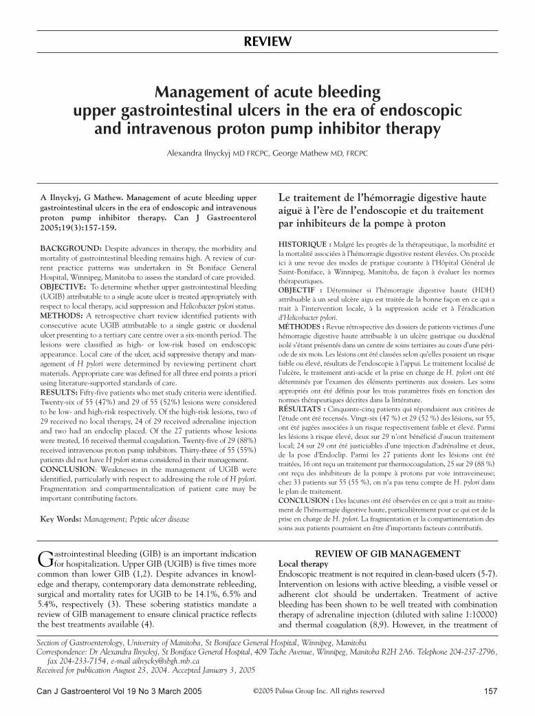
Can J Gastroenterol Vol 19 No 3 March 2005 157 Management of acute bleeding upper gastrointestinal ulcers in the era of endoscopic and intravenous proton pump inhibitor therapy Alexandra Ilnyckyj MD FRCPC, George Mathew MD, FRCPC Section of Gastroenterology, University of Manitoba, St Boniface General Hospital, Winnipeg, Manitoba Correspondence: Dr Alexandra Ilnyckyj, St Boniface General Hospital, 409 Tache Avenue, Winnipeg, Manitoba R2H 2A6. Telephone 204-237-2796, fax 204-233-7154, e-mail [email protected] Received for publication August 23, 2004. Accepted January 3, 2005 A Ilnyckyj, G Mathew. Management of acute bleeding upper gastrointestinal ulcers in the era of endoscopic and intravenous proton pump inhibitor therapy. Can J Gastroenterol 2005;19(3):157-159. BACKGROUND: Despite advances in therapy, the morbidity and mortality of gastrointestinal bleeding remains high. A review of cur- rent practice patterns was undertaken in St Boniface General Hospital, Winnipeg, Manitoba to assess the standard of care provided. OBJECTIVE: To determine whether upper gastrointestinal bleeding (UGIB) attributable to a single acute ulcer is treated appropriately with respect to local therapy, acid suppression and Helicobacter pylori status. METHODS: A retrospective chart review identified patients with consecutive acute UGIB attributable to a single gastric or duodenal ulcer presenting to a tertiary care centre over a six-month period. The lesions were classified as high- or low-risk based on endoscopic appearance. Local care of the ulcer, acid suppressive therapy and man- agement of H pylori were determined by reviewing pertinent chart materials. Appropriate care was defined for all three end points a priori using literature-supported standards of care. RESULTS: Fifty-five patients who met study criteria were identified. Twenty-six of 55 (47%) and 29 of 55 (52%) lesions were considered to be low- and high-risk respectively. Of the high-risk lesions, two of 29 received no local therapy, 24 of 29 received adrenaline injection and two had an endoclip placed. Of the 27 patients whose lesions were treated, 16 received thermal coagulation. Twenty-five of 29 (88%) received intravenous proton pump inhibitors. Thirty-three of 55 (55%) patients did not have H pylori status considered in their management. CONCLUSION: Weaknesses in the management of UGIB were identified, particularly with respect to addressing the role of H pylori. Fragmentation and compartmentalization of patient care may be important contributing factors. Key Words: Management; Peptic ulcer disease Le traitement de l’hémorragie digestive haute aiguë à l’ère de l’endoscopie et du traitement par inhibiteurs de la pompe à proton HISTORIQUE : Malgré les progrès de la thérapeutique, la morbidité et la mortalité associées à l’hémorragie digestive restent élevées. On procède ici à une revue des modes de pratique courante à l’Hôpital Général de Saint-Boniface, à Winnipeg, Manitoba, de façon à évaluer les normes thérapeutiques. OBJECTIF : Déterminer si l’hémorragie digestive haute (HDH) attribuable à un seul ulcère aigu est traitée de la bonne façon en ce qui a trait à l’intervention locale, à la suppression acide et à l’éradication d’Helicobacter pylori. MÉTHODES : Revue rétrospective des dossiers de patients victimes d’une hémorragie digestive haute attribuable à un ulcère gastrique ou duodénal isolé s’étant présentés dans un centre de soins tertiaires au cours d’une péri- ode de six mois. Les lésions ont été classées selon qu’elles posaient un risque faible ou élevé, résultats de l’endoscopie à l’appui. Le traitement localisé de l’ulcère, le traitement anti-acide et la prise en charge de H. pylori ont été déterminés par l’examen des éléments pertinents aux dossiers. Les soins appropriés ont été définis pour les trois paramètres fixés en fonction des normes thérapeutiques décrites dans la littérature. RÉSULTATS : Cinquante-cinq patients qui répondaient aux critères de l’étude ont été recensés. Vingt-six (47 %) et 29 (52 %) des lésions, sur 55, ont été jugées associées à un risque respectivement faible et élevé. Parmi les lésions à risque élevé, deux sur 29 n’ont bénéficié d’aucun traitement local; 24 sur 29 ont été justiciables d’une injection d’adrénaline et deux, de la pose d’Endoclip. Parmi les 27 patients dont les lésions ont été traitées, 16 ont reçu un traitement par thermocoagulation, 25 sur 29 (88 %) ont reçu des inhibiteurs de la pompe à protons par voie intraveineuse; chez 33 patients sur 55 (55 %), on n’a pas tenu compte de H. pylori dans le plan de traitement. CONCLUSION : Des lacunes ont été observées en ce qui a trait au traite- ment de l’hémorragie digestive haute, particulièrement pour ce qui est de la prise en charge de H. pylori. La fragmentation et la compartimentation des soins aux patients pourraient en être d’importants facteurs contributifs. G astrointestinal bleeding (GIB) is an important indication for hospitalization. Upper GIB (UGIB) is five times more common than lower GIB (1,2). Despite advances in knowl- edge and therapy, contemporary data demonstrate rebleeding, surgical and mortality rates for UGIB to be 14.1%, 6.5% and 5.4%, respectively (3). These sobering statistics mandate a review of GIB management to ensure clinical practice reflects the best treatments available (4). REVIEW OF GIB MANAGEMENT Local therapy Endoscopic treatment is not required in clean-based ulcers (5-7). Intervention on lesions with active bleeding, a visible vessel or adherent clot should be undertaken. Treatment of active bleeding has been shown to be well treated with combination therapy of adrenaline injection (diluted with saline 1:10000) and thermal coagulation (8,9). However, in the treatment of REVIEW ©2005 Pulsus Group Inc. All rights reserved
Transcript of Management of acute bleeding upper gastrointestinal ulcers in...
Can J Gastroenterol Vol 19 No 3 March 2005 157
Management of acute bleeding upper gastrointestinal ulcers in the era of endoscopic
and intravenous proton pump inhibitor therapy
Alexandra Ilnyckyj MD FRCPC, George Mathew MD, FRCPC
Section of Gastroenterology, University of Manitoba, St Boniface General Hospital, Winnipeg, ManitobaCorrespondence: Dr Alexandra Ilnyckyj, St Boniface General Hospital, 409 Tache Avenue, Winnipeg, Manitoba R2H 2A6. Telephone 204-237-2796,
fax 204-233-7154, e-mail [email protected] for publication August 23, 2004. Accepted January 3, 2005
A Ilnyckyj, G Mathew. Management of acute bleeding uppergastrointestinal ulcers in the era of endoscopic and intravenousproton pump inhibitor therapy. Can J Gastroenterol2005;19(3):157-159.
BACKGROUND: Despite advances in therapy, the morbidity and
mortality of gastrointestinal bleeding remains high. A review of cur-
rent practice patterns was undertaken in St Boniface General
Hospital, Winnipeg, Manitoba to assess the standard of care provided.
OBJECTIVE: To determine whether upper gastrointestinal bleeding
(UGIB) attributable to a single acute ulcer is treated appropriately with
respect to local therapy, acid suppression and Helicobacter pylori status.
METHODS: A retrospective chart review identified patients with
consecutive acute UGIB attributable to a single gastric or duodenal
ulcer presenting to a tertiary care centre over a six-month period. The
lesions were classified as high- or low-risk based on endoscopic
appearance. Local care of the ulcer, acid suppressive therapy and man-
agement of H pylori were determined by reviewing pertinent chart
materials. Appropriate care was defined for all three end points a priori
using literature-supported standards of care.
RESULTS: Fifty-five patients who met study criteria were identified.
Twenty-six of 55 (47%) and 29 of 55 (52%) lesions were considered
to be low- and high-risk respectively. Of the high-risk lesions, two of
29 received no local therapy, 24 of 29 received adrenaline injection
and two had an endoclip placed. Of the 27 patients whose lesions
were treated, 16 received thermal coagulation. Twenty-five of 29 (88%)
received intravenous proton pump inhibitors. Thirty-three of 55 (55%)
patients did not have H pylori status considered in their management.
CONCLUSION: Weaknesses in the management of UGIB were
identified, particularly with respect to addressing the role of H pylori.
Fragmentation and compartmentalization of patient care may be
important contributing factors.
Key Words: Management; Peptic ulcer disease
Le traitement de l’hémorragie digestive hauteaiguë à l’ère de l’endoscopie et du traitementpar inhibiteurs de la pompe à proton
HISTORIQUE : Malgré les progrès de la thérapeutique, la morbidité et
la mortalité associées à l’hémorragie digestive restent élevées. On procède
ici à une revue des modes de pratique courante à l’Hôpital Général de
Saint-Boniface, à Winnipeg, Manitoba, de façon à évaluer les normes
thérapeutiques.
OBJECTIF : Déterminer si l’hémorragie digestive haute (HDH)
attribuable à un seul ulcère aigu est traitée de la bonne façon en ce qui a
trait à l’intervention locale, à la suppression acide et à l’éradication
d’Helicobacter pylori.
MÉTHODES : Revue rétrospective des dossiers de patients victimes d’une
hémorragie digestive haute attribuable à un ulcère gastrique ou duodénal
isolé s’étant présentés dans un centre de soins tertiaires au cours d’une péri-
ode de six mois. Les lésions ont été classées selon qu’elles posaient un risque
faible ou élevé, résultats de l’endoscopie à l’appui. Le traitement localisé de
l’ulcère, le traitement anti-acide et la prise en charge de H. pylori ont été
déterminés par l’examen des éléments pertinents aux dossiers. Les soins
appropriés ont été définis pour les trois paramètres fixés en fonction des
normes thérapeutiques décrites dans la littérature.
RÉSULTATS : Cinquante-cinq patients qui répondaient aux critères de
l’étude ont été recensés. Vingt-six (47 %) et 29 (52 %) des lésions, sur 55,
ont été jugées associées à un risque respectivement faible et élevé. Parmi
les lésions à risque élevé, deux sur 29 n’ont bénéficié d’aucun traitement
local; 24 sur 29 ont été justiciables d’une injection d’adrénaline et deux,
de la pose d’Endoclip. Parmi les 27 patients dont les lésions ont été
traitées, 16 ont reçu un traitement par thermocoagulation, 25 sur 29 (88 %)
ont reçu des inhibiteurs de la pompe à protons par voie intraveineuse;
chez 33 patients sur 55 (55 %), on n’a pas tenu compte de H. pylori dans
le plan de traitement.
CONCLUSION : Des lacunes ont été observées en ce qui a trait au traite-
ment de l’hémorragie digestive haute, particulièrement pour ce qui est de la
prise en charge de H. pylori. La fragmentation et la compartimentation des
soins aux patients pourraient en être d’importants facteurs contributifs.
Gastrointestinal bleeding (GIB) is an important indicationfor hospitalization. Upper GIB (UGIB) is five times more
common than lower GIB (1,2). Despite advances in knowl-edge and therapy, contemporary data demonstrate rebleeding,surgical and mortality rates for UGIB to be 14.1%, 6.5% and5.4%, respectively (3). These sobering statistics mandate areview of GIB management to ensure clinical practice reflectsthe best treatments available (4).
REVIEW OF GIB MANAGEMENTLocal therapyEndoscopic treatment is not required in clean-based ulcers (5-7).Intervention on lesions with active bleeding, a visible vessel oradherent clot should be undertaken. Treatment of activebleeding has been shown to be well treated with combinationtherapy of adrenaline injection (diluted with saline 1:10000)and thermal coagulation (8,9). However, in the treatment of
REVIEW
©2005 Pulsus Group Inc. All rights reserved
Ilnyckyj.qxd 3/1/2005 11:12 AM Page 157

lesions with a visible vessel, combination treatment has notbeen shown to be more effective than using thermal coagulationalone (8,9). There is ongoing debate regarding the ideal man-agement of lesions with an adherent clot (10-13).
Acid suppression therapyStudies support the use of intravenous proton pump therapyin the high-risk lesion that has been endoscopically treated(14-19).
Helicobacter pylori
All patients presenting with an ulcerative lesion of the upperGI tract should be examined for the presence of Helicobacterpylori infection. The most sensitive and specific testing isaccomplished through histological examination of a gastricbiopsy taken at the time of endoscopy. The sample can be testedfor urease production if H pylori is not identified by the pathol-ogist. A less desirable approach consists of measuring antibodylevels against H pylori. All patients who test positive forH pylori in the setting of an acute ulcer presentation shouldreceive eradicative therapy at some point.
AIMThe aim of the present study was to determine whether treat-ment of UGIB attributable to acute ulcers was consistent withcontemporary management practices (4).
METHODSThere are two tertiary teaching hospitals in Winnipeg, Manitoba
(population 750,000). Both centres have a dedicated GI bleed
service which provides 24 h consultation and endoscopic inter-
vention for patients presenting with acute GIB. The service is
staffed by gastroenterologists and general surgeons. Patients pre-
senting with GIB requiring hospitalization are admitted under the
care of a clinical teaching unit, either medical or surgical. The GI
bleed service functions in the capacity of a consultative team.
A retrospective chart review for a six-month period dated
January to June 2002 was undertaken. International Classification of
Diseases, Ninth Revision (ICD-9) (20) codes on patient discharge
summaries were used by the medical records department to extract
charts. The ICD-9 codes extracted included GI bleed (578), peptic
ulcer disease (536), gastric ulcer (531) and duodenal ulcer (532).
Charts were reviewed by a senior GI Fellow and data were col-
lected using a standardized method. The Fellow reviewed the
entire hospital chart with specific attention to the GI bleed team
consultation, the endoscopy report, emergency room record if
present, doctors orders, chart notes made by the GI bleed team,
medication records, discharge summary, discharge recommenda-
tions and pathology and microbiology reports.
To meet entry criteria, the endoscopy report had to document
an acute gastric or duodenal ulcer as the source of the bleeding.
Patients whose source of bleeding were diagnosed as a nonulcer
source, multiple sources or unknown source were excluded.
The present review aimed to determine how the ulcer was
treated with regard to the three issues relevant to the management
of UGIB; specifically:
• local treatment (source document: endoscopy report);
• acid suppressive therapy (source documents: physician
order sheets and drug administration sheets); and
• H pylori testing and eradication (source documents:
GI consultation record, pathology report, microbiology
report, drug administration sheets, chart progress notes,
discharge summary and discharge recommendations to
primary care physician).
High-risk lesions were defined as lesions with adherent clot,
visible vessel or actively bleeding ulcer. Low-risk lesions included
clean-based ulcers or ulcers with heme stains.
RESULTSTwo hundred eighty charts were identified by medical recordswhich fulfilled the previously stated ICD-9 codes during thestudy interval. A review of these charts identified 55 patientsthat met entry criteria; specifically, an acute GI bleed referredto the GI bleeding service which was attributable to a gastricor duodenal ulcer. Of the 55 patients, 42 were treated by a gas-troenterologist and 13 by a surgeon.
Twenty-four of the 26 patients diagnosed with clean-based(low-risk) ulcers received no endoscopic intervention; tworeceived endoscopic therapy (injection) (Table 1). Twenty-four of the 29 patients diagnosed with high-risk lesions weretreated with injection therapy, which consisted of a variabledose of adrenaline diluted with saline 1:10000. Sixteen of 29high-risk lesions were treated with electrical or thermal energy.Another two high-risk lesions were treated with a hemoclip.Two of the 29 patients diagnosed with high-risk lesions did notreceive any endoscopic therapy (Table 1).
Eighteen of 55 patients (33%) had H pylori biopsies takenat the time of their endoscopy. An additional seven of55 patients had H pylori addressed in some manner; either sero-logical testing was obtained or recommended, or empiricalantibiotic therapy was given or recommended in the future.Thirty of 55 patients (55%) presenting with an UGIB attribut-able to an upper GI ulcer did not have an H pylori biopsyobtained, serology requested or empirical therapy recommended.
Twenty-five of 29 patients with high-risk ulcers and five of26 patients with low-risk ulcers received proton pump
Ilnyckyj and Mathew
Can J Gastroenterol Vol 19 No 3 March 2005158
TABLE 1Endoscopic therapy by risk of lesion
Lesion Total (n=55)
Low risk, n (%) 26 (47)
No treatment 24 of 26 (92)
Injection adrenaline 2 of 26 (8)
High risk, n (%) 29 (52)
No treatment 2 of 29 (7)
Injection adrenaline 24 of 29 (83)
Thermal coagulation 16 of 27 (55)
Endoclip 2 of 29 (7)
TABLE 2Acid suppression
Acid suppression Total (n=55)
IV PPI for high-risk ulcer, n (%) 25 of 29 (86)
IV PPI for low-risk ulcer, n (%) 5 of 26 (19)
PPI therapy total (oral or IV), n (%) 53 of 55 (96)
IV PPI Intravenous proton pump inhibitor
Ilnyckyj.qxd 3/1/2005 11:12 AM Page 158
inhibitor therapy intravenously (Table 2). The intravenousdosage was a bolus of 80 mg of pantoprazole followed by anintravenous infusion of 8 mg/h. The duration of the infusionwas not reviewed.
The all-cause mortality in the present study was 11% (six of55 patients). All deaths occurred in patients who suffered withmultiple medical comorbidities. The mean age of these sixpatients was 78 years. The mean time to death was 42 daysafter admission (range two to 115 days).
DISCUSSIONThe present review overwhelmingly demonstrates that physi-cians are treating UGIB attributable to an acute ulcer appro-priately with respect to local therapy and acid suppression.
Unfortunately, H pylori testing and treatment is not maxi-mized. Failure to undertake biopsy acquisition for H pylori hasbeen described by other workers (21). In this national databaseof 8000 patients with acute upper gastrointestinal ulcers, 33%did not have biopsies taken for H pylori. Of note, the databaseincluded only patients with nonbleeding acute ulcers, whereasall of our patients were presenting with bleeding ulcers. Thismay explain the higher rate of nonacquisition in our study.
The omission of H pylori biopsy acquisition is especially dis-appointing because eradication of H pylori is the greatestadvancement in peptic ulcer disease management to date. Wespeculate that this may be attributable to compartmentalized
care. During presentation, UGIB management is focused onvolume repletion and stabilization of the patient. Duringendoscopy, local therapy is foremost; acquisition of the biopsyfor H pylori may be undermined. The disparity between thenational database review and our own data support this specu-lation. Thereafter, hospitalization may not afford opportunitiesto review the issues pertinent to UGIB. Either short or pro-longed hospital stay may detract from the importance ofH pylori testing and treatment if H pylori biopsy was notobtained during the gastroscopy. As well, fragmentation of careamong the endoscopist, the admitting physician and the familyphysician may further contribute to omission of H pylori testingand treatment.
CONCLUSIONSDespite the management of acute GIB by a dedicated bleedservice, our review revealed significant weaknesses in the man-agement of this common and costly disease. High-risk lesions arenot uniformly treated with endoscopic interventions. Of greatermagnitude is the failure in acquiring an H pylori biopsy, or under-taking serological testing or advising empirical therapy in thosein whom biopsy was not obtained. Compartmentalization andfragmentation of care may contribute to oversight. Regardless,the GI bleed consultant must be vigilant to ensure H pylori sta-tus and treatment is addressed because the organism is integralto the pathogenesis of peptic ulcer disease.
Management of acute UGI bleed with endoscopy and IV PPI
Can J Gastroenterol Vol 19 No 3 March 2005 159
REFERENCES1. Longstreth GF. Epidemiology of hospitalization for acute upper
gastrointestinal hemorrhage: A population-based study. Am JGastroenterol 1995; 90:206-10.
2. Longstreth GF. Epidemiology and outcome of patients hospitalizedwith acute lower gastrointestinal hemorrhage: A population-basedstudy. Am J Gastroenterol 1997; 92:419-24.
3. Barkun A, Sabbah S, Enns R, et al; for the RUGBE Investigators.The Canadian Registry on Nonvariceal Upper GastrointestinalBleeding and Endoscopy (RUGBE): Endoscopic hemostasis andproton pump inhibition are associated with improved outcomes in areal-life setting. Am J Gastroenterol 2004;99:1238-46.
4. Barkun A, Fallone CA, Chiba N, et al; for the Nonvariceal UpperGI Bleeding Consensus Conference Group. A Canadian clinicalpractice algorithm for the management of patients with nonvaricealupper gastrointestinal bleeding. Can J Gastroenterol 2004;18:605-9.
5. Laine L, Peterson WL. Bleeding peptic ulcer. N Engl J Med1994;331:717-27.
6. Laine L, Cohen H, Brodhead J, Cantor D, Garcia F, Mosquera M.Prospective evaluation of immediate versus delayed refeeding andprognostic value of endoscopy in patients with uppergastrointestinal hemorrhage. Gastroenterology 1992;102:314-6.
7. Longstreth GF, Feitelberg SP. Successful outpatient management ofacute upper gastrointestinal hemorrhage: Use of practice guidelinesin a large patient series. Gastrointest Endosc 1998; 47:219-22.
8. Chung SS, Lau JY, Sung JJ, et al. Randomised comparison betweenadrenaline injection alone and adrenaline injection plus heat probetreatment for actively bleeding ulcers. BMJ 1997;314:1307-11.
9. Jensen DM, Kovacs T, Randall G, Smith J, Freeman M, Jutabha R.Prospective study of thermal coagulation (Gold probe) versuscombination injection and thermal treatment of high risk patientswith severe ulcer or Mallory Weiss bleeding. Gastrointest Endosc1994;40:25. (Abst)
10. Bleau BL, Gostout CJ, Sherman KE, et al. Recurrent bleeding frompeptic ulcer associated with adherent clot: A randomized studycomparing endoscopic treatment with medical therapy. GastrointestEndosc 2002;56:1-6.
11. Bini EJ, Cohen J. Endoscopic treatment of adherent clots
significantly reduces early rebleeding in patients with peptic ulcerdisease. Gastrointest Endosc 1999;49:164. (Abst)
12. Jensen DM, Kovacs TO, Jutabha R, et al. Randomized trial ofmedical or endoscopic therapy to prevent recurrent ulcerhemorrhage in patients with adherent clots. Gastroenterology2002;123:407-13.
13. Laine L. Management of ulcers with adherent clots.Gastroenterology 2002;123:632-6.
14. Lin HJ, Lo WC, Lee FY, Perng CL, Tseng GY. A prospectiverandomized comparative trial showing that omeprazole preventsrebleeding in patients with bleeding peptic ulcer after successfulendoscopic therapy. Arch Intern Med 1998;158:54-8.
15. Lau JY, Sung JJ, Lee KK, et al. Effect of intravenous omeprazole onrecurrent bleeding after endoscopic treatment of bleeding pepticulcers. N Engl J Med 2000;343:310-6.
16. Javid G, Masoodi I, Zargar SA, et al. Omeprazole as adjuvanttherapy to endoscopic combination injection sclerotherapy fortreating bleeding peptic ulcer. Am J Med 2001;111:280-4.
17. Lee KK, You JH, Wong IC, et al. Cost-effectiveness analysis ofhigh-dose omeprazole infusion as adjuvant therapy to endoscopictreatment of bleeding peptic ulcer. Gastrointest Endosc2003;57:160-4.
18. Hasselgren G, Lind T, Lundell L, et al. Continuous intravenousinfusion of omeprazole in elderly patients with peptic ulcerbleeding. Results of a placebo-controlled multicenter study. Scand JGastroenterol 1997;32:328-33.
19. Schaffalitzky de Muckadell OB, Havelund T, Harling H, et al.Effect of omeprazole on the outcome of endoscopically treatedbleeding peptic ulcers. Randomized double-blind placebo-controlled multicentre study. Scand J Gastroenterol 1997;32:320-7.
20. World Health Organization. International Classification ofDiseases, 9th Revision. Geneva: World Health Organization, 1978.
21. Harewood GC, Holub JL, Lieberman DA. Biopsy specimenacquisition in patients with newly diagnosed peptic ulcer disease asdetermined from a national endoscopic database. GastrointestEndosc 2004;59:664-9.
Ilnyckyj.qxd 3/1/2005 11:12 AM Page 159

Submit your manuscripts athttp://www.hindawi.com
Stem CellsInternational
Hindawi Publishing Corporationhttp://www.hindawi.com Volume 2014
Hindawi Publishing Corporationhttp://www.hindawi.com Volume 2014
MEDIATORSINFLAMMATION
of
Hindawi Publishing Corporationhttp://www.hindawi.com Volume 2014
Behavioural Neurology
EndocrinologyInternational Journal of
Hindawi Publishing Corporationhttp://www.hindawi.com Volume 2014
Hindawi Publishing Corporationhttp://www.hindawi.com Volume 2014
Disease Markers
Hindawi Publishing Corporationhttp://www.hindawi.com Volume 2014
BioMed Research International
OncologyJournal of
Hindawi Publishing Corporationhttp://www.hindawi.com Volume 2014
Hindawi Publishing Corporationhttp://www.hindawi.com Volume 2014
Oxidative Medicine and Cellular Longevity
Hindawi Publishing Corporationhttp://www.hindawi.com Volume 2014
PPAR Research
The Scientific World JournalHindawi Publishing Corporation http://www.hindawi.com Volume 2014
Immunology ResearchHindawi Publishing Corporationhttp://www.hindawi.com Volume 2014
Journal of
ObesityJournal of
Hindawi Publishing Corporationhttp://www.hindawi.com Volume 2014
Hindawi Publishing Corporationhttp://www.hindawi.com Volume 2014
Computational and Mathematical Methods in Medicine
OphthalmologyJournal of
Hindawi Publishing Corporationhttp://www.hindawi.com Volume 2014
Diabetes ResearchJournal of
Hindawi Publishing Corporationhttp://www.hindawi.com Volume 2014
Hindawi Publishing Corporationhttp://www.hindawi.com Volume 2014
Research and TreatmentAIDS
Hindawi Publishing Corporationhttp://www.hindawi.com Volume 2014
Gastroenterology Research and Practice
Hindawi Publishing Corporationhttp://www.hindawi.com Volume 2014
Parkinson’s Disease
Evidence-Based Complementary and Alternative Medicine
Volume 2014Hindawi Publishing Corporationhttp://www.hindawi.com