Management of a giant omphalocele with an external skin closure system
4
Management of a giant omphalocele with an external skin closure system Robert Baird ⁎ , Suad Gholoum, Jean-Martin Laberge, Pramod Puligandla Division of Pediatric Surgery, The Montreal Children's Hospital, McGill University, Montreal, Quebec, Canada H3H 1P3 Received 25 December 2009; revised 30 April 2010; accepted 3 May 2010 Key words: Giant omphalocele; Abdominal closure device Abstract Background/Purpose: The management of neonates with giant omphalocele remains challenging and multiple strategies have been described. We present the case of a 34-week-old neonate with isolated giant omphalocele managed with an external surgical skin closure system as a component of a staged closure strategy. Case Presentation: An Inuit boy of 34 weeks gestation was born by urgent Caesarean delivery at an affiliated obstetrical hospital with a giant ruptured omphalocele and loss of abdominal domain. He was transferred to our institution and a silastic silo was fashioned and placed in the operating room. He returned to the operating room several times and was treated by placement of a combined Gore-Tex (WL Gore and Associates, Flagstaff, Ariz)/silastic inlay mesh. An eschar formed over this temporary closure, and we elected to place a dynamic skin closure device to continue gradual bedside reduction. The initial abdominal wall defect was 8.5 cm in transverse diameter and was reduced to 4.5 cm over 3 weeks. Complete closure was subsequently achieved without the need for skin grafting. Discussion: The use of a dynamic reduction skin closure device has not been documented previously in the pediatric population or in the context of a congenital defect. We describe the use of an external surgical skin closure device in the context of the staged closure of a giant neonatal omphalocele and postulate that such a device may prove useful in the treatment of other congenital tissue defects. © 2010 Elsevier Inc. All rights reserved. The optimal management of neonates with giant ompha- locele remains controversial. The resultant visceroabdominal disproportion often excludes the possibility of a primary repair, and multiple, staged closure strategies have been described. The use of skin flaps was originally described by Gross [1], whereas newer publications favor the use of absorbable [2] or nonabsorbable [3] mesh to achieve closure. Adult surgeons have been increasingly using external closure devices to aid in abdominal closure after damage control laparotomy [4,5]; a phase IV trial of this strategy was due to begin on July 2009 (ClinicalTrials.gov Identifier: NCT00754156). We present the case of a 34-week-old neonate with isolated giant omphalocele managed with an external surgical skin closure system as a component of a staged closure strategy. 1. Case presentation An Inuit boy of 34 weeks gestation was born to an 18- year-old mother at an affiliated obstetrical hospital with a ⁎ Corresponding author. Tel.: +1 514 412 4438; fax: +1 514 412 4289. www.elsevier.com/locate/jpedsurg 0022-3468/$ – see front matter © 2010 Elsevier Inc. All rights reserved. doi:10.1016/j.jpedsurg.2010.05.004 Journal of Pediatric Surgery (2010) 45, E17–E20
-
Upload
robert-baird -
Category
Documents
-
view
215 -
download
2
Transcript of Management of a giant omphalocele with an external skin closure system

www.elsevier.com/locate/jpedsurg
Journal of Pediatric Surgery (2010) 45, E17–E20
Management of a giant omphalocele with an external skinclosure systemRobert Baird⁎, Suad Gholoum, Jean-Martin Laberge, Pramod Puligandla
Division of Pediatric Surgery, The Montreal Children's Hospital, McGill University, Montreal, Quebec, Canada H3H 1P3
Received 25 December 2009; revised 30 April 2010; accepted 3 May 2010
0d
Key words:Giant omphalocele;Abdominal closure device
AbstractBackground/Purpose: The management of neonates with giant omphalocele remains challenging andmultiple strategies have been described. We present the case of a 34-week-old neonate with isolatedgiant omphalocele managed with an external surgical skin closure system as a component of a stagedclosure strategy.Case Presentation: An Inuit boy of 34 weeks gestation was born by urgent Caesarean delivery at anaffiliated obstetrical hospital with a giant ruptured omphalocele and loss of abdominal domain. He wastransferred to our institution and a silastic silo was fashioned and placed in the operating room. Hereturned to the operating room several times and was treated by placement of a combined Gore-Tex(WL Gore and Associates, Flagstaff, Ariz)/silastic inlay mesh. An eschar formed over this temporaryclosure, and we elected to place a dynamic skin closure device to continue gradual bedside reduction.The initial abdominal wall defect was 8.5 cm in transverse diameter and was reduced to 4.5 cm over3 weeks. Complete closure was subsequently achieved without the need for skin grafting.Discussion: The use of a dynamic reduction skin closure device has not been documented previously inthe pediatric population or in the context of a congenital defect. We describe the use of an externalsurgical skin closure device in the context of the staged closure of a giant neonatal omphalocele andpostulate that such a device may prove useful in the treatment of other congenital tissue defects.© 2010 Elsevier Inc. All rights reserved.
The optimal management of neonates with giant ompha-locele remains controversial. The resultant visceroabdominaldisproportion often excludes the possibility of a primaryrepair, and multiple, staged closure strategies have beendescribed. The use of skin flaps was originally described byGross [1], whereas newer publications favor the useof absorbable [2] or nonabsorbable [3] mesh to achieveclosure. Adult surgeons have been increasingly usingexternal closure devices to aid in abdominal closure afterdamage control laparotomy [4,5]; a phase IV trial of this
⁎ Corresponding author. Tel.: +1 514 412 4438; fax: +1 514 412 4289.
022-3468/$ – see front matter © 2010 Elsevier Inc. All rights reserved.oi:10.1016/j.jpedsurg.2010.05.004
strategy was due to begin on July 2009 (ClinicalTrials.govIdentifier: NCT00754156). We present the case of a34-week-old neonate with isolated giant omphalocelemanaged with an external surgical skin closure system as acomponent of a staged closure strategy.
1. Case presentation
An Inuit boy of 34 weeks gestation was born to an 18-year-old mother at an affiliated obstetrical hospital with a
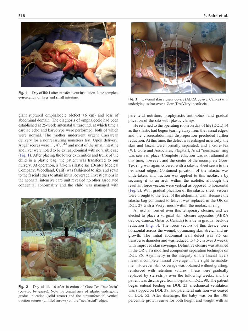
Fig. 1 Day of life 1 after transfer to our institution. Note completeevisceration of liver and small intestine. ig. 3 External skin closure device (ABRA device, Canica) with
nderlying eschar over a Gore-Tex/Vicryl neofascia.
E18 R. Baird et al.
giant ruptured omphalocele (defect N6 cm) and loss ofabdominal domain. The diagnosis of omphalocele had beenestablished at 25-week antenatal ultrasound, at which time acardiac echo and karyotype were performed, both of whichwere normal. The mother underwent urgent Caesareandelivery for a nonreassuring nonstress test. Upon delivery,Apgar scores were 11, 45, 710 and most of the small intestineand liver were noted to be extraabdominal with no visible sac(Fig. 1). After placing the lower extremities and trunk of thechild in a plastic bag, the patient was transferred to ournursery. At operation, a 7.5-cm silastic sac (Bentec MedicalCompany, Woodland, Calif) was fashioned to size and sewnto the fascial edges to attain initial coverage. Investigations inthe neonatal intensive care unit revealed no other associatedcongenital abnormality and the child was managed with
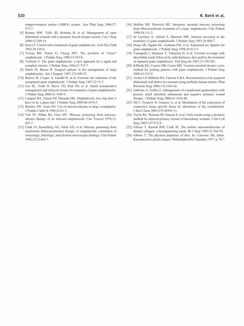
Fig. 2 Day of life 16 after insertion of Gore-Tex “neofascia”(covered by gauze). Note the central area of silastic undergoinggradual plication (solid arrow) and the circumferential verticaltraction sutures (unfilled arrows) on the “neofascial” edges.
Fu
parenteral nutrition, prophylactic antibiotics, and gradualplication of the silo with plastic clamps.
He returned to the operating room on day of life (DOL) 14as the silastic had begun tearing away from the fascial edges,and the visceroabdominal disproportion precluded furtherreduction. At this time, the defect was enlarged inferiorly, theskin and fascia were formally separated, and a Gore-Tex(WL Gore and Associates, Flagstaff, Ariz) “neofascia” ringwas sewn in place. Complete reduction was not attained atthis time, however, and the center of the incomplete Gore-Tex ring was again covered with a silastic sheet sewn to theneofascial edges. Continued plication of the silastic wasundertaken, and traction was applied to this neofascia bysecuring it to an arch within the isolette, although theresultant force vectors were vertical as opposed to horizontal(Fig. 2). With gradual plication of the silastic sheet, viscerawere brought to the level of the abdominal wall. Because thesilastic bag continued to tear, it was replaced in the OR onDOL 27 with a Vicryl mesh within the neofascial ring.
An eschar formed over this temporary closure, and weelected to place a surgical skin closure apparatus (ABRAdevice, Canica, Ontario, Canada) to aide in gradual bedsidereduction (Fig. 3). The force vectors of this device werehorizontal across the wound, optimizing skin stretch and in-growth. The initial abdominal wall defect was 8.5 cmtransverse diameter and was reduced to 4.5 cm over 3 weeks,with improved skin coverage. Definitive closure was attainedin the OR via a modified component separation technique onDOL 86. Asymmetry in the integrity of the fascial layersmeant incomplete fascial coverage in the right hemiabdo-men. However, skin coverage was obtained without grafting,reinforced with retention sutures. These were graduallyreplaced by steri-strips over the following weeks, and thepatient was discharged from hospital on DOL 98. The patientbegan enteral feeding on DOL 23, mechanical ventilationwas stopped on DOL 38, and parenteral nutrition was ceasedon DOL 52. After discharge, the baby was on the 10thpercentile growth curve for both height and weight with an
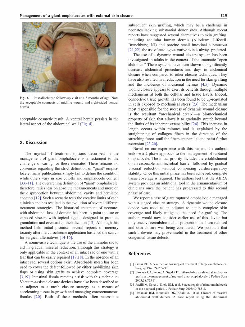
Fig. 4 Post-discharge follow-up visit at 4.5 months of age. Notethe acceptable cosmesis of midline wound and right-sided ventralhernia.
E19Management of a giant omphaloceles with external skin closure
acceptable cosmetic result. A ventral hernia persists in thelateral aspect of the abdominal wall (Fig. 4).
2. Discussion
The myriad of treatment options described in themanagement of giant omphalocele is a testament to thechallenge of caring for these neonates. There remains noconsensus regarding the strict definition of “giant” ompha-locele; many publications simply fail to define the conditionwhile others vary in size cutoffs and omphalocele content[3,6-11]. The overarching definition of “giant” omphalocele,therefore, relies less on absolute measurements and more onthe disproportion between abdominal cavity and herniatedcontents [12]. Such a scenario tests the creative limits of eachclinician and has resulted in the evolution of several differenttreatment strategies. The historical treatment of neonateswith abdominal loss-of-domain has been to paint the sac orexposed viscera with topical agents designed to promotegranulation and eventual epithelialization [13]. Although thismethod held initial promise, several reports of mercurytoxicity after mercurochrome application hastened the searchfor surgical alternatives [14-16].
A noninvasive technique is the use of the amniotic sac toaid in gradual visceral reduction, although this strategy isonly applicable in the context of an intact sac or one with atear that can be easily repaired [17,18]. In the absence of anintact sac, several options exist. Absorbable mesh has beenused to cover the defect followed by either mobilizing skinflaps or using skin grafts to achieve complete coverage[3,19]. Intestinal fistula remains a risk with this technique.Vacuum-assisted closure devices have also been described asan adjunct to a mesh closure strategy as a means ofaccelerating tissue in-growth and managing enterocutaneousfistulas [20]. Both of these methods often necessitate
subsequent skin grafting, which may be a challenge inneonates lacking substantial donor sites. Although recentreports have suggested several alternatives to skin grafting,including acellular human dermis (Alloderm, Lifecell,Branchburg, NJ) and porcine small intestinal submucosa[21,22], the use of autologous native skin is always preferred.
The use of a dynamic wound closure system has beeninvestigated in adults in the context of the traumatic “openabdomen.” These systems have been shown to significantlydecrease abdominal procedures and days to abdominalclosure when compared to other closure techniques. Theyhave also resulted in a reduction in the need for skin graftingand the incidence of incisional hernias [4,5]. Dynamicwound closure appears to exert its benefits through multiplemechanisms at both the cellular and tissue levels. Indeed,connective tissue growth has been found to be up-regulatedin cells exposed to mechanical stress [23]. The mechanismmost responsible for the success of dynamic wound closureis the resultant “mechanical creep”—a biomechanicalproperty of skin that allows it to gradually stretch beyondthe limits of its inherent extensibility [24]. This increase inlength occurs within minutes and is explained by thestraightening of collagen fibers in the direction of thestretching force, until the fibers are parallel and resist furtherextension [25,26].
Based on our experience with this patient, the authorsendorse a 2-phase approach to the management of rupturedomphalocele. The initial priority includes the establishmentof a reasonable antimicrobial barrier followed by gradualvisceral reduction without comprising cardiorespiratorystability. Once this initial phase has been achieved, completetissue coverage is required. The authors feel that the ABRAsystem provides an additional tool in the armamentarium ofclinicians once the patient has progressed to this secondphase of care.
We report a case of giant ruptured omphalocele managedwith a staged closure strategy. A dynamic wound closuredevice was used as an adjunct to attain complete skincoverage and likely mitigated the need for grafting. Theauthors would now consider earlier use of this device butonly once visceroabdominal disproportion had been reducedand skin closure was being considered. We postulate thatsuch a device may prove useful in the treatment of othercongenital tissue defects.
References
[1] Gross RE. A new method for surgical treatment of large omphaloceles.Surgery 1948;24:277-92.
[2] Bawazir OA, Wong A, Sigalet DL. Absorbable mesh and skin flaps orgrafts in the management of ruptured giant omphalocele. J Pediatr Surg2003;38:725-8.
[3] Pacilli M, Spitz L, Kiely EM, et al. Staged repair of giant omphalocelein the neonatal period. J Pediatr Surg 2005;40:785-8.
[4] Urbaniak RM, Khuthaila DK, Khalil AJ, et al. Closure of massiveabdominal wall defects. A case report using the abdominal
E20 R. Baird et al.
reapproximation anchor (ABRA) system. Ann Plast Surg 2006;57:573-7.
[5] Reimer MW, Yelle JD, Reitsma B, et al. Management of openabdominal wounds with a dynamic fascial closure system. Can J Surg2008;51:209-14.
[6] Soave F. Conservative treatment of giant omphalocele. Arch Dis Child1963;38:130-4.
[7] Towne BH, Peters G, Chang JHT. The problem of “Giant”omphalocele. J Pediatr Surg 1980;15:543-8.
[8] Yazbeck S. The giant omphalocele: a new approach for a rapid andcomplete closure. J Pediatr Surg 1986;21:715-7.
[9] Hatch EI, Baxter R. Surgical options in the management of largeomphaloceles. Am J Surgery 1987;153:449-52.
[10] Barlow B, Cooper A, Gandhi R, et al. External silo reduction of theunruptured giant omphalocele. J Pediatr Surg 1987;22:75-7.
[11] Lee SL, Todd D, Beyer TD, Kim SS, et al. Initial nonoperativemanagement and delayed closure for treatment of giant omphaloceles.J Pediatr Surg 2006;41:1846-9.
[12] Campos BA, Tatsuo ES, Miranda ME. Omphalocele: how big does ithave to be a giant one? J Pediatr Surg 2009;44:1474-5.
[13] Beasley SW, Jones PG. Use of mercurochrome in large exomphalos.J Pediatr Child H 1986;22:61-3.
[14] Yeh TF, Pildes RS, Firor HV. Mercury poisoning from mercuro-chrome therapy of an infected omphalocele. Clin Toxicol 1978;13:463-7.
[15] Clark JA, Kasselberg AG, Glick AD, et al. Mercury poisoning frommerbromin (Mercurochrome) therapy of omphalocele: correlation oftoxicologic, histologic, and electron microscopic findings. Clin Pediatr1982;21(7):445-7.
[16] Mullins ME, Horowitz BZ. Iatrogenic neonatal mercury poisoningfrom Mercurochrome treatment of a large omphalocele. Clin Pediatr1999;38:111-2.
[17] de Lorimier A, Adzick S, Harrison MR. Amnion inversion in thetreatment of giant omphalocele. J Pediatr Surg 1991;26:804-7.
[18] Hong AR, Sigalet DL, Guttman FM, et al. Sequential sac ligation forgiant omphalocele. J Pediatr Surg 1994;29:413-5.
[19] Yamagishi J, Ishimaru Y, Takayasu H, et al. Visceral coverage withabsorbable mesh followed by split-thickness skin graft in the treatmentof ruptured giant omphalocele. Ped Surg Int 2007;23:199-201.
[20] Kilbride KE, Cooney DR, Custer MD. Vacuum-assisted closure: a newmethod for treating patients with giant omphalocele. J Pediatr Surg2006;41:212-5.
[21] Archer LP, Billmire DA, Falcone Jr RA. Reconstruction of an acquiredabdominal wall defect in a neonate using acellular human dermis. PlastReconstr Surg 2006;118:163e-6e.
[22] Gabriela A, Gollin G. Management of complicated gastroschisis withporcine small intestinal submucosa and negative pressure woundtherapy. J Pediatr Surg 2006;41:1836-40.
[23] Ott C, Iwanciw D, Graness A, et al. Modulation of the expression ofconnective tissue growth factor by alterations of the cytoskeleton.J Biol Chem 2003;278:44305-11.
[24] Taylor RC, Reitsma BJ, Sarazin S, et al. Early results using a dynamicmethod for delayed primary closure of fasciotomy wounds. J Am CollSurg 2003;197:872-8.
[25] Gibson T, Kenedi RM, Craik JE. The mobile microarchitecture ofdermal collagen: a bioengineering study. Br J Surg 1965;52:764-70.
[26] Gibson T. The physical properties of skin. In: Converse JM, editor.Reconstructiveplastic surgery. Philadelphia (Pa): Saunders; 1977. p. 70-7.