Mammography
26
ن م ح ر ل ها ل ل ما س ب م ي ح ر ل ا
-
Upload
- -
Category
Health & Medicine
-
view
63 -
download
6
Transcript of Mammography

الرحمن الله بسمالرحيم

RAD242
Medical Imaging Equipment's

Mammography

الزيدي عبدالرحمن
الوليعي عبدالله
المدالله عبدالمحسن
المحمود عبدالعزيز

Topics :• Introduction
• X-ray tube
• Compression and breast support plate
• Types of mammography

INTRODUCTION
What is mammography?
What is general components of the equipment?

Mammography • Mammography is a radiographic examination that
is designed for detecting breast pathology ( particularly breast cancer ).
Breast cancer screening with mammography assists in detecting cancer at an earlier, and is an important clinical procedure because approximately one in eight women will develop breast cancer over their lifetime.


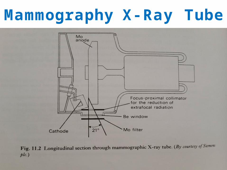
Mammography X-Ray Tube

Mammography X-Ray Tube
Target materials consist of three main types.
• Molybdenum• Specialized Tungsten• Rhodium
• Molybdenum– the best material to be used in mammography,– allows production of low energy spectrums of radiation– low kVp (26-40kVp)
• Tungsten and Rhodium are used for higher beam needs, in dense breast tissues.

Filtration•Materials that are placed in the path of the X-ray
beam in order to absorb those X-rays with energies above and below the desired spectrum.
• Tube filtration types:•Molybdenum (Mo) (best used for lower kVp)• Rhodium (Rh)• Yttrium• Aluminum (used for above 30 kVp)

CollimationCollimator – used to shape radiation field :
• Recall, smaller radiation field means less scatter, collimate when you can!
• Smaller exposed area, better for patient dose

Cont.
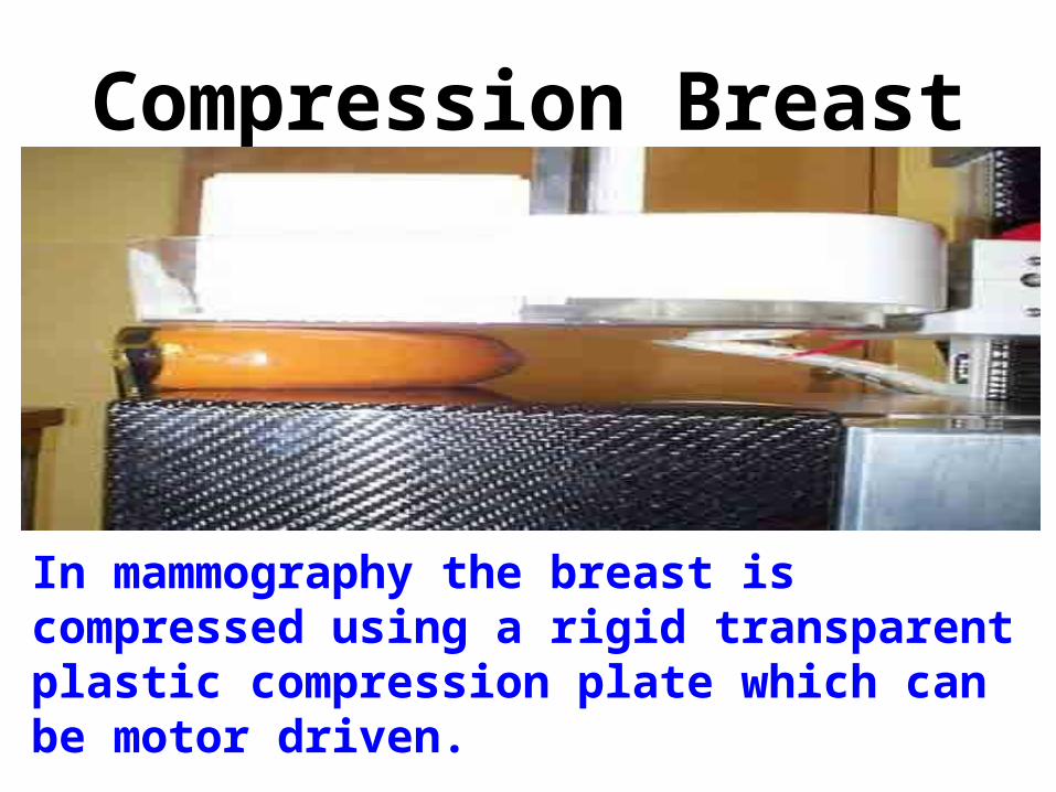
Compression Breast
In mammography the breast is compressed using a rigid transparent plastic compression plate which can be motor driven.

Breast compression Why we apply breast compression?

Cont….• Better spatial resolution. The breast is broughtcloser to the imaging receptor so that magnification and focal spot blurring is reduced.
• Reduced movement blur, even at the relatively long exposure times.
• Less scattered radiation in the image. The beam path length through the breast is shorter, so there is less material to do the scattering.

Cont.• The reduced path length makes practicable theuse of lower energy (less penetrating) X-rayspectra. This gives greater subject contrast.
• Small areas of pathology buried in glandulartissue can be better visualized, as malignanttissues tend to be firmer.

Breast support plate • It’s the plate that hold the breast :
• Two parts:• Upper part made from carbon fiber(free
absorption)
• Lower part made from lead (safe the patient abdomen from radiation hazard )

Types of mammography • There are two main types of mammography:
1- Screening mammogram:

cont .2- Diagnostic mammogram:
Advances 1. Digital mammography:
•also called full-field digital mammography (FFDM), is a mammography system in which
•the x-ray film is replaced by solid-state detectors that convert x-rays into electrical signals.

Advances 2. computed aided detection:
• that can be obtained from either a conventional film mammogram or a digitally acquired mammogram.
3. breast tomosynthesis:
•also called three-dimensional (3-D) breast imaging, is a mammography system where the x-ray tube moves in an arc over the breast during the exposure.

1. THE ESSENTIAL PHYSICS OF MEDICAL IMAGING, THIRD EDITION.
2. CHESNES’ EQUIPMENT FOR STUDENT RADIOGRAPHERS FOURTH EDITION.
References :

Thank you
Have a Nice Day