
Mammalian Circulation: Components and Control AP Biology Unit 6.
30
Mammalian Circulation: Components and Control AP Biology Unit 6
-
Upload
milo-logan -
Category
Documents
-
view
218 -
download
0
Transcript of Mammalian Circulation: Components and Control AP Biology Unit 6.
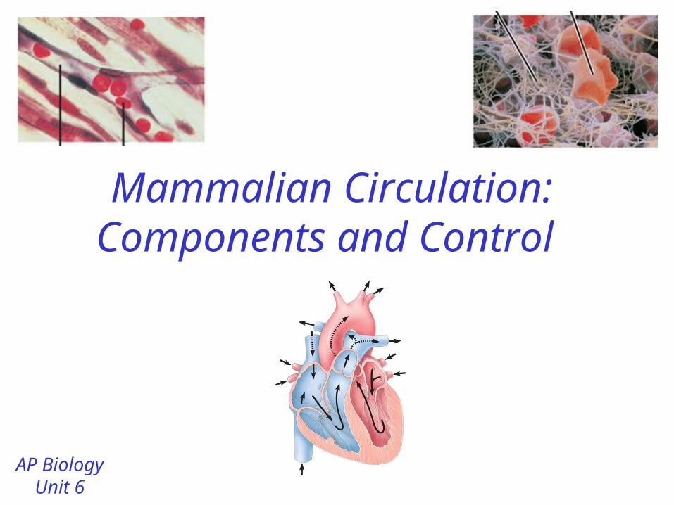
Mammalian Circulation: Components and Control
AP BiologyUnit 6

Mammalian Heart
• 4 chambered heart (2 atria, 2 ventricles)
• Valves = flaps that keep chambers of the heart closed at the right time
• Valves are needed to build pressure in heart and prevent back-flow of blood.
Right side Left side

Atrioventricular (AV) Valves
• Located between the atria and ventricles
• Tricuspid Valve – Between the right atrium
and right ventricle– 3 flaps
• Bicuspid (Mitral) valve– Between the left atrium
and left ventricle– 2 flaps
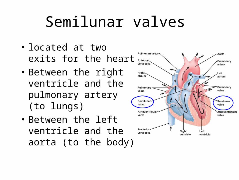
Semilunar valves
• located at two exits for the heart
• Between the right ventricle and the pulmonary artery (to lungs)
• Between the left ventricle and the aorta (to the body)

Pathway of Blood
• Do you remember the pathway of blood through the body and the heart?
• Use these terms: Right Atrium, Left Atrium, Right Ventricle, Left Ventricle, Pulmonary Artery, Pulmonary Vein, Aorta, Lung Capillaries, Capillaries in Top or Bottom of Body, Anterior / Posterior Vena Cava
• Start where the blood first leaves the heart to go to the body
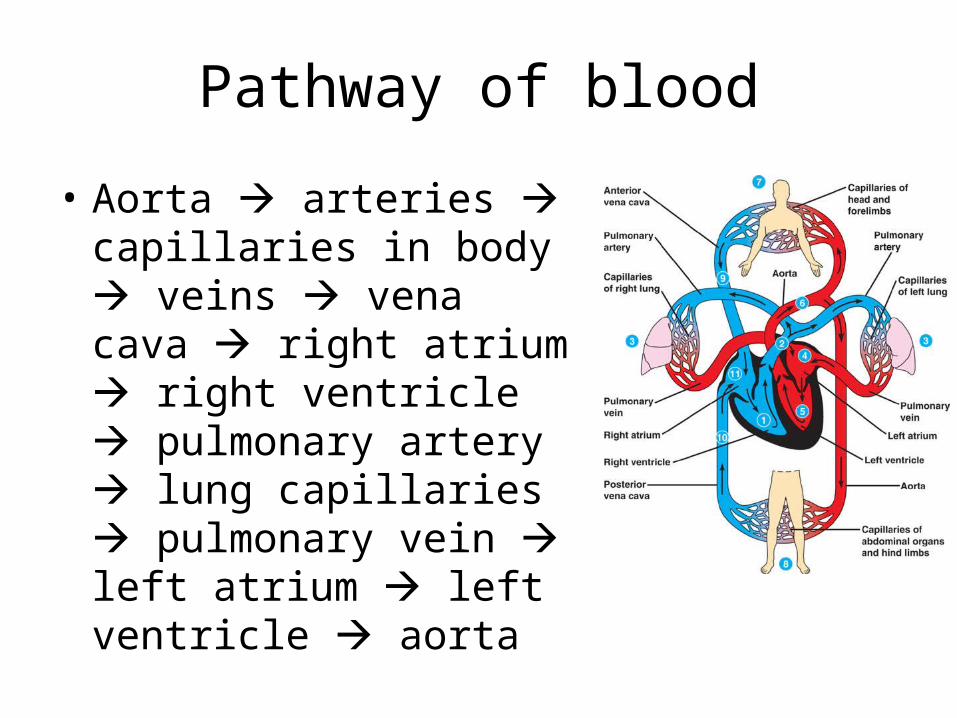
Pathway of blood
• Aorta arteries capillaries in body veins vena cava right atrium right ventricle pulmonary artery lung capillaries pulmonary vein left atrium left ventricle aorta
Questions…
• Where does the blood have the highest O2
concentration? – Just after leaving lungs (where it picked up O2)
• Where does the blood have the highest CO2
concentration? – Just before getting to the lungs (hasn’t dropped
off the CO2 waste yet)

Heartbeat• The heart beat is controlled by
electrical signals generated in specific cells in the heart = self excitation
• Sinoatrial (SA) node = a group of specialized cells that initiates the heartbeat– Also called the pacemaker of
the heart– generates electrical impulses
that cause both atria to contract
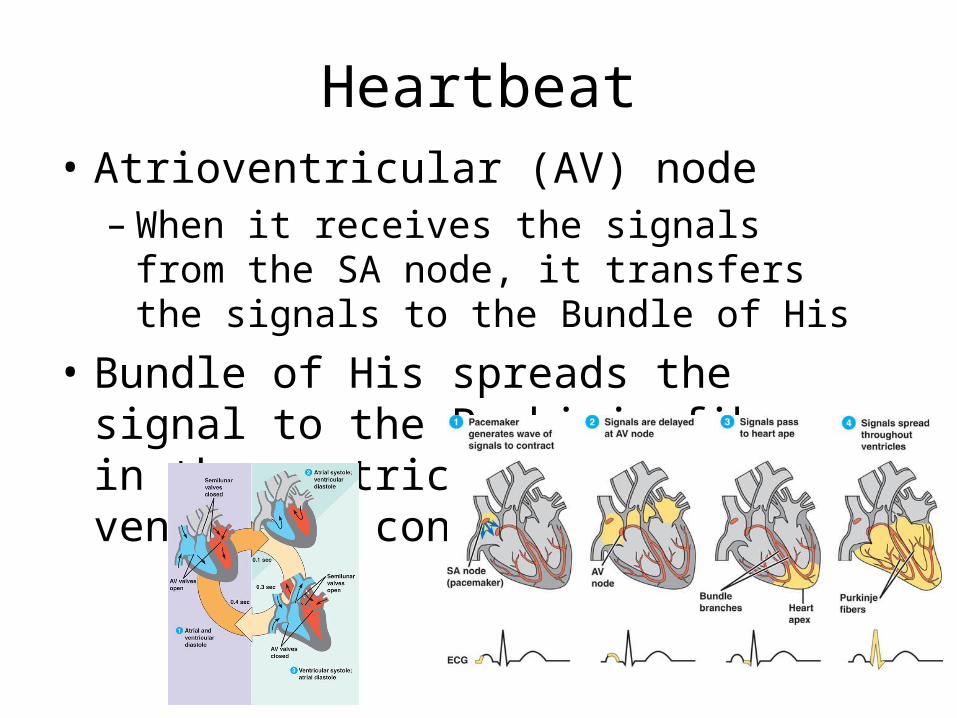
Heartbeat• Atrioventricular (AV) node
– When it receives the signals from the SA node, it transfers the signals to the Bundle of His
• Bundle of His spreads the signal to the Purkinje fibers in the ventricles both ventricles contract
Blood Flow through Vessels
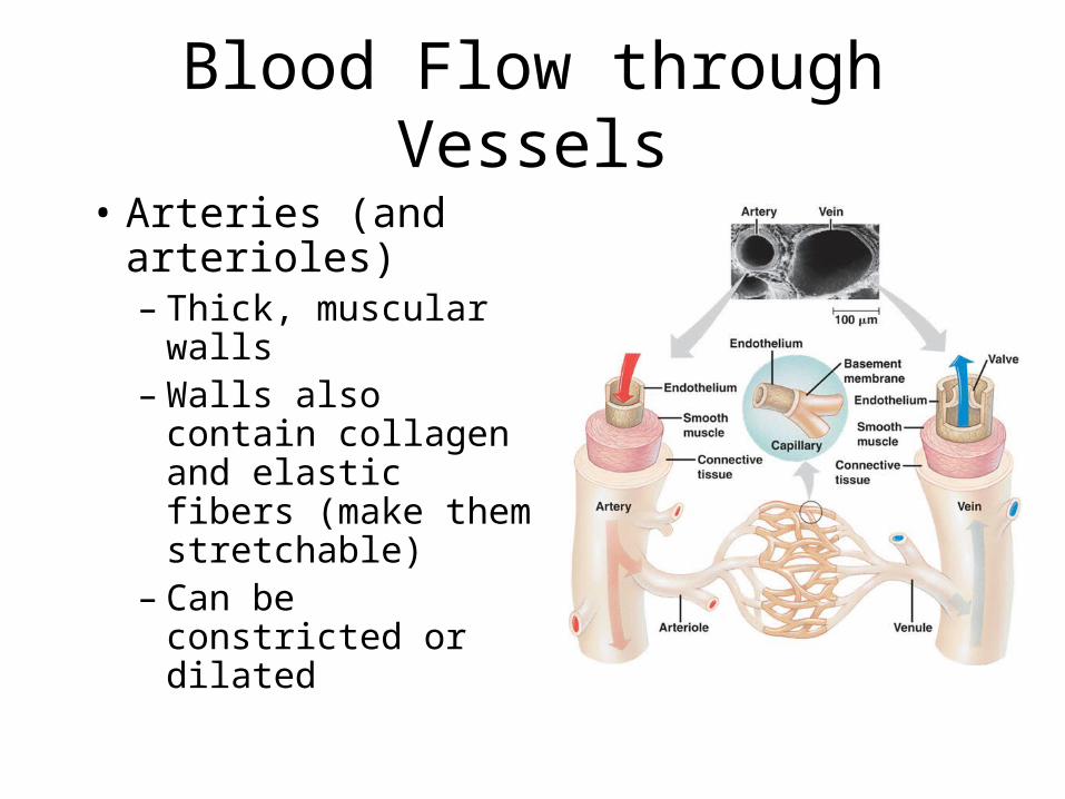
• Arteries (and arterioles) – Thick, muscular
walls– Walls also contain
collagen and elastic fibers (make them stretchable)
– Can be constricted or dilated
Blood Flow through Vessels
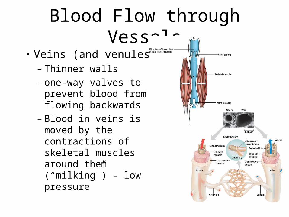
• Veins (and venules)– Thinner walls – one-way valves to
prevent blood from flowing backwards
– Blood in veins is moved by the contractions of skeletal muscles around them (“milking”) – low pressure
Blood Flow through Vessels
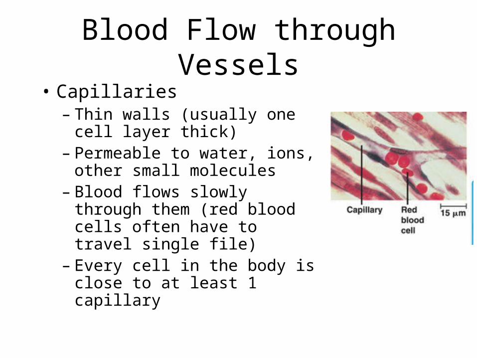
• Capillaries– Thin walls (usually one cell
layer thick)– Permeable to water, ions, other
small molecules – Blood flows slowly through
them (red blood cells often have to travel single file)
– Every cell in the body is close to at least 1 capillary

Capillaries • Capillaries exchange materials
between the blood and the interstitial fluid
• Blood pressure and osmotic pressure drive the movement of molecules into and out of capillaries – Blood pressure forces water and
solutes out (on the artery side)– Osmotic pressure (due to the
proteins left in the capillaries) causes fluid to flow back into the capillaries by osmosis
Question…
• How does the structure of each type of vessel support its function?– Arteries– thick walls can withstand pressure
from heart pumping blood– Veins- valves help prevent backflow since the
heart is too far away to provide forward pressure
– Capillaries- very thin walls allow for easy exchange with the interstitial fluid

Blood Pressure
• Blood pressure = the force being applied to the blood vessel walls (from blood).
• 2 phases of the cardiac cycle– Systole = when the heart
muscle is contracting– Diastole = when the heart
muscle is relaxed (between contractions)

Blood Pressure
• Systolic Pressure = pressure in arteries when heart contracts
• Diastolic Pressure = pressure in arteries between contractions

Question…
• Giraffes need higher blood pressure. Why?
• Since they are taller, they need more pressure to get the blood all the way to the top of their bodies
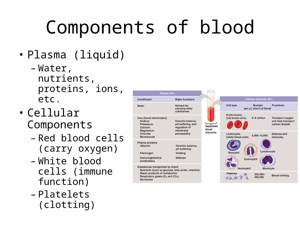
Components of blood
• Plasma (liquid)– Water, nutrients,
proteins, ions, etc.
• Cellular Components– Red blood cells
(carry oxygen)– White blood cells
(immune function)
– Platelets (clotting)

Differentiation of Blood Cells
• Blood cells (RBC, WBC and platelets) all develop from stem cells in the red bone marrow.
• Erythropoietin (EPO) is a hormone that promotes the production of erythrocytes (RBC) – Synthesized in the kidneys
Question…
• When the body is not receiving enough O2,
what will happen to EPO levels? – They increase to create more RBC to carry O2

Control of Circulation
• Heart rate is controlled by – Nerve impulses sent to SA and AV Nodes
• Parasympathetic division- slows heartbeat down
• Sympathetic division – speeds up heart beat – Hormones (adrenaline/epinephrine)– Body temperature – Oxygen requirements due to exercise
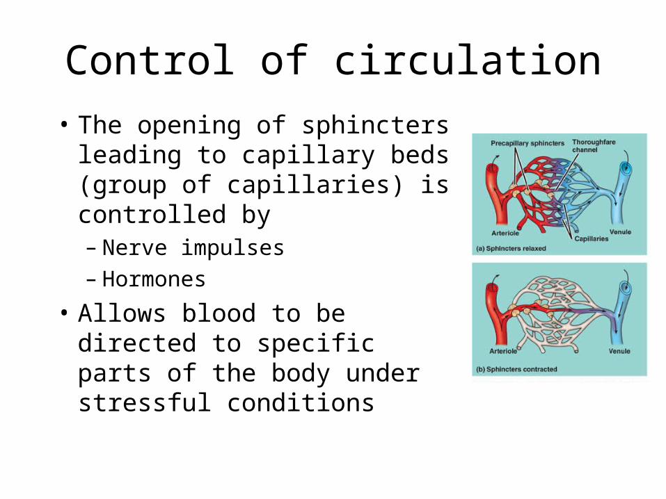
Control of circulation
• The opening of sphincters leading to capillary beds (group of capillaries) is controlled by – Nerve impulses – Hormones
• Allows blood to be directed to specific parts of the body under stressful conditions

Control of Circulation
• The Lymphatic System also plays a role in controlling circulation – Lymph = fluid in lymphatic system (like
interstitial fluid, high in water and other nutrients)
– Fluids flow out and into lymph capillaries via blood pressure and osmotic pressure
– Maintains blood volume so blood pressure can remain constant

Question…
• How is the lymph connected to the digestive system? – Lacteals are lymph vessels in the villi that
absorb some nutrients from the small intestines.

Blood clotting
• Platelets begin the clotting reaction – damage in the blood vessel wall
exposed collagen fibers
– Platelets stick the collagen release substances to make other platelets sticky
• Clotting factors = released by platelets to activate enzymes needed for clotting
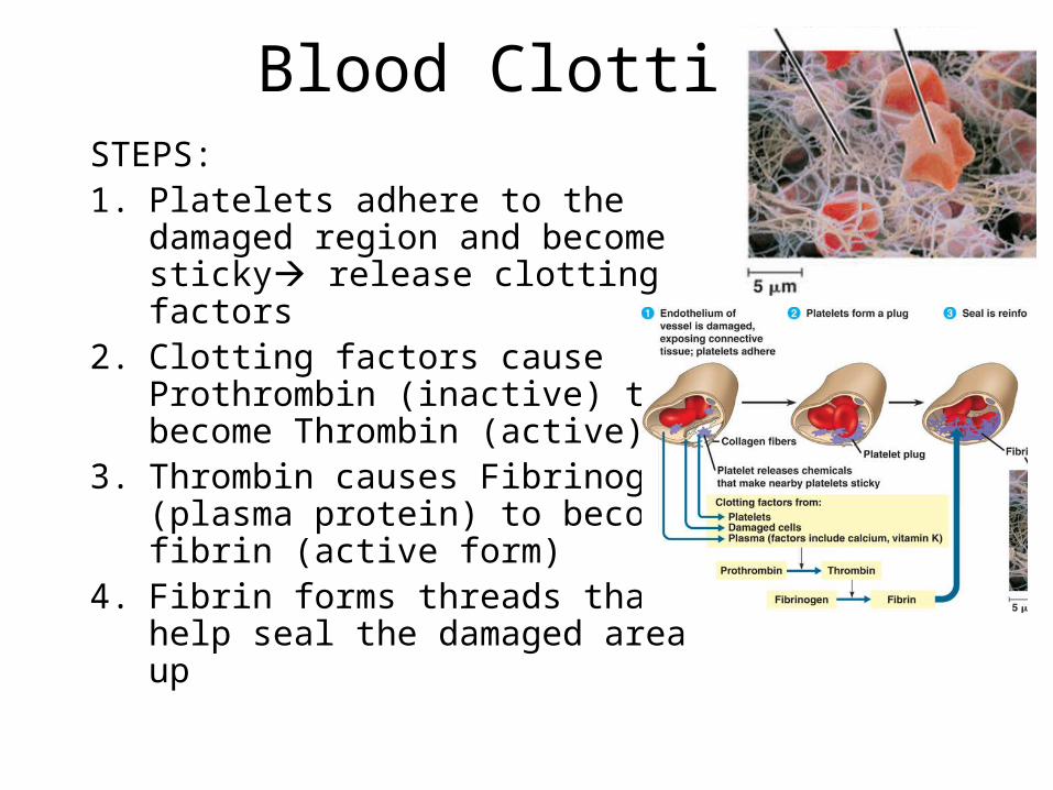
Blood ClottingSTEPS:1. Platelets adhere to the damaged
region and become sticky release clotting factors
2. Clotting factors cause Prothrombin (inactive) to become Thrombin (active)
3. Thrombin causes Fibrinogen (plasma protein) to become fibrin (active form)
4. Fibrin forms threads that help seal the damaged area up
Cardiovascular Disease
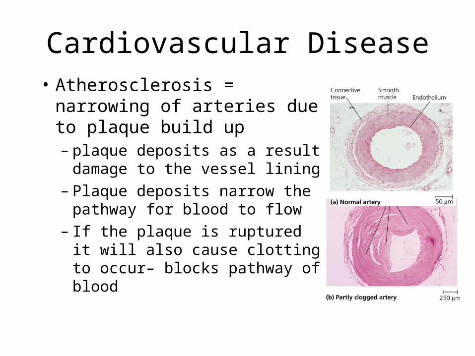
• Atherosclerosis = narrowing of arteries due to plaque build up– plaque deposits as a result
damage to the vessel lining – Plaque deposits narrow the
pathway for blood to flow– If the plaque is ruptured it will
also cause clotting to occur– blocks pathway of blood

Cardiovascular Disease• Atherosclerosis can lead to heart attack or
stroke – Heart attack – blockage in the arteries that supply
the heart with blood– Stroke = blockage in an artery in the brain
• Relationship between high blood pressure and heart disease? – High blood pressure will damage the lining of the
arteries causes plaque to deposit in the damaged areas.
Heart Disease and Cholesterol
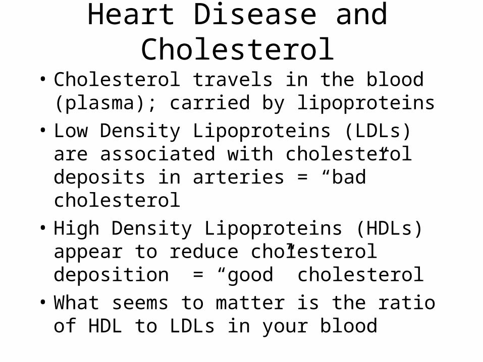
• Cholesterol travels in the blood (plasma); carried by lipoproteins
• Low Density Lipoproteins (LDLs) are associated with cholesterol deposits in arteries = “bad” cholesterol
• High Density Lipoproteins (HDLs) appear to reduce cholesterol deposition = “good” cholesterol
• What seems to matter is the ratio of HDL to LDLs in your blood

Cholesterol and lifestyle choices
• While there is a genetic component to cholesterol levels, lifestyle choices also influence it– Exercise increases HDL levels, lowers LDL
levels – Smoking increases LDL and lowers HDL levels


