Malinosculation-bronchopulmonary sequestration and beyond
40
January-March 2015 Volume 25 | Issue 1 Speaker: Dr.Subrata Nag
-
Upload
pgt-radiology -
Category
Education
-
view
115 -
download
1
Transcript of Malinosculation-bronchopulmonary sequestration and beyond
January-March 2015Volume 25 | Issue 1
Speaker: Dr.Subrata Nag

Congenital bronchopulmonary vascular malformations (BPVMs) include a broad spectrum of disorders that involve abnormalities in the form of disruptions of normal communication and/or presence of abnormal communication between one or more of the three main systems of the lung, namely, the airways, arteries, and veins.
The establishment of abnormal communications by means of small openings or anastomoses is termed as malinosculation.

In 1946, Pryce used the term “sequestration” to describethe abnormal lung that was “disconnected” or “secluded”from the normal bronchial tree and had anomaloussystemic arterial supply
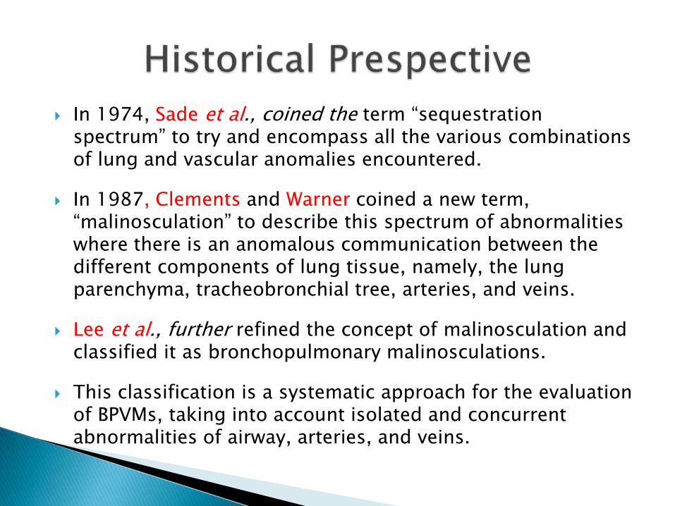
In 1974, Sade et al., coined the term “sequestration spectrum” to try and encompass all the various combinations of lung and vascular anomalies encountered.
In 1987, Clements and Warner coined a new term, “malinosculation” to describe this spectrum of abnormalities where there is an anomalous communication between the different components of lung tissue, namely, the lung parenchyma, tracheobronchial tree, arteries, and veins.
Lee et al., further refined the concept of malinosculation and classified it as bronchopulmonary malinosculations.
This classification is a systematic approach for the evaluation of BPVMs, taking into account isolated and concurrent abnormalities of airway, arteries, and veins.


This group includes isolated airway abnormalities in the proximal and distal tracheobronchial tree with normal arterial and venous systems.

Proximal lesions
• Tracheal stenosis
• Tracheal cysts
• Tracheal diverticulum
• Tracheal bronchus
Distal leisions
• Bronchial branching
• Bronchogenic cyst
• Bronchial atresia
• Cpam
• Supernumerary bronchus
• Displaced bronchus
• Accessory cardiac
bronchus
• Tracheal bronchus

Figure (A and B): Isolated bronchial malinosculation – Trachealbronchus. Axial (A) and coronal (B) chest sections in lung windowshow the origin of tracheal bronchus (white arrow) supplying the apical segment of the right upper lobe above the level of carina
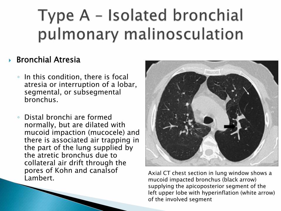
Axial CT chest section in lung window shows a mucoid impacted bronchus (black arrow) supplying the apicoposterior segment of the left upper lobe with hyperinflation (white arrow) of the involved segment
Bronchial Atresia
◦ In this condition, there is focal atresia or interruption of a lobar, segmental, or subsegmentalbronchus.
◦ Distal bronchi are formed normally, but are dilated with mucoid impaction (mucocele) and there is associated air trapping in the part of the lung supplied by the atretic bronchus due to collateral air drift through the pores of Kohn and canalsofLambert.
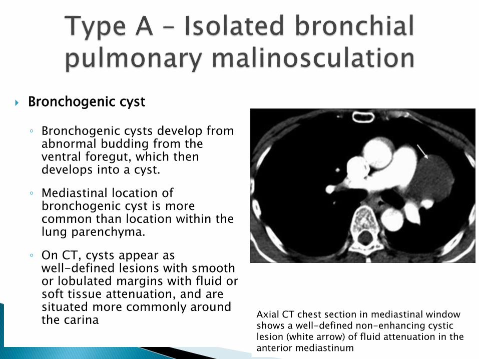
Axial CT chest section in mediastinal window shows a well-defined non-enhancing cystic lesion (white arrow) of fluid attenuation in the anterior mediastinum
Bronchogenic cyst
◦ Bronchogenic cysts develop from abnormal budding from the ventral foregut, which then develops into a cyst.
◦ Mediastinal location of bronchogenic cyst is more common than location within the lung parenchyma.
◦ On CT, cysts appear as well-defined lesions with smooth or lobulated margins with fluid or soft tissue attenuation, and are situated more commonly around the carina

Congenital pulmonary airway malformations (CPAMs)
◦ Represent developmental anomalies of the lower respiratory tract which result from abnormalities in the bronchial branching.
◦ These are hamartomatous lesions with cystic and adenomatoidcomponents arising from the tracheobronchial tree.
◦ Five types are described .
◦ They usually communicate with the bronchi

Type 0 arises from trachea or bronchi and involves whole lungRAREST,FATAL
Type 1 arises from distal bronchi or proximalbronchiolesCOMMONEST,MALIGNANCY ASSOCIATED
Type 2 multiple cysts 0.5-2 cm in diameter with intervening solid-appearing areas
Type 3 Alveolar origin; can have small cysticareas (>0.5 cm) with solid tissue or are mostly solid appearing
Type 4 Acinar origin. Large air-filled or fluid-filledcysts up to 10 cmASSOCIATED WITH PNEUMOTHORAX

Congenital pulmonary airway malformation. Axial (A) and coronal (B) CT chest sections in lung window show a large multiseptatedcystic lesion (arrows) with a single dominant cyst in the left lower lobe with associated mediastinal shift

Axial (A) and coronal (B) CT chest sections in lung window show hyperinflated left upper lobe (arrows) with attenuated lung markings and herniation across the midline
Congenital lobar emphysema◦ anomaly of the lower respiratory
tract, where there is hyperinflation of one or more lobes of the lungs.
◦ Extrinsic obstruction can be caused by vascular anomalies,mediastinal masses, or foregut cysts which can compress the bronchus.
◦ Developmental anomaly in the bronchial wall cartilage leading to collapse of the airway during expiration or intraluminal lesions like meconium plugsor mucosal folds could also cause bronchial obstruction.

This group includes isolated arterial abnormalities with normal airways and normal venous system.
Interrupted pulmonary artery
◦ In this condition, a portion of the proximal part of pulmonary artery is absent with intact distal vascular network within the lungs.
◦ The intrapulmonary arteries receive oxygenated blood from systemic artery collaterals.
◦ Usually, the interruption occurs on the side opposite to the aortic arch.

Congenital interruption of pulmonary artery. Axial CT chest mediastinal (A, B) and lung window (C) sections show absent right pulmonary artery with normal left pulmonary artery (thin white arrow) and prominent bronchial arteries (thick white arrow) at the right hilum supplying the right lung
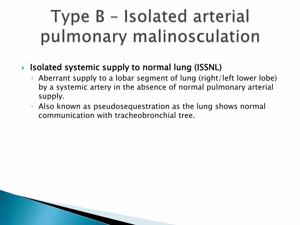
Isolated systemic supply to normal lung (ISSNL)
◦ Aberrant supply to a lobar segment of lung (right/left lower lobe) by a systemic artery in the absence of normal pulmonary arterial supply.
◦ Also known as pseudosequestration as the lung shows normal communication with tracheobronchial tree.
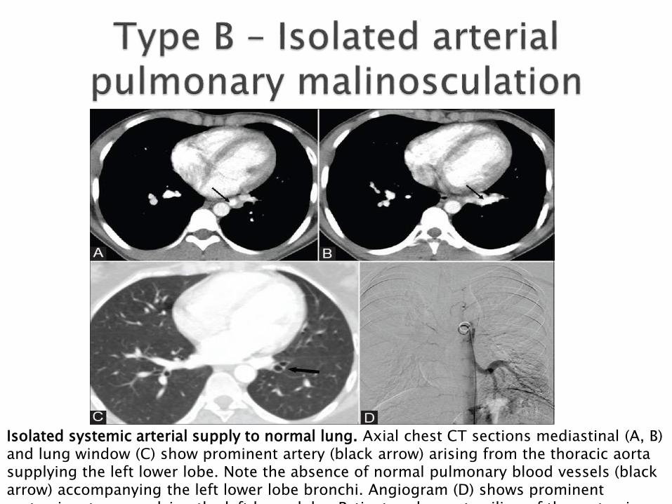
Isolated systemic arterial supply to normal lung. Axial chest CT sections mediastinal (A, B) and lung window (C) show prominent artery (black arrow) arising from the thoracic aorta supplying the left lower lobe. Note the absence of normal pulmonary blood vessels (black arrow) accompanying the left lower lobe bronchi. Angiogram (D) shows prominent systemic artery supplying the left lower lobe. Patient underwent coiling of the systemic
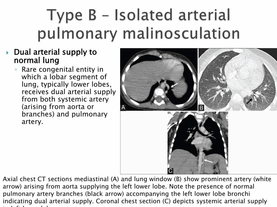
Axial chest CT sections mediastinal (A) and lung window (B) show prominent artery (white arrow) arising from aorta supplying the left lower lobe. Note the presence of normal pulmonary artery branches (black arrow) accompanying the left lower lobe bronchi indicating dual arterial supply. Coronal chest section (C) depicts systemic arterial supply to left lower lobe
Dual arterial supply to normal lung◦ Rare congenital entity in
which a lobar segment of lung, typically lower lobes, receives dual arterial supply from both systemic artery (arising from aorta or branches) and pulmonary artery.

Includes isolated malinosculation of the pulmonary vein with normal bronchial and arterial system
• Partial anomalous pulmonary venous drainage (PAPVC)
• Total anomalous pulmonary venous drainage (TAPVC)
• Meandering pulmonary vein
• AUSPV
• Scimitar variant

Partial anomalous pulmonary venous connection
◦ one of the pulmonary veins (superior or inferior) does not drain into the left atrium.
◦ most commonly occurs on the right with the right superior pulmonary vein draining into the right atrium or the superior vena cava (SVC).
◦ The leftsuperior pulmonary vein drains into the brachiocephalic vein or coronary sinus

Partial anomalous pulmonary venous drainage (PAPVC). Axial CT sections in mediastinal window (A-E) show anomalous drainage of the left superior pulmonary vein (thin white arrow) into the left brachiocephalic vein (thick white arrow)

Total anomalous pulmonary venous connection◦ four pulmonary veins do not drain into the left atrium
◦ classified into four types
Type 1 Supracardiac is the most common type. The pulmonary veins form a single vertical vein which most commonly drains into the left brachiocephalic vein.
Type 2 Cardiac in which the pulmonary veins drain into the coronary sinus or the right atrium
Type 3 Infracardiac type drains into an infra-diaphragmatic vessel, either in the systemic or the portal venous circulation. This type is prone for obstruction, usually at the level of the diaphragm
Type 4 Mixed type in which the pulmonary veins drain into different locations
Meandering pulmonary vein (pseudo-scimitar)◦ anomalously coursing pulmonary vein (typically right or left
inferior pulmonary vein) draining a part of one lung follows a circuitous route through the lung parenchyma before finally opening into the left atrium.
◦ On chest radiograph, this anomalously coursing vein can be mistaken for scimitar vein which opens into IVC.
◦ CT can demonstrate the anomalous coursing vein opening into the left atrium.
Anomalous unilateral single pulmonary vein◦ In this condition, a single pulmonary vein drains an entire lung
and terminates into left atrium.
Scimitar variant ◦ In this condition, an anomalous pulmonary vein terminates into
both IVC and left atrium

Abnormality of the pulmonary artery and the airways.
Intralobar sequestration◦ Pulmonary sequestration is defined as a segment of nonfunctioning lung
that does not communicate with the tracheobronchial tree and is supplied by an anomalous systemic artery.
◦ Intralobar sequestration does not have a separate pleural covering,
◦ separated from normal bronchial tree
◦ has a systemic arterial supply
◦ drains into the pulmonary vein.
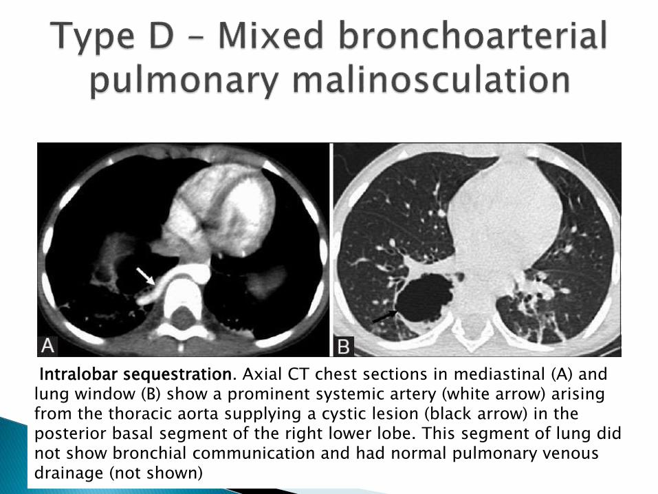
Intralobar sequestration. Axial CT chest sections in mediastinal (A) and lung window (B) show a prominent systemic artery (white arrow) arising from the thoracic aorta supplying a cystic lesion (black arrow) in the posterior basal segment of the right lower lobe. This segment of lung did not show bronchial communication and had normal pulmonary venous drainage (not shown)

Combination of malinosculations of pulmonary airways and pulmonary vein, but with a normal pulmonary arterial supply.
Any of the pulmonary airway malinosculations (Type A) associated with an abnormal pulmonary venous connection (Type C) is included in this type.
◦ Combination of CPAM with PAPVC
◦ Scimitar vein in combination with hypoplasia of lung and no systemic arterial supply
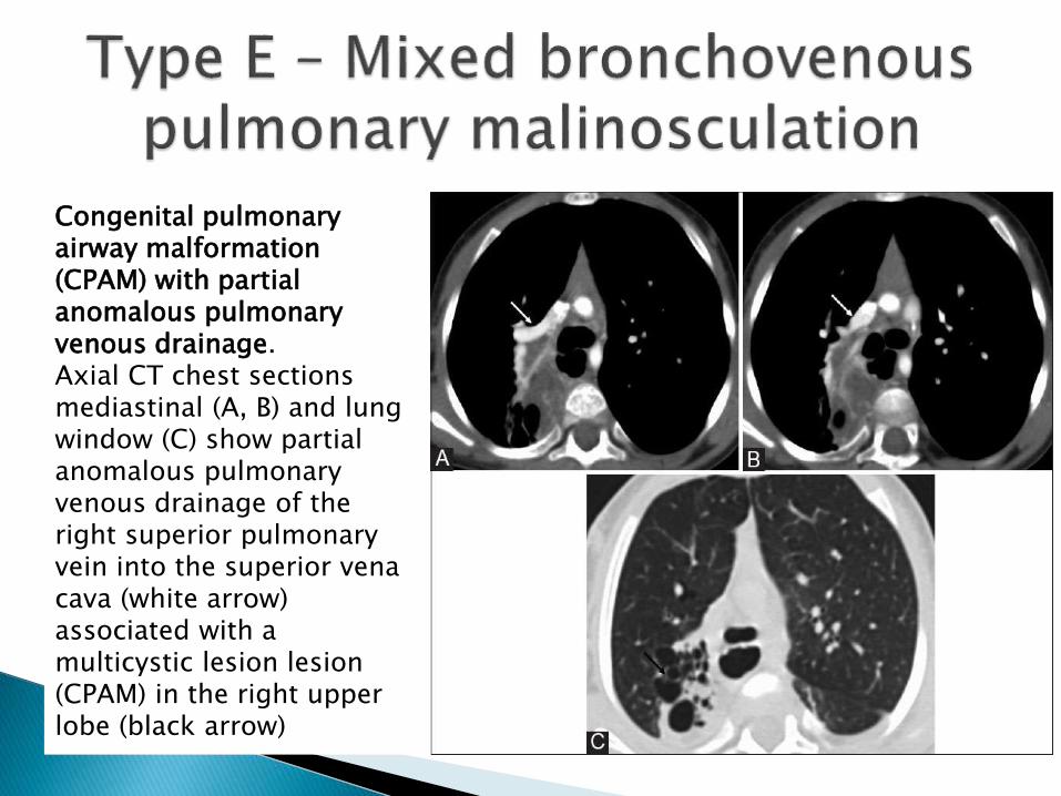
Congenital pulmonary airway malformation (CPAM) with partial anomalous pulmonary venous drainage. Axial CT chest sections mediastinal (A, B) and lung window (C) show partial anomalous pulmonary venous drainage of the right superior pulmonary vein into the superior vena cava (white arrow) associated with a multicystic lesion lesion(CPAM) in the right upper lobe (black arrow)

Pulmonary hypoplasia and scimitar vein. Chest radiograph (A) shows mediastinalshift to the right side with prominent scimitar vein (arrow) in the right lung base. CT axial section (B) shows scimitar vein draining the right lung into the IVC (arrow) with mediastinal shift to the right due to lung hypoplasia. No anomalous systemic artery was seen to diagnose scimitar syndrome

Combination of malinosculations of pulmonary arteries and vein, but with a normal pulmonary airway system.
Arteriovenous malformation (AVM)
◦ Pulmonary AVMs or fistulas show abnormal communication between pulmonary arteries and veins.
◦ They are more often located subpleurally or in lower lobes.
◦ They can be classified as simple AVM when only single feeding artery is seen or complex AVM when multiple feeding arteries are seen.
◦ On chest radiographs, rounded or ovoid opacities can be seen subpleurally with feeding artery and draining vein seen as "rabbit ears."

AVMAxial CT chest mediastinalwindow (A, B) shows a nidus(thin white arrow) of prominent tortuous tangle of vessels in the right upper lobe supplied by a prominent pulmonary artery and drained by prominent pulmonary vein (thick white arrows). 3D volume-rendered image (C) confirms the vascular nature of the lesion with prominent supplying pulmonary artery and vein

Combination of malinosculations of pulmonary airways, arteries, and vein
• Classical scimitar syndrome • Extralobar sequestration
• Pulmonary hypoplasia • Pulmonary aplasia
• Pulmonary agenesis • Combination of type A, B, & C

Meandering pulmonary vein with right lung hypoplasia and systemic arterial supply to lung. Chest CT axial sections (A-E) in mediastinal window show anomalous course of the right inferior pulmonary vein (white arrow) taking a circuitous course through the right lung before draining into the left atrium (meandering pulmonary vein) and a prominent systemic artery (black arrow) arising from the aorta supplying the right lower lobe. 3D volume-rendered image (F) confirms systemic supply to the right lower lobe (black arrow) and meandering right inferior pulmonary vein (white arrow)

Scimitar syndrome◦ Anomalous pulmonary venous drainage of the right lung to the IVC
◦ Systemic arterial supply of the right lower lobe from aorta
◦ Hypoplasia of the right lung with resultant cardiac dextroposition
◦ Right pulmonary artery hypoplasia.

Extralobar sequestration◦ combined bronchoarteriovenous malinosculation
◦ isolation from tracheobronchial tree
◦ systemic arterial supply
◦ drains into systemic veins

Pulmonary
agenesis
• Complete
absence of lung,
bronchial, and
vascular supply
on one side
Pulmonary aplasia
• The presence of
small
rudimentary
bronchus which
ends as a blind
pouch
Pulmonary
hypoplasia
• Reduced lung
volume
• Decrease in the
number of
bronchial
divisions,
number of alveoli
• Reduced size of
pulmonary
vessels

Congenital bronchopulmonary vascular malformations include
◦ a broad spectrum of disorders
◦ abnormal communication or anastomoses between one or more of the three main systems of the lung, namely, airways, arteries, and veins.
Each disease entity can show a spectrum of variations on imaging in its pulmonary/systemic arterial and venous supply.
Classification of BPVM according to the components involved helps provide a systematic approach for evaluation of each disease entity.
Contrast-enhanced CT is very helpful in elaborating the anomalies of different components