Malignant Melanoma - podiatryinstitute.com · Malignant Melanoma Jenna Song, DPM Ingie El-Khashab,...
5
Malignant Melanoma Jenna Song, DPM Ingie El-Khashab, DPM INTRODUCTION Malignant melanoma is an increasingly important public health problem in the US and worldwide. The incidence of melanoma has been increasing faster than that of any other cancer in the US and is now the fifth most common cancer in the US (1). It is estimated that 73,870 men and women were diagnosed with melanoma in 2015, and it is responsible for 1-2% of all cancer deaths and actually 9,940 men and women died with melanoma in 2015 (1). Incidence rates are higher in women than in men before age 50, but the incidence of melanoma increases significantly in men by age 65 or older (1). Although the incidence of, and mortality from, melanoma continues to increase, the rate of increase appears to be slowing. The factors contributing to this slowing are an increased public awareness and education, better prevention, and earlier detection (2). Melanoma is the most common malignancy seen in the foot and the most common locations are the soles of the feet, thumb, or great toe. The term “acral lentiginous melanoma” refers to a melanoma of the hands and feet that account for 3-15% of all melanomas (3). Barnes et al reviewed the 1,018 patients prospectively who were seen for a primary cutaneous melanoma of the lower extremity over a period of 20 years and they found that melanomas of the foot were associated with a poorer prognosis for survival than melanomas of other parts of the lower extremity. They also mentioned that the specific location of the tumor on the foot was a significant factor in survival rate where the plantar and subungual lesions were associated with a poorer rate of survival than lesions of the dorsum (4). Fortin et al retrospectively reviewed patients who had a malignant melanoma of the foot or ankle over a period of 15 years, and reported the shorter survival rate for the patients who had a plantar or subungual lesion. They related that this might be the result of an initial misdiagnosis that noted to be the higher prevalence at the plantar and subungual sites (3). The most common misdiagnoses are benign nevus, subungual or traumatic hematoma, verruca, dermatofibroma, hallux paronychia, and pyogenic granuloma (3-5). RISK FACTORS Melanoma is primarily a disease of Caucasians with at least a 10-fold and a 7-fold increase in the incidence of melanoma compared with African American and American Hispanics, respectively (6). Phenotype plays a significant role in disease prevalence. Typically, fair-skinned individuals with red or blonde hair and blue eyes, or those who burn easily are more likely to develop a melanoma (1, 7). Most melanomas arise de novo and therefore they are not the result of malignant transformation from existing skin lesion but melanoma occasionally can develop within certain precursor lesions, such as melanocytic nevi and dysplastic nevi (8, 9). The total number of nevi and its presence are important independent risk factors for the development of melanoma (10, 11). Studies have demonstrated that dysplastic nevi are reported in up to 34-56% of patients with melanoma (10), but the development of potential precursors to melanoma is inhibited by the regular use of sunscreen (12). The sun exposure/UV radiation is closely linked to the development of melanomas and it is the most significant environmental factor contributing to melanoma (2). Multiple studies have demonstrated a significant correlation between tanning bed use and the risk of developing melanoma. The International Agency for Research on Cancer and the National Institutes of Health recognized the UV exposure from tanning beds as a carcinogenic risk (7, 13). The risk of melanoma increases substantially with each tanning bed use and inversely increases with age (13, 14). Westerdahl et al found a higher risk for developing a melanoma in individuals younger than age 36 years and the greater risk was associated with the use of commercial tanning devices and their use during the winter months (15, 16). This is an interesting observation because the skin of most individuals is known to be less adapted to UV radiation during the winter. In Australia, the country with the highest incidence of melanoma worldwide, legislation was been passed to ban tanning beds by the year 2014 (7). Melanoma can be a familial disease and it has been estimated that up to 12% of cases of cutaneous melanoma are familial (2). A family history of melanoma increases an individual’s risk of melanoma by a factor of 3-8 compared with individuals who have no family history (6). CHAPTER 24
Transcript of Malignant Melanoma - podiatryinstitute.com · Malignant Melanoma Jenna Song, DPM Ingie El-Khashab,...

Malignant Melanoma
Jenna Song, DPMIngie El-Khashab, DPM
INTRODUCTION
Malignant melanoma is an increasingly important public health problem in the US and worldwide. The incidence of melanoma has been increasing faster than that of any other cancer in the US and is now the fi fth most common cancer in the US (1). It is estimated that 73,870 men and women were diagnosed with melanoma in 2015, and it is responsible for 1-2% of all cancer deaths and actually 9,940 men and women died with melanoma in 2015 (1). Incidence rates are higher in women than in men before age 50, but the incidence of melanoma increases signifi cantly in men by age 65 or older (1). Although the incidence of, and mortality from, melanoma continues to increase, the rate of increase appears to be slowing. The factors contributing to this slowing are an increased public awareness and education, better prevention, and earlier detection (2).
Melanoma is the most common malignancy seen in the foot and the most common locations are the soles of the feet, thumb, or great toe. The term “acral lentiginous melanoma” refers to a melanoma of the hands and feet that account for 3-15% of all melanomas (3). Barnes et al reviewed the 1,018 patients prospectively who were seen for a primary cutaneous melanoma of the lower extremity over a period of 20 years and they found that melanomas of the foot were associated with a poorer prognosis for survival than melanomas of other parts of the lower extremity. They also mentioned that the specifi c location of the tumor on the foot was a signifi cant factor in survival rate where the plantar and subungual lesions were associated with a poorer rate of survival than lesions of the dorsum (4). Fortin et al retrospectively reviewed patients who had a malignant melanoma of the foot or ankle over a period of 15 years, and reported the shorter survival rate for the patients who had a plantar or subungual lesion. They related that this might be the result of an initial misdiagnosis that noted to be the higher prevalence at the plantar and subungual sites (3). The most common misdiagnoses are benign nevus, subungual or traumatic hematoma, verruca, dermatofi broma, hallux paronychia, and pyogenic granuloma (3-5).
RISK FACTORS
Melanoma is primarily a disease of Caucasians with at least a 10-fold and a 7-fold increase in the incidence of melanoma compared with African American and American Hispanics, respectively (6). Phenotype plays a signifi cant role in disease prevalence. Typically, fair-skinned individuals with red or blonde hair and blue eyes, or those who burn easily are more likely to develop a melanoma (1, 7).
Most melanomas arise de novo and therefore they are not the result of malignant transformation from existing skin lesion but melanoma occasionally can develop within certain precursor lesions, such as melanocytic nevi and dysplastic nevi (8, 9). The total number of nevi and its presence are important independent risk factors for the development of melanoma (10, 11). Studies have demonstrated that dysplastic nevi are reported in up to 34-56% of patients with melanoma (10), but the development of potential precursors to melanoma is inhibited by the regular use of sunscreen (12).
The sun exposure/UV radiation is closely linked to the developme nt of melanomas and it is the most signifi cant environmental factor contributing to melanoma (2). Multiple studies have demonstrated a signifi cant correlation between tanning bed use and the risk of developing melanoma. The International Agency for Research on Cancer and the National Institutes of Health recognized the UV exposure from tanning beds as a carcinogenic risk (7, 13). The risk of melanoma increases substantially with each tanning bed use and inversely increases with age (13, 14). Westerdahl et al found a higher risk for developing a melanoma in individuals younger than age 36 years and the greater risk was associated with the use of commercial tanning devices and their use during the winter months (15, 16). This is an interesting observation because the skin of most individuals is known to be less adapted to UV radiation during the winter. In Australia, the country with the highest incidence of melanoma worldwide, legislation was been passed to ban tanning beds by the year 2014 (7).
Melanoma can be a familial disease and it has been estimated that up to 12% of cases of cutaneous melanoma are familial (2). A family history of melanoma increases an individual’s risk of melanoma by a factor of 3-8 compared with individuals who have no family history (6).
CHAPTER 24
102
DIAGNOSIS
Melanomas were often recognized only when they were large, ulcerated, and at more advanced stage because the distinction between a benign nevus and an early melanoma can be quite diffi cult (17). But early detection is key to reduce the risk and mortality that can be done by performing a through physical examination. The history should include assessment of the risk factors as described above.
The ABCD acronym (asymmetry, border irregularity, color variegation, diameter >6 mm) for melanoma screening (Figure 1) was devised in 1985 by professors at New York University Dermatology Department to provide the public and primary health care professionals with a useful and memorable mnemonic to aid in the early recognition of malignant melanoma. This was expanded to ABCDE, which includes a criterion for “evolving lesions” including any changes with respect to size, shape, symptoms (e.g., itching, tenderness), surface (e.g., bleeding), or shades of color (17, 18). Evidence has accumulated that the addition of “E” will substantially improve and enhance the ability of physicians and public to recognize melanomas at earlier stages (18).
Biopsy is the most accurate and confi rmative diagnosis. The purpose of the biopsy is not only to confi rm a diagnosis of melanoma but also to determine the maximum depth of penetration (7). There are 3 different types of technique: excisional, incisional, or shave biopsy (19). Excisional is to excise the lesion totally, with a 1 to 2 mm margin of clinically
normal skin with the depth of the biopsy sample extending completely through the dermis and some subcutaneous fat. Incisional is a full-thickness biopsy that does not remove the entire lesion, but merely removes enough tissue to obtain a diagnosis. The simplest form of incisional biopsy is a punch biopsy and this is the preferable biopsy technique because it is simple and easy. The recommendation for the proper technique is to perform through the thickest area of the lesion and obtain multiple biopsy samples for large lesions to avoid a sampling error. This is performed with a disposable instrument; usually measuring 2-8 mm in diameter. A punch at least 4 mm in size should be performed to obtain an adequate tissue for accurate pathologic evaluation (2, 19).
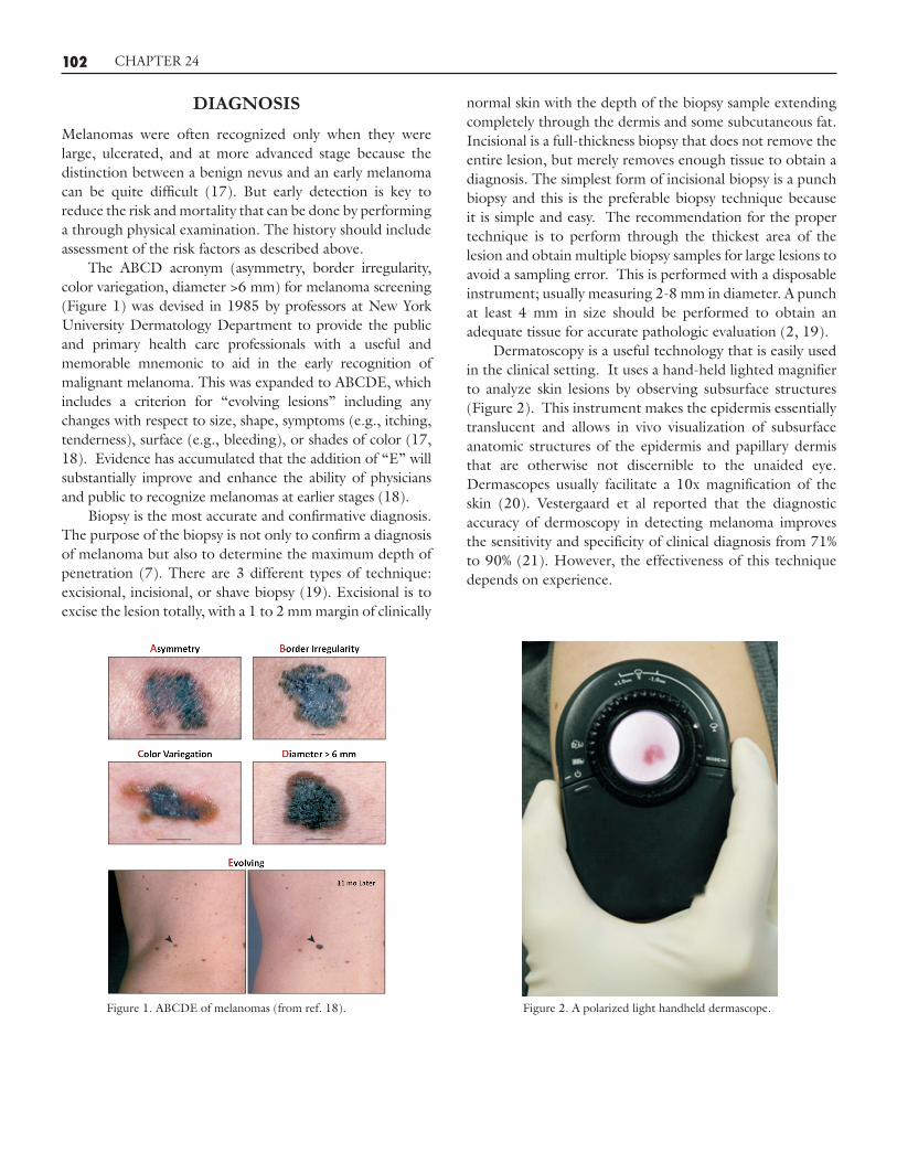
Dermatoscopy is a useful technology that is easily used in the clinical setting. It uses a hand-held lighted magnifi er to analyze skin lesions by observing subsurface structures (Figure 2). This instrument makes the epidermis essentially translucent and allows in vivo visualization of subsurface anatomic structures of the epidermis and papillary dermis that are otherwise not discernible to the unaided eye. Dermascopes usually facilitate a 10x magnifi cation of the skin (20). Vestergaard et al reported that the diagnostic accuracy of dermoscopy in detecting melanoma improves the sensitivity and specifi city of clinical diagnosis from 71% to 90% (21). However, the effectiveness of this technique depends on experience.
CHAPTER 24
Figure 1. ABCDE of melanomas (from ref. 18). Figure 2. A polarized light handheld dermascope.

103 CHAPTER 24
PROGNOSIS
The prognosis of an individual patient can vary widely and depends largely on the stage of the melanoma at the time of diagnosis. In an effort to better predict survival in these patients, a number of variables have been found to infl uence survival. The single most important prognostic factor for overall survival is the status of the regional lymph nodes (7, 22, 23). Jansen et al reviewed 200 patients with a cutaneous melanoma who had undergone a sentinel node biopsy and found that the overall survival at 3 years was 93% if the sentinel node was negative and 67% if it was positive (24). The chance of mortality from melanoma increases substantially if there is metastasis to regional lymph nodes (7).
Another prognostic factor that consistently correlates with survival is Breslow thickness (2). This classifi cation was described by Alexander Breslow in 1970, and recognized the depth of invasion as a vertical thickness of the melanoma in millimeters; the prognosis worsens as the thickness of the melanoma increases. Using Breslow thickness, melanomas are commonly considered to be thin (<1 mm), of intermediate thickness (>1-4 mm), or thick (>4 mm) (4). Diagnostic decisions are also based on the Breslow classifi cation.
Clark level is a known classifi cation system as well for the prognostic variable based on the level of invasion into the anatomic layers of the skin. Clark level 1 represents melanoma in situ, limited to the epidermis with no metastatic potential; level 2 is melanoma that extends into the papillary dermis; level 3 is fi lling the papillary dermis; level 4 is to the reticular dermis; and level 5 is invasion of the subcutaneous fat (2, 7, 24, 25).
Both Breslow thickness and Clark level are important prognostic variables but a recommendation from the American Joint Committee on Cancer (AJCC) is to use Breslow thickness solely and eliminate the Clark level from the melanoma staging protocol to simplify the staging system (25, 26). Breslow thickness actually has became the system of choice because many studies have been reported as showing that Breslow thickness retains its high level of prognostic signifi cance and has been shown to be a more accurate method of predicting prognosis (4, 25, 26). Conversely, there are also many studies that show that both staging systems are high prognostic for melanomas (25).
Ulceration has been recognized as a very robust predictor of prognosis along with other variables (7, 26, 27). There is no clear reason but ulceration appears to be a phenotypic marker for worse tumor biology and greater propensity for invasion and metastasis. Patients with ulcerated melanomas have a worse prognosis than those with nonulcerated melanomas (26). Baade et al reviewed 28,654 patients who were diagnosed with a single invasive melanoma between 1995 and 2008 and found that survival is 7-fold higher with patients without ulceration (28).
Mitotic rate is recognized as an important and independent prognostic factor in the fi nal version of the AJCC Melanoma Staging and Classifi cation recommendations (26). Mitotic rate is the most recently validated prognostic factor and is very important to consider. A threshold of mitotic rate at least 1/mm2 is considered the most signifi cant correlation with survival, and its rate declines as mitotic rate increases (26).
The AJCC has proposed an updated clinical and pathologic staging system in 2009 for cutaneous melanoma based on new data, which have accumulated in recent years (Figure 3). The original AJCC Staging Manual has included all the prognostic factors as discussed above but the newest version of staging system has references from an expanded sample size of the melanoma staging database and newly added mitotic rate.
TREATMENT
The standard treatment for melanoma is a surgical wide excision with margins if detected early. Noting the aggressive nature of melanoma recognized by William Norris in 1875, the recommendation is to remove the melanoma with appropriate margins (7). What constitutes adequate margins, when excising a melanoma, has been a point of contention for years. There are recommendations in the literature regarding the margins of excision for primary melanomas. The recent American Academy of Dermatology guidelines and others like the National Institutes of Health consensus group recommend 5 mm margins for in situ, 1 cm margins for lesions <2 mm thick, and 2 cm margins for lesions *2 mm thick (29, 30). In contrast to these groups, others have suggested that 1 cm margins are adequate for most lesions up to 2 mm in thickness and 2 cm margins for the greater thickness (2). According to many studies, 2 cm margins may be the adequate margin for the large thickness lesion whether they are >2 mm or *2 mm but interestingly retrospective analysis suggests that there is no advantage for margins wider than 2 cm (7). Nonetheless, performing wider margin excision will be appropriate for advanced melanomas when the surgeon decides it is needed due to the high risk of recurrence.
As mentioned previously, the status of the regional lymph node is the most important prognostic factor for survival. Therefore, the sentinel lymph node biopsy and following lymphadenectomy (LND) for a positive biopsy sample are essential. The questions arise as to what are the indications for sentinel lymph node biopsy. The literature has identifi ed that ulceration and mitotic rate should be considered when deciding whether or not to perform sentinel lymph node

104
biopsy. Due to the powerful overall prognostic signifi cance of ulceration, sentinel lymph node biopsy should be considered for all ulcerated melanomas, regardless of thickness (7). Likewise, a mitotic rate greater than 1/mm2 is an indication for sentinel lymph node biopsy as it has been shown to be an important predictor of nodal metastasis (7, 26). Referring a patient to an appropriate surgeon for sentinel lymph node biopsy or LND is necessary after the full evaluation of the patient and its result if the pathology report of the lesion is available. To increase the reliability of the sentinel lymph node biopsy, it has been suggested that the defi nitive removal of the lesion should be done at the same time as the sentinel node biopsy, since extensive surgery and repair might disrupt the lymphatic drainage, making lymphoscintigraphy unreliable (2). Lastly, decision for an appropriate adjuvant therapy including systemic and/or radiation therapy will be determined in addition to the primary treatment (such as surgery) by an oncologist.
CHAPTER 24
Figure 3A. TNM staging categories for cutaneous melanoma (from ref. 26).
Classification Thickness (mm) Ulceration Status/Mitoses
NA a: Without ulceration and
mitosis < 1/mm2 b: With ulceration or
mitosis � 1/mm2 a: Without ulceration b: With ulceration a: Without ulceration b: With ulceration a: Without ulceration b: With ulceration
T Tis
T1 T2
T3
T4
NA � 1.00 1.01 – 2.00 2.01 – 4.00 > 4.00
N No. of Metastatic Nodes Nodal Metastatic Burden
N0 N1 N2 N3
0 1 2 – 3 4+ metastatic nodes, or matted nodes,
or in transit metastases/satellites with metastatic nodes
NA a: Micrometastasis* b: Macrometastasis† a: Micrometastasis* b: Macrometastasis† c: In transit metastases/satellites
without metastatic nodes
M Site Serum LDH M0 M1a M1b M1c
Mo distant metastases Distant skin, subcutaneous, or nodal metastases Lung metastases All other visceral metastases Any distant metastases
NA Normal Normal Normal Elevated
Abbreviations: NA, not applicable; LDH, lactate dehydrogenase. * Micrometastases are diagnosed after sentinel lymph node biopsy. † Macrometastases are defined as clinically detectable nodal metastases
confirmed pathologically.
Figure 3B. Anatomic stage groupings for cutaneous melanoma (from ref. 26).
Clinical Staging* Pathologic Staging† T N M T N M
0 IA IB IIA IIB IIC III IV
Tis N0 M0 T1a N0 M0 T1b N0 M0 T2a N0 M0 T2b N0 M0 T3a N0 M0 T3b N0 M0 T4a N0 M0 T4b N0 M0 Any T N > N0 M0 Any T Any N M1
0 IA IB IIA IIB IIC IIIA IIIB IIIC IV
Tis N0 M0 T1a N0 M0 T1b N0 M0 T2a N0 M0 T2b N0 M0 T3a N0 M0 T3b N0 M0 T4a N0 M0 T4b N0 M0 T1-4a N1a M0 T1-4a N2a M0 T1-4b N1a M0 T1-4b N2a M0 T1-4a N1b M0 T1-4a N2b M0 T1-4a N2c M0 T1-4b N1b M0 T1-4b N2b M0 T1-4b N2c M0 Any T N3 M0 Any T Any N M1
* Clinical staging includes microstaging of the primary melanoma and clinical/ radiographic evaluation for metastases. By convention, it should be used after complete excision of the primary melanoma with clinical assessment for regional and distant metastases.
† Pathologic staging includes microstaging of the primary melanoma and pathologic information about the regional lymph nodes after partial (i.e. sentinel node biopsy) or complete lymphadenectomy. Pathologic stage 0 or stage IA patients are the exception; they do not require pathologic evaluation of their lymph nodes.
CASE REPORT
A 73-year-old female presented to the clinic with a history of a pigmented lesion in the plantar lateral aspect of the left foot of several months duration. On clinical examination, there was a 2.2 x 2 cm pigmented ulcerated lesion under the bottom of the lateral foot with irregular borders in the ulcer margins (Figure 4). A punch biopsy was taken from 2 different sites of the lesion and the histopathologic examination showed a malignant melanoma with a Breslow depth of 4.5 mm. The patient was immediately referred to a general surgeon and oncologist and a surgical wide excision of the tumor (4 cm margins) and sentinel lymph node biopsy were performed. Complete inguinal lymph node dissection was performed after the positive result of the sentinel lymph node biopsy. Fortunately, no metastasis to regional lymph nodes was found. Currently, the patient is receiving adjunctive therapy and is being closely monitored by the oncologist and general surgeon.

105 CHAPTER 24
In conclusion, malignant melanoma is an increasingly important public health problem in the US and worldwide and its incidence continues to rise. A through history and physical examination performed by the clinician and an understanding of the risk factors associated with melanoma are crucial for making an appropriate diagnosis. A multidisciplinary approach focused on early detection and aggressive treatment may help to lower mortality from malignant melanoma.
REFERENCES 1. American Cancer Society. Cancer Facts & Figures 2015. Atlanta:
American Cancer Society; 2015. 2. Lang PG. Current concepts in the management of patients with
melanoma. Am J Clin Derm 2002;3:401-26. 3. Fortin PT, Freiberg AA, Rees R, Sondak VK, Johnson TM.
Malignant melanoma of the foot and ankle. J Bone Joint Surg Am 1995;77;1396-403.
4. Barnes BC, Seigler HF, Saxby TS, Kocher MS, Harrelson JM. Melanoma of the foot. J Bone Joint Surg Am 1994;76:892-98.
5. Pack GT, Oropeza R. Subungual melanoma. Surg Gynec Obstet 1967;124:571-82.
6. Goldstein BG, Goldstein AO. Diagnosis and management of malignant melanoma. Am Family Phys 2001;63:1359-68.
7. Dunki-Jacobs EM, Callender GG, McMasters KM. Current management of melanoma. Curr Prob Surg 2013;50:351-82.
8. Goldstein AM, Tucker MA. Dysplastic nevi and melanoma: cancer epidemiology, biomarkers & prevention.Oncology 2013;22:528-32.
9. Duffy K, Grossman D. The dysplastic nevus: from historical perspective to management in the modern era: part I historical, histologic, and clinical aspects. J Am Acad Dermatol 2012;67:1e-16.
10. Tucker MA. Melanoma epidemiology. Hematol Oncol Clin North Am 2009;23:383-95.
11. Snels DG, Hille ET, Gruis NA, et al. Risk of cutaneous malignant melanoma in patients with nonfamilial atypical nevi from a pigmented lesions clinic. J Am Acad Dermatol 1999;40:686-93.
12. Russak JE, Rigel DS. Risk Factors for the Development of Primary Cutaneous Melanoma. Dermatol Clin 2012;30:363-8.
13. The International Agency for Research on Cancer Working Group on Artifi cial Ultraviolet (UV) Light and Skin Cancer. The association of use of sunbeds with cutaneous malignant melanoma and other skin cancers: a systematic review. Int J Cancer 2007;120:1116-22.
14. Russak JE, Rigel DS. Risk factors for the development of primary cutaneous melanoma. Dermatol Clin 2012;30:363-8.
15. Westerdahl J, Olsson H, Masback A, Ingvar C, Jonsson N, Brandt L, et al. Use of sunbeds or sunlamps and malignant melanoma in southern Sweden. Am J Epidemiol 1994;140:691-9.
16. Westerdahl J, Ingvar C, Masbak A, Jonsson N, Olsson H. Risk of cutaneous malignant melanoma in relation to use of sunbeds: further evidence for UV-A carcinogenicity. Br J Cancer 2000;82:1593-99.
17. Montella A, Gavin A, Middleton R, Autier P, Boniol M. Cutaneous melanoma mortality starting to change: a study of trends in Northern Ireland. Eur J Cancer 2009;45: 2360-6.
18. Abbasi NR, Shaw HM, Rigel DS, Friedman RJ, McCarthy WH, et al. Early diagnosis of cutaneous melanoma: revisiting the ABCD Criteria. JAMA 2004;292:2771-6.
19. Bichakjian CK, Halpern AC, Johnson TM, et al. Guidelines of care for the management of primary cutaneous melanoma. J Am Acad Dermatol 2011;65:1032-47.
20. Rigel DS, Russak J, and Friedman R. The evolution of melanoma diagnosis: 25 Years beyond the ABCDs. Cancer 2010;60:301-16.
21. Vestergaard ME, Macaskill P, Holt PE, Menzies SW. Dermoscopy compared with naked eye examination for the diagnosis of primary melanoma: a meta-analysis of studies performed in a clinical setting. Br J Dermatol 2008;159:669-76.
22. Scolyer RA, Murali R, McCarthy SW, Thompson JF. Pathologic examination of sentinel lymph nodes from melanoma patients. Semin Diag Path 2008;25:100-11.
23. Pawlik TM, Ross MI, Prieto VG, Ballo MT, Johnson MM, et al. Assessment of the role of sentinel lymph node biopsy for primary cutaneous desmoplastic melanoma. Cancer 2006;106:900-6.
24. Jansen LO, Nieweg E, Peterse JL, Hoefnagel CA, Olmos RA, Kroon BB. Reliability of sentinel lymph node biopsy for staging melanoma. Brit J Surg 2000;87:484-9.
25. Marghoob AA, Koenig K, Bittencourt FV, Kopf AW, Bart RS. Breslow thickness and Clark level in melanoma. Cancer 2000;88:589-95.
26. Balch CM, Gershenwald JE, Soong SJ, Thompson JF, Atkins MB, Byrd DR, et al. Final version of 2009 AJCC melanoma staging and classifi cation. J Clin Oncol 2009;27:6199-206.
27. Lyth J, Hansson J, Ingvar C, Mansson-Brahme E, Naredi P, Stierner U, et al. Prognostic subclassifi cations of T1 cutaneous melanomas based on ulceration, tumour thickness and Clark’s level of invasion: results of a population-based study from the Swedish melanoma register. Brit J Derm 2013;168:779-86.
28. Baade PD, Royston P, Youl PH, Weinstock MA, Geller A, Aitken JF. Prognostic survival model for people diagnosed with invasive cutaneous melanoma. BMC Cancer 2015;15:27.
29. NIH Consensus Conference. Diagnosis and treatment of early melanoma. JAMA 1992;268:1314-9. 30. Bichakjian CK, Halpern AC, Johnson TM, Hood AF, Grichnik JM, Swetter SM, et al. Guidelines of care for the management of primary cutaneous melanoma. J Am Acad Derm 2011;65:1032-47.
Figure 4. Clinical appearance of the lesion.