
Malignant Disease of the Breast Part One · Etiological factors, genetics and epidemiology of...
83
Malignant Disease of the Breast – Part One Scotty Rieder Dr. S. Latosinsky September 28 th , 2016
Transcript of Malignant Disease of the Breast Part One · Etiological factors, genetics and epidemiology of...

Malignant Disease of the Breast
– Part One
Scotty Rieder
Dr. S. Latosinsky
September 28th, 2016

Objectives Medical Expert: Etiological factors, genetics and epidemiology of breast cancer Anatomy of breast, blood supply, lymphatic drainage, surrounding neuro-vascular structures. Presentation, histology and management of DCIS Presentation, histology and management of LCIS Histological classification of invasive breast carcinoma Grading and tumor prognosis Role of hormone receptors in breast carcinoma Staging of breast carcinoma Surgical management of breast cancer , SLNB role Collaborator: Imaging and diagnostic techniques of malignant breast disease Health Advocate: Role of screening mammography Manager: Breast cancer screening Genetic screening for breast cancer

Epidemiology • Most commonly diagnosed cancer worldwide
• 280,000 new cases diagnosed in the USA annually, with approximately 40,000 deaths per year
• The American Cancer Society estimates that 1 in 8 women will be diagnosed with breast cancer in their lifetime
• Highly heterogeneous in tumour biology

Risk Factors • Increasing Age
• Gender (100 times greater risk in women)
• Race
• Weight
• Height
• Estrogen
• Breast Density
• Age of menarche
• Nulliparity
• Family history
• Lifestyle (Smoking, fat intake, alcohol all show weak relationships)
• Abortion DOES NOT increase risk
Risk Prediction Models • There are three leading models for risk prediction in breast cancer: BRCAPRO, IBIS and BOADICEA
• IBIS has the best predictability of the models
• IBIS accounts for both genetic and nongenetic factors, and is useable in clinic and family practice settings
• It may overestimate risk in women with atypical hyperplasia
• http://ibis.ikonopedia.com/
Breast Cancer Genetics • Majority of breast cancers are sporadic
• 5-10% of women presenting with breast cancer will have a hereditary form
• Most commonly identified genetic loci are BRCA1/2 mutations
• Breast cancers may also be associated with inherited syndromes (Li-Fraumeni, Cowden)
• Risk assessment, testing and management of patients with these mutations is different than average risk patients


Breast Screening
You are speaking to your mother. She has just received her notice from the Ontario Breast Screening Program recommending that she go for a screening mammogram. She is reluctant to attend. A recent article she read indicates there are concerns with mammographic screening in average risk women and that screening may cause more harm than good. She wants to know your opinion.
What are the breast screening guidelines for average risk women in Ontario? What would you tell her regarding the benefits of mammographic screening?
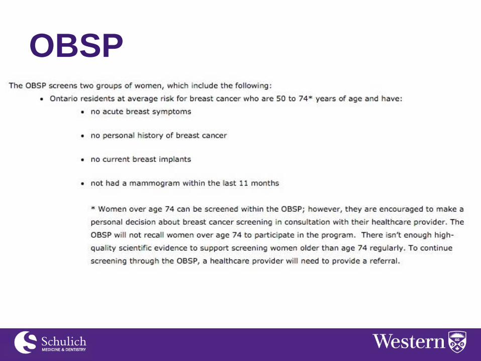
OBSP

Out of any group of 1,000 50-year-old women today, about five are
likely to die of breast cancer in the next 10 years.
If all 1,000 women received mammograms at age 50 and every two
years for the next decade, though, the number of deaths might decline
by only one — to four, the collected research shows.

Mammogram 1cm
CBE 2cm

The Canadian Task Force on Preventive
Health Care CMAJ 2011 For (average-risk) women aged 50–69 years, we
recommend routinely screening with mammography
every two to three years. (Weak recommendation;
moderate-quality evidence)
Weak recommendations result when:
• the balance between desirable and undesirable
effects is small
• the quality of evidence is lower
• there is more variability in the values and preferences
of patients.
Are we over-treating DCIS?
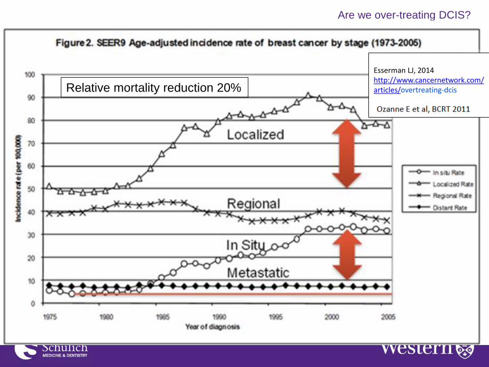
Are we over-treating DCIS?
Esserman LJ, 2014 http://www.cancernetwork.com/ articles/overtreating-dcis
Relative mortality reduction 20%

What do you discuss?
Screening 230 average risk women 50-69 over 20
years would*:
• avert 1 BC death
• ? less treatment
• ~ 100 false positives
• ~ 40 biopsies
• 3-(5) over-diagnoses
•15-20 women will develop breast cancer Independent UK Panel on Breast Cancer Screening, Lancet. 2012;380 Canadian Task Force on Preventive Health Care, CMAJ. 2011;183


Breast Screening
• Breast self-exam -not recommended Preventive health care, 2001 update: Should women be routinely taught breast
self-examination to screen for breast cancer?N Baxter, Canadian Task Force on Preventive Health CareCanadian Medical Association Journal 164 (13), 1837-1846
• Physician routine breast exam -not recommended - Canadian breast screening studies
• 50-60 randomized to PE + mammography vs PE • 40-50 randomized to PE + mammography vs observation
• Screening MRI
– No evidence – Only in Ontario for lifetime risk >25%

Breast MRI
• Identify occult primary in patient with axillary-
positive disease
• Identify residual disease in cases of positive
margins after lumpectomy
• Biopsy-proven Paget’s disease and normal
US and mammography

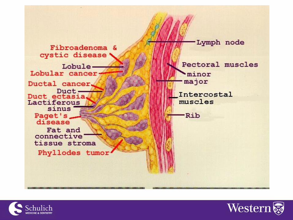
Anatomy • Skin - Margins of the breast
• Muscles
• Nodal Basins
– Axillary
– Supraclavicular
– Internal mammary • Axilla
-Nerves
• Intercostal
• Lateral pectoral nv bundle
• Thoracodorsal nv bundle
• Long thoracic
Local, regional, systemic

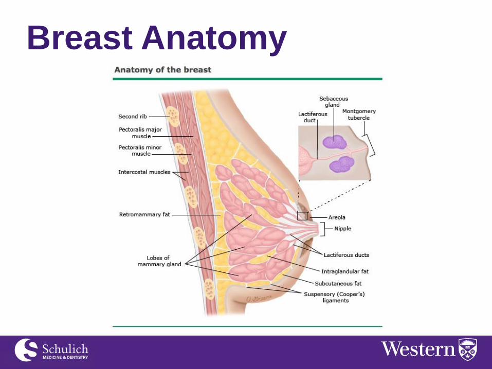
Breast Anatomy

Breast Anatomy

Breast Anatomy


Case
• 42 year old woman, otherwise healthy, woman
is referred to you for a suspicious lesion
(BIRADS 4) in her left breast
• She undergoes stereotactic biopsy and the
pathology is LCIS
• Plan?

LCIS
• Non-invasive lesions that arise from lobules
and terminal breast ducts
• Not identified clinically, radiographically or by
gross pathology
• Serves as an indicator of increased risk of
breast cancer development in either breast
• 80-90% of LCIS diagnosed in pre-menopausal
women

LCIS
• Absolute risk of breast cancer development is
1% per year and double that of average-risk
women
• Chemoprevention with tamoxifen over 5 years
has been shown to reduce cancer risk by half
over 10 years in trials

High risk breast lesions
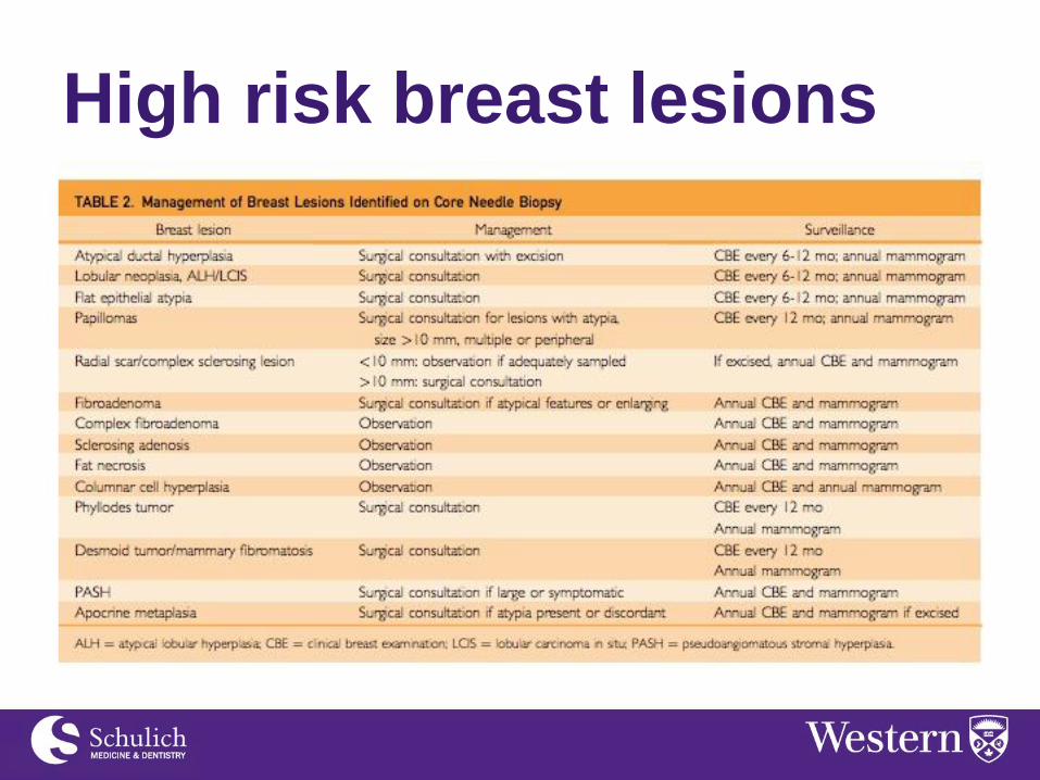
High risk breast lesions

Case • 54 year old women referred to your clinic for a
small cluster of microcalcifications in right
breast picked up screening mammography
• Medical history is HTN, Hypothyroidism
• Her mother was diagnosed with breast cancer
at the age of 68, no other family history
• Plan?

Case • Biopsy reveals ER/PR positive DCIS
• She has been reading about the treatment
options online and is concerned about
needing surgery, and is also asking what are
the chances she will need radiotherapy
• How do you counsel this woman?

DCIS

DCIS and MRI
Pilewski, Cancer 2014;
Are we over-treating DCIS?
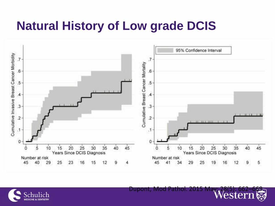
Dupont, Mod Pathol. 2015 May; 28(5): 662–669.
Natural History of Low grade DCIS

DCIS Surgery
• Lumpectomy +/- radiation preferred
• Margins - SSO, ASTRO JCO August 15, 2016 • 0-2mm recurrence 18%, 2mm 9% • 2mm recommended
The “holy grail” of margins
>1cm are achieved in <10%
of patients

DCIS Margins
• ‘gaps’, most commonly seen in
low grade DCIS, whereas high
grade tends to have a continuous growth pattern
Suggestion:
• No tumour at the inked margin is OK
• Minimum 3mm margin if want to try and avoid
radiation based on recent trials (low +
intermediate grade)
Are we over-treating DCIS?
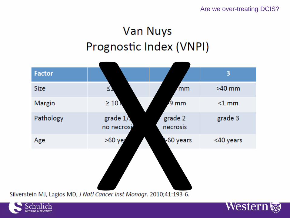
Are we over-treating DCIS?

EBCTCG, JNCI Monograph 2010; 41
Are we over-treating DCIS?
50% invasive for both groups
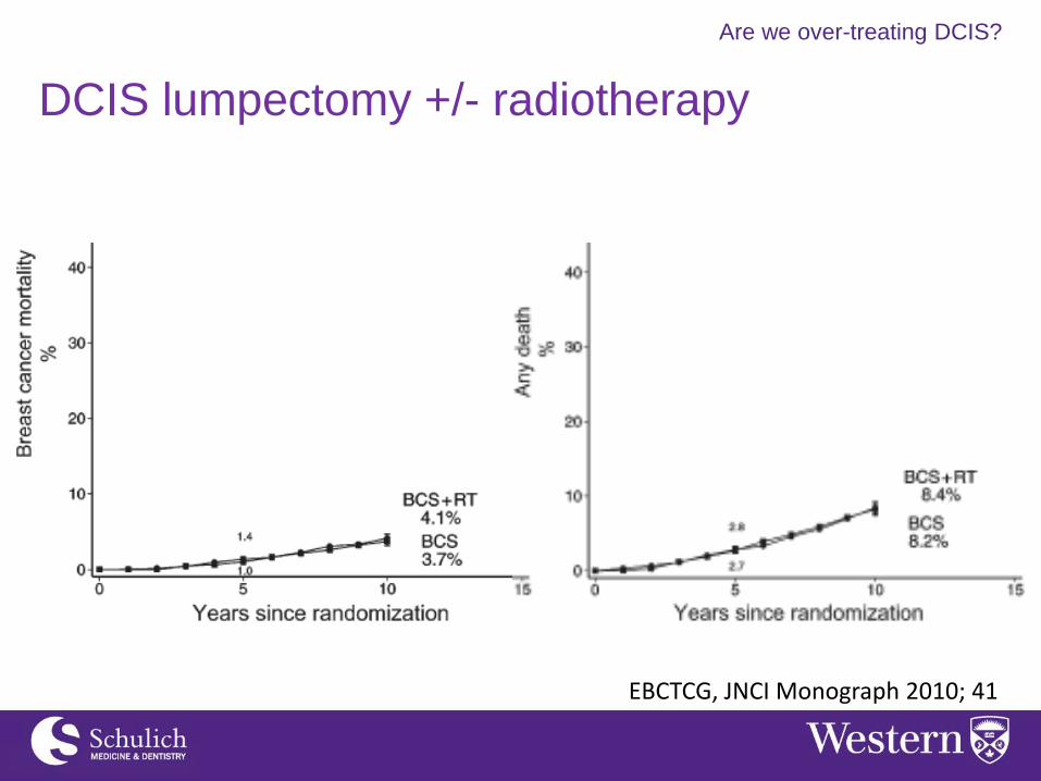
DCIS lumpectomy +/- radiotherapy

EBCTCG, JNCI Monograph 2010; 41
Are we over-treating DCIS?
50% invasive for both groups
DCIS lumpectomy +/- radiotherapy
EBCTCG, JNCI Monograph 2010; 41
Are we over-treating DCIS?
DCIS lumpectomy +/- radiotherapy

EBCTCG, JNCI Monograph 2010; 41
Are we over-treating DCIS?
50% invasive for both groups
DCIS lumpectomy +/- radiotherapy

BCS Alone: RTOG 9804
McCormick, JCO 2015; 33
Are we over-treating DCIS?

DCIS Score
Solin, JNCI 2013;105
Are we over-treating DCIS?

Solin, JNCI 2013;105
Ipsilateral Breast Events Ipsilateral Invasive Breast Events
Recurrence Score
Rakovitch, SABCS 2014
Are we over-treating DCIS?

Solin, JNCI 2013;105
Ipsilateral Breast Events
DCIS Recurrence Score
Are we over-treating DCIS?
Rakovitch, BCRT (2015) 152:389–398

Tamoxifen and DCIS
NNT to Prevent 1 Breast Event
NNT to Prevent 1 Invasive Breast Event
BCS alone 13 28
BCS + Radiation 17 33
Staley, The Breast 2014; 23
Are we over-treating DCIS?
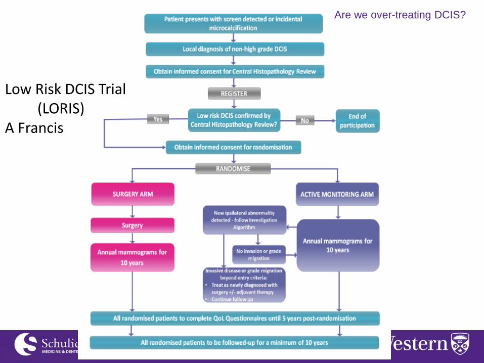
Low Risk DCIS Trial (LORIS) A Francis
Are we over-treating DCIS?

DCIS + SN Biopsy
• recommended for mastectomy patients
• can be considered in BCS patients with
palpable or larger high grade lesions.
SLN in
DCIS with
BCS
Chin-Lenn, Ann Surg Onc
2014; 21 Alberta 61%
Nicholson, EJSO 2015; 41
UK
(Sloane
project)
24%
Are we over-treating DCIS?

Tamoxifen and DCIS
NNT to Prevent 1 Breast Event
NNT to Prevent 1 Invasive Breast Event
BCS alone 13 28
BCS + Radiation 17 33
Staley, The Breast 2014; 23
Are we over-treating DCIS?

Classification of Breast Ca
• Infiltrating ductal – 76%
• Infiltrating lobular – 8%
• Ductal/lobular – 7%
• Mucinous (colloid) – 2.5%
• Tubular/Medullary/Papillary – 1% each

Infiltrating ductal
• Vast majority of breast cancers
• Pathologically graded into 3 categories: well,
moderately and poorly differentiated
• Often associated with DCIS, and the amount
of DCIS present is an important prognostic
factor in patients treated with breast-
conserving surgery
Infiltrating lobular
• Second most common breast cancer
• Rates rising in NA, may be associated with
post-menopausal hormone use
• Appear later and usually are well-differentiated
• Some evidence suggests a better prognosis
for most sub-types compared to ductal

Mucinous (Colloid)
• Rare (1-2% of all breast cancers)
• Soft gelatinous appearance, usually a well-
circumscribed lesion
• Better prognosis than ductal

Medullary
• Account for ~1% of breast cancers
• Often are poorly-differentiated, are common in
younger women, and are associated with
BRCA1
• Despite their aggressive appearance,
prognosis is more favourable compared to
ductal
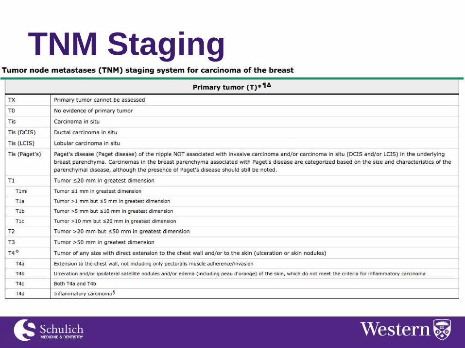
TNM Staging
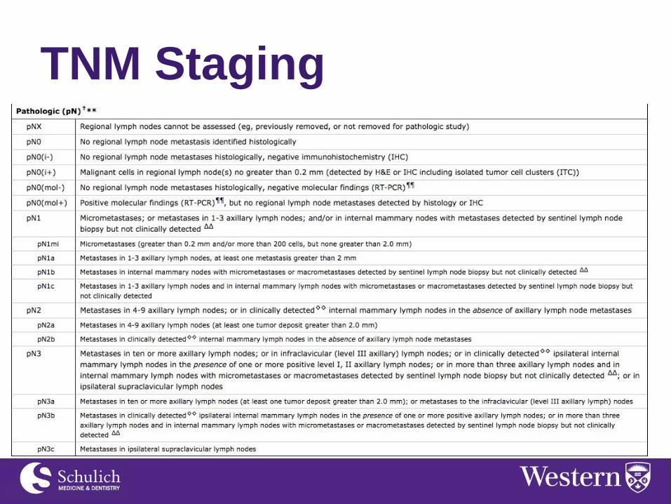
TNM Staging

TNM Staging

Hormone Receptors
• ER/PR receptor positive disease accounts for
~75% of breast cancers
• Adjuvant hormone therapy has demonstrated
benefit in these patients
• Agents used are tamoxifen and aromatase
inhibitors

Tamoxifen
• Selective Estrogen Receptor Modulator
(SERM)
• Agent of choice for premenopausal women
• Important side-effect is risk of venous
thrombosis


Aromatase Inhibitors
• Inhibit peripheral conversion of androgens to
estrogen
• In post-menopausal women, is superior to
tamoxifen for both recurrence and mortality
• Side-effects include AI-associated MSK
syndrome and long term bone density
decrease
• Anastrazole, Letrozole, Exemestane
Tratuzumab (Herceptin)
• Interferes with HER2/neu receptor, stopping
cells that overexpress HER2 from proliferation
• Administered for 12 months with adjuvant
chemotherapy and has overall survival benefit
for patients with HER2 positive disease
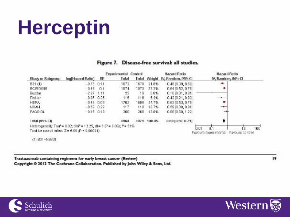
Herceptin

“Surgical” Management
(Duff School Case)
• 50 yo F
• New right breast mass . Routine normal mammogram 9 months before. Core biopsy shows invasive mammary carcinoma. Grade 3 ER 10%, PR -, Her2 +. Node positive
• Mother BC 48 died. Maternal aunt BC 54 died.
• Otherwise well, no operations.
• On exam, moderate-large breasts, nothing on inspection, 4 cm mobile mass RUQ, 3cm palpable node, no supraclavicular lymphadenopathy.
• She wants bilateral mastectomies + reconstruction

Further Investigations
• CT Scan
• Bone scan
• MRI of breast?
– COMICE trial for affected breast Turnbull, Lancet 2010; 375: 563–71
– Contralateral breast? • Genetics referral
– If BRCA negative will affect decision making?

“What tormented Ivan Ilych most was the
deception, the lie, which for some reason they all
accepted, that he was not dying but was simply ill,
and that he only need keep quiet and undergo a
treatment and then something very good would
result.”
Leo Tolstoy, The Death of Ivan Ilych 1886

Prognosis
• 25,000 breast cancer cases/year
• 5,000 breast cancer deaths/year
• 1 out of 5 women die of breast cancer


Surgery - breast
• Mastectomy vs breast conserving therapy
– NSABP B-07 • Contra-lateral breast?
– What is the benefit? • Reconstruction?
– Immediate vs delayed
– CCO guidelines for breast reconstruction

Lumpectomy + Radiation Therapy
Mastectomy +/- Reconstruction
Survival SAME
Local Recurrence @ 10 yrs No RT RT If node positive
40% 10% ESSENTIALLY
<5%, not 0 SAME**
Radiation Yes No (Yes if node positive)**
Margin* Re-excision 20% No
Cosmetic Result 75% Good to excellent Lousy (flat), 70% Good with reconstruction
Systemic Therapy SAME
} NSABP B07*
* Rates are half of historical rates due to use of systemic therapy ** Ragaz, N Engl J Med 1997; 337:956-962, 1997 Overgaarde N Engl J Med 1997; 337:949-955, 1997

Pathologic Extent of
Disease
Holland, R., Veling, S. H., Mravunac, M., & Hendriks, J. H. (1985). Histologic multifocality of Tis, T1-2 breast carcinomas. Implications for clinical trials of breast-conserving surgery. Cancer, 56(5), 979-990.
59% 42%
17% 10%
1 cm
2 cm
3 cm
4 cm

Lumpectomy + Radiation Therapy
Mastectomy +/- Reconstruction
Survival SAME
Local Recurrence @ 10 yrs No RT RT If node positive
40% 10% ESSENTIALLY
<5%, not 0 SAME
Radiation Yes No (Yes if node positive)*
Margin* Re-excision 20% No
Cosmetic Result 75% Good to excellent Lousy, 75% Good with reconstruction
Systemic Therapy SAME
} NSABP b07
Surgery - breast
• Mastectomy vs breast conserving therapy
– NSABP B-07 • Contra-lateral breast?
– What is the benefit? • Reconstruction?
– Immediate vs delayed
– CCO guidelines for breast reconstruction

Neo-adjuvant chemotherapy • Safety – lots of studies, no benefit to chemo first but safe (eg NSABP B-27)
• Clip
• Mastectomy → lumpectomy, better cosmetic result
• Prediction of successful lumpectomy (MD Anderson)
- Lobular not very successful, triple negative CR 25%
• Prognostic value
• Rare progression on chemotherapy
• Buys time
Surgical decision making
-Organizing surgery
• Node negative
-Medical oncology needs to agree chemotherapy candidate
timing of sentinel node biopsy
Surgery - Axilla
• Standard of care in node positive core is AND
– 6 week recovery
– 15% chronic lymphedema
– 10% chronic pain • If nodes clinically negative after systemic
therapy options on study

Radiation
• Will get radiation regardless of breast surgery due to positive nodes
• Radiation to:
– breast or chest wall – regional nodes
• Internal mammary • Supraclavicular • Undissected axilla
• Immediate reconstruction – autologous only, implant contra-indicated.

Systemic Therapy
For this patient:
• AC followed by taxane – 4 cycles of each
(6mos)
• Herceptin (1 year) – starts with or just after
chemo
• Hormonal therapy – 5-10 years after radiation

Nodal Surgery - then until now • Halstead – 1882 first in NA
• NSABP B04 -Fisher, NEJM,347,2002– first published 1981 - nodal treatment (observation vs radiation vs axillary node dissection) did not seem to make a difference (results ignored)
• Late 1990’s Sentinel Node Biopsy – Krag, Giuliani - NSABP B32 completed but taken up as new standard prior to results
• ACOSOG Z11 – Giuliani JAMA. 2011,305(6):569-75 -early stage breast cancer (not stage 3) and <3 positive sentinel nodes do not need AND
• AMAROS – Donkers Lancet 2014 15:12, 1303–1310 -early stage breast cancer and positive sentinel nodes can be given axillary radiation, do not need AND
• MA 20 – Whelan NEJM. 2015;373(4):307-16 - radiation to regional nodes in addition to the breast in patients with node positive disease and BCT.
– Most patients had AND (no axillary radiation) but those with <10 nodes (sentinel node biopsy) received axillary radiation
• ACOSOG Z1071 – Broughey JAMA. 2013;310(14):1455-61. - node positive patients with neoadjuvant chemotherapy, followed by sentinel node biopsy.
– False negative rate >10% so recommended against unless 3 or more nodes identified – OK if get 3 negative sentinel nodes
• Future for node positive patients, neoadjuvant chemotherapy NSABP B51, Alliance

Lymphedema
AMAROS – Donkers Lancet 2014 15:12, 1303–1310

Thanks for your attention!


![Manuallymphaticdrainageforlymphedemafollowingbreast cancertreatment… · 2019. 1. 8. · [Intervention Review] Manual lymphatic drainage for lymphedema following breast cancer treatment](https://static.fdocuments.net/doc/165x107/6122b14d4796fe601d43a8c0/manuallymphaticdrainageforlymphedemafollowingbreast-cancertreatment-2019-1-8.jpg)