malaria
17
BioMed Central Page 1 of 17 (page number not for citation purposes) Malaria Journal Open Access Research Cerebrospinal fluid and serum biomarkers of cerebral malaria mortality in Ghanaian children Henry B Armah 1,2 , Nana O Wilson 1 , Bismark Y Sarfo 3 , Michael D Powell 1 , Vincent C Bond 1 , Winston Anderson 4 , Andrew A Adjei 2 , Richard K Gyasi 2 , Yao Tettey 2 , Edwin K Wiredu 2 , Jon Eric Tongren 5 , Venkatachalam Udhayakumar 5 and Jonathan K Stiles* 1 Address: 1 Morehouse School of Medicine, Department of Microbiology, Biochemistry and Immunology, Atlanta, Georgia, USA, 2 University of Ghana Medical School, Department of Pathology, Accra, Ghana, 3 Noguchi Memorial Institute for Medical Research, Department of Parasitology, Legon, Ghana, 4 Howard University, Department of Biology, Washington DC, USA and 5 Centers for Disease Control and Prevention (CDC), National Center for Zoonotic, Vector-Borne and Enteric Diseases, Division of Parasitic Diseases, Malaria Branch, Atlanta, GA, USA Email: Henry B Armah - [email protected]; Nana O Wilson - [email protected]; Bismark Y Sarfo - [email protected]; Michael D Powell - [email protected]; Vincent C Bond - [email protected]; Winston Anderson - [email protected]; Andrew A Adjei - [email protected]; Richard K Gyasi - [email protected]; Yao Tettey - [email protected]; Edwin K Wiredu - [email protected]; Jon Eric Tongren - [email protected]; Venkatachalam Udhayakumar - [email protected]; Jonathan K Stiles* - [email protected] * Corresponding author Abstract Background: Plasmodium falciparum can cause a diffuse encephalopathy known as cerebral malaria (CM), a major contributor to malaria associated mortality. Despite treatment, mortality due to CM can be as high as 30% while 10% of survivors of the disease may experience short- and long-term neurological complications. The pathogenesis of CM and other forms of severe malaria is multi-factorial and appear to involve cytokine and chemokine homeostasis, inflammation and vascular injury/repair. Identification of prognostic markers that can predict CM severity will enable development of better intervention. Methods: Postmortem serum and cerebrospinal fluid (CSF) samples were obtained within 2–4 hours of death in Ghanaian children dying of CM, severe malarial anemia (SMA), and non-malarial (NM) causes. Serum and CSF levels of 36 different biomarkers (IL-1β, IL-1ra, IL-2, IL-4, IL-5, IL-6, IL-7, IL-8, IL-9, IL-10, IL-12 (p70), IL-13, IL-15, IL-17, Eotaxin, FGF basic protein, CRP, G-CSF, GM-CSF, IFN-γ, TNF-α, IP-10, MCP-1 (MCAF), MIP-1α, MIP-1β, RANTES, SDF-1α, CXCL11 (I-TAC), Fas-ligand [Fas-L], soluble Fas [sFas], sTNF-R1 (p55), sTNF-R2 (p75), MMP-9, TGF-β1, PDGF bb and VEGF) were measured and the results compared between the 3 groups. Results: After Bonferroni adjustment for other biomarkers, IP-10 was the only serum biomarker independently associated with CM mortality when compared to SMA and NM deaths. Eight CSF biomarkers (IL-1ra, IL-8, IP-10, PDGFbb, MIP-1β, Fas-L, sTNF- R1, and sTNF-R2) were significantly elevated in CM mortality group when compared to SMA and NM deaths. Additionally, CSF IP-10/PDGFbb median ratio was statistically significantly higher in the CM group compared to SMA and NM groups. Conclusion: The parasite-induced local cerebral dysregulation in the production of IP-10, 1L-8, MIP-1β, PDGFbb, IL-1ra, Fas- L, sTNF-R1, and sTNF-R2 may be involved in CM neuropathology, and their immunoassay may have potential utility in predicting mortality in CM. Published: 12 November 2007 Malaria Journal 2007, 6:147 doi:10.1186/1475-2875-6-147 Received: 6 June 2007 Accepted: 12 November 2007 This article is available from: http://www.malariajournal.com/content/6/1/147 © 2007 Armah et al; licensee BioMed Central Ltd. This is an Open Access article distributed under the terms of the Creative Commons Attribution License (http://creativecommons.org/licenses/by/2.0 ), which permits unrestricted use, distribution, and reproduction in any medium, provided the original work is properly cited.
description
kedokteran
Transcript of malaria

BioMed CentralMalaria Journal
ss
Open AcceResearchCerebrospinal fluid and serum biomarkers of cerebral malaria mortality in Ghanaian childrenHenry B Armah1,2, Nana O Wilson1, Bismark Y Sarfo3, Michael D Powell1, Vincent C Bond1, Winston Anderson4, Andrew A Adjei2, Richard K Gyasi2, Yao Tettey2, Edwin K Wiredu2, Jon Eric Tongren5, Venkatachalam Udhayakumar5 and Jonathan K Stiles*1Address: 1Morehouse School of Medicine, Department of Microbiology, Biochemistry and Immunology, Atlanta, Georgia, USA, 2University of Ghana Medical School, Department of Pathology, Accra, Ghana, 3Noguchi Memorial Institute for Medical Research, Department of Parasitology, Legon, Ghana, 4Howard University, Department of Biology, Washington DC, USA and 5Centers for Disease Control and Prevention (CDC), National Center for Zoonotic, Vector-Borne and Enteric Diseases, Division of Parasitic Diseases, Malaria Branch, Atlanta, GA, USA
Email: Henry B Armah - [email protected]; Nana O Wilson - [email protected]; Bismark Y Sarfo - [email protected]; Michael D Powell - [email protected]; Vincent C Bond - [email protected]; Winston Anderson - [email protected]; Andrew A Adjei - [email protected]; Richard K Gyasi - [email protected]; Yao Tettey - [email protected]; Edwin K Wiredu - [email protected]; Jon Eric Tongren - [email protected]; Venkatachalam Udhayakumar - [email protected]; Jonathan K Stiles* - [email protected]
* Corresponding author
AbstractBackground: Plasmodium falciparum can cause a diffuse encephalopathy known as cerebral malaria (CM), a major contributorto malaria associated mortality. Despite treatment, mortality due to CM can be as high as 30% while 10% of survivors of thedisease may experience short- and long-term neurological complications. The pathogenesis of CM and other forms of severemalaria is multi-factorial and appear to involve cytokine and chemokine homeostasis, inflammation and vascular injury/repair.Identification of prognostic markers that can predict CM severity will enable development of better intervention.
Methods: Postmortem serum and cerebrospinal fluid (CSF) samples were obtained within 2–4 hours of death in Ghanaianchildren dying of CM, severe malarial anemia (SMA), and non-malarial (NM) causes. Serum and CSF levels of 36 differentbiomarkers (IL-1β, IL-1ra, IL-2, IL-4, IL-5, IL-6, IL-7, IL-8, IL-9, IL-10, IL-12 (p70), IL-13, IL-15, IL-17, Eotaxin, FGF basic protein,CRP, G-CSF, GM-CSF, IFN-γ, TNF-α, IP-10, MCP-1 (MCAF), MIP-1α, MIP-1β, RANTES, SDF-1α, CXCL11 (I-TAC), Fas-ligand[Fas-L], soluble Fas [sFas], sTNF-R1 (p55), sTNF-R2 (p75), MMP-9, TGF-β1, PDGF bb and VEGF) were measured and the resultscompared between the 3 groups.
Results: After Bonferroni adjustment for other biomarkers, IP-10 was the only serum biomarker independently associated withCM mortality when compared to SMA and NM deaths. Eight CSF biomarkers (IL-1ra, IL-8, IP-10, PDGFbb, MIP-1β, Fas-L, sTNF-R1, and sTNF-R2) were significantly elevated in CM mortality group when compared to SMA and NM deaths. Additionally, CSFIP-10/PDGFbb median ratio was statistically significantly higher in the CM group compared to SMA and NM groups.
Conclusion: The parasite-induced local cerebral dysregulation in the production of IP-10, 1L-8, MIP-1β, PDGFbb, IL-1ra, Fas-L, sTNF-R1, and sTNF-R2 may be involved in CM neuropathology, and their immunoassay may have potential utility in predictingmortality in CM.
Published: 12 November 2007
Malaria Journal 2007, 6:147 doi:10.1186/1475-2875-6-147
Received: 6 June 2007Accepted: 12 November 2007
This article is available from: http://www.malariajournal.com/content/6/1/147
© 2007 Armah et al; licensee BioMed Central Ltd. This is an Open Access article distributed under the terms of the Creative Commons Attribution License (http://creativecommons.org/licenses/by/2.0), which permits unrestricted use, distribution, and reproduction in any medium, provided the original work is properly cited.
Page 1 of 17(page number not for citation purposes)

Malaria Journal 2007, 6:147 http://www.malariajournal.com/content/6/1/147
BackgroundMalaria is an important neglected disease and one of themost important global health problems, potentiallyaffecting more than one third of the world's population.Cerebral malaria (CM) is a deadly complication of Plasmo-dium falciparum infection, associated with a 10–14% mor-tality rate and approximately 1–2 million annual deathsamong young children predominantly in sub-SaharanAfrica and Southeast Asia, yet its pathogenesis remainsincompletely understood. In Ghana, malaria has a widespectrum of presentations: from asymptomatic carriers tomild malaria to multifactorial severe disease, includingCM and severe malarial anemia (SMA) [1-5].
CM, a clinically complex syndrome of coma and poten-tially reversible encephalopathy, is associated withincreased levels of proinflammatory cytokines like tumornecrosis factor (TNF)-α, interferon (IFN)-γ and lympho-toxin [6,7], and increasingly recognized long-term seque-lae in survivors [7-15]. Although the physiopathology ofCM has been extensively investigated, the exact cellularand molecular basis of the neuropathology is still unclear.Recent studies have shown that mechanical blockagecaused by sequestration of parasitized red blood cells(pRBCs), leukocytes and platelets [3,4,8-12], secretion ofcytokines and chemokines [2,6,7,13], angiogenic failure[14,15], immune status and the genetic background of thehost, and parasite factors [7,16] are involved in the patho-genesis of CM. However, it is generally accepted that twomajor factors are involved: (i) metabolic insufficienciesdue to the sequestration of pRBCs, leukocytes and plate-lets within brain vessels via upregulated adhesion mole-cules [3,4,8-12], and (ii) immunological reactions withthe local involvement of T cells and monocytes activatedby Plasmodium antigens [7,11]. These two major mecha-nisms appear to act together under the control ofcytokines [7], and chemokines [2] to exacerbate CM.
Postmortem data in humans and murine models of CMshow that neuronal damage in brain tissue occurs in CM,although the parasites remain confined to the intravascu-lar space (with no contact with neurons). This stronglysuggests that the blood-brain barrier (BBB) is perturbed,and thus the BBB represents a key interface between theintraerythrocytic stages of the parasite and the humanhost. The functional and morphological evidence sup-ports mild-to-moderate impairment of the BBB, butwhether this is sufficient to cause neurological complica-tions such as CM is inconclusive [16,17]. Mechanicalblockage could occur from the ability of pRBC's to adhereto unparasitized erythrocytes and endothelial cells, andsequester in the deep cerebral microvasculature [3,4,8-12]. Parasite sequestration alone, however, cannotaccount for fatal CM pathogenesis as there is evidence thatsurvivors of CM have the same degree of sequestration
during comas as those succumbing to disease. It is evidentthat host immune factors play an important role in thepathogenesis of CM. [8,16]. Although parasite sequestra-tion during severe malaria is central to pathogenesis ofsevere malaria, the role of cytokines, chemokines, apop-totic, and angiogenic factors in exacerbating disease sever-ity remains unclear.
The balance between specific cytokines and chemokinesproduced in response to infection with Plasmodium falci-parum is thought to play an important role in CM andother forms of severe malaria. Severe malaria has beenassociated with high TNF-α plasma levels in conjunctionwith increased production of IFN-γ and IL-1β [18,19] anddecreased production of anti-inflammatory cytokines,notably IL-10 and TGF-β [1,19-22]. Pro-inflammatoryTh1-type cytokines (eg. TNF-α, IFN-γ, interleukin IL-1β,and IL-6) are thought to be critical to the control of exo-erythrocytic and erythrocytic Plasmodium falciparum infec-tion [23,24], but their exaggerated production may alsocontribute to organ damage, particularly in the brain. It iswidely accepted that anti-inflammatory Th2-typecytokines down-regulate Th1-derived cytokines. Th2-typecytokines, such as IL-10, has been shown to regulate Th1-cytokines and prevent CM in some animal models [18].The regulation of TNF-α levels by IL-10 appears to con-tribute to the prevention of severe malarial anemia inhumans [1,20,21]. However, the role that IL-10 plays maydepend on its levels, since very high levels of IL-10 havebeen associated with severe malaria in humans [19] andsome animal models [25]. Therefore, cytokines appear tomaintain a delicate balance between the control of infec-tion and contribution to disease in falciparum malariainfection. However, the expression of Th1 and Th2cytokines in CSF, either from the peripheral circulationvia the BBB or from neuronal immune cells (glia) has notbeen adequately addressed.
Chemokines, or chemoattractant cytokines, and their cor-responding receptors have also been shown to mediatemobilization and coordination of immune responses tomalaria. Chemokines have lympho-chemotactic activityand modulate many infectious and inflammatory dis-eases, including malaria. The recent demonstration of leu-kocyte sequestration, in addition to pRBC sequestration[3,4,8-12], within brain vessels in human CM suggests amore important role for leukocytes, including eosi-nophils, in CM immunopathology than previouslythought. Thus, chemokines, including eotaxin, may playan important role in human CM by attracting leukocytesto sequestration sites. Chemokines are less well studied insevere malaria, but recent studies have associated severemalaria infection with increased production of chemok-ines of the C-C or β subfamily, including regulated uponactivation, normal T cell expressed and secreted
Page 2 of 17(page number not for citation purposes)

Malaria Journal 2007, 6:147 http://www.malariajournal.com/content/6/1/147
(RANTES), monocyte chemotactic protein (MCP)-1, mac-rophage inflammatory protein (MIP)-1α, MIP-1β, and IL-8 [2,13,26-28]. MCP-1, MIP-1α and MIP-1β are potentchemoattractants for monocytes to produce TNF-α and IL-6. IL-8 preferentially recruits neutrophils and plays animportant role in inflammatory diseases. Recently, lowlevels of RANTES have been associated with severemalaria [26,27,29], and specifically associated with mor-tality in children with CM [26]. The low levels of RANTESin severe malaria have been associated with malaria-induced thrombocytopenia [26,29], given that plateletsare a major reservoir of RANTES in the peripheral circula-tion. In contrast, increased mRNA and protein expressionof RANTES and CCR5 was found in localized brainregions of children dying of CM [2]. Interferon inducibleprotein 10 (IP-10) is a member of the CXC or α subfamilyof chemokines, and is induced in response to IFN-γattracting activated Th1 cells [30]. IP-10 levels have beenshown to increase in cultured intervillous blood mononu-clear cells isolated from placenta's infected with malaria[31,32], although no studies have characterized IP-10 lev-els in human cerebral malaria. Hanum and colleaguesrecently demonstrated the induction of IP-10 expressionin the brain of both CM-susceptible (C57BL/6) and CM-resistant (BALB/c) mice as early as 24 hours post-infectionwith Plasmodium berghei ANKA, and in KT-5 astrocyte cellline in vitro upon stimulation with a crude antigen ofmalaria parasites [33]. Currently, the role of chemokinesin clinical severity and outcome of malaria, especially thedevelopment of CM and SMA in children remains poorlydefined.
Angiogenic factors, long implicated as prognostic factorsin cerebral ischemia or stroke [34], have been suggested toplay a role in the petechial hemorrhages and BBB dysfunc-tion associated with CM pathology [14,15]. Vascularendothelial growth factor (VEGF) stimulates endothelialcell growth and migration as well as enhancing vascularpermeability. VEGF levels (more VEGF+ astrocytes) werehigher in CM patients as compared with controls in apost-mortem immunohistology study of CM patients[15]. Platelets, that accumulate with pRBCs in the brainmiscrovasculature in CM patients [10,12], are also impli-cated in CM pathology through TGF-β induced apoptosisin TNF activated human brain endothelial cells [35,36].Platelet derived growth factor (PDGF) is another ang-iogenic factor that stimulates vascular growth, and hasbeen implicated as a neuroprotective factor inducingregeneration of damaged axons and neuronal growth afterischemia [37]. These angiogenic factors play a dominantrole in the recovery from stroke and may be applicable inCM due to their effect on the endothelium, which is cen-tral to CM pathology. These angiogenic factors most prob-ably impact the regenerative potential of the parasite-
induced BBB damage, rather than impacting neoangio-genesis, since CM is an acute neurological syndrome.
Parasite-induced apoptosis in the host may also mediatethe severity of malaria. High levels of Fas-Ligand in sera ofhuman [38,39], monkey [40], and mice [41] are associ-ated with severity of malaria. Lymphocytes and macro-phages express increased levels of Fas and Fas-Ligandduring an acute Plasmodium chabaudi infection [40].TNFR2-deficient mice are resistant to experimental cere-bral malaria (ECM), Fas-deficient mice showed 50%reduction in ECM incidence, and TNFR1-deficient miceshowed the least reduction in ECM incidence [42-46]. Lpr& Gld mice, deficient in Fas & Fas-Ligand, are protectedfrom fatal ECM [47]. The presence of these apoptotic fac-tors in CSF and serum, and their relevance in CM and CM-associated mortality has not been fully investigated. Therapid reversibility of the clinical symptoms of CM suggeststhat tissue necrosis is unlikely to occur [9,16,48], makingapoptosis a more likely pathogenic mechanism.
Recently, new strategies including magnetic resonanceimaging and ophthalmological evaluation of childrenwith CM have been proposed as clinically useful predic-tors of CM severity, but their reliability is being evaluated.The study hypothesis was that parasite-induced dysregula-tion in the levels of inflammatory, apoptotic and ang-iogenic factors at the time of CM death would predictmortality risk of CM. The goal of this study was to identifyfactors that are tightly associated with CM mortality inGhanaian children for further development as biomarkersof CM disease. The present study employed a highthroughput multiplexed immunoassay to evaluate thepredictive value of serum and CSF levels of key immu-nomodulators (inflammatory, apoptotic and angiogenicproteins) in determining mortality risk in severe malariain Ghanaian children. We investigated the serum and CSFprofiles of 36 different biomarkers (IL-1β, IL-1ra, IL-2, IL-4, IL-5, IL-6, IL-7, IL-8, IL-9, IL-10, IL-12 (p70), IL-13, IL-15, IL-17, Eotaxin, FGF basic protein, CRP, G-CSF, GM-CSF, IFN-γ, TNF-α, IP-10, MCP-1 (MCAF), MIP-1α, MIP-1β, RANTES, SDF-1α, CXCL11 (I-TAC), Fas-ligand [Fas-L],soluble Fas [sFas], sTNF-R1 (p55), sTNF-R2 (p75), MMP-9, TGF-β1, PDGF bb and VEGF) in order to identify theimmune factors which influence progression to fatal out-comes associated with CM.
MethodsCase SelectionThe post-mortem serum and CSF samples investigatedwere collected from children who died during the peakmalaria season of 2005 (i.e. June-August), after beingadmitted to the Emergency Unit at the Department ofChild Health, KorIe-Bu Teaching Hospital, Accra, Ghana.Only children for who detailed clinical and laboratory
Page 3 of 17(page number not for citation purposes)
Malaria Journal 2007, 6:147 http://www.malariajournal.com/content/6/1/147
records and a clinically certified cause of death were avail-able were included in the study. Samples were only col-lected only after written informed consent from parents orguardians of the deceased child. Nineteen (19) deceasedchildren meeting the above inclusion criteria had serumand CSF samples removed at autopsy within 2–4 hours ofdeath. Cadavers were moved immediately after consenthad been given, from the Emergency Unit to the hospital'smortuary, for storage at 4°C. A full autopsy, with removalof serum and cerebrospinal fluid samples, was conductedon each consented case. At autopsy, blood samples of 2–5 mL were obtained by left ventricular aspiration, and CSFsamples of 1–2 mL were obtained by lumbar or cisternpuncture. Blood samples for serum testing were collectedin a BD Vacutainer® CPT Cell Preparation Tube [BD Diag-nostics, New Jersey, USA], gently inverted 8–10 times, andthen centrifuged for 20 minutes at 1500 rcf. These specialtubes with polyester gel and density gradient liquid sepa-rated the blood into three distinct layers, namely serum,blood mononuclear cell and red blood cell layers. Theseparated serum and CSF were pipetted into aliquots andfrozen at -70°C until testing was performed. Paired serumand CSF samples from all 19 children were available fortesting.
The gross findings of the full autopsy and the examinationof the brain, the observations on the brain smears and his-tological sections, and the data from the clinical and diag-nostic-laboratory records of each subject were togetherused to classify the 19 cadavers into three illness groups.Nine (9) of the cadavers were categorized as cerebralmalaria (CM), 5 as malaria complicated by severe anemia/severe malarial anemia (SMA), and 5 as non-malaria(NM) deaths. To be considered a case of CM while alive, achild had to fulfill the World Health Organization's defi-nition of severe malaria [49], have a Blantyre coma scoreof ≤ 2; have a Plasmodium falciparum parasitemia, and haveno other clinically evident cause of unconsciousness. Atautopsy, the CM cases had a slaty-grey discoloration of thebrain, white-matter petechial hemorrhages in the brain,and/or parasitized erythrocytes and/or malaria pigment inthe cerebral microvasculature. While alive, the SMA caseshad also clinically fulfilled the World Health Organiza-tion's definition of severe malaria [49], and also had aPlasmodium falciparum parasitemia, but were found tohave no more than 5 g hemoglobin/dl and to remain con-scious until shortly (< 2 hours) before their death. Atautopsy, the brains of the SMA cases showed no slaty-graydiscoloration, white-matter petechial hemorrhages, orparasitized erythrocytes and malaria pigment in the cere-bral microvasculature, but all the internal organs of thesecases had moderate to severe pallor. None of the NM cases(included as non-malarial controls) had been found clin-ically parasitemic, had normal biochemical and micro-biological assessment of the CSF, and, at autopsy, none
showed any slaty-grey discoloration of the brain, liver orspleen, or any white-matter petechial hemorrhages, or anyparasitized erythrocytes, or any malaria pigment in his orher cerebral microvasculature, and no other gross ormicroscopic evidence of central nervous system (CNS)pathology. The study was approved by the Ethical andProtocol Review Committee of the University of GhanaMedical School (Accra, Ghana), and the InstitutionalReview Boards of both the Morehouse School of Medicine(Atlanta, GA) and the Noguchi Memorial Institute forMedical Research (Accra, Ghana).
Multiplexed Microsphere ImmunoassayThe 19 paired serum and CSF samples were evaluatedsimultaneously for 27 different circulating cytokines (IL-1β, IL-1ra, IL-2, IL-4, IL-5, IL-6, IL-7, IL-8, IL-9, IL-10, IL-12 (p70), IL-13, IL-15, IL-17, Eotaxin, FGF basic protein,G-CSF, GM-CSF, IFN-γ, IP-10, MCP-1 (MCAF), MIP-1α,MIP-1β, PDGF bb, RANTES, TNF-α and VEGF) using acommercially available multiplex colorimetric bead-based cytokine immunoassay coupled with the Luminex™system (Austin, TX) and human-specific bead sets (Bio-Rad, San Diego, CA), according to the manufacturer'sinstructions. The results were interpolated from 5-param-eter-fit standard curves generated using the relevantrecombinant human proteins (BioRad). Samples weretested at a 1:4 dilution.
Enzyme-Linked Immunosorbent AssayThe 19 paired plasma and CSF samples were evaluated forthe 9 other immune markers not available on the Multi-plex-Luminex immunoassay system (TGF-β1 [latent andbioactive], sTNF-R2 (p75), sTNF-R1 (p55), sFas, Fas-L,SDF-1α, CXCL11 (I-TAC), MMP-9 and CRP) were meas-ured by standard commercially available solid-phasesandwich ELISA kits, using human-specific primary andsecondary antibodies (Biosource, R&D, and BD Pharmin-gen, San Diego, CA). The results were interpolated from 5-parameter-fit standard curves generated using the relevantrecombinant human proteins (Biosource, R&D, and BDPharmingen). Samples were tested at a 1:4 dilution.
Statistical AnalysisDemographic variables were compared across the 3 studygroups using analysis of variance (for continuous varia-bles) and chi-squared testing (for categorical variables).Non-parametric test (Mann-Whitney rank sum test) wasused for individual immune biomarker analysis betweenthe 3 study comparison groups, while multivariate analy-ses (Least Squares with Bonferonni correction) was usedto determine immune biomarker significance between the3 groups after modeling and controlling for covariates(age, sex, and parasitemia). Correlations betweenimmune biomarker levels were assessed by Spearman'srank correlation. Box plots representing medians with
Page 4 of 17(page number not for citation purposes)

Malaria Journal 2007, 6:147 http://www.malariajournal.com/content/6/1/147
25th and 75th percentiles, bars for 10th and 90th percentiles,and values outside the 10th and 90th percentiles of biomar-ker concentrations were plotted as points. Statistical sig-nificance for each biomarker was set at a two tailed P <0.05, and Spearman's rank coefficients ρ > 0.25. TheSTATA™ (College Station, TX, USA) and SAS™ (Cary, NC,USA) Statistical Software were used to calculate statisticsand plot graphs.
ResultsClinical and Diagnostic Characteristics of the Study ParticipantsThe demographic, parasitological, and hematologicalcharacteristics of the 19 children (10 male, 9 female)investigated are summarized in Table 1. Children withSMA (5; 3 male, 2 female) were significantly (P = 0.021)younger than children with CM (9; 4 male, 5 female) andNM (5; 3 male, 2 female) (Table 1). Anemia (hemoglobin< 11 g/dl) was found among all the 3 study groups, butsevere anemia (hemoglobin < 5 g/dl) was not found in theCM and NM groups. As expected, children with SMA hadsignificantly (P < 0.01) lower hemoglobin levels than chil-dren with CM and NM, and CM children in turn hadlower levels than children with NM (Table 1). All 5 chil-dren in the NM group were aparasitemic. The parasitedensity was significantly (P < 0.001) higher in childrenwith SMA than CM (Table 1). Children with CM had sig-nificantly (P < 0.001) lower platelet count than childrenwith SMA and NM, and SMA children in turn had lowerlevels than children with NM (Table 1).
The 5 non-malaria (NM) cases investigated were made upof a case each of severe bronchopneumonia, severe gastro-enteritis, abdominal tuberculosis, purulent bacterial peri-tonitis, and acute lymphoblastic leukemia. All 9 CM casesinvestigated had seizures, hypertonicity or posturing oflimbs, and 4 CM cases had clinical and biochemical aci-dosis. None of the CM cases had hemoglobinuria, jaun-dice or renal failure. Although 4 of the 5 SMA casesinvestigated had hemoglobinuria, there was no clinical orbiochemical evidence of renal failure. None of the SMAcases had clinical evidence of jaundice, an abnormalbleeding tendency, or meningitis or any other focus of
infection. The CSF samples obtained from all 19 casesinvestigated were biochemically and microbiologicallynormal. No parasitized erythrocytes or sequestered mono-nuclear leukocytes were detected when the brain tissues ofthe 5 non-malaria cases studied were subjected to cytolog-ical and histological examination. Sequestered parasitizederythrocytes were seen, in the cytological and histologicalpreparations, in brain microvessels of all the fatal malariacases studied (CM and SMA), and sequestered mononu-clear leukocytes (monocytes and lymphocytes) were seenin the preparations from 7 of the 9 CM cases but none ofthose from the SMA cases. In the cytological and histolog-ical preparations of the brain samples from the fatalmalaria cases, the intra-erythrocytic malarial parasiteswere either nonpigmented or pigmented, and malaria pig-ment was found in intra-erythrocytic and intraleukocyticlocations as well as lying free within the microvessels. Theextent of sequestration and the distribution of malarialpigment in the cerebrum, cerebellum, brainstem, whitematter and grey matter of each brain from the fatalmalaria cases appeared identical.
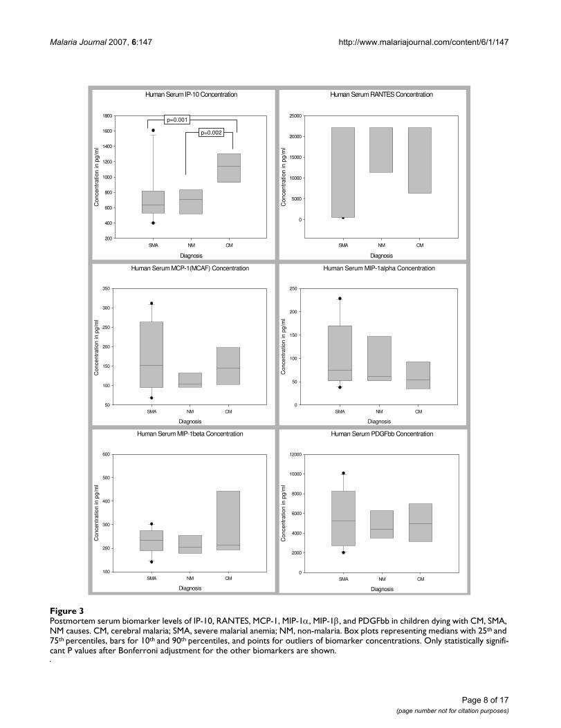
Serum Levels of Biomarkers in Children with CM, SMA, and NMPair wise comparisons were used to determine levels ofsignificance of the differences between the serum biomar-ker levels of the 3 disease groups after controlling for age,sex and parasitemia. The serum levels of 35 biomarkers(IL-1β, IL-1ra, IL-2, IL-4, IL-5, IL-6, IL-7, IL-8, IL-9, IL-10,IL-12 (p70), IL-13, IL-15, IL-17, Eotaxin, FGF basic pro-tein, CRP, G-CSF, GM-CSF, IFN-γ, TNF-α, MCP-1 (MCAF),MIP-1α, MIP-1β, RANTES, SDF-1α, CXCL11 (I-TAC), Fas-L, sFas, sTNF-R1 (p55), sTNF-R2 (p75), MMP-9, TGF-β1,PDGF bb and VEGF) demonstrated marginal changes, butdid not show statistically significant differences betweenthe 3 disease groups, after Bonferroni adjustment for theother biomarkers (Figures 1, 2, 3). However, serum levelof IP-10 was independently predictive of CM mortalitywhen compared to SMA and NM deaths. The serum levelof IP-10 was significantly higher in children with CMcompared with those children with SMA (P = 0.001) andNM (P = 0.002) (Figure 3).
Table 1: Demographic, parasitological, and hematological characteristics of the study participants
CHARACTERISTIC CM SMA NM P value
No. of children 9 5 5Gender (male/female) 4/5 3/2 3/2 0.249
Age (months) 61.2 (3.1) 14.6 (1.2) 79.0 (4.3) 0.021Parasite density (/µL) 51,604 (9,468) 195,003 (23,613) 0 < 0.001Hemoglobin level (g/
dL)7.2 (0.1) 3.7 (0.1) 7.9 (0.1) < 0.01
Platelet count (×103/µL) 170.8 (12.7) 288.4 (19.5) 330.7 (27.3) < 0.001
CM, cerebral malaria; SMA, severe malarial anemia; NM, non-malaria; P < 0.05 considered statistically significant.
Page 5 of 17(page number not for citation purposes)

Malaria Journal 2007, 6:147 http://www.malariajournal.com/content/6/1/147
Page 6 of 17(page number not for citation purposes)
Postmortem serum biomarker levels of IL-1β, IL-1ra, IL-6, IL-8, TNF-α, and IFN-γ in children dying with CM, SMA, NM causesFigure 1Postmortem serum biomarker levels of IL-1β, IL-1ra, IL-6, IL-8, TNF-α, and IFN-γ in children dying with CM, SMA, NM causes. CM, cerebral malaria; SMA, severe malarial anemia; NM, non-malaria. Box plots representing medians with 25th and 75th per-centiles, bars for 10th and 90th percentiles, and points for outliers of biomarker concentrations. Only statistically significant P values after Bonferroni adjustment for the other biomarkers are shown.
Human Serum IL-1b Concentration
Diagnosis
SMA NM CM
Concen
tration
in p
g/m
l
0
20
40
60
80
100
120
Human Serum IL-1ra Concentration
Diagnosis
SMA NM CM
Concen
tration
in p
g/m
l
0
1000
2000
3000
4000
5000
6000
7000
Human Serum IL-6 Concentration
Diagnosis
SMA NM CM
Con
ce
ntr
ation
in p
g/m
l
0
50
100
150
200
250
300
Human Serum IL-8 Concentration
Diagnosis
SMA NM CM
Con
ce
ntr
ation
in p
g/m
l
0
20
40
60
80
100
120
140
160
Human Serum IFNgamma Concentration
Diagnosis
SMA NM CM
Con
ce
ntr
ation
in p
g/m
l
0
500
1000
1500
2000
2500
3000
Human Serum TNFalpha Concentration
Diagnosis
SMA NM CM
Con
ce
ntr
ation
in p
g/m
l
0
50
100
150
200
250
300

Malaria Journal 2007, 6:147 http://www.malariajournal.com/content/6/1/147
Page 7 of 17(page number not for citation purposes)
Postmortem serum biomarker levels of IL-10, TGF-βx, sTNF-R1, sTNF-R2, sFas, and Fas-L in children dying with CM, SMA, NM causesFigure 2Postmortem serum biomarker levels of IL-10, TGF-βx, sTNF-R1, sTNF-R2, sFas, and Fas-L in children dying with CM, SMA, NM causes. CM, cerebral malaria; SMA, severe malarial anemia; NM, non-malaria. Box plots representing medians with 25th and 75th percentiles, bars for 10th and 90th percentiles, and points for outliers of biomarker concentrations. Only statistically signifi-cant P values after Bonferroni adjustment for the other biomarkers are shown.
Human Serum IL-10 Concentration
Diagnosis
SMA NM CM
Con
cent
ratio
n in
pg/
ml
0
20
40
60
80
100
Human Serum TGF beta1 Concentration
Diagnosis
SMA NM CM
Con
cent
ratio
n in
pg/
ml
0
2e+4
4e+4
6e+4
8e+4
1e+5
Human Serum sTNFR1 (p55) Concentration
Diagnosis
SMA NM CM
Con
cent
ratio
n in
ng/
ml
0
10
20
30
40
50
Human Serum sTNFR2 (p75) Concentration
Diagnosis
SMA NM CM
Con
cent
ratio
n in
ng/
ml
20
40
60
80
100
120
140
Human Serum sFas Concentration
Diagnosis
SMA NM CM
Con
cent
ratio
n in
pg/
ml
6000
8000
10000
12000
14000
16000
18000
20000
22000
24000
26000
Human Serum Fas Ligand Concentration
Diagnosis
SMA NM CM
Con
cent
ratio
n in
pg/
ml
0
200
400
600
800
1000
1200
Malaria Journal 2007, 6:147 http://www.malariajournal.com/content/6/1/147
Page 8 of 17(page number not for citation purposes)
Postmortem serum biomarker levels of IP-10, RANTES, MCP-1, MIP-1α, MIP-1β, and PDGFbb in children dying with CM, SMA, NM causesFigure 3Postmortem serum biomarker levels of IP-10, RANTES, MCP-1, MIP-1α, MIP-1β, and PDGFbb in children dying with CM, SMA, NM causes. CM, cerebral malaria; SMA, severe malarial anemia; NM, non-malaria. Box plots representing medians with 25th and 75th percentiles, bars for 10th and 90th percentiles, and points for outliers of biomarker concentrations. Only statistically signifi-cant P values after Bonferroni adjustment for the other biomarkers are shown.
Human Serum IP-10 Concentration
Diagnosis
SMA NM CM
Con
cen
tration
in
pg/m
l
200
400
600
800
1000
1200
1400
1600
1800p=0.001
p=0.002
Human Serum RANTES Concentration
Diagnosis
SMA NM CM
Con
cen
tration
in
pg/m
l
0
5000
10000
15000
20000
25000
Human Serum MCP-1(MCAF) Concentration
Diagnosis
SMA NM CM
Co
nce
ntr
ation
in
pg/m
l
50
100
150
200
250
300
350
Human Serum MIP-1alpha Concentration
Diagnosis
SMA NM CM
Con
ce
ntr
ation
in p
g/m
l
0
50
100
150
200
250
Human Serum MIP-1beta Concentration
Diagnosis
SMA NM CM
Co
nce
ntr
atio
n in
pg
/ml
100
200
300
400
500
600
Human Serum PDGFbb Concentration
Diagnosis
SMA NM CM
Co
nce
ntr
atio
n in
pg/m
l
0
2000
4000
6000
8000
10000
12000

Malaria Journal 2007, 6:147 http://www.malariajournal.com/content/6/1/147
CSF Levels of Biomarkers in Children with CM, SMA, and NMPair wise comparisons were used to determine levels ofsignificance of the differences between the CSF biomarkerlevels of the 3 disease groups after controlling for age, sexand parasitemia (Table 2). The CSF levels of 27 biomark-ers (IL-1β, IL-2, IL-4, IL-5, IL-6, IL-7, IL-9, IL-10, IL-12(p70), IL-13, IL-15, IL-17, Eotaxin, FGF basic protein,CRP, G-CSF, GM-CSF, IFN-γ, TNF-α, MCP-1 (MCAF),MIP-1α, RANTES, SDF-1α, CXCL11 (I-TAC), MMP-9,TGF-β1, and VEGF) did not differ significantly betweenthe three disease groups, after Bonferroni adjustment (Fig-ures 4, 5, 6). The CSF levels of 9 biomarkers (IL-1ra, IL-8,IP-10, PDGFbb, MIP-1β, sFas, Fas-Ligand, sTNF-R1, andsTNF-R2) were independently predictive of CM mortalitywhen compared to SMA and NM deaths (Figures 4, 5, 6).The CSF levels of IL-1ra, IL-8, IP-10, MIP-1β, sFas, Fas-Lig-and, sTNF-R1, and sTNF-R2 were significantly higher inchildren with CM compared with those children withSMA and NM. On the contrary, PDGFbb was significantlylower in children with CM compared with those childrenwith SMA and NM (Figures 4, 5, 6) (Table 2). These 9biomarkers that were independently predictive of CMmortality can be grouped into four major categories suchas cytokines (IL-8), cytokine receptors (IL-1ra, sTNF-R1,and sTNF-R2), chemokines (MIP-1β and IP-10), apop-totic (sFas and Fas-L), and angiogenic factors (PDGFbb).
Serum Biomarker Ratios in Children with CM, SMA, and NMSerum pro-inflammatory or angiostatic to anti-inflamma-tory or angiogenic cytokine median ratios were deter-mined and compared between the 3 disease groups (Table3). Serum TNF-α/IL-10 median ratio was higher in CMgroup compared to the SMA and NM groups, but the dif-ference was not significantly different (Table 3). Similarly,the serum TNF-α/IL-8, TNF-α/PDGFbb, IP-10/IL-10, IP-
10/1L-8, and IP-10/PDGFbb median ratios were consist-ently higher in the CM group compared to the SMA andNM groups, but the differences were not statistically sig-nificant (Table 3).
CSF Biomarker Ratios in Children with CM, SMA, and NMCSF pro-inflammatory or angiostatic to anti-inflamma-tory or angiogenic cytokine median ratios were deter-mined and compared between the 3 disease groups (Table4). CSF IP-10/PDGFbb median ratio was significantlyhigher in the CM group compared to the SMA and NMgroups (Table 4). However, the CSF TNF-α/IL-10, TNF-α/IL-8, TNF-α/PDGFbb, IP-10/IL-10, and IP-10/1L-8median ratios varied between the 3 disease groups, but thedifferences were not statistically significant (Table 4).
Correlation between Serum and CSF Levels of BiomarkersThe relationships of the serum and CSF levels of individ-ual inflammatory markers with each other were examinedusing Spearman's rank correlational analyses. With theexception of IP-10, sTNF-R1 and sTNF-R2, there was nosignificant association between the serum and CSF levelsof the other inflammatory markers studied (Spearman's ρ< 0.25; P > 0.05). There was a strong positive correlationbetween the serum and CSF levels of IP-10, sTNF-R1 andsTNF-R2 (Spearman's ρ = 0.58–0.82; all P < 0.0001) [datanot shown].
Correlation between Biomarker Levels and Clinical CharacteristicsThe relationships of individual inflammatory markerswith each other, and with parasite density, hemoglobinlevel, and platelet count were examined using Spearman'srank correlational analyses. With the exception of IP-10and MIP-1α, there was no significant association betweenthe serum levels of the other inflammatory markers stud-ied (Spearman's ρ < 0.25; P > 0.05). A moderately strong
Table 2: Comparison of Least Squares (Predicted) Means by Category, Controlled for Covariates (age, sex and parasitemia) For Biomarkers Showing Overall Statistically Significant Differences between the three Study Groups
BIOMARKER OVERALL P VALUE P VALUE FOR PAIRED GROUPS
CM vs. NM CM vs. SMA SMA vs. NM
Serum IP-10 0.005 0.002 0.001 NSSCSF IP-10 0.005 0.001 0.004 NSSCSF IL-8 0.0005 0.0001 0.001 NSS
CSF MIP-1β 0.0005 0.0001 0.001 NSSCSF PDGFbb 0.008 0.0002 0.01 NSS
CSF IL-1ra 0.002 0.0004 0.005 NSSCSF Fas-L 0.04 0.002 NSS NSS
CSF sTNF-R1 0.0001 0.00001 0.0002 NSSCSF sTNF-R2 0.001 0.0001 0.002 NSS
Statistically significant differences in biomarkers between the three disease groups determined by multivariate analyses [Comparison of Least Squares (Predicted) with Bonferonni correction] after modeling and controlling for covariates (age, sex, and parasitemia); P < 0.05 considered statistically significant; NSS, not statistically significant.
Page 9 of 17(page number not for citation purposes)

Malaria Journal 2007, 6:147 http://www.malariajournal.com/content/6/1/147
Page 10 of 17(page number not for citation purposes)
Postmortem CSF biomarker levels of IL-1β, IL-1ra, IL-6, IL-8, TNF-α, and IFN-γ in children dying with CM, SMA, NM causesFigure 4Postmortem CSF biomarker levels of IL-1β, IL-1ra, IL-6, IL-8, TNF-α, and IFN-γ in children dying with CM, SMA, NM causes. CM, cerebral malaria; SMA, severe malarial anemia; NM, non-malaria. Box plots representing medians with 25th and 75th per-centiles, bars for 10th and 90th percentiles, and points for outliers of biomarker concentrations. Only statistically significant P values after Bonferroni adjustment for the other biomarkers are shown.
Human CSF IL-1beta Concentration
Diagnosis
SMA NM CM
Con
cen
tration
in
pg/m
l
0
5
10
15
20
25
30
35
Human CSF IL-1ra Concentration
Diagnosis
SMA NM CM
Con
cen
tration
in
pg/m
l
0
100
200
300
400
p=0.0004
p=0.005
Human CSF IL-6 Concentration
Diagnosis
SMA NM CM
Co
nce
ntr
ation in
pg/m
l
0
10
20
30
40
50
60
70
Human CSF IL-8 Concentration
Diagnosis
SMA NM CM
Co
nce
ntr
ation in
pg/m
l
0
1
2
3
4
5
6
7
p=0.001
p=0.0001
Human CSF TNFalpha Concentration
Diagnosis
SMA NM CM
Con
ce
ntr
ation
in p
g/m
l
0
2
4
6
8
10
12
14
Human CSF IFNgamma Concentration
Diagnosis
SMA NM CM
Con
ce
ntr
ation
in p
g/m
l
0
10
20
30
40
50

Malaria Journal 2007, 6:147 http://www.malariajournal.com/content/6/1/147
Page 11 of 17(page number not for citation purposes)
Postmortem CSF biomarker levels of IL-10, TGF-βx, sTNF-R1, sTNF-R2, sFas, and Fas-L in children dying with CM, SMA, NM causesFigure 5Postmortem CSF biomarker levels of IL-10, TGF-βx, sTNF-R1, sTNF-R2, sFas, and Fas-L in children dying with CM, SMA, NM causes. CM, cerebral malaria; SMA, severe malarial anemia; NM, non-malaria. Box plots representing medians with 25th and 75th
percentiles, bars for 10th and 90th percentiles, and points for outliers of biomarker concentrations. Only statistically significant P values after Bonferroni adjustment for the other biomarkers are shown.
Human CSF IL-10 Concentration
Diagnosis
SMA NM CM
Con
cen
tra
tion in
pg
/ml
0
10
20
30
40
50
60
Human CSF TGF beta1 Concentration
Diagnosis
SMA NM CM
Con
cen
tration
in
pg/m
l
0
1000
2000
3000
4000
Human CSF sTNFR1 (p55) Concentration
Diagnosis
SMA NM CM
Con
ce
ntr
ation
in n
g/m
l
0
1
2
3
4
5
p=0.00001
p=0.0002
Human CSF sTNFR2 (p75) Concentration
Diagnosis
SMA NM CM
Con
ce
ntr
ation
in n
g/m
l
0
5
10
15
20
25
p=0.0001
p=0.002
Human CSF sFas Concentration
Diagnosis
SMA NM CM
Co
nce
ntr
atio
n in
pg
/ml
0
50
100
150
200
250
Human CSF Fas Ligand Concentration
Diagnosis
SMA NM CM
Con
ce
ntr
ation
in p
g/m
l
0
2
4
6
8
10
12
14
16
p=0.002

Malaria Journal 2007, 6:147 http://www.malariajournal.com/content/6/1/147
Page 12 of 17(page number not for citation purposes)
Postmortem CSF biomarker levels of IP-10, RANTES, MCP-1, MIP-1α, MIP-1β, and PDGFbb in children dying with CM, SMA, NM causesFigure 6Postmortem CSF biomarker levels of IP-10, RANTES, MCP-1, MIP-1α, MIP-1β, and PDGFbb in children dying with CM, SMA, NM causes. CM, cerebral malaria; SMA, severe malarial anemia; NM, non-malaria. Box plots representing medians with 25th and 75th percentiles, bars for 10th and 90th percentiles, and points for outliers of biomarker concentrations. Only statistically signifi-cant P values after Bonferroni adjustment for the other biomarkers are shown.
Human CSF IP-10 Concentration
Diagnosis
SMA NM CM
Con
cen
tra
tion in
pg
/ml
0
20
40
60
80
100
120
140p=0.004
p=0.001
Human CSF RANTES Concentration
Diagnosis
SMA NM CM
Con
cen
tration
in
pg/m
l
0
50
100
150
200
250
300
350
Human CSF MCP-1(MCAF) Concentration
Diagnosis
SMA NM CM
Con
ce
ntr
ation
in p
g/m
l
0
20
40
60
80
100
120
140
160
Human CSF MIP-1alpha Concentration
Diagnosis
SMA NM CM
Con
ce
ntr
ation
in p
g/m
l
0
1
2
3
4
Human CSF MIP-1beta Concentration
Diagnosis
SMA NM CM
Con
ce
ntr
ation
in p
g/m
l
0
20
40
60
80
100
120
Human CSF PDGFbb Concentration
Diagnosis
SMA NM CM
Con
ce
ntr
ation
in p
g/m
l
0
5
10
15
20
25
30
35
p=0.01
p=0.0002p=0.0001
p=0.001

Malaria Journal 2007, 6:147 http://www.malariajournal.com/content/6/1/147
positive correlation was seen between the serum levels ofIP-10 and MIP-1α (Spearman's ρ = 0.40; P = 0.001). Withthe exception of IL-1ra, IL-8, IP-10, PDGFbb, MIP-1β,sTNF-R1, and sTNF-R2, there was no significant associa-tion between the CSF levels of the other inflammatorymarkers studied (Spearman's ρ < 0.25; P > 0.05). The CSFlevel of PDGFbb correlated strongly and inversely withlevels of IL-1ra, IL-8, IP-10, MIP-1β, sTNF-R1, and sTNF-R2 (Spearman's ρ = 0.61–0.76; all P < 0.0001). A moder-ately strong positive correlation was seen between the CSFlevels of IL-1ra and sTNF-R1, IL-1ra and sTNF-R2, IL-1raand IL-8, IL-1ra and 1P-10, IL-1ra and MIP-1β, and sTNF-R1 and sTNF-R2 (Spearman's ρ = 0.41–0.48; all P <0.001). A weak positive correlation was seen between theCSF levels of IP-10 and MIP-1β, IP-10 and sTNF-R1, IP-10and sTNF-R2, IP-10 and IL-8, MIP-1β and IL-8, MIP-1βand sTNF-R1, MIP-1β and sTNF-R2, IL-8 and sTNF-R1,and IL-8 and sTNF-R2 (Spearman's ρ = 0.26–0.37; all P <0.05). Finally, there was no significant associationbetween parasite density, hemoglobin level, and plateletcount, and the serum and CSF levels of all the inflamma-tory markers studied (data not shown).
DiscussionThe present study examined a broad range of disease asso-ciated inflammatory mediators, including cytokines,chemokines, and markers of apoptosis and angiogenesis,in postmortem serum and CSF samples of children with
CM, SMA, and NM. The study was conducted in an area ofmoderate Plasmodium falciparum transmission where allthe life-threatening complications of malaria occur,namely, coma, severe anemia, and respiratory distress [1-5]. Although post-mortem studies have provided a wealthof detailed information they reflect, at best, pathology ata single time point after death in the most severely illpatients and may be potentially biased by post-mortemartifacts (agonal changes that may simulate disease-induced pathology). The concurrent studying of theseinflammatory, apoptotic and angiogenic biomarkers inappropriate time-matched post-mortem controls fromother disease causes helps place the results in context. Dueto these limitations, further studies would be required toconfirm the functional roles of these host factors in CM.
Cerebral malaria (CM) is a major life-threatening compli-cation of Plasmodium falciparum infection in humans. Themechanisms underlying the fatal cerebral complicationsare still not fully understood. However, two predominanthypotheses are generally proposed to explain the neu-ropathology of CM, namely the sequestration and immu-nological hypotheses. The sequestration hypotheticalmodel suggest that the adhesion of pRBCs to the cerebralvasculature leads to obstruction of the microcirculation,metabolic depletion, BBB breakdown, and alteration inbrain function resulting in coma [16]. The immunologicalhypothetical model suggest that hyperimmune responses
Table 3: Comparison ofSelected Serum Biomarker Median Ratios between the three Study Groups
PRO-INFLAMMATORY OR ANGIOSTATIC vs. ANTI-INFLAMMATORY OR ANGIOGENIC BIOMARKER MEDIAN RATIO
CM SMA NM OVERALL P VALUE
TNF-α : IL-10 4.11 3.22 3.18 NSSTNF-α : IL-8 2.48 2.29 2.26 NSSTNF-α : PDGFbb 1.64 × 10⁻² 1.54 × 10⁻² 1.56 × 10⁻² NSSIP-10 : IL-10 34.85 18.86 23.23 NSSIP-10 : IL-8 2.31 × 10⁻¹ 1.27 × 10⁻¹ 1.63 × 10⁻¹ NSSIP-10 : PDGFbb 57.52 26.41 32.73 NSS
Median ratios of serum levels of pro-inflammatory/angiostatic versus anti-inflammatory/angiogenic biomarkers were compared between the 3 disease groups; P < 0.05 considered statistically significant; NSS, not statistically significant.
Table 4: Comparison of Selected CSF Biomarker Median Ratios between the three Study Groups
PRO-INFLAMMATORY OR ANGIOSTATIC vs. ANTI-INFLAMMATORY OR ANGIOGENIC BIOMARKER MEDIAN RATIO
CM SMA NM OVERALL P VALUE
TNF-α : IL-10 3.33 × 10⁻² 7.51 × 10⁻² 2.04 × 10⁻¹ NSSTNF-α : IL-8 7.76 × 10⁻² 7.52 × 10⁻¹ 6.67 × 10⁻¹ NSSTNF-α : PDGFbb 5.05 × 10⁻² 7.56 × 10⁻² 1.53 × 10⁻² NSSIP-10 : IL-10 10.28 2.07 3.14 NSSIP-10 : IL-8 23.08 20.12 10.29 NSSIP-10 : PDGFbb 15.35 2.28 2.32 × 10⁻² 0.003
Median ratios of CSF levels of pro-inflammatory/angiostatic versus anti-inflammatory/angiogenic biomarkers were compared between the three disease groups; P < 0.05 considered statistically significant; NSS, not statistically significant.
Page 13 of 17(page number not for citation purposes)

Malaria Journal 2007, 6:147 http://www.malariajournal.com/content/6/1/147
(originally evolved for the destruction of the parasite andprotection of the host) and Th1/Th2 cytokine or chemok-ine dysregulation results in localized recruitment ofimmune effectors cells (T cells, monocytes, etc) and BBBimpairment resulting in the development of cerebral com-plications [7]. However, recent studies indicate that para-site induced apoptosis and tissue degeneration, as well asangiogenic factors may be involved in the pathogenesis ofCM [14,15,36,38]. Understanding the cytokine/chemok-ine cascade, parasite induced apoptotic pathways, anddysregulation of angiogenic factors in CM patients willelucidate the underlying pathogenesis and identify poten-tial predictive prognostic biomarkers for CM mortality.
In the present study, evidence is provided indicating thatthe serum levels of various cytokines and chemokines arealtered in children with CM compared to SMA and NM.The elevated serum level of IP-10 is particularly remarka-ble since it was the only independent predictor of CMmortality. Eight (8) CSF inflammatory biomarkers (IL-1ra, IL-8, IP-10, PDGFbb, MIP-1β, Fas-L, sTNF-R1, andsTNF-R2) were independently predictive of CM mortality,when compared to SMA and NM deaths. The significantincrease in CSF levels of IL-1ra, IL-8, IP-10, MIP-1β, sTNF-R1, and sTNF-R2 in CM compared to SMA and NM sug-gests a critical role for the brain parenchymal expressionof these biomarkers in CM pathogenesis and mortality. Inthe present study, both the serum and CSF levels ofRANTES were not predictive of CM mortality, althoughlow serum levels of RANTES have recently been associatedwith mortality in Ugandan children with CM [26].
TNF-R1 and TNF-R2 are key mediators of the classicalextrinsic apoptotic pathway, as well as in inflammation.The increased expression of sTNF-R1 and sTNF-R2 in CSFof CM non-survivors when compared to SMA and NMsuggests that parasite-induced apoptosis in host CNS iscritical to CM pathogenesis and mortality. Recent studiesin murine experimental CM have shown that the TNFreceptor super family also plays a role in CM pathogenesis[42-47]. Mice deficient in TNF-R2 (TNF-R2-/-) and Fas(Fas-/-) survived significantly longer than wild type inexperimental CM, and TNFR2-/- mice survived the longestin the absence of anti-malarial treatment [42,47]. Addi-tionally, the serum levels of sTNF-R1 and sTNF-R2, whichact as binding proteins for TNF, were elevated in patientswith acute Plasmodium falciparum malaria compared to thelevels in convalescent children and in healthy controls[38,39].
Platelet derived growth factor is a key factor that mediatesvascular smooth muscle cell proliferation and serves aneuroprotective role by inducing regeneration of dam-aged axons and neuronal growth after ischemia [37]. Inthis study, a significant decline in PDGFbb production
was independently predictive of CM mortality. Therefore,it seems that the down regulation of this angiogenicgrowth factor and upregulation of apoptotic factors in CMpatients may be a result of parasite-induced damage ordepletion of cells producing PDGF, a highly angiostaticmicroenvironment with high levels of proinflammatorycytokines/chemokines (notably IP-10), or even an uni-dentified parasite-derived factor that initiates/exacerbatesthe inflammatory and apoptotic cascades.
IFN-inducible protein of 10 kDa (IP-10) is a chemokineinduced by IFN-γ and TNF-α. Although IP-10 was initiallyshown to have chemotactic activity for activated Th1 lym-phocytes, there is growing evidence implicating thischemokine in both infectious and non-infectious causesof neuronal injury, dementia and inhibition of angiogen-esis [50-54]. This is the first report demonstrating that sig-nificant elevation of serum and CSF levels of IP-10 isassociated with CM mortality. Our finding suggests thatIP-10 plays a major role in the CM immunopathology,and begs for further study in other endemic settings. Inter-estingly, Plasmodium berghei ANKA infection induced IP-10 and monocyte chemotactic protein (MCP)-1 geneexpression in the brain of both CM-susceptible (C57BL/6)and CM-resistant (BALB/c) mice as early as 24 hours post-infection [33]. Additionally, the expression of IP-10 andMCP-1 genes in KT-5, an astrocyte cell line, was inducedin vitro upon stimulation with a crude antigen of malariaparasites, suggesting astrocytes as the potential cellularsource of cytokine and chemokine expression in brainparenchyma in response to plasmodial infection [33].Therefore, in response to plasmodial infection, the cellsthat produce these inflammatory mediators may be differ-ent in the brain (microglia and astrocytes) and peripheralcirculation (platelets, monocytes, and lymphocytes), andtheir effects may also differ in the two areas.
This study has revealed new associations, underlyingpathogenic events, between different biomarkers and CMmortality in Ghanaian children that may be applicable toother malaria endemic populations. The most importantfinding demonstrates the association between the eleva-tion of serum and CSF factors involved in the classicalextrinsic apoptotic pathways (such as IP-10, TNF-α-sTNF-R1-sTNF-R2 and Fas-L) and the reduction of growth fac-tors that confer endothelial and neuronal cell protection(such as PDGF) with CM mortality. We propose the fol-lowing hypothesis to explain our observations. It appearsthat both inflammatory and apoptotic mechanisms maybe triggered locally in the human brain during CM thatresult in the damage of the constituent cells of the BBB(glial cells, astrocytes, and endothelial cells) and possiblyneurons. Additionally, this localized BBB damage may befurther exacerbated by the significantly decreased levels ofneuroprotective angiogenic growth factors (such PDGF),
Page 14 of 17(page number not for citation purposes)

Malaria Journal 2007, 6:147 http://www.malariajournal.com/content/6/1/147
induced by the angiostatic effects of the elevated local CSFlevels of IP-10, ultimately resulting in death.
Furthermore, we propose that TNF-α and other proin-flammatory factors which are activated following therelease of malaria antigens after schizont rupture mayinduce the local production of IP-10 by the constituentcells of the BBB (glial cells, astrocytes, and endothelialcells) [33]. Subsequently, IP-10 in concert with TNF-αmay induce apoptosis of endothelial cells leading to BBBbreakdown. Additionally, activated circulating immu-nomodulator cells (T cells, monocytes, etc) attracted tothe BBB by IP-10, may also play a pathogenic role in thisprocess. The significantly decreased production of PDGFmay further inhibit angiogenesis and negatively impactthe regeneration of damaged endothelial cells and bloodcapillaries at the BBB. Recently, elevated CSF level of IP-10has been demonstrated in viral meningitis [54]. ElevatedCSF level of IP-10 has been shown to be significantly cor-related with the neuropsychiatric impairment in HIV-associated dementia [55]. Furthermore, mouse studieshave demonstrated that the HIV-1 virus encoded proteingp120 directly activates astrocytes to produce IP-10 usinga novel mechanism independent of IFN-γ and STAT-1pathway of IP-10 induction [52]. Therefore, elevatedserum and CSF levels of IP-10 may be an important path-ogenic factor in CM neuropathology, as observed in otherinfectious disease models. Most CM deaths occur within24 hours of admission before antimalarials have had timeto kill the parasites [9,16,48], hence new interventionsthat address pathophysiological processes causing theseearly deaths is a public health priority, in addition toaddressing the public-health problems resulting indelayed presentation to hospital and ensuring childrenreceive prompt and appropriate resuscitation. Thus, thisstudy provides new insights into the processes leading tocerebral malaria and mortality associated with it.
ConclusionThis study has demonstrated an association between CMassociated mortality with elevated serum and CSF levels ofapoptotic factors (IP-10, IL-1ra, sTNFR1, sTNFR2, sFas)and reduced serum and CSF levels of neuroprotective ang-iogenic growth factors (PDGFbb). The observations sup-port recent reports that implicate parasite-inducedapoptosis and angiogenic factors in CM neuropathology.Further studies in other endemic areas to confirm thesefindings are necessary.
AbbreviationsBBB, Blood-Brain Barrier;
CM, cerebral malaria;
CNS, central nervous system;
CRP, C-reactive protein;
CSF, cerebrospinal fluid;
ECM, experimental cerebral malaria;
FGF, fibroblast growth factor;
G-CSF, granulocyte colony stimulating factor;
GM-CSF, granulocyte-monocyte colony stimulating fac-tor;
IFN, Interferon;
IL, Interleukin;
IP, Interferon inducible protein 10;
MCP, monocyte chemotactic protein;
MIP, macrophage inflammatory protein;
MMP, matrix metalloproteinase;
NM, non-malaria;
PDGF, platelet derived growth factor;
pRBC, parasitized red blood cell;
RANTES, regulated upon activation, normal T cellexpressed and secreted;
SDF, stromal differentiation factor;
SMA, severe malarial anemia;
TGF, transforming growth factor;
TNF, Tumor Necrosis Factor;
VEGF, vascular endothelial growth factor;
Competing interestsThe author(s) declare that they have no competing inter-ests.
Authors' contributionsHBA performed the autopsies and sample collection,immunoassays, data analysis and drafting of the manu-script.NOW and BYS participated in the performance ofimmunoassay, data analysis and drafting of the manu-script. MDP, VCB, JET and VU, participated in the per-formance of proteomics analysis.WA, AAA, RKG, YT and
Page 15 of 17(page number not for citation purposes)

Malaria Journal 2007, 6:147 http://www.malariajournal.com/content/6/1/147
EKW participated in the design and coordination of thestudy, and supervised the autopsies and sample collec-tion. JKS conceived of the study, participated in its designand coordination, and revised the manuscript for impor-tant intellectual content. All authors read and approvedthe final manuscript.
AcknowledgementsThis investigation received financial support from the WHO/UNDP/TDR Collaborative Research Grant (A00524) and National Institutes of Health grant numbers NIH-RCMI (RR03034), NIH-NIGM-MBRS (SO6GM08248), and NIH-FIC (R21TW006804-01).
References1. Kurtzhals JA, Adabayeri V, Goka BQ, Akanmori BD, Oliver-Commey
JO, Nkrumah FK, Behr C, Hviid L: Low plasma concentrations ofinterleukin 10 in severe malarial anaemia compared withcerebral and uncomplicated malaria. Lancet 1998,351(9118):1768-1772.
2. Sarfo BY, Singh S, Lillard JW, Quarshie A, Gyasi RK, Armah H, AdjeiAA, Jolly P, Stiles JK: The cerebral-malaria-associated expres-sion of RANTES, CCR3 and CCR5 in post-mortem tissuesamples. Ann Trop Med Parasitol 2004, 98(3):297-303.
3. Armah H, Dodoo AK, Wiredu EK, Stiles JK, Adjei AA, Gyasi RK, Tet-tey Y: High-level cerebellar expression of cytokines and adhe-sion molecules in fatal, paediatric, cerebral malaria. Ann TropMed Parasitol 2005, 99(7):629-647.
4. Armah H, Wired EK, Dodoo AK, Adjei AA, Tettey Y, Gyasi R:Cytokines and adhesion molecules expression in the brain inhuman cerebral malaria. Int J Environ Res Public Health 2005,2(1):123-131.
5. Awandare GA, Goka B, Boeuf P, Tetteh JK, Kurtzhals JA, Behr C,Akanmori BD: Increased levels of inflammatory mediators inchildren with severe Plasmodium falciparum malaria withrespiratory distress. J Infect Dis 2006, 194(10):1438-1446.
6. Brown H, Turner G, Rogerson S, Tembo M, Mwenechanya J,Molyneux M, Taylor T: Cytokine expression in the brain inhuman cerebral malaria. J Infect Dis 1999, 180(5):1742-1746.
7. Hunt NH, Grau GE: Cytokines: accelerators and brakes in thepathogenesis of cerebral malaria. Trends Immunol 2003,24(9):491-499.
8. Silamut K, Phu NH, Whitty C, Turner GD, Louwrier K, Mai NT, Simp-son JA, Hien TT, White NJ: A quantitative analysis of the micro-vascular sequestration of malaria parasites in the humanbrain. Am J Pathol 1999, 155(2):395-410.
9. Taylor TE, Fu WJ, Carr RA, Whitten RO, Mueller JS, Fosiko NG,Lewallen S, Liomba NG, Molyneux ME: Differentiating the pathol-ogies of cerebral malaria by postmortem parasite counts.Nat Med 2004, 10(2):143-145.
10. Grau GE, Mackenzie CD, Carr RA, Redard M, Pizzolato G, Allasia C,Cataldo C, Taylor TE, Molyneux ME: Platelet accumulation inbrain microvessels in fatal pediatric cerebral malaria. J InfectDis 2003, 187(3):461-466.
11. Wassmer SC, Combes V, Grau GE: Pathophysiology of cerebralmalaria: role of host cells in the modulation of cytoadhesion.Ann N Y Acad Sci 2003, 992:30-38.
12. Wassmer SC, Lepolard C, Traore B, Pouvelle B, Gysin J, Grau GE:Platelets reorient Plasmodium falciparum-infected erythro-cyte cytoadhesion to activated endothelial cells. J Infect Dis2004, 189(2):180-189.
13. Sarfo BY, Armah HB, Irune I, Adjei AA, Olver CS, Singh S, Lillard JWJr., Stiles JK: Plasmodium yoelii 17XL infection up-regulatesRANTES, CCR1, CCR3 and CCR5 expression, and inducesultrastructural changes in the cerebellum. Malar J 2005, 4:63.
14. Deininger MH, Kremsner PG, Meyermann R, Schluesener HJ: Differ-ential cellular accumulation of transforming growth factor-beta1, -beta2, and -beta3 in brains of patients who died withcerebral malaria. J Infect Dis 2000, 181(6):2111-2115.
15. Deininger MH, Winkler S, Kremsner PG, Meyermann R, SchluesenerHJ: Angiogenic proteins in brains of patients who died withcerebral malaria. J Neuroimmunol 2003, 142(1-2):101-111.
16. Gitau EN, Newton CR: Review Article: blood-brain barrier infalciparum malaria. Trop Med Int Health 2005, 10(3):285-292.
17. Brown H, Hien TT, Day N, Mai NT, Chuong LV, Chau TT, Loc PP,Phu NH, Bethell D, Farrar J, Gatter K, White N, Turner G: Evidenceof blood-brain barrier dysfunction in human cerebralmalaria. Neuropathol Appl Neurobiol 1999, 25(4):331-340.
18. de Kossodo S, Grau GE: Profiles of cytokine production in rela-tion with susceptibility to cerebral malaria. J Immunol 1993,151(9):4811-4820.
19. Lyke KE, Burges R, Cissoko Y, Sangare L, Dao M, Diarra I, Kone A,Harley R, Plowe CV, Doumbo OK, Sztein MB: Serum levels of theproinflammatory cytokines interleukin-1 beta (IL-1beta), IL-6, IL-8, IL-10, tumor necrosis factor alpha, and IL-12(p70) inMalian children with severe Plasmodium falciparum malariaand matched uncomplicated malaria or healthy controls.Infect Immun 2004, 72(10):5630-5637.
20. Othoro C, Lal AA, Nahlen B, Koech D, Orago AS, Udhayakumar V:A low interleukin-10 tumor necrosis factor-alpha ratio isassociated with malaria anemia in children residing in aholoendemic malaria region in western Kenya. J Infect Dis1999, 179(1):279-282.
21. May J, Lell B, Luty AJ, Meyer CG, Kremsner PG: Plasma inter-leukin-10:Tumor necrosis factor (TNF)-alpha ratio is associ-ated with TNF promoter variants and predicts malarialcomplications. J Infect Dis 2000, 182(5):1570-1573.
22. Esamai F, Ernerudh J, Janols H, Welin S, Ekerfelt C, Mining S, ForsbergP: Cerebral malaria in children: serum and cerebrospinalfluid TNF-alpha and TGF-beta levels and their relationshipto clinical outcome. J Trop Pediatr 2003, 49(4):216-223.
23. Schofield L, Villaquiran J, Ferreira A, Schellekens H, Nussenzweig R,Nussenzweig V: Gamma interferon, CD8+ T cells and antibod-ies required for immunity to malaria sporozoites. Nature1987, 330(6149):664-666.
24. Vreden SG, van den Broek MF, Oettinger MC, Verhave JP, MeuwissenJH, Sauerwein RW: Cytokines inhibit the development of liverschizonts of the malaria parasite Plasmodium berghei invivo. Eur J Immunol 1992, 22(9):2271-2275.
25. Kobayashi F, Ishida H, Matsui T, Tsuji M: Effects of in vivo admin-istration of anti-IL-10 or anti-IFN-gamma monoclonal anti-body on the host defense mechanism against Plasmodiumyoelii yoelii infection. J Vet Med Sci 2000, 62(6):583-587.
26. John CC, Opika-Opoka R, Byarugaba J, Idro R, Boivin MJ: Low Levelsof RANTES Are Associated with Mortality in Children withCerebral Malaria. J Infect Dis 2006, 194(6):837-845.
27. Ochiel DO, Awandare GA, Keller CC, Hittner JB, Kremsner PG,Weinberg JB, Perkins DJ: Differential regulation of beta-chem-okines in children with Plasmodium falciparum malaria.Infect Immun 2005, 73(7):4190-4197.
28. Burgmann H, Hollenstein U, Wenisch C, Thalhammer F, Looareesu-wan S, Graninger W: Serum concentrations of MIP-1 alpha andinterleukin-8 in patients suffering from acute Plasmodiumfalciparum malaria. Clin Immunol Immunopathol 1995, 76(1 Pt1):32-36.
29. Were T, Ouma C, Otieno RO, Orago AS, Ong'echa JM, Vulule JM,Keller CC, Perkins DJ: Suppression of RANTES in children withPlasmodium falciparum malaria. Haematologica 2006,91(10):1396-1399.
30. Farber JM: Mig and IP-10: CXC chemokines that target lym-phocytes. J Leukoc Biol 1997, 61(3):246-257.
31. Chaisavaneeyakorn S, Moore JM, Otieno J, Chaiyaroj SC, Perkins DJ,Shi YP, Nahlen BL, Lal AA, Udhayakumar V: Immunity to placentalmalaria. III. Impairment of interleukin(IL)-12, not IL-18, andinterferon-inducible protein-10 responses in the placentalintervillous blood of human immunodeficiency virus/malaria-coinfected women. J Infect Dis 2002, 185(1):127-131.
32. Suguitan AL Jr., Leke RG, Fouda G, Zhou A, Thuita L, Metenou S,Fogako J, Megnekou R, Taylor DW: Changes in the levels ofchemokines and cytokines in the placentas of women withPlasmodium falciparum malaria. J Infect Dis 2003,188(7):1074-1082.
33. Hanum PS, Hayano M, Kojima S: Cytokine and chemokineresponses in a cerebral malaria-susceptible or -resistantstrain of mice to Plasmodium berghei ANKA infection: earlychemokine expression in the brain. Int Immunol 2003,15(5):633-640.
Page 16 of 17(page number not for citation purposes)
http://www.ncbi.nlm.nih.gov/entrez/query.fcgi?cmd=Retrieve&db=PubMed&dopt=Abstract&list_uids=9635949
http://www.ncbi.nlm.nih.gov/entrez/query.fcgi?cmd=Retrieve&db=PubMed&dopt=Abstract&list_uids=9635949
http://www.ncbi.nlm.nih.gov/entrez/query.fcgi?cmd=Retrieve&db=PubMed&dopt=Abstract&list_uids=9635949
http://www.ncbi.nlm.nih.gov/entrez/query.fcgi?cmd=Retrieve&db=PubMed&dopt=Abstract&list_uids=8409439
http://www.ncbi.nlm.nih.gov/entrez/query.fcgi?cmd=Retrieve&db=PubMed&dopt=Abstract&list_uids=8409439
http://www.ncbi.nlm.nih.gov/entrez/query.fcgi?cmd=Retrieve&db=PubMed&dopt=Abstract&list_uids=9841855
http://www.ncbi.nlm.nih.gov/entrez/query.fcgi?cmd=Retrieve&db=PubMed&dopt=Abstract&list_uids=9841855
http://www.ncbi.nlm.nih.gov/entrez/query.fcgi?cmd=Retrieve&db=PubMed&dopt=Abstract&list_uids=9841855
http://www.ncbi.nlm.nih.gov/entrez/query.fcgi?cmd=Retrieve&db=PubMed&dopt=Abstract&list_uids=3120015
http://www.ncbi.nlm.nih.gov/entrez/query.fcgi?cmd=Retrieve&db=PubMed&dopt=Abstract&list_uids=3120015
http://www.ncbi.nlm.nih.gov/entrez/query.fcgi?cmd=Retrieve&db=PubMed&dopt=Abstract&list_uids=1516619
http://www.ncbi.nlm.nih.gov/entrez/query.fcgi?cmd=Retrieve&db=PubMed&dopt=Abstract&list_uids=1516619
http://www.ncbi.nlm.nih.gov/entrez/query.fcgi?cmd=Retrieve&db=PubMed&dopt=Abstract&list_uids=1516619
http://www.ncbi.nlm.nih.gov/entrez/query.fcgi?cmd=Retrieve&db=PubMed&dopt=Abstract&list_uids=7606866
http://www.ncbi.nlm.nih.gov/entrez/query.fcgi?cmd=Retrieve&db=PubMed&dopt=Abstract&list_uids=7606866
http://www.ncbi.nlm.nih.gov/entrez/query.fcgi?cmd=Retrieve&db=PubMed&dopt=Abstract&list_uids=7606866
http://www.ncbi.nlm.nih.gov/entrez/query.fcgi?cmd=Retrieve&db=PubMed&dopt=Abstract&list_uids=9060447

Malaria Journal 2007, 6:147 http://www.malariajournal.com/content/6/1/147
Publish with BioMed Central and every scientist can read your work free of charge
"BioMed Central will be the most significant development for disseminating the results of biomedical research in our lifetime."
Sir Paul Nurse, Cancer Research UK
Your research papers will be:
available free of charge to the entire biomedical community
peer reviewed and published immediately upon acceptance
cited in PubMed and archived on PubMed Central
yours — you keep the copyright
Submit your manuscript here:http://www.biomedcentral.com/info/publishing_adv.asp
BioMedcentral
34. Hayashi T, Deguchi K, Nagotani S, Zhang H, Sehara Y, Tsuchiya A,Abe K: Cerebral ischemia and angiogenesis. Curr Neurovasc Res2006, 3(2):119-129.
35. Wassmer SC, Combes V, Candal FJ, Juhan-Vague I, Grau GE: Plate-lets potentiate brain endothelial alterations induced by Plas-modium falciparum. Infect Immun 2006, 74(1):645-653.
36. Wassmer SC, de Souza JB, Frere C, Candal FJ, Juhan-Vague I, GrauGE: TGF-beta1 released from activated platelets can induceTNF-stimulated human brain endothelium apoptosis: a newmechanism for microvascular lesion during cerebralmalaria. J Immunol 2006, 176(2):1180-1184.
37. Krupinski J, Issa R, Bujny T, Slevin M, Kumar P, Kumar S, Kaluza J: Aputative role for platelet-derived growth factor in angiogen-esis and neuroprotection after ischemic stroke in humans.Stroke 1997, 28(3):564-573.
38. Kern P, Dietrich M, Hemmer C, Wellinghausen N: Increased levelsof soluble Fas ligand in serum in Plasmodium falciparummalaria. Infect Immun 2000, 68(5):3061-3063.
39. Molyneux ME, Engelmann H, Taylor TE, Wirima JJ, Aderka D, WallachD, Grau GE: Circulating plasma receptors for tumour necrosisfactor in Malawian children with severe falciparum malaria.Cytokine 1993, 5(6):604-609.
40. Matsumoto J, Kawai S, Terao K, Kirinoki M, Yasutomi Y, Aikawa M,Matsuda H: Malaria infection induces rapid elevation of thesoluble Fas ligand level in serum and subsequent T lym-phocytopenia: possible factors responsible for the differ-ences in susceptibility of two species of Macaca monkeys toPlasmodium coatneyi infection. Infect Immun 2000,68(3):1183-1188.
41. Helmby H, Jonsson G, Troye-Blomberg M: Cellular changes andapoptosis in the spleens and peripheral blood of miceinfected with blood-stage Plasmodium chabaudi chabaudiAS. Infect Immun 2000, 68(3):1485-1490.
42. Lucas R, Juillard P, Decoster E, Redard M, Burger D, Donati Y, GiroudC, Monso-Hinard C, De Kesel T, Buurman WA, Moore MW, DayerJM, Fiers W, Bluethmann H, Grau GE: Crucial role of tumornecrosis factor (TNF) receptor 2 and membrane-boundTNF in experimental cerebral malaria. Eur J Immunol 1997,27(7):1719-1725.
43. Piguet PF, Kan CD, Vesin C, Rochat A, Donati Y, Barazzone C: Roleof CD40-CVD40L in mouse severe malaria. Am J Pathol 2001,159(2):733-742.
44. Piguet PF, Kan CD, Vesin C: Role of the tumor necrosis factorreceptor 2 (TNFR2) in cerebral malaria in mice. Lab Invest2002, 82(9):1155-1166.
45. Stoelcker B, Hehlgans T, Weigl K, Bluethmann H, Grau GE, MannelDN: Requirement for tumor necrosis factor receptor 2expression on vascular cells to induce experimental cerebralmalaria. Infect Immun 2002, 70(10):5857-5859.
46. Ohno T, Kobayashi F, Nishimura M: Fas has a role in cerebralmalaria, but not in proliferation or exclusion of the murineparasite in mice. Immunogenetics 2005, 57(3-4):293-296.
47. Potter SM, Chan-Ling T, Rosinova E, Ball HJ, Mitchell AJ, Hunt NH: Arole for Fas-Fas ligand interactions during the late-stage neu-ropathological processes of experimental cerebral malaria.Journal of Neuroimmunology 2006, 173(1-2):96.
48. Idro R, Jenkins NE, Newton CR: Pathogenesis, clinical features,and neurological outcome of cerebral malaria. Lancet Neurol2005, 4(12):827-840.
49. Severe falciparum malaria. World Health Organization,Communicable Diseases Cluster. Trans R Soc Trop Med Hyg2000, 94 Suppl 1:S1-90.
50. Sheng WS, Hu S, Ni HT, Rowen TN, Lokensgard JR, Peterson PK:TNF-alpha-induced chemokine production and apoptosis inhuman neural precursor cells. J Leukoc Biol 2005,78(6):1233-1241.
51. Sui Y, Potula R, Pinson D, Adany I, Li Z, Day J, Buch E, Segebrecht J,Villinger F, Liu Z, Huang M, Narayan O, Buch S: Microarray analysisof cytokine and chemokine genes in the brains of macaqueswith SHIV-encephalitis. J Med Primatol 2003, 32(4-5):229-239.
52. Asensio VC, Maier J, Milner R, Boztug K, Kincaid C, Moulard M, Phil-lipson C, Lindsley K, Krucker T, Fox HS, Campbell IL: Interferon-independent, human immunodeficiency virus type 1 gp120-mediated induction of CXCL10/IP-10 gene expression byastrocytes in vivo and in vitro. J Virol 2001, 75(15):7067-7077.
53. Galimberti D, Schoonenboom N, Scheltens P, Fenoglio C, BouwmanF, Venturelli E, Guidi I, Blankenstein MA, Bresolin N, Scarpini E:Intrathecal chemokine synthesis in mild cognitive impair-ment and Alzheimer disease. Arch Neurol 2006, 63(4):538-543.
54. Lahrtz F, Piali L, Nadal D, Pfister HW, Spanaus KS, Baggiolini M, Fon-tana A: Chemotactic activity on mononuclear cells in the cer-ebrospinal fluid of patients with viral meningitis is mediatedby interferon-gamma inducible protein-10 and monocytechemotactic protein-1. Eur J Immunol 1997, 27(10):2484-2489.
55. Kolb SA, Sporer B, Lahrtz F, Koedel U, Pfister HW, Fontana A: Iden-tification of a T cell chemotactic factor in the cerebrospinalfluid of HIV-1-infected individuals as interferon-gammainducible protein 10. J Neuroimmunol 1999, 93(1-2):172-181.
Page 17 of 17(page number not for citation purposes)
http://www.ncbi.nlm.nih.gov/entrez/query.fcgi?cmd=Retrieve&db=PubMed&dopt=Abstract&list_uids=9056612
http://www.ncbi.nlm.nih.gov/entrez/query.fcgi?cmd=Retrieve&db=PubMed&dopt=Abstract&list_uids=9056612
http://www.ncbi.nlm.nih.gov/entrez/query.fcgi?cmd=Retrieve&db=PubMed&dopt=Abstract&list_uids=8186373
http://www.ncbi.nlm.nih.gov/entrez/query.fcgi?cmd=Retrieve&db=PubMed&dopt=Abstract&list_uids=8186373
http://www.ncbi.nlm.nih.gov/entrez/query.fcgi?cmd=Retrieve&db=PubMed&dopt=Abstract&list_uids=9247583
http://www.ncbi.nlm.nih.gov/entrez/query.fcgi?cmd=Retrieve&db=PubMed&dopt=Abstract&list_uids=9247583
http://www.ncbi.nlm.nih.gov/entrez/query.fcgi?cmd=Retrieve&db=PubMed&dopt=Abstract&list_uids=9247583
http://www.ncbi.nlm.nih.gov/entrez/query.fcgi?cmd=Retrieve&db=PubMed&dopt=Abstract&list_uids=9368600
http://www.ncbi.nlm.nih.gov/entrez/query.fcgi?cmd=Retrieve&db=PubMed&dopt=Abstract&list_uids=9368600


![MALARIA [Descriptive Epidemiology of Malaria] Dr …wp.cune.org/.../11/MALARIA-descriptive-epidemiology-of-malaria.pdfMALARIA [Descriptive Epidemiology of Malaria] Dr Adeniyi Mofoluwake](https://static.fdocuments.net/doc/165x107/5ac17de07f8b9ad73f8cf6b2/malaria-descriptive-epidemiology-of-malaria-dr-wpcuneorg11malaria-descriptive-epidemiology-of-.jpg)