
Major Trauma · 2013-07-23 · Major Trauma Fourth Edition EDITED BY David V. Skinner Consultant in...
25
Transcript of Major Trauma · 2013-07-23 · Major Trauma Fourth Edition EDITED BY David V. Skinner Consultant in...


Major TraumaFourth Edition


Major TraumaFourth Edition
E D I T E D B Y
David V. SkinnerConsultant in Emergency Medicine
John Radcliffe Hospital
Oxford, UK
Peter A. DriscollConsultant in Accident and Emergency Medicine
Hope Hospital
Salford, UK
A John Wiley & Sons, Ltd., Publication

This edition first published 2013, © 1991, 1996, 2000, 2013 by Blackwell Publishing Ltd
BMJ Books is an imprint of BMJ Publishing Group Limited, used under licence by Blackwell Publishing which was acquired by John Wiley
& Sons in February 2007. Blackwell’s publishing programme has been merged with Wiley’s global Scientific, Technical and Medical
business to form Wiley-Blackwell.
Registered office: John Wiley & Sons Ltd, The Atrium, Southern Gate, Chichester, West Sussex, PO19 8SQ, UK
Editorial offices: 9600 Garsington Road, Oxford, OX4 2DQ, UK
The Atrium, Southern Gate, Chichester, West Sussex, PO19 8SQ, UK
111 River Street, Hoboken, NJ 07030-5774, USA
For details of our global editorial offices, for customer services and for information about how to apply for permission to reuse the
copyright material in this book, please see our website at www.wiley.com/wiley-blackwell.
The right of the author to be identified as the author of this work has been asserted in accordance with the Copyright, Designs and Patents
Act 1988.
All rights reserved. No part of this publication may be reproduced, stored in a retrieval system, or transmitted, in any form or by any
means, electronic, mechanical, photocopying, recording or otherwise, except as permitted by the UK Copyright, Designs and Patents Act
1988, without the prior permission of the publisher.
Wiley also publishes its books in a variety of electronic formats. Some content that appears in print may not be available in electronic books.
Designations used by companies to distinguish their products are often claimed as trademarks. All brand names and product names used
in this book are trade names, service marks, trademarks or registered trademarks of their respective owners. The publisher is not associated
with any product or vendor mentioned in this book. This publication is designed to provide accurate and authoritative information in
regard to the subject matter covered. It is sold on the understanding that the publisher is not engaged in rendering professional services. If
professional advice or other expert assistance is required, the services of a competent professional should be sought.
The contents of this work are intended to further general scientific research, understanding, and discussion only and are not intended and
should not be relied upon as recommending or promoting a specific method, diagnosis, or treatment by physicians for any particular
patient. The publisher and the author make no representations or warranties with respect to the accuracy or completeness of the contents
of this work and specifically disclaim all warranties, including without limitation any implied warranties of fitness for a particular purpose.
In view of ongoing research, equipment modifications, changes in governmental regulations, and the constant flow of information relating
to the use of medicines, equipment, and devices, the reader is urged to review and evaluate the information provided in the package insert
or instructions for each medicine, equipment, or device for, among other things, any changes in the instructions or indication of usage and
for added warnings and precautions. Readers should consult with a specialist where appropriate. The fact that an organisation or website is
referred to in this work as a citation and/or a potential source of further information does not mean that the author or the publisher
endorses the information, the organisation or website may provide or recommendations it may make. Further, readers should be aware
that internet websites listed in this work may have changed or disappeared between when this work was written and when it is read. No
warranty may be created or extended by any promotional statements for this work. Neither the publisher nor the author shall be liable for
any damages arising herefrom.
Library of Congress Cataloging-in-Publication Data
ABC of major trauma / edited by David Skinner and Peter Driscoll. – 4th ed.
p. ; cm.
Includes bibliographical references and index.
ISBN 978-0-7279-1859-8 (pbk. : alk. paper)
I. Skinner, David V. II. Driscoll, P.A. (Peter A.), 1955-
[DNLM: 1. Wounds and Injuries–therapy. 2. Emergencies. 3. Emergency Medical Services–methods. WO 700]
617.1′026–dc23
2011043617
A catalogue record for this book is available from the British Library.
Wiley also publishes its books in a variety of electronic formats. Some content that appears in print may not be available in electronic books.
Cover image: iStock (17389284)
Cover design by Meaden Creative
Set in 9.25/12 Minion by Laserwords Private Limited, Chennai, India
Printed in
1 2013

Contents
List of Contributors, vii
Foreword, xi
Preface, xiii
Acknowledgements, xv
List of Abbreviations, xvii
1 Initial Assessment and Management: Primary Survey and Resuscitation, 1David V. Skinner and Peter A. Driscoll
2 Initial Assessment and Management: Secondary Survey, 8Rachael Pery-Johnston and David V. Skinner
3 The Upper Airway, 13David Watson and Catherine Peters
4 Thoracic Trauma, 18Andrew Blyth
5 Hypovolaemic Shock, 29Jerry P. Nolan and Rick Pullinger
6 Head Injuries, 35Lisa E. Munro-Davies
7 Maxillofacial Trauma, 44Steve R. Watt-Smith, Sarah J. Wilson and Karen A. Eley
8 Spine and Spinal Cord Injury, 52Andrew Swain and Andre Cromhout
9 Abdominal Trauma, 67Munawar Al-Mudhaffar and Philip Hormbrey
10 The Urinary Tract, 71Jaskarn Rai, Ajith Malalasekera, Timothy Terry and Anthony Deane
11 Limb Injuries, 78Rohit Kotnis, Nigel Rossiter and Keith Willett
12 Eye Injuries, 85John Elston and Andrew Gibson
13 Medical Problems in Trauma Patients, 90Tom Hughes
v
vi Contents
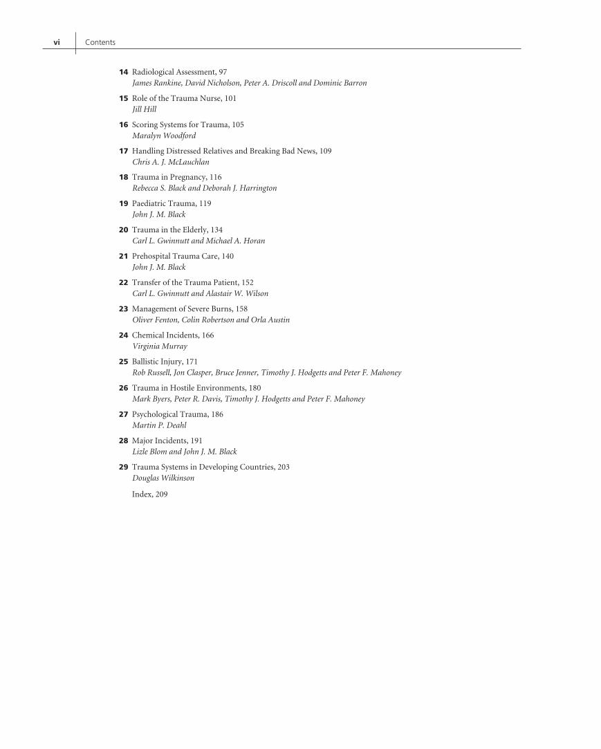
14 Radiological Assessment, 97James Rankine, David Nicholson, Peter A. Driscoll and Dominic Barron
15 Role of the Trauma Nurse, 101Jill Hill
16 Scoring Systems for Trauma, 105Maralyn Woodford
17 Handling Distressed Relatives and Breaking Bad News, 109Chris A. J. McLauchlan
18 Trauma in Pregnancy, 116Rebecca S. Black and Deborah J. Harrington
19 Paediatric Trauma, 119John J. M. Black
20 Trauma in the Elderly, 134Carl L. Gwinnutt and Michael A. Horan
21 Prehospital Trauma Care, 140John J. M. Black
22 Transfer of the Trauma Patient, 152Carl L. Gwinnutt and Alastair W. Wilson
23 Management of Severe Burns, 158Oliver Fenton, Colin Robertson and Orla Austin
24 Chemical Incidents, 166Virginia Murray
25 Ballistic Injury, 171Rob Russell, Jon Clasper, Bruce Jenner, Timothy J. Hodgetts and Peter F. Mahoney
26 Trauma in Hostile Environments, 180Mark Byers, Peter R. Davis, Timothy J. Hodgetts and Peter F. Mahoney
27 Psychological Trauma, 186Martin P. Deahl
28 Major Incidents, 191Lizle Blom and John J. M. Black
29 Trauma Systems in Developing Countries, 203Douglas Wilkinson
Index, 209

List of Contributors
Munawar Al-MudhaffarSpecialist Registrar in Emergency Medicine
Department of Emergency Medicine
John Radcliffe Hospital
Oxford, UK
Orla AustinDepartment of Plastic Surgery
Pinderfields Hospital
Wakefield, UK
Dominic BarronConsultant Radiologist
Leeds General Infirmary
Leeds, UK
John J.M. BlackMedical Director, South Central Ambulance Service;
Consultant in Emergency Medicine
John Radcliffe Hospital
Oxford, UK
Rebecca S. BlackDepartment of Obstetrics
John Radcliffe Hospital
Oxford, UK
Lizle BlomConsultant in Emergency Medicine
Royal Berkshire Hospital
Reading, UK
Andrew BlythRoyal Berkshire Hospital
Reading, UK
Mark ByersMinistry of Defence
London, UK
Jon ClasperDefence Professor, Orthopaedics and Trauma
Royal Centre for Defence Medicine
Birmingham, UK
Andre CromhoutEmergency Physician
Emergency Department
Welllington Hospital
Wellington, New Zealand
Peter R. DavisConsultant in Emergency Medicine
Defence Medical Services and Honorary Consultant
Southern General Hospital
Glasgow, UK
Martin P. DeahlConsultant Psychiatrist
South Staffordshire and Shropshire Healthcare NHS Foundation Trust
Stafford, UK
Anthony DeaneConsultant Urologist
William Harvey Hospital
Ashford;
Buckland Hospital
Dover, UK
Peter A. DriscollConsultant in Accident and Emergency Medicine
Department of Emergency Medicine
Hope Hospital
Salford, UK
Karen A. EleyResearch Fellow
University of Oxford
Oxford, UK
John ElstonConsultant Ophthalmologist
Oxford Eye Hospital
Oxford University Hospitals NHS Trust
Oxford, UK
vii

viii List of Contributors
Oliver FentonDepartment of Plastic Surgery
Pinderfields Hospital
Wakefield, UK
Andrew GibsonConsultant Ophthalmologist
The James Cook University Hospital
Middlesbrough, UK
Carl L. GwinnuttConsultant Anaesthetist
Hope Hospital
Salford, UK
Deborah J. HarringtonDepartment of Obstetrics
John Radcliffe Hospital
Oxford, UK
Jill HillSenior Sister
Emergency Department
John Radcliffe Hospital
Oxford, UK
Timothy J. HodgettsChief Medical Officer
NATO Allied Rapid Reaction Corps;
Honorary Professor of Emergency Medicine
University of Birmingham
Birmingham, UK
Michael A. HoranHope Hospital
Salford, UK
Philip HormbreyConsultant in Emergency Medicine
Department of Emergency Medicine
John Radcliffe Hospital
Oxford, UK
Tom HughesConsultant in Emergency Medicine
John Radcliffe Hospital, Oxford;
Consultant in Emergency Medicine and Clinical
Director of Emergency Care
Hinchingbrooke Hospital, Huntingdon;
Hon. Senior Lecturer in Emergency Medicine
University of Oxford
Oxford, UK
Bruce JennerTrauma Nurse
Royal Air Force
UK
Rohit KotnisSpecialist Registrar in Trauma and Orthopaedics
John Radcliffe Hospital
Oxford, UK
Peter F. MahoneyDefence Professor Anaesthetics and Critical Care
Royal Centre for Defence Medicine
Birmingham, UK
Ajith MalalasekeraDepartment of Urology
Leicester General Hospital
Leicester, UK
Chris A.J. McLauchlanConsultant in Emergency Medicine
Emergency Department
Royal Devon and Exeter Hospital
Exeter, UK
Lisa E. Munro-DaviesConsultant in Emergency Medicine
University Hospitals Bristol NHS Foundation Trust
Bristol, UK
Virginia MurrayConsultant Medical Toxicologist and Environmental Public Health
Head of Extreme Events and Health Protection
Health Protection Agency
London, UK
David NicholsonConsultant Radiologist
Department of Emergency Medicine
Hope Hospital
Salford, UK
Jerry P. NolanConsultant Anaesthetist
Department of Anaesthesia and Intensive Care Medicine
Royal United Hospital
Bath, UK
Rachael Pery-JohnstonDepartment of Emergency Medicine
Princess Alexandra Hospital
Brisbane, QLD, Australia
Catherine PetersConsultant in Critical Care Medicine and Anaesthesia
Department of Intensive Care
Homerton University Hospital
London, UK

List of Contributors ix
Rick PullingerConsultant in Emergency Medicine
Department of Emergency Medicine
John Radcliffe Hospital
Oxford, UK
Jaskarn RaiDepartment of Urology
Leicester General Hospital
Leicester, UK
James RankineConsultant Radiologist
Leeds General Infirmary
Leeds, UK
Colin RobertsonConsultant in Accident and Emergency Medicine
Department of Emergency Medicine
Royal Infirmary of Edinburgh
Edinburgh, UK
Nigel RossiterConsultant Trauma and Orthopaedic Surgeon
Basingstoke General Hospital;
Royal Defence Medical College
Basingstoke, UK
Rob RussellRoyal Army Medical Corps Consultant
Emergency Medicine
Royal Centre for Defence Medicine
Birmingham, UK
David V. SkinnerConsultant in Emergency Medicine
John Radcliffe Hospital
Oxford, UK
Andrew SwainSenior Lecturer and Consultant in Emergency Medicine
University of Otago
Wellington, New Zealand
Timothy TerryConsultant Urologist
Department of Urology
Leicester General Hospital
Leicester, UK
David WatsonHonorary Professor of Intensive Care Education
Department of Medical Education
Homerton University Hospital
London, UK
Steve R. Watt-SmithConsultant Maxillofacial Surgeon
Honorary Clinical Senior Lecturer
Oxford University Hospitals NHS Trust
University of Oxford
Oxford, UK
Douglas WilkinsonConsultant Anaesthetist in Intensive Care
Oxford University Hospitals NHS Trust
Oxford, UK
Keith WillettProfessor of Trauma Surgery
John Radcliffe Hospital
Oxford, UK
Alastair W. WilsonConsultant in Emergency Medicine
Royal London Hospital
London, UK
Sarah J. WilsonConsultant in Emergency Medicine
Wexham Park Hospital
Slough, UK
Maralyn WoodfordExecutive Director
The Trauma Audit and Research Network
University of Manchester
Manchester, UK


Foreword
As a contributor and user of this book, I am delighted to seea fourth edition published. It represents a most useful core textfor those seeking a contemporary practical guide to assess anddeliver the best trauma care for those patients who are ‘‘candidatemajor traumas’’. All counties and healthcare systems are differentand some changing, but the core principles of management arecommon. This text, through its breadth of expert contributors,succinctly describes these. Reading each chapter feels like you’vejust had a really good tutorial on the subject and represents a
very efficient method of acquiring knowledge. I would certainlyrecommend you keep a copy of this publication close by whetherpreparing for your on-call day or a teaching session.
Keith WillettProfessor of Trauma Surgery
John Radcliffe HospitalOxford, UK
xi


Preface
This edition of the ABC of Major Trauma has had a long gestation,being 12 years since the third edition.
Trauma care continues to evolve and improve both in the ‘frontline’ and nationally with the development of Trauma Centres andsupporting networks. Prevention is also playing its part, with deathson the roads continuing to fall.
There is, however, no room for complacency and we hope thisfourth edition will remind our readership of the crucial importanceof a thorough, stepwise assessment of the trauma patient and thatattention to detail, not least in spinal care, can avoid the devastatingconsequences of the ‘second’ injury.
This edition sees extensive revision of all its chapters and the addi-tion of further material. At the time of publishing all informationis current.
The book is aimed at all clinicians involved in front line traumacare, paramedics, hospital doctors and nurses as well as those
members of the ‘team’, crucial to optimal management, includingradiographers, radiologists and laboratory staff.
Chapter 29 reminds us of the excellent facilities available to usin UK practice. However, this chapter also shows us that simplemanoeuvres can be life saving in the Third World environment.
The continuing conflicts around the world involving UK armedforces has resulted in improved trauma management in theseconflict zones. Lessons learnt and techniques developed have beenshared with civilian clinicians to the benefit of patients. Manyauthors in this edition have put themselves in ‘harms way’ tomanage victims of conflict. This edition is dedicated to them.
David V. Skinner
xiii


Acknowledgements
I would like to acknowledge the help and guidance afforded to meby all at Wiley-Blackwell during the course of the production ofthis fourth edition of the ABC of Major Trauma.
I would like in particular to thank Adam Gilbert, Vicki Donald,Ilaria Meliconi, Laura Quigley, Kate Newell, Cathryn Gates and
Helen Harvey whose support, encouragement, good humour andpure professionalism have seen this task to completion.
David V. Skinner
xv


List of Abbreviations
5-HT 5-hydroxytryptamineABC airway, breathing, circulationACE angiotensin-converting enzyme inhibitorAIC ambulance incident commanderAIS Abbreviated Injury ScaleAP anteroposteriorAPLS Advanced Paediatric Life SupportARDS acute respiratory distress syndromeASD acute stress disordersATLS Advanced Trauma Life SupportATP adenosine triphosphateBATLS Battlefield Advanced Trauma Life SupportBP blood pressureBVM bag-valve-maskCAT computed axial tomographyCBF cerebral blood flowCBT cognitive-behavioural therapyCCS casualty clearing stationCHaPD Chemical Hazards and Poisons DivisionCOPD chronic obstructive pulmonary diseaseCPB cardiopulmonary bypassCPP cerebral perfusion pressureCPR cardiopulmonary resuscitationCRT capillary refill timeCSF cerebrospinal fluidCT computed tomographyCXR chest X-rayDCLHb diaspirin cross-linked haemoglobin solutionDIC disseminated intravascular coagulationDPL diagnostic peritoneal lavageDVT deep venous thrombosisECG electrocardiogramECMO extracorporeal membranous oxygenationED emergency departmentEEG electroencephalogramET endotrachealETA expected time of arrivalETCO2 end-tidal carbon dioxide concentrationFAST focused assessment with sonography in traumaFBC full blood countFFP fresh frozen plasmaFiO2 fraction of inspired oxygen
FRC functional residual capacityGCS Glasgow Coma ScaleGDP Gross Domestic ProductGP general practitionerHART hazardous area response teamHBOC haemoglobin-based oxygen carrierHCT hospital co-ordination teamHCVR hypercapnic ventilatory responseHPA Health Protection
Agency/hypothalamo-pituitary-adrenalHR heart rateHVR hypoxic ventilatory responseICP intracranial pressureICU intensive care unitI/E inspiratory/expiratoryIED improvised explosive deviceIO intraosseousIPE individual protective equipmentISS Injury Severity ScoreITU intensive therapy unitIV intravenousMAP mean arterial pressureMERIT medical emergency incident response teamMIC medical incident commanderMODS multiorgan dysfunction syndromeMRI magnetic resonance imagingNAI non-accidental injuryNGT nasogastric tubeNICE National Institute for Health and Clinical ExcellenceNPIS National Poisons Information ServiceNSAID non-steroidal anti-inflammatory drugPEEP positive end-expiratory pressurePPE personal protective equipmentPPH postpartum haemorrhagePTA post-traumatic amnesiaPTC Primary Trauma CarePTSD post-traumatic stress disorderRSI rapid-sequence inductionRTA road traffic accidentRTC road traffic crashRTS Revised Trauma ScoreSaO2 oxygen saturation
xvii

xviii List of Abbreviations
SCIWORA spinal cord injury without radiological abnormalitySHO senior house officerSIGN Scottish Intercollegiate Guidelines NetworkSIRS systemic inflammatory response syndromeSXR plain radiograph of the skullTARN Trauma Audit and Research Network
TNF tumour necrosis factorUS ultrasoundUSAR urban search and rescueWHO World Health OrganizationZPP zone of partial preservation

CHAPTER 1
Initial Assessment and Management:Primary Survey and Resuscitation
David V. Skinner1 and Peter A. Driscoll2
1John Radcliffe Hospital, Oxford, UK2Hope Hospital, Salford, UK
OVERVIEW
• Initial management of trauma victims requires a team approachin which each member carries out a specific task. Collectively,the team should aim to treat all the immediately life-threateningconditions and identify the need for surgery early.
• The ABC (airway, breathing, circulation) approach provides anoptimal system whereby urgent, potentially life-threateningconditions are dealt with first.
• The critically injured patient requires a calm rapid response tohis/her injuries, in the field, resuscitation room and operatingtheatre. If prehospital personnel, the resuscitation room teamand its leader, as well as the appropriate surgeons can deliverthis, then lives will be saved and unnecessary deaths avoided.Any deaths that do occur will have been unavoidable. The teamshould also be aware of this and suitably debriefed.
Morbidity and mortality in seriously injured patients, managedin UK hospitals, remain higher than necessary. Recognition ofthis problem over the last 25 years has seen a variety of initia-tives designed to improve the situation, including the introductionof Advanced Trauma Life Support (ATLS) to clinical practice,the widespread use of the auditing tool TARN (Trauma Auditand Research Network), and the deployment of multidisciplinarytrauma teams to manage trauma victims in emergency depart-ment (ED) resuscitation rooms. Increasingly, consultant-deliveredservices, where available, will further enhance care.
For each individual patient, however, survival and reduction oflong-term disability depend on the rapid deployment of skilledprehospital clinicians (paramedics and/or doctors), the skills andexperience of the receiving clinicians (trauma team) and the humanand other resources available round the clock to deal with patientinjuries in a timely and effective fashion.
Most seriously injured patients seen in UK EDs have sufferedblunt trauma. This, by its very nature, presents its own uniqueset of difficulties for the clinician, not least because serious life-threatening injuries may be initially covert, especially in the young.Prehospital clinicians may not recognise potential problems; this
ABC of Major Trauma, Fourth Edition. Edited by David V. Skinner and Peter A. Driscoll.© 2013 Blackwell Publishing Ltd. Published 2013 by Blackwell Publishing Ltd.
may be further compounded by a failure of recognition by thereceiving hospital, leading to inappropriate triage. Lone juniordoctors may then find themselves assessing a deteriorating traumapatient in an unmonitored area of the ED, leading to potentialcatastrophe.
All ED doctors should therefore be ATLS trained and encouragedto have a very low threshold for ‘upgrading’ such patients withoutdelay to the resuscitation room for a team response. Such upgradeshould include not only the deteriorating patient, but also those inwhom the mechanism of injury suggests the possibility of seriousproblems. In the authors’ experience, most problems arise from afailure to understand, or take note of, the mechanism’s injuriouspotential, rather than poor management of an overtly seriouslyinjured patient.
Comprehensive management protocols (usually ATLS) must befollowed to the letter. Short cuts expose patients to risk whichwill lead some into difficulty. The ‘experienced’ clinician’s personalopinion must be outweighed every time by the multitude of expe-rienced clinicians who devised the protocol. Such protocols arefrequently driven by the need to avoid the errors of the past.
The introduction of trauma centres will hopefully produce afurther improvement in trauma care but in the end, individualclinicians, either working alone or as trauma team leaders, bear theresponsibility for ensuring optimum care.
Effective ED care depends on the following.
• Safe, accurate receipt of prehospital information regarding thetrauma victim or victims.
• Assembly of a competent trauma team, competently led, anddressed in protective clothing.
• The team’s ability to identify immediately life-threatening prob-lems and begin their correction.
• Limiting investigations and interventions to those crucial toaddressing life-threatening problems.
• Ready availability of all investigation modalities, and a suitablyurgent response by labs, radiology, intensive therapy units (ITU)and theatres.
• The additional ability to sensibly allocate resources when amultivictim response is needed.
Trauma centre ‘feeder units’ will not have the resources andmanpower to provide a full trauma team response 24 hours a day,
1
2 ABC of Major Trauma
7 days a week. In spite of this and given the difficulty of completetriage accuracy in the prehospital field, seriously injured patientswill continue to arrive at such feeder centres. It is crucial thereforethat such patients are managed in a logical way, based on ATLS,before possible onward transfer to a trauma centre. The maindifference will be that the resuscitation phase will take longer giventhe reduced numbers in the trauma team.
Where a trauma team can be made available round the clockthen it is in the patient’s interests that it should be deployed. Thefollowing text suggests one way in which such a team should bedeveloped and deployed. Individual centres will decide on the exactcomposition of such teams and comparative national data willidentify the optimum team size and composition.
The trauma team
PersonnelThe trauma team (Figure 1.1) should initially comprise four doctors,five nurses and a radiographer. The medical team consists of a teamleader, an ‘airway’ doctor and two ‘circulation’ doctors. The nursingteam comprises a team leader, an ‘airway’ nurse, two ‘circulation’nurses and a ‘relatives’ nurse.
Team members’ rolesExamples of paired roles and tasks are given below but assignmentsmay vary among units depending on the resources available. Toavoid chaos, no more than six people should be touching the patient.The other team members must keep well back. The objectives ofthe trauma team are shown in Box 1.1.
Box 1.1 Objectives of the trauma team
• Identify and correct life-threatening injuries.• Commence resuscitation.• Determine the nature and extent of other injuries.• Prioritise investigation/treatment needs.• Prepare and transport the patient to a place of continuing care.
Figure 1.1 Trauma team in action.
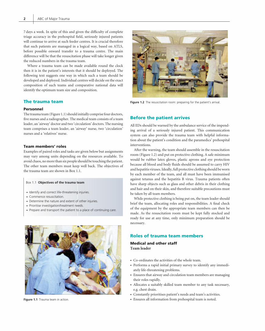
Figure 1.2 The resuscitation room: preparing for the patient’s arrival.
Before the patient arrives
All EDs should be warned by the ambulance service of the impend-ing arrival of a seriously injured patient. This communicationsystem can also provide the trauma team with helpful informa-tion about the patient’s condition and the paramedics’ prehospitalinterventions.
After the warning, the team should assemble in the resuscitationroom (Figure 1.2) and put on protective clothing. A safe minimumwould be rubber latex gloves, plastic aprons and eye protectionbecause all blood and body fluids should be assumed to carry HIVand hepatitis viruses. Ideally, full protective clothing should be wornby each member of the team, and all must have been immunisedagainst tetanus and the hepatitis B virus. Trauma patients oftenhave sharp objects such as glass and other debris in their clothingand hair and on their skin, and therefore suitable precautions mustbe taken by all team members.
While protective clothing is being put on, the team leader shouldbrief the team, allocating roles and responsibilities. A final checkof the equipment by the appropriate team members can then bemade. As the resuscitation room must be kept fully stocked andready for use at any time, only minimum preparation should benecessary.
Roles of trauma team members
Medical and other staffTeam leader
• Co-ordinates the activities of the whole team.• Performs a rapid initial primary survey to identify any immedi-
ately life-threatening problems.• Ensures that airway and circulation team members are managing
their roles rapidly.• Allocates a suitably skilled team member to any task necessary,
e.g. chest drain.• Constantly prioritises patient’s needs and team’s activities.• Ensures all information from prehospital team is noted.

Initial Assessment and Management: Primary Survey and Resuscitation 3
• Ensures that other specialist clinicians are urgently alerted as soonas their need is identified.
Airway doctor
• Clears and secures the airway while taking appropriate cervicalspine precautions.
• Inserts central and arterial lines if required.
Circulation doctors
• Assist in the removal of the patient’s clothes.• Establish peripheral intravenous infusions and take blood samples
for investigations.• Carry out other procedures depending on their skill level.
Radiographer
• Takes three standard X-ray films on all patients subjected to blunttrauma: chest, pelvis and lateral cervical spine.
Nursing staffTeam leader
• Co-ordinates the nursing team and liaises with the medical teamleader.
• Records clinical findings, laboratory results, intravenous fluid anddrug infusion, and the vital signs as called out by the circulationnurse.
• Prepares sterile packs for procedures.• Assists the circulation nurses and brings extra equipment as
necessary.
Airway nurse
• Assists in securing the airway and the cervical spine.• Establishes a rapport with the patient in the resuscitation room.
Ideally all information should be fed through this nurse to thepatient.
Circulation nurses
• Assist in the removal of the patient’s clothes.• Assist with starting intravenous infusions, blood bottle labelling
and other tasks allocated to the circulation doctor.• Measure the vital signs and connect the patient to the monitors.
Relatives’ nurse
• Cares for the patient’s relatives.
Reception and transfer
The team leader should meet the patient and prehospital team in theambulance bay and accompany them to the resuscitation room.The essential prehospital information required by the trauma teamis shown in Box 1.2. The nursing team leader should start the stopclock so that accurate times can be recorded.
Box 1.2 Essential prehospital information
• Nature of the incident.• Number, age and sex of the casualties.• The patient’s complaints, priorities and injuries.• Airway, ventilatory and circulatory status.• The conscious level and spinal status.• Estimated time of arrival.
The transfer of the patient from stretcher to trolley must be co-ordinated to avoid rotation of the spinal column or exacerbation ofpre-existing injuries (see Chapter 8). Team members should alsocheck that lines and leads are free so that they do not becomedisconnected or snagged.
Primary survey and resuscitation
The objectives of this phase are to identify and treat any immediatelylife-threatening condition (Box 1.3). Each patient should be assessedin the same way, and the appropriate tasks performed automaticallyand simultaneously by the team. It is vital that problems areanticipated and prepared for, rather than reacted to. If the patientdeteriorates at any stage, the medical team leader must reassess thepatient, beginning again with the airway.
Box 1.3 Objectives
• Primary survey and resuscitation
Airway and cervical spine controlBreathingCirculation and haemorrhage controlDysfunction of the central nervous systemExposure and environmental control
• Secondary survey• Definitive care
As previously suggested, the team leader should perform a rapidprimary survey to identify immediately life-threatening conditions.This should take no longer than 1–2 min. The management ofindividual problems identified in this rapid primary survey isdetailed below. The tasks are allocated to team members andtake place concurrently rather than in a stepwise approach as laidout below.
Airway management, with cervical spineprotectionAssume that the cervical spine has been damaged if there is ahistory of a high-speed impact, head injury, neck pain or anypositive neurology. If the ambulance service have immobilised thenassume a C-spine injury until proven otherwise. The doctor dealingwith the airway should talk to the patient with the neck immobileand if the patient replies appropriately with a normal voice, then

4 ABC of Major Trauma
the airway is patent and the brain is being perfused adequately withoxygenated blood. If there is no reply, the patient’s airway shouldbe checked, cleared and managed appropriately. This is the first andpre-eminent priority.
The complications of alcohol ingestion and possible injuries tothe chest and abdomen increase the chance of regurgitation. If thepatient does vomit and is on a spinal board, the trolley should betipped head down by 20◦ and the vomit sucked away with a rigidsucker as it appears in the mouth. If not on a spinal board then logroll the patient and suck out.
Progressively interventionist manoeuvres should be employed asnecessary including chin lift/jaw thrust, Guedel airway (Figure 1.3)insertion (although these can precipitate vomiting) and deploymentof a nasopharyngeal airway (Figure 1.4) which is preferred (lesslikely to cause vomiting) provided that there is no evidence of abase of skull fracture.
Apnoeic patients require ventilation with a bag-valve-maskdevice initially but this may lead to gastric distension with airand can induce vomiting, so early intubation should be considered.Orotracheal intubation with in-line stabilisation of the neck is
Figure 1.3 Guedel airway.
Figure 1.4 Nasopharyngeal airway.
Figure 1.5 Patient with rigid collar in place.
recommended, rather than nasotracheal intubation. If this provesimpossible (rarely) then a surgical airway must be provided.
Once the airway has been cleared and secured, every patientshould receive 100% oxygen at a flow rate of 15 L/min. The neckmust then be examined for wounds, tracheal position, venousdistension, surgical emphysema and laryngeal crepitus. Confirma-tion of the security of the cervical spine, using a semi-rigid collar,sand bags and tape, is crucial. The only exception is the restlessand thrashing patient. Here the cervical spine can be damaged byimmobilising the head and neck while allowing the rest of the bodyto move. Suboptimal immobilisation with just a semi-rigid collar istherefore accepted (Figure 1.5).
BreathingListed in Box 1.4 are five immediately life-threatening thoracicconditions that must be urgently identified and treated during theprimary survey and resuscitation phase (see Chapter 4).
Box 1.4 Immediately life-threatening thoracic conditions
• Tension pneumothorax.• Cardiac tamponade.• Open chest wound.• Massive haemothorax.• Flail chest.
All clothes covering the front and sides of the chest must beremoved. The respiratory rate, effort and symmetry must be noted.These are sensitive indicators of underlying pulmonary contu-sion, haemothorax, pneumothorax and fractured ribs. The teamleader must examine both sides of the chest for bruising, abra-sions, open wounds and evidence of penetrating trauma. Cardiactamponade after trauma is usually associated with a penetrat-ing injury. The team leader should also remember that becauseof intercostal muscle spasm, paradoxical breathing is seen witha flail chest only if the segment is large or central, or whenthe patient’s muscles become fatigued. The patient with a flail

Initial Assessment and Management: Primary Survey and Resuscitation 5
chest usually has a rapid, shallow, symmetrical respiratory patterninitially.
After inspection, the chest should be auscultated and percussedto assess symmetry of ventilation and resonance. As listening overthe anterior chest detects mainly air movement in the large air-ways, it is recommended that the medical team leader also listensover the axillae to gain a more accurate assessment of pulmonaryventilation. A tension pneumothorax or massive haemothorax canthus be identified. Early chest X-ray (CXR) is crucial: clinicalexamination in the context of major trauma is unreliable. Pneu-mothorax or haemothorax should be treated by inserting a chestdrain with a gauge of >28 in the fifth intercostal space just anteriorto the midaxillary line. This enables air and fluid to be drainedbut should always be preceded by intravenous lines. During exam-ination of the chest, the patient should be attached to a pulseoximeter. Common causes of inadequate ventilation are shownin Box 1.5.
Box 1.5 Common causes of inadequate ventilation
• Bilateral
– Obstruction of the upper respiratory tract– Leak between the face and mask
• Unilateral
– Pneumothorax– Haemothorax– Intubation of the right main bronchus– Foreign body in a main bronchus, significant lung contusion
Circulation and haemorrhage controlThe medical team leader will look for clinical signs of shock (seeChapter 5), apparent with tachycardia, poor capillary refill andperipheral perfusion. It is important to remember that up to 30%loss of blood volume produces tachycardia and reduces the pulsepressure, but the blood pressure may stay within normal limits(particularly in the young).
There is a consistent fall in the systolic blood pressure only whenmore than 30% of the blood volume has been lost.
The circulation doctor must control any major external haem-orrhage by direct pressure. Tourniquets are usually only used whenthe affected limb is deemed unsalvageable.
A pelvic splint should be used when there is a suspected pelvicfracture and this may already have been applied by the prehospitalteam.
Concurrently with the above, two wide-bore (14–l6 gauge)peripheral lines must be inserted, preferably in the antecubitalfossae. If this is impossible, venous access should be gained by avenous cutdown or by inserting a short, wide-bore central lineinto the femoral or subclavian vein. If a subclavian approach isused and a chest drain is already in place, the central line mustbe inserted on the same side. As central vein cannulation can
cause serious injury, it should be carried out only by experiencedpersonnel.
Once the first cannula is in position, 20 mL of blood should bedrawn for group, type or full cross-match, full blood count, andmeasurement of urea and electrolyte concentrations. An arterialsample should also be taken for blood gas and pH analysis, but thiscan wait until the end of the primary survey. While venous access isbeing gained, a circulation nurse must measure the blood pressureand record the rate, volume and regularity of the pulse. An auto-matic blood pressure recorder and electrocardiogram (ECG) mon-itor should also be attached to the patient. In seriously ill patients,palpating femoral and carotid pulses is a quick and reliable methodof establishing whether there is some cardiac output when no bloodpressure can be recorded, either automatically or otherwise.
In the UK, the type of fluid initially given to injured patientsto maintain fluid balance depends on departmental policy. Somestart with colloid while others use crystalloid such as physiologicalsaline. It is therefore important for team leaders to know the localpolicy. The aim of fluid management in a hypotensive resuscitationshould be to restore critical organ perfusion until haemorrhage thatis amenable to surgery is stemmed. Therefore the initial approachin a standard adult trauma victim is to give 1 L of warm colloid(or 2 L of crystalloid) and then reassess the patient. Rememberthat the best colloid is blood and, where necessary, this should begiven as soon as possible. This underlines the importance of earlycross-match and an effective chain between the resuscitation room,labs and back again!
When there is a limited response to the fluid bolus, or after amajor injury, blood is urgently required. To reduce the incidenceof hypothermia, all fluids must be warmed before use.
In reassessing the circulatory state one of three responses will beseen (Figure 1.6).
• The vital signs return to normal after infusion of less than 1 Lof colloid solution (or 2 L of physiological saline). Such patientshave lost less than 20% of their blood volume and are probablynot actively bleeding.
• The vital signs initially improve with the infusion but then dete-riorate. These patients are actively bleeding and have usually lostmore than 20% of their blood volume. They require transfu-sion with typed blood and the source of the bleeding must becontrolled. This often requires surgery.
• The vital signs do not improve at all. This suggests either that theshock has not been caused by hypovolaemia or that the patient isbleeding faster than blood is being infused. History, mechanismof injury and physical findings will help to distinguish betweenthese possibilities.
Measurement of the central venous pressure and, in particular, itschange after a fluid bolus may assist in diagnosis. There are a limitednumber of anatomical sites of bleeding: external, into the chest orabdomen, or around a fractured pelvis or long bones. Skilled FAST(focused assessment with sonography in trauma) ultrasound willconfirm intra-abdominal blood and clinical examination and CXRwill identify the others.


