Mailing address: Wei-Cai Yang, Key Laboratory - Plant Physiology
37
1 Running head: SWA2 is essential for gametogenesis Corresponding author: Mailing address: Wei-Cai Yang, Key Laboratory of Molecular and Developmental Biology Institute of Genetics and Developmental Biology Chinese Academy of Sciences Datun Road, Chaoyang District Beijing 100101, China Tel: +861062551272; Fax: +861062551272; E-mail: [email protected] Journal research area: Systems biology, molecular biology, and gene regulation Plant Physiology Preview. Published on September 4, 2009, as DOI:10.1104/pp.109.142414 Copyright 2009 by the American Society of Plant Biologists www.plantphysiol.org on April 3, 2019 - Published by Downloaded from Copyright © 2009 American Society of Plant Biologists. All rights reserved.
Transcript of Mailing address: Wei-Cai Yang, Key Laboratory - Plant Physiology

1
Running head: SWA2 is essential for gametogenesis
Corresponding author:
Mailing address: Wei-Cai Yang,
Key Laboratory of Molecular and Developmental Biology
Institute of Genetics and Developmental Biology
Chinese Academy of Sciences
Datun Road, Chaoyang District
Beijing 100101, China
Tel: +861062551272; Fax: +861062551272; E-mail: [email protected]
Journal research area: Systems biology, molecular biology, and gene regulation
Plant Physiology Preview. Published on September 4, 2009, as DOI:10.1104/pp.109.142414
Copyright 2009 by the American Society of Plant Biologists
www.plantphysiol.orgon April 3, 2019 - Published by Downloaded from Copyright © 2009 American Society of Plant Biologists. All rights reserved.
2
SLOW WALKER2, a NOC1/Mak21 Homologue, Is Essential for Coordinated Cell
Cycle Progression during Female Gametophyte Development in Arabidopsis
Na Li a,b,1, Li Yuan a,b,1, Naiyou Liu a,b, Dongqiao Shi a, Xinran Lia, Zuoshun Tang
a, Jie Liu a, Venkatesan Sundaresan c, and Wei-Cai Yanga*
a Key Laboratory of Molecular and Developmental Biology, Institute of Genetics and
Developmental Biology, Chinese Academy of Sciences, 1 West Beichen Road,
Chaoyang District, Beijing 100101, China;
b Graduate University of Chinese Academy of Sciences, 19 Yuquan Road, Beijing
100039, China;
c Plant Biology and Agronomy, Life Sciences Addition 1002, University of California,
Davis, California 95616
1 These authors contributed equally to this work.
*Corresponding author; e-mail: [email protected]; fax 86-10-62551272
www.plantphysiol.orgon April 3, 2019 - Published by Downloaded from Copyright © 2009 American Society of Plant Biologists. All rights reserved.

3
Footnotes
This work was supported by grants from the Ministry of Science and Technology
(2007CB108702), the National Science Foundation of China (30830063), and State
Key Laboratory of Crop Biology at Shandong Agricultural University, China.
www.plantphysiol.orgon April 3, 2019 - Published by Downloaded from Copyright © 2009 American Society of Plant Biologists. All rights reserved.

4
ABSTRACT
Morphogenesis requires the coordination of cell growth, division, and cell
differentiation. Female gametogenesis in flowering plants, where a single haploid
spore undergoes continuous growth and nuclear division without cytokinesis to form
an eight-nucleate coenocytic embryo sac before cellularization, provides a good
system to study the genetic control of such processes in multicellular organisms. Here
we reported the characterization of an Arabidopsis female gametophyte mutant, slow
walker2 (swa2), in which the progression of the mitotic cycles and the synchrony of
female gametophyte development were impaired, causing an arrest of female
gametophytes at two-, four- or eight-nucleate stages. Delayed pollination test showed
that a portion of the mutant ovules were able to develop into functional embryo sacs
and could be fertilized. SWA2 encodes a nucleolar protein homologous to yeast
NOC1/MAK21 that, together with NOC2, is involved in pre-ribosome export from
the nucleus to cytoplasm. Similarly, SWA2 can physically interact with a putative
Arabidopsis NOC2 homologue. SWA2 is expressed ubiquitously throughout the plant,
at high levels in actively dividing tissues and gametophytes. Together, we conclude
that SWA2 most likely plays a role in ribosome biogenesis that is essential for the
coordinated mitotic progression of the female gametophyte.
Key words: Arabidopsis, Gametophyte, SWA2, Nucleolar protein, Ribosome
biogenesis
www.plantphysiol.orgon April 3, 2019 - Published by Downloaded from Copyright © 2009 American Society of Plant Biologists. All rights reserved.

5
INTRODUCTION
Morphogenesis requires tightly coordinated coupling of cellular activities, such as
cell growth, cell division and differentiation. In past decades, significant progresses
on cell cycle control have been achieved mostly in single-celled organisms and
cultured mammalian cells. The elucidation of the cyclin/CDK checkpoint control, for
example, provides insight into molecular mechanisms on how and when cells divide.
Mechanisms coupling cell growth to environmental and developmental signals have
also been investigated. Ribosome biogenesis, a key for rapid cell growth, is coupled
with nutrient availability and stress signals via the TOR signaling pathway
(Wullischleger et al., 2006; Warner et al., 2001). However, questions such as how cell
senses intrinsic cellular homeostatic signals remain to be addressed. For example,
how ribosome dynamics and translational activities are measured and coupled to
cytokinesis and cell differentiation, especially in the context of development of
multicellular organisms.
Female gametogenesis in Arabidopsis is a unique system to address such question
in multicellular organisms. During female gametogenesis, the haploid functional
megaspore undergoes continuous cell growth and three cycles of consecutive nuclear
division without cytokinesis, giving rise to a giant eight-nucleate, coenocytic cell-the
embryo sac. The size of the embryo sac increases about six-fold without cytokinesis
until it reaches its maximum during the gametogenesis in maize (Dow and
Mascarenhas, 1991). The two polar nuclei migrate toward the micropylar half of the
embryo sac and eventually fuse to give rise to a diploid nucleus of the central cell. As
the polar nuclei migrate, cellularization takes place simultaneously to divide the
coenocytic embryo sac into seven cells of four cell fates: three antipodal cells, two
synergid cells, one egg cell and one central cell (Drews et al., 1998; Grossniklaus and
Schneitz, 1998; Yang and Sundaresan, 2000; Wilson and Yang, 2004). Obviously, its
haploid nature and coupling of cell growth, division, and cell fates provided the
female gametophyte as a nice system to investigate how these cellular activities are
coordinated in development.
www.plantphysiol.orgon April 3, 2019 - Published by Downloaded from Copyright © 2009 American Society of Plant Biologists. All rights reserved.

6
The temporal and spatial control of cell growth, the mitotic division cycles and
cell fate specification during female gametogenesis has been the focus of sexual plant
reproduction research. Recently, genetic studies have identified gametophytic
mutations that start to shed light on the genetic and molecular control of these
processes. Mutations in genes involved in diverse cellular functions, including
ANDARTA (Howden et al., 1998), GAMETOPHYTIC FACTOR1 (GFA1) (Christensen
et al., 1997), HADAD (HDD) (Moore et al., 1997), LETHAL OVULE2 (LO2)
(Sheridan and Huang 1997), LYSOPHOSPHATIDYL ACYLTRANSFERASE (LPAT)
(Kim et al., 2005), NOMEGA (Kwee and Sundaresan, 2003), PROLIFERA (PRL)
(Springer et al., 1995), SLOW WALKER1 (SWA1) (Shi et al., 2005), SUCCINATE
DEHYDROGENASE (SDH1) (Leon et al., 2007), and TISTRYA (Howden et al., 1998),
all result in defective gametophytic cell divisions, implying that the progression of
mitotic cycle is critical for the formation of a functional female gametophyte. Loss of
function mutations in the Arabidopsis RETINOBLASTOMA-RELATED PROTEIN1
(RBR1), a key negative regulator controlling G1/S transition of the cell cycle, result in
uncontrolled nuclear proliferation and cell fates, giving rise to embryo sacs with
supernumerary nuclei that are irregular in size and partially enclosed by cell-wall-like
structures (Ebel et al., 2004). Loss of functions in CKI1 (Hejatko et al., 2003),
DIANA/AGL61 (DIA) (Bemer et al., 2008), AGAMOUS-LIKE80 (AGL80) (Portereiko
et al., 2006), and NUCLEAR FUSION DEFECTIVE1 (Portereiko et al., 2006) affect
polar nuclear fusion and central cell development.
Recently, accumulating data suggest a key role of nucleolus in cell survival and
proliferation (Cockell and Gasser, 1999; Shaw and Doonan, 2005). A number of
nucleolar proteins are discovered to be involved in linking the cell proliferation
control and ribosome biogenesis in yeasts (Srivastava and Pollard, 1999; Du and
Stillman, 2002; Jorgensen et al., 2002; Zhang et al., 2002; Bernstein et al., 2007).
Mutations in genes involved in RNA processing, including SWA1 (Shi et al., 2005),
GFA1/CLO1 and ATO (Moll et al., 2008; Liu et al., 2009; Yagi et al., 2009), lead to
slow progression of the division cycle during female gametogenesis. Intriguingly,
www.plantphysiol.orgon April 3, 2019 - Published by Downloaded from Copyright © 2009 American Society of Plant Biologists. All rights reserved.
7
mutation in LACHESIS (LIS), coding for a putative splicing factor, promotes egg cell
fate in the synergid and the central cell with the expense of the synergid and central
cell fate (Groß-Hardt et al., 2007), suggesting that LIS plays a pivotal role in
suppressing the egg cell fate in the synergid and the central cell as well as the central
cell fate in antipodal cells. Similarly, cell fate changes have also been observed in
gfa1/clo1 and ato mutant (Moll et al., 2008). These data imply that RNA processing
and ribosome biogenesis play a key role in coordinating cell cycle progression and
cell fate. Here, we report the genetic and molecular characterization of a slow walker2
(swa2) mutation that impairs cell growth and cell division in Arabidopsis. SWA2
encodes a nucleolar protein homologous to yeast NOC1/MAK21 that is essential for
ribosome biogenesis in yeast. We also showed that SWA2 interacts physically with
NOC2 homologues in yeast cells. Together, these data indicated that SWA2 is most
likely involved in ribosome biogenesis and essential for cell cycle progression in
female gametophyte development in Arabidopsis.
RESULTS
Isolation and Genetic Characterization of the swa2 Mutant
To identify mutations affected cell growth and division during female
gametophyte development, a screen for distorted Mendelian segregation was carried
out as described previously (Springer et al., 1995; Sundaresan et al., 1995; Pagnussat
et al., 2005). One mutant, swa2, was isolated which exhibited an aberrant KanR to
KanS segregation ratio of 1:1 (KanR: KanS=661:663) and siliques from swa2
heterozygous plants contained about 31.5% (n=964 ovules) aborted ovules (Figure 1),
suggesting that it is likely defective in gametophytic function.
To analyze the transmission of swa2 through female germ lines, we performed
crosses between swa2 and wild type plant and traced the presence of Ds insertion in
the F1 progeny. When the heterozygous mutant was used as the egg donor, the
transmission efficiency to the mutation was 19% (n=759), indicating that the mutation
has a strong defect in female gametophytes. In addition, ~6% (n=964) of selfed F1
www.plantphysiol.orgon April 3, 2019 - Published by Downloaded from Copyright © 2009 American Society of Plant Biologists. All rights reserved.

8
seeds displayed embryo arrest before globular stage, and no homozygous plants were
obtained. This suggested that the mutant is homozygous lethal. Therefore, we use
swa2 to represent the heterozygous mutant (Ds/+) in this paper. Overall, the mutant
plants are morphologically normal except that they bore shorter siliques than the
wild-type.
Synchrony of Female Gametophyte Development Is Impaired in swa2
To characterize the mutant phenotype, we examined ovule development in
wild-type and swa2 plants using confocal laser scanning microscopy (CLSM). Female
gametophyte (FG) development in Arabidopsis is divided into seven distinct stages
(Christensen et al., 1997; Christensen et al., 2002), as showed in Figure 2. The
functional megaspore (FG1, Figure 2A) undergoes mitosis to give rise to a
two-nucleate embryo sac (FG2, Figure 2B). A central vacuole is formed between the
two nuclei and pushes them apart from each other (FG3, Figure 2C). The two nuclei
undergo a second division to give rise to a four-nucleate embryo sac at early FG4
(Figure 2D). At this time, the division plane between the two chalazal nuclei lies
parallel to the chalazal-micropylar axis. Then, two of the four nuclei migrate so that
the division plane between them is orthogonal to the axis (Figure 2E). Another round
of division takes place to give rise to an eight-nucleate embryo sac (FG5, Figure 2F).
The two polar nuclei, one from each pole, migrate toward the micropylar half of the
developing female gametophyte (Figure 2G) and eventually fuse to form the central
cell (FG6, Figure 2H). The three antipodal cells degenerate just before fertilization
and the mature female gametophyte consists of two synergid cells, one egg cell and
one central cell (FG7, Figure 2I). In wild-type plants, ovule development in a pistil is
synchronous with only narrow range of variations ( Christensen et al., 1997; Shi et al.,
2005). At 24 hrs after emasculation (HAE), most ovules in a wild-type pistil were
found in FG6 or FG7. However, in the mutant pistils we found that about half of the
ovules were at FG6 or FG7 representing the wild-type ovules and the other half were
arrested at different stages, including the FG3, FG4, and FG5 (Figure 2, J-L). These
data indicated that the cell cycle progression in the mutant female gametophyte was
www.plantphysiol.orgon April 3, 2019 - Published by Downloaded from Copyright © 2009 American Society of Plant Biologists. All rights reserved.

9
impaired.
To further investigate whether the synchronous development of female
gametophytes in the mutant pistils was affected, we performed a detailed study of
synchrony of wild-type and mutant ovules. Pistils from the same inflorescence were
opened sequentially and ovules from each pistil were dissected out and checked for
their development stages. The numbers of ovules at each stage were counted. The
result is summarized in Table 1 and 2. In the wild-type plant, most ovules within the
same pistil are often at either one or two adjacent developmental stages (Table 1).
This observation is consistent with previous studies (Christensen et al., 1997; Shi et
al., 2005), which indicates that the embryo sac development within a pistil is
synchronous to a large extent. However, in the mutant plants, the development of the
embryo sacs are asynchronous, ovules within the same pistil are arrested at several
developmental stages (Table 2). These data suggested that the progression of the
gametophytic nuclear division was retarded in the mutant embryo sacs.
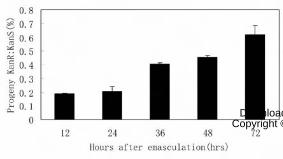
To investigate whether the retarded female gametophyte in the mutant ovule is
able to form functional embryo sac, we performed delayed pollination test according
to Shi et al. (2005). Pistils of swa2 plants were emasculated at floral stage 12c (Smyth
et al., 1990) and pollinated with pollens from wild-type plants at 12, 24, 36, 48, or 72
h after emasculation, respectively. F1 seeds from three independent plants of each
group were collected and examined for KanR:KanS ratio. As the pollination was
postponed, the KanR:KanS ratio of the F1 progeny increased from 18.8% when
pollinated at 12HAE to 60.7% when pollinated at 72HAE (Figure 3), indicating that
more mutant ovules were fertilized and produced seeds. These results suggested that
although the mutant ovules develop more slowly than their wild-type counterpart,
they have the potential to develop into functional female gametophytes and could be
fertilized.
In conclusion, the swa2 mutant displayed retarded progression of the
gametophytic division cycles and asynchronous development of the female
gametophyte. The mutant embryo sacs reach mature stage and could be fertilized by
delayed pollination.
www.plantphysiol.orgon April 3, 2019 - Published by Downloaded from Copyright © 2009 American Society of Plant Biologists. All rights reserved.

10
Pollen Development Is Defective in swa2 Mutant
To analyze whether the mutation also affected the male germ line, we performed
crosses between wild-type and swa2 plant and traced the presence of Ds insertion in
the F1 progeny. When the heterozygous mutant was used as the pollen donor, the
transmission efficiency was 82% (n=1374), indicated that the mutation has a slight
effect in male gametophytes.
To further clarify the defect in pollen, 4’,6-diamino-2-phenylindole (DAPI)
staining was performed to check male gametophytic cell cycle progression. At
anthesis, wild-type pollen completed mitosis II (PMII) and display a typical tricellular
configuration that the vegetative cytoplasm contains one vegetative nucleus and two
highly condensed sperm nuclei (Figure 4A). Wild-type plants showed less than 1%
aberrant pollen with disrupted positioning or aberrant appearance of the nuclei. In the
mutant, about 9.4% (n=832) pollen showed abnormal cell cycle, of which 5.3% pollen
grains were arrested at the bicellular stage (Figure 4B) and 4.1% mutant pollen
completed PMII, but the sperm nuclei were less condensed and appeared
thread-shaped (Figure 4B, 4C). These data indicated that the mutation also affects cell
cycle in pollen, mainly causes the slowing-down of pollen mitosis, although to a less
extent compared to that in female gametophytes.
SWA2 Encodes a NOC1 Homologue
Thermal asymmetric interlaced PCR (Liu et al., 1995; Grossniklaus et al., 1998)
was used to isolate the genomic sequences flanking the Ds element. Sequence
analysis revealed that the Ds was inserted at 27 bp upstream the ATG of At1g72440
(Figure 5A). Southern blot analysis showed that a single Ds element was inserted in
the genome of swa2 (data not shown). To confirm whether the mutant phenotype of
swa2 was indeed caused by the Ds insertion of At1g72440, a 9024 bp genomic
fragment from -827 bp upstream of the start codon to 2928 bp downstream of the stop
codon of At1g72440 was cloned to pCAMBIA1301 and introduced into the
heterozygous swa2 plants. 29 transgenic lines were obtained by kanamycin and
www.plantphysiol.orgon April 3, 2019 - Published by Downloaded from Copyright © 2009 American Society of Plant Biologists. All rights reserved.

11
hygromycin double selection. Seed set of these T1 plants were restored to 76%~90%
in independent lines, compared with 62.5% in swa2 plants. Ten independent lines
were randomly chosen for further statistical analysis. Progeny of these lines showed a
KanR:KanS ratio of 1.77-2.85 compared with 1.0 in the mutant. Furthermore, we
obtained several T3 plants which were completely resistant to kanamycin and
hygromycin. Siliques of these plants showed full seed set, indicative of complete
complementation. These data confirmed that the mutant phenotype was indeed caused
by the loss of function of At1g72440.
We then searched for additional T-DNA insertion lines in the At1g72440 gene. A
T-DNA insertion line, salk_016552, was identified in which the T-DNA was inserted
into the last intron of At1g72440 (Figure 5A). This mutant displayed 43.5% (n=989)
seed abortion and about 8% defective pollen with abnormal nuclei (Figure 4, D-F).
Morphologically, the embryo sac phenotype of salk_016552 is the same as the Ds
insertion line (not shown). These data together with the complementation result
demonstrated that At1g72440 corresponds to SWA2 gene.
To determine the gene structure of At1g72440, cDNA was isolated by RT-PCR
from Arabidopsis ecotype Landsberg erecta and Columbia. Sequencing result
revealed that the second exon was 39 bp shorter compared to the predicted cDNA
sequence of At1g72440 at TAIR database (http://www.arabidopsis.org). Thus the
SWA2 gene encodes a putative protein of 1043 amino acids (Figure 5B). Sequence
analysis with BLAST and SMART revealed that SWA2 contains a N-terminal JmjN
domain found in Jumonji transcription factor family, a central nucleic acid-binding
domain (resides 97-1018) possibly involved in ribosome biogenesis, and a C-terminal
nuclear localization signal (residues 1023-1040) (Figure 5B). Within the nucleic acid
binding domain, there are several motifs such as the CBF (residues 520-772),
TOPEUc (residues 130-335) that are present in the C-terminal of eukaryotic DNA
topoisomerase, and DEXDc motif found in DEAD and DEAH box helicases involved
in RNA metabolism, and divergent HEAT repeats involved in ribosome synthesis and
export (Dlakic and Tollervey, 2004). BLAST analysis indicated that SWA2 is a
single-copy gene in Arabidopsis and the SWA2 protein shares high homology with
www.plantphysiol.orgon April 3, 2019 - Published by Downloaded from Copyright © 2009 American Society of Plant Biologists. All rights reserved.

12
proteins from many eukaryotic species. Phylogenetic analysis showed that the yeast
NOC1 (Mak21) protein is the most similar to SWA2 (Figure 5C). They share 31%
identity and 53% similarity at the amino acid level (Figure 5D). In yeast, NOC1
(Mak21) is a nucleolar protein involved in nuclear export of pre-ribosomes (Edskes et
al., 1998; Milkereit et al., 2001).
SWA2 Protein Is Localized in the Nucleolus
To determine the subcellular localization of SWA2, a C-terminal translational
fusion of SWA2 with DsRed2 driven by SWA2 native promoter was cloned into
pCAMBIA1300 and introduced into swa2 plants. Transgenic plants selected by
hygromycin and kanamycin double selection showed rescued KanR:KanS segregation
ratio and seed set (data not shown), indicating that the fusion protein functionally
complemented the mutant phenotype. Confocal laser scanning microscopy revealed
that the fusion protein was localized in the nucleolus of root cells at interphase (Figure
6). These results demonstrated that SWA2 is a nucleolar protein, consistent with its
putative role in pre-ribosome transport.
SWA2 Interacts with NOC2 Homologue in Yeast
Since SWA2 is homologous to NOC1 and localized to the nucleolus, it might be
involved in nucleolar function such as ribosome biogenesis. In Saccharomyces
cerevisiae, NOC1/Mak21 interacts with NOC2 and is required for ribosome
maturation and transport (Milkereit et al., 2001). There are two NOC2 homologues in
Arabidopsis, At2g18220 and At3g55510, and the former is more similar to NOC2. To
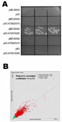
investigate whether SWA2 plays a similar role as NOC1, we tested whether SWA2
interacts with Arabidopsis NOC2 homologues using yeast two-hybrid assay. Full
length SWA2 coding sequence (CDS) was constructed into pGBKT7, and truncated
At2g18220 cDNA and full length At3g55510 cDNA were constructed into pGADGH,
respectively. Yeast cells contransformed with pAD-AT2G18220 and pBD-SWA2 grew
well in Trp-, Leu-, and His- dropout medium supplemented with 10 mM 3-amino-1, 2,
4- triazole (3-AT). However, cells transformed with pAD-At3g55510 and pBD-SWA2
www.plantphysiol.orgon April 3, 2019 - Published by Downloaded from Copyright © 2009 American Society of Plant Biologists. All rights reserved.

13
did not grow (Figure 7A). These data suggested that SWA2 physically interacts with
At2g18220, but not with At3g55510, in yeast cells. Expression pattern analysis using
available microarray data sets (https://www.genevestigator.ethz.ch; Zimmermann et
al., 2004) showed high correlation of the expression profiles between SWA2 and
At2g18220 (Figure 7B), which suggests that the two proteins may interact with each
other in planta.
Expression Pattern of SWA2 Gene
To investigate the expression pattern of SWA2 gene in different organs, RT-PCR
was performed with total RNA from roots, stems, leaves, inflorescences, siliques and
seedlings. A single band with the expected size was detected in RNAs from all tissues,
with the highest expression level in inflorescences, seedlings and leaves (Figure 8A).
These data is consistent with the microarray data available at the
GENEVESTIGATOR (Figure 8B, https://www.genevestigator.ethz.ch).
To further study the expression pattern of SWA2, a PSWA2: SWA2:GUS reporter
system is used to monitor its expression. The full length 9024 bp genomic sequence of
SWA2 was fused in frame with GUS reporter gene and subcloned into pCAMBIA1300.
The construct was introduced into A. thaliana Landsberg erecta plants. In T2
transgenic plants, GUS activity was detected in nucleolus in actively dividing tissues,
such as root tips, lateral root primordia, shoot apexes, young leaves, inflorescences
and pollen grains (Figure 8, C-E). During female gametophyte development, strong
GUS activity was detected in gametophytic nucleus from one-nucleate to
two-nucleate stages (Figure 8, F and G). At four-nucleate stages (FG4) the GUS
activity became much weaker (data not show). And in the mature embryo sac just
before fertilization, only central cell showed strong GUS staining (Figure 8H),
indicating that SWA2 expresses differentially in mature embryo sac.
DISCUSSION
The coordination of cell growth, division and differentiation is fundamental to
development in multicellular organisms. However, mechanisms that couples growth
www.plantphysiol.orgon April 3, 2019 - Published by Downloaded from Copyright © 2009 American Society of Plant Biologists. All rights reserved.

14
and division, for example, have been investigated mainly in single cell organisms or
cultured cells. The developmental process of haploid female gametophyte in
Arabidopsis provides us an excellent system to address how cell growth and division
are coupled, and the biological significance of such coupling to development
(Grossniklaus and Schneitz, 1998; Yang and Sundaresan, 2000). We and others have
previously isolated mutations that disrupted the progression of the mitotic division
cycle during female gametogenesis in Arabidopsis (Moore et al., 1997; Christensen et
al., 1998; Shi et al., 2005). The characterization of these mutations is starting to shed
light on how cell growth, division, and cell fate are coupled (Shi et al., 2005;
Groß-Hardt et al., 2007; Moll et al., 2008; Liu et al., 2009; Yagi et al., 2009).
SWA2 Is Most Likely Involved in Ribosome Biogenesis in Plants
SWA2 encodes a nucleolar protein which shows high homology to yeast
NOC1/Mak21. In yeast, it was reported that NOC1 interacts with NOC2 and is
involved in pre-ribosome biogenesis and nucleolar export (Edskes et al., 1998;
Milkereit et al., 2001). NOC1 is a conserve protein during the evolution and there is
only one homologue in almost all eukaryotes, so it is likely that its function in
ribosome biogenesis is also conserved. The high homology with NOC1, its nucleolar
localization, and interaction with an Arabidopsis NOC2 homologue, all suggest that
SWA2 is most likely involved in ribosome biogenesis in plants. However, in our
experiments SWA2 failed to functionally complement a yeast noc1/mak21 mutant
(data not show). This might be due to divergence of the protein structure of the NOC1
and/or NOC2 component of the ribosome exporting complex although it is
functionally conserved during evolution. It was reported that the human homologue of
NOC, CBF, could neither functionally complement the yeast noc1 mutation (Edskes et
al., 1998). This suggested that the human CBF and the yeast NOC1 are either
functionally different or have diverged in structure.
Previously, we identified a group of female gametophytic mutants that displayed
delayed progression of the division cycle were identified, and designated slow walker
www.plantphysiol.orgon April 3, 2019 - Published by Downloaded from Copyright © 2009 American Society of Plant Biologists. All rights reserved.
15
(swa) mutations. swa2, also named as embryo sac development arrest 25 (eda25) in
the large scale screen for female gametophytic mutations (Pagnussat et al., 2005), is
phenotypically similar to swa1 (Shi et al., 2005). Both swa1 and swa2 mutations
disrupted the progression of female gametophytic division cycle, showed a retarded
development phenotype. As SWA2, SWA1 also encodes a nucleolar protein with a
WD40 domain that is involved in 18S pre-rRNA processing in Arabidopsis (Shi et al.,
2005). These suggest that they may act in the same pathway to modulate ribosome
biogenesis during female gametophyte development.
Nucleolar Function Is Essential for Progression of the Division Cycle during
Female Gametogenesis in Plants
The nucleolus may be not just the site of rDNA transcription and ribosome
biogenesis as our conventional perception (Raška et al., 2004, Boisvert et al., 2007).
More and more studies showed that nucleolus is also involved in controlling mitosis,
cell cycle progression and cell proliferation (Cockell and Gasser, 1999;
Carmo-Fonseca et al., 2002; Shaw and Doonan 2005). Nucleolar proteomics data
showed that nucleolus contains proteins related to these functions (Leung et al., 2003;
Coute et al., 2006). In yeast, nucleolar proteins were found functioning in
coordination of cell proliferation, DNA replication and ribosome biogenesis (Wade et
al., 2001; Du and Stillman, 2002; Zhang et al., 2002). Mutations of these nucleolar
proteins causes delayed or arrested cell cycle progression. Consistently, in plant
several mutations in genes coding for nucleolar proteins, such as SWA1 (Shi et al.,
2005), TORMOZ (Griffith et al., 2007), DOMINO1 (Lahmy et al., 2004), LIS
(Groß-Hardt et al., 2007), and GFA1/CLO and ATO (Moll et al., 2008; Liu et al., 2009;
Yagi et al., 2009), causing defectiveness in ribosome biogenesis or nucleolar function,
have been shown to affect cell proliferation during female gametophyte and embryo
development. More intriguingly, gametic cell fates are altered in lis (Groß-Hardt et al.,
2007) and gfa1/clo/ato (Moll et al., 2008) mutants. These suggest a key role of
nucleolar function in female gametophyte development in plants.
Ribosome biogenesis and dynamics are central for cell growth, it was estimated
www.plantphysiol.orgon April 3, 2019 - Published by Downloaded from Copyright © 2009 American Society of Plant Biologists. All rights reserved.

16
that yeast cells must synthesize more than 2000 ribosomes and transport about 1000
ribosomal proteins from cytoplasm to the nucleolus per minute (Warner et al., 2001).
Therefore, the coordination of rRNA transcription with ribosomal protein synthesis
and transportation must be tightly regulated. More importantly, ribosome biogenesis
must be coordinated with cell state, cell division, and development. Elucidation of
genetic mechanisms governing these processes will shed light on the understanding of
how cell senses intrinsic cellular activities in a developmental context.
MATERIAL AND METHODS
Plant Material and Growth Conditions
The swa2 mutant was isolated from a genetic screen of Ds insertion lines as
described previously (Sundarensan et al., 1995; Shi et al., 2005). Seeds were sterilized
with 20% bleach for 5-10 min, then washed five times in sterilized water and
germinated on MS agar plates. For antibiotic selection, 50 mg/L kanamycin and/or 20
mg/L hygromycin were supplemented as required. Seeds were stratified in darkness at
4� for three days before grown in growth chambers at 22±2� under a
16-h-light/8-h-dark cycle. Plant transformation was performed by Agrobacterium
tumefaciens-mediated infiltration (Bechtold and Pelletier, 1998).
Phenotypic Analysis by Confocal Laser Scanning Microscopy
Confocal observation of ovules was performed as described previously
(Christesen et al., 1998; Shi et al., 2005). Inflorescences were fixed in 4%
glutaraldehyde in 12.5 mM cacodylate (pH6.9) overnight at room temperature. The
tissue was then dehydrated through a conventional ethanol series with 30 min per step.
The dehydrated tissue was cleared in 2:1 (v/v) benzyl benzoate: benzyl alcohol for 1h.
Pistils were dissected, mounted with immersion oil and observed using a Zeiss
LSM510 META laser scanning microscope (Zeiss, Jena, Germany) with a 488-nm
argon laser and an LP 530 filter.
www.plantphysiol.orgon April 3, 2019 - Published by Downloaded from Copyright © 2009 American Society of Plant Biologists. All rights reserved.

17
Reciprocal Crosses and Delayed Pollination Test
Reciprocal crosses between wild type and swa2 plants were performed as
described (Yang et al., 1999). Delayed pollination test was performed according to Shi
et al. (2005) with slight modifications. Anthers of swa2 plants were removed at stage
12c (Smyth et al., 1990) and pollinated with wild-type pollen after at 12, 24, 36, 48, or
72 h after emasculation, respectively. The F1 seedlings were scored for kanamycin
resistance on MS plate according to Sundaresan et al. (1995).
Pollen Analysis
For light and fluorescent microscopy of pollen, specimens were observed using a
Zeiss Axioskop II microscope, and images were acquired with a Cannon PowerShot
G6 (Cannon, Japan). Staining assay with 4’, 6-diamino-2-phenylindole (DAPI) was
performed as described previously (Johnson-Brousseau and McCormick, 2004) and
DAPI concentration was at 1μg/ml.
Molecular Cloning and Genetic Complementation
The Ds flanking sequences were isolated by thermal asymmetric interlaced PCR
as described (Liu et al., 1995; Yang et al., 1999). For the complementation construct,
two genome fragments were amplified by KOD plus polymerase (TOYOBO) using
primers P290CPF-KPN
(5’-GGGGTACCCCTCCAAAACCAAAGGCCCATAACC-3’)/P290G-M-SAL-PST
(5’-GTCGACATCGAAAATTTAATAACAAAAGAA-3’) for fragment 290CPa and
P290CPRV-PST
(5’-AACTGCAGCTTCTGAGAGTTCGTCGGAAACAGC-3’)/P290G-M-SAL-KPN
(5’-GTCGACACACTGTATAGTAAATTTTTTTGT-3’) for 290CPb. The two
fragments were first cloned into pGEM-T Easy (Promega) and then subcloned into
pCAMBIA1301 (CAMBIA; www.cambia.org.au) at KpnI/SalI and SalI/PstI sites,
respectively to produce p1301-290CP containing the full-length genomic fragment
form from -827 bp upstream of the ATG start condon to 2928 bp downstream of the
stop codon of At1g72440. The construct was introduced into swa2 plants by
www.plantphysiol.orgon April 3, 2019 - Published by Downloaded from Copyright © 2009 American Society of Plant Biologists. All rights reserved.

18
Agrobacterium-mediated infiltration (Bechtold and Pelletier, 1998).
SWA2-DsRed2 Construct and Subcellular Localization
The DsRed2 coding sequence was amplified from pDsRed2 (Clontech) using
primers PDsRed2F-KPN (5’-GGGGTACCATGGCCTCCTCCGAGAACGTCA-3’)
and PDsRed2-RV-SAC
(5’-GGGGAAGCTTGAGCTCTACAGGAACAGGTGGTGGCGGC-3’) and inserted
into pCAMBIA 1300 at the KpnI and SacI sites. The 2928 bp fragment downstream of
the stop codon of SWA2 was amplified using primers P2903UTR+Sstup
(5’-GGAGAGCTCGAAGCAAGACTTGTTGCTTG-3’) and P290CPRV-ECOR
(5’-GGAATTCCTTCTGAGAGTTCGTCGGAAACAGC-3’) and inserted into the
construct pCAMBIA1300-DsRed2 at SacI and EcoRI sites. The 6093 bp promoter
and coding region of SWA2 was amplified using primers P290CPF-KPN
(5’-GGGGTACCCCTCCAAAACCAAAGGCCCATAACC-3’) and
P290STOPRV-KPN (5’-GGGGTACCCTCTGATGCTTTAGACTTCTTCTTT-3’) and
inserted into the above construct at the KpnI site to produce
pCAMBIA1300-Pswa2:SWA2:DsRed2. Root tips of transgenic plants were stained
with DAPI as described previously (Shi et al., 2005) and observed with LSM510
META confocal microscope (Zeiss).
Yeast Two-hybrid Assay
The full length CDS of SWA2 was amplified by PCR using the primers
P290C-BD-NCO-F (5’-GGACCATGGACATGTCAAAGATAAAGCCTTT-3’) and
P290C-PWM101-PSTDOWN (5’-GGACTGCAGTTACTCTGATGCTTTAGACT-3’)
and cloned into pGBKT7 (Clontech) at NcoI and PstI sites to give rise to pBD-SWA2.
The cDNA fragment of At2g18220 and full length CDS of At3g55510 were amplified
using primers P820ADF4-NDE
(5’-GGAATTCCATATGAAGCGTGGGAAGAAGGTGAAATCTAA-3’) in
combination with P820ADR4-XHO
(5’-CCGCTCGAGAAGCTACTCGGGCCTTCTTCTTCTT-3’) and P510ADF-NDE
www.plantphysiol.orgon April 3, 2019 - Published by Downloaded from Copyright © 2009 American Society of Plant Biologists. All rights reserved.

19
(5’-GGAATTCCATATGGGTAAGCTGGGGAAGAAAGCTA-3’) in combination
with P510ADRV-XHO
(5’-CCGCTCGAGTCACTTCTTCTTCTTCTTGTTTGTTC-3’), respectively. The
fragments were cloned into pGADGH at NdeI and XhoI sites. Yeast transformation
was performed as described previously (Xie et al., 1999). The transformed cells were
transferred to -Leu/-Trp/-His DO supplement (BD) plates supplemented with 1-10
mM 3-AT.
RNA Isolation and RT-PCR Analysis
Total RNA was isolated using TRIzol reagent (Invitrogen) and digested with
RNase-free DNase I (Takara, Beijing, China). 1 μg total RNA was used as templates
to transcribe single-stranded cDNA by reverse transcriptase AMV (Takara). Mock
controls without reverse transcriptase were performed simultaneously to detect
genomic DNA contamination. One microliter of the synthesized cDNA and control
products were used for PCR. Primers P290RTF1
(5’-CTCTGAATGGTACAACGATG-3’) in combination with P290RTR1
(5’-CAGCCTCGTCAGTGGAAACA-3’) was used for detection of SWA2 expression
and primers PEIF4AF (5’-ATGGCAGGACCGCACCGGA-3’) in combination with
PEIF4ARV (5’-GCATGTCAAAAACACGACCGGGAGTTCC-3’) was used for
amplification of the EIF4A gene as an internal control. PCR products were analyzed
on 1% agarose gel.
Express Analysis Using GUS Reporter System
The GUS coding sequence was amplified from pWM101 (Ding et al., 2006) using
primers PGUSF-KPN (5’-GGGGTACCATGTTACGTCCTGTAGAAAC-3’) and
PGUSRV-PST (5’-AACTGCAGTCATTGTTTGCCTCCCTGCT-3’) and inserted into
pCAMBIA 1300 at the KpnI and PstI sites to produce pCAMBIA1300-GUS. The
6093 bp fragment containing the promoter plus coding region and the fragment
containing the 2928 bp sequence downstream of the SWA2 stop codon were amplified
with primer pairs P290CPF-KPN
www.plantphysiol.orgon April 3, 2019 - Published by Downloaded from Copyright © 2009 American Society of Plant Biologists. All rights reserved.

20
(5’-GGGGTACCCCTCCAAAACCAAAGGCCCATAACC-3’), P290STOPRV-KPN
(5’-GGGGTACCCTCTGATGCTTTAGACTTCTTCTTT-3’) and P2903UTR-F-PST
(5’-AACTGCAGGAAGCAAGACTTGTTGCTTGCTATG-3’),
P290-STOPRV-KPN-GUS
(5’-GGGGTACCCTCTGATGCTTTAGACTTCTTCTTT-3’), respectively. The
resulting products were cloned into pCAMBIA1300-GUS at KpnI and PstI sites in the
correct directions to produce the PSWA2: SWA2: GUS construct. GUS activity assay of
transgenic plants was performed as described previously (Ding et al., 2006).
ACKNOWLEDGEMENTS
We thank Dr. Yan Guo and Dr. Fiyi Zhao at National Institute of Biological
Science, Beijing, for technical assistance in confocal microscopy.
LITERATURE CITED
Bechtold N, Pelletier G (1998) In planta Agrobacterium-mediated transformation of adult Arabidopsis
thaliana plants by vacuum infiltration. Methods Mol Biol 82: 259-266
Bemer M, Wolters-Arts M, Grossniklaus U, Angenent GC. 2008. The MADS domain protein DIANA
acts together with AGAMOUS-LIKE80 to specify the central cell in Arabidopsis ovules. Plant Cell
20:2088–101.
Bernstein KA, Bleichert F, Bean JM, Cross FR, Baserga SJ (2007) Ribosome biogenesis is sensed
at the Start cell cycle checkpoint. Mol Biol Cell 18: 953-964
Boisvert FM, van Koningsbruggen S, Navascués J, Lamond AI (2007) The multifunctional
nucleolus. Nat Rev Mol Cell Bio 8: 574-585
Christensen CA, Gorsich SW, Brown RH, Jones LG, Brown J, Shaw JM, Drews GN (2002)
Mitochondrial GFA2 is required for synergid cell death in Arabidopsis. Plant Cell 14: 2215-2232
Christensen CA, King Ej, jordan JR, and Drews GN (1997) Megagametogenesis in Arabisopsis
wild type and the Gf mutant. Sex Plant Reprod 10: 49-64
Christensen CA, Subramanian S, Drews GN (1998) Identification of gametophytic mutations
affecting female gametophyte development in Arabidopsis. Dev.Biol. 202: 136-151
Cockell M, Gasser SM (1999) Nuclear compartments and gene regulation. Curr Opin Genet Dev 9:
199-205
Coute Y, Burgess JA, Diaz JJ, Chichester C, Lisacek F, Greco A, Sanchez JC (2006) Deciphering
the human nucleolar proteome. Mass Spectrom Rev 25: 215-234
Ding YH, Liu NY, Tang ZS, Liu J, Yang WC (2006) Arabidopsis GLUTAMINE-RICH PROTEIN23
is essential for early embryogenesis and encodes a novel nuclear PPR motif protein that interacts
with RNA polymerase II subunit III. Plant Cell 18: 815-830
www.plantphysiol.orgon April 3, 2019 - Published by Downloaded from Copyright © 2009 American Society of Plant Biologists. All rights reserved.

21
Dlakic M, Tollervey D (2004) The Noc proteins involved in ribosome synthesis and export contain
divergent HEAT repeats. RNA. 10: 351-354
Drews GN, Lee D, Christensen CA (1998) Genetic analysis of female gametophyte development and
function. Plant Cell 10: 5-17
Du YC, Stillman B (2002) Yph1p, an ORC-interacting protein: potential links between cell
proliferation control, DNA replication, and ribosome biogenesis. Cell 109: 835-848
Ebel C, Mariconti L, Gruissem W (2004) Plant retinoblastoma homologues control nuclear
proliferation in the female gametophyte. Nature 429: 776-780
Edskes HK, Ohtake Y, Wickner RB (1998) Mak21p of Saccharomyces cerevisiae, a homolog of
human CAATT-binding protein, is essential for 60 S ribosomal subunit biogenesis. J Biol Chem
273: 28912-28920
Groß-Hardt R, Kagi C, Baumann N, Moore JM, Baskar R, Gagliano WB, Jurgens G,
Grossniklaus U (2007) LACHESIS restricts gametic cell Fate in the female gametophyte of
Arabidopsis. PLoS Biol 5: e47
Grossniklaus U, Vielle-Calzada J-P, Hoeppner MA, Gagliano WB. (1998). Maternal control of
embryogenesis by MEDEA, a polycomb group gene in Arabidopsis. Science 280:446–450.
Grossniklaus U, Schneitz K (1998) The molecular and genetic basis of ovule and megagametophyte
development. Semin Cell Dev Biol 9: 227-238
Hejatko J, Pernisova M, Eneva T, Palme K, Brzobohaty B (2003) The putative sensor histidine
kinase CKI1 is involved in female gametophyte development in Arabidopsis. Mol Genet
Genomics 269: 443-453
Howden R, Park SK, Moore JM, Orme J, Grossniklaus U, Twell D (1998) Selection of
T-DNA-tagged male and female gametophytic mutants by segregation distortion in Arabidopsis.
Genetics 149: 621-631
Imbriano C, Bolognese F, Gurtner A, Piaggio G, Mantovani R (2001) HSP-CBF is an
NF-Y-dependent coactivator of the heat shock promoters CCAAT boxes. J Biol Chem 276:
26332-26339
Johnson-Brousseau SA, McCormick S (2004) A compendium of methods useful for characterizing
Arabidopsis pollen mutants and gametophytically-expressed genes. Plant J 39: 761-775
Jorgensen P, Nishikawa JL, Breitkreutz BJ, Tyers M (2002) Systematic identification of pathways
that couple cell growth and division in yeast. Science 297: 395-400
Kim HU, Li Y, Huang AH (2005) Ubiquitous and endoplasmic reticulum-located lysophosphatidyl
acyltransferase, LPAT2, is essential for female but not male gametophyte development in
Arabidopsis. Plant Cell 17: 1073-1089
Kwee HS, Sundaresan V (2003) The NOMEGA gene required for female gametophyte development
encodes the putative APC6/CDC16 component of the Anaphase Promoting Complex in
Arabidopsis. Plant J 36: 853-866
Leon G, Holuigue L, Jordana X (2007) Mitochondrial complex II Is essential for gametophyte
development in Arabidopsis. Plant Physiol 143: 1534-1546
Leung AK, Andersen JS, Mann M, Lamond AI (2003) Bioinformatic analysis of the nucleolus.
Biochem J 376: 553-569
Liu M, Yuan L, Liu NY, Shi DQ, Liu J, Yang WC (2009) GAMETOPHYTIC FACTOR1, involved in
www.plantphysiol.orgon April 3, 2019 - Published by Downloaded from Copyright © 2009 American Society of Plant Biologists. All rights reserved.

22
pre-mRNA splicing, is essential for megagametogenesis and embryogenesis in Arabidopsis. J
Integr Plant Biol 51: 261-271
Liu YG, Mitsukawa N, Oosumi T, Whittier RF (1995) Efficient isolation and mapping of
Arabidopsis thaliana T-DNA insert junctions by thermal asymmetric interlaced PCR. Plant J 8:
457-463
Lum LS, Sultzman LA, Kaufman RJ, Linzer DI, Wu BJ (1990) A cloned human
CCAAT-box-binding factor stimulates transcription from the human hsp70 promoter. Mol Cell
Biol 10: 6709-6717
Milkereit P, Gadal O, Podtelejnikov A, Trumtel S, Gas N, Petfalski E, Tollervey D, Mann M, Hurt
E, Tschochner H (2001) Maturation and intranuclear transport of pre-ribosomes requires Noc
proteins. Cell 105: 499-509
Moore JM, Calzada JP, Gagliano W, Grossniklaus U (1997) Genetic characterization of hadad, a
mutant disrupting female gametogenesis in Arabidopsis thaliana. Cold Spring Harb Symp Quant
Biol 62: 35-47
Pagnussat GC, Yu HJ, Ngo QA, Rajani S, Mayalagu S, Johnson CS, Capron A, Xie LF, Ye D,
Sundaresan V (2005) Genetic and molecular identification of genes required for female
gametophyte development and function in Arabidopsis. Development 132: 603-614
Portereiko MF, Lloyd A, Steffen JG, Punwani JA, Otsuga D, Drews GN (2006) AGL80 is required
for central cell and endosperm development in Arabidopsis. Plant Cell 18: 1862-1872
Portereiko MF, Sandaklie-Nikolova L, Lloyd A, Dever CA, Otsuga D, Drews GN (2006)
NUCLEAR FUSION DEFECTIVE1 encodes the Arabidopsis RPL21M protein and is required for
karyogamy during female gametophyte development and fertilization. Plant Physiol 141: 957-965
Shaw P, Doonan J (2005) The nucleolus. Playing by different rules? Cell Cycle 4: 102-105
Shi DQ, Liu J, Xiang YH, Ye D, Sundaresan V, Yang WC (2005) SLOW WALKER1, essential for
gametogenesis in Arabidopsis, encodes a WD40 protein involved in 18S ribosomal RNA
biogenesis. Plant Cell 17: 2340-2354
Smyth DR, Bowman JL, Meyerowitz EM (1990) Early flower development in Arabidopsis. Plant
Cell 2: 755-767
Springer PS, McCombie WR, Sundaresan V, Martienssen RA (1995) Gene trap tagging of
PROLIFERA, an essential MCM2-3-5-like gene in Arabidopsis. Science 268: 877-880
Srivastava M, Pollard HB (1999) Molecular dissection of nucleolin's role in growth and cell
proliferation: new insights. FASEB J 13: 1911-1922
Sundaresan V, Springer P, Volpe T, Haward S, Jones JD, Dean C, Ma H, Martienssen R (1995)
Patterns of gene action in plant development revealed by enhancer trap and gene trap transposable
elements. Genes Dev 9: 1797-1810
Wade C, Shea KA, Jensen RV, McAlear MA (2001) EBP2 is a member of the yeast RRB regulon, a
transcriptionally coregulated set of genes that are required for ribosome and rRNA biosynthesis.
Mol Cell Biol 21: 8638-8650
Warner JR, Vilardell J, Sohn JH (2001) Economics of ribosome biosynthesis. Cold Spring Harb
Symp Quant Biol 66: 567-574
Wilson ZA, Yang C (2004) Plant gametogenesis: conservation and contrasts in development.
Reproduction 128: 483-492
Wullischleger S, Loewith R, Hall MN (2006) TOR signaling in growth and metabolism. Cell
124: 471-484
www.plantphysiol.orgon April 3, 2019 - Published by Downloaded from Copyright © 2009 American Society of Plant Biologists. All rights reserved.
23
Xie Q, Sanz-Burgos AP, Guo H, Garcia JA, Gutierrez C (1999) GRAB proteins, novel members of
the NAC domain family, isolated by their interaction with a geminivirus protein. Plant Mol Biol 39:
647-656
Yagi N, Takeda S, Matsumoto N, Okada K (2009) VAJ/GFA1/CLO is involved in the directional
control of floral organ growth. Plant Cell Physiol 50: 515 - 527
Yang WC, Sundaresan V (2000) Genetics of gametophyte biogenesis in Arabidopsis. Curr Opin Plant
Biol 3: 53-57
Yang WC, Ye D, Xu J, Sundaresan V (1999) The SPOROCYTELESS gene of Arabidopsis is required
for initiation of sporogenesis and encodes a novel nuclear protein. Genes Dev 13: 2108-2117
Zhang Y, Yu Z, Fu X, Liang C (2002) Noc3p, a bHLH protein, plays an integral role in the initiation
of DNA replication in budding yeast. Cell 109: 849-860
www.plantphysiol.orgon April 3, 2019 - Published by Downloaded from Copyright © 2009 American Society of Plant Biologists. All rights reserved.
24
FIGURE LEGENDS
Figure 1. Phenotype of the swa2 plant.
A. A swa2 silique showing aborted ovules (black arrows).
B. A wild-type silique showing a full seed set. Bar=100 μm
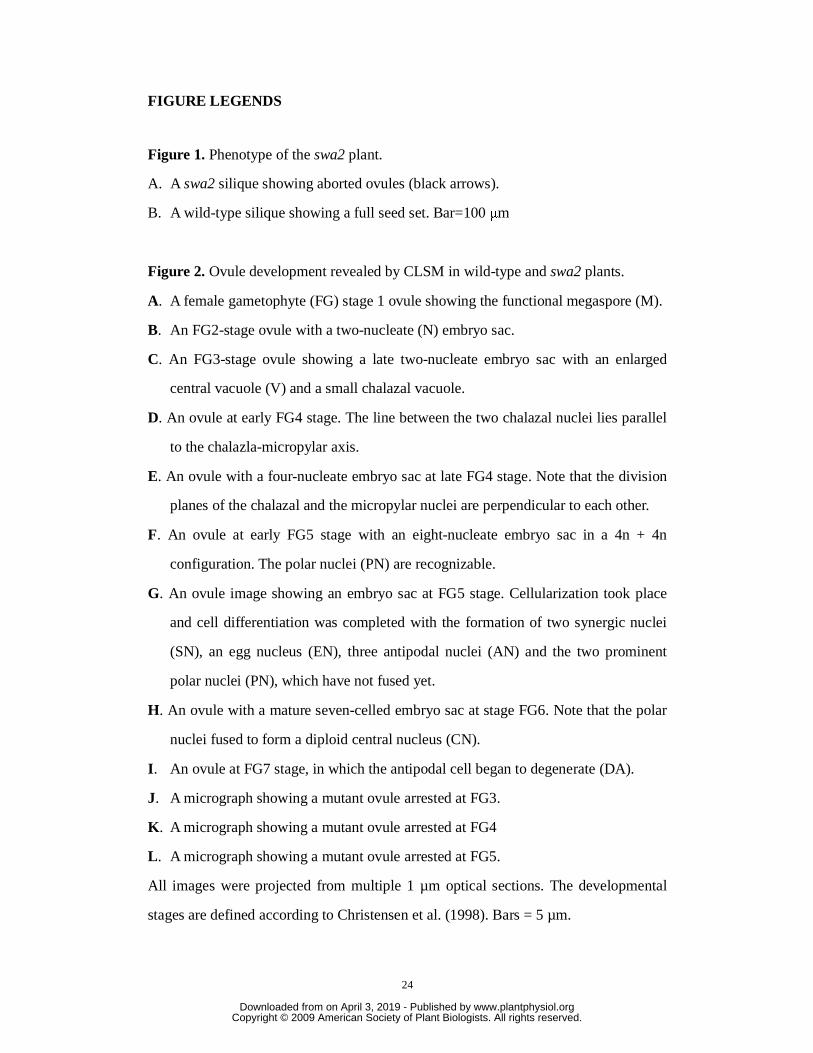
Figure 2. Ovule development revealed by CLSM in wild-type and swa2 plants.
A. A female gametophyte (FG) stage 1 ovule showing the functional megaspore (M).
B. An FG2-stage ovule with a two-nucleate (N) embryo sac.
C. An FG3-stage ovule showing a late two-nucleate embryo sac with an enlarged
central vacuole (V) and a small chalazal vacuole.
D. An ovule at early FG4 stage. The line between the two chalazal nuclei lies parallel
to the chalazla-micropylar axis.
E. An ovule with a four-nucleate embryo sac at late FG4 stage. Note that the division
planes of the chalazal and the micropylar nuclei are perpendicular to each other.
F. An ovule at early FG5 stage with an eight-nucleate embryo sac in a 4n + 4n
configuration. The polar nuclei (PN) are recognizable.
G. An ovule image showing an embryo sac at FG5 stage. Cellularization took place
and cell differentiation was completed with the formation of two synergic nuclei
(SN), an egg nucleus (EN), three antipodal nuclei (AN) and the two prominent
polar nuclei (PN), which have not fused yet.
H. An ovule with a mature seven-celled embryo sac at stage FG6. Note that the polar
nuclei fused to form a diploid central nucleus (CN).
I. An ovule at FG7 stage, in which the antipodal cell began to degenerate (DA).
J. A micrograph showing a mutant ovule arrested at FG3.
K. A micrograph showing a mutant ovule arrested at FG4
L. A micrograph showing a mutant ovule arrested at FG5.
All images were projected from multiple 1 µm optical sections. The developmental
stages are defined according to Christensen et al. (1998). Bars = 5 µm.
www.plantphysiol.orgon April 3, 2019 - Published by Downloaded from Copyright © 2009 American Society of Plant Biologists. All rights reserved.

25
Figure 3. Segregation ratio of F1 progeny from the delayed pollination test. Swa2
plants were emasculated at floral stage 12c (Smyth et al., 1990) and pollinated with
pollens from wild-type plants at 12h, 24h, 36h, 48h, or 72h after emasculation. KanR:
KanS ratios of the F1 progeny of each group were analyzed.
Figure 4. The pollen showed defective cell cycle progression in the swa2 mutant
A. A micrograph showing wild-type pollen grains at maturity. Note the condensed
sperm nuclei.
B. A micrograph showing swa2 pollen grains arrested at either bicellular stage (green
arrow), or tricellular stage with sperm nuclei defective in chromosome condensing
(red arrow).
C. A micrograph showing a mutant pollen grain with thread-like sperm nuclei (red
arrow).
D. A micrograph showing a pollen grain of salk_016552 line arrested at bicellular
stage (green arrow).
E. A micrograph showing pollen grains of salk_016552 arrested at bicellular stage
(green arrow) compared to the wild-type pollen grain.
F. A micrograph showing thread-like nuclei of pollen grain of salk_016552 (red
arrow).
Bars=10 μm.
Figure 5. Molecular characterization of the SWA2.
A. Diagram of the insertion positions of Ds (swa2) and T-DNA (Salk_016552) in
SWA2. The black box indicates the exons of the SWA2 gene. The nucleotide
numbers are consistent with those in BAC clone T10D10.
B. Predicted SWA2 amino acid sequences. The shaded amino acids representing the
putative Nucleic-acid-binding domain possibly involved in ribosomal biogenesis
and the divergent HEAT repeats are underlined. Boxed columns show the
www.plantphysiol.orgon April 3, 2019 - Published by Downloaded from Copyright © 2009 American Society of Plant Biologists. All rights reserved.

26
conserved CBF domain. The underlined amino acids at the C terminal show the
predicted nuclear localization signal.
C. Phylogenetic tree of SWA2 with its homologs from other organisms.
D. Alignment of the SWA2 protein with its homologs from Oryza sativa (cv
japonica), yeast, human and mouse. Identical amino acids are shown with white
letters in black boxes, and similar amino acids are shown with shaded boxes.
Figure 6. Subcellular localization of SWA2-DsRed2 fusion protein in Arabidopsis
root cells.
A. Confocal image of a transgenic root cell under the DsRed2 channel showing
SWA2-DsRed2 localization (red).
B. The same image as A showing DNA visualized with DAPI staining (blue).
C. The same image as A under bright field illumination.
D. Merged image of A, B, and C showing the nucleolar localization of
SWA2-DsRed2. Bar=5 µm
Figure 7. SWA2 interacts with Arabidopsis NOC2 homologue in yeast cells.
A. SWA2 interacts with AT2G18220 but not AT3G55510 in yeast cells. For each
transformation, three independent transformants were streaked on a plate
containing synthetic dropout selection medium that lacked Trp, Leu, and His
supplemented with 10 mM 3-AT.
B. Correlation of the expression profiles of SWA2 and AT2G18220. Data were
retrieved from the public GENEVESTIGATOR microarray data set
(https://www.genevestigator.ethz.ch; Zimmermann et al., 2004).
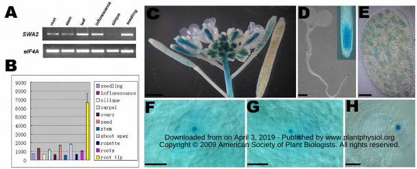
Figure 8. Expression pattern of the SWA2 gene as revealed by RT-PCR analysis (A),
RNA profiling (B) and transgenic plants expressing PSWA2-SWA2:GUS reporter (C-H).
A. Tissue-specific expression of SWA2 using RT-PCR analysis. RT-PCR was
performed on total RNAs from different tissues, including roots, stems, leaves,
inflorescences, siliques, and seedlings as indicated. After 32 cycles, the resulting
www.plantphysiol.orgon April 3, 2019 - Published by Downloaded from Copyright © 2009 American Society of Plant Biologists. All rights reserved.

27
products were stained with ethidium bromide and analyzed by gel electrophoresis.
EIF4A RNA was used as an internal template control.
B. Expression profiles of SWA2 in various organs. The y axis represents the
expression level. Data used in this analysis were retrieved from the public
GENEVESTIGATOR microarray data set (https://www.genevestigator.ethz.ch;
Zimmermann et al., 2004).
C. A micrograph showing GUS activity specifically detected in floral meristem and
young siliques of PSWA2-SWA2:GUS transgenic Arabidopsis. Bar = 1 mm.
D. A micrograph showing GUS activity in the shoot apex, leaf primordium, lateral
root primordia, and root meristem in a 7-d-old PSWA2-SWA2:GUS transgenic
seedling. Bar = 1 mm.
E. A micrograph showing GUS activity in pollen grains. Bar = 10μm.
F. A micrograph showing GUS activity in a one-nucleate embryo sac. Bar = 5μm.
G. A micrograph showing GUS activity in an early two-nucleate embryo sac. Bar = 5
μm.
H. A micrograph showing GUS activity in the central cell nucleus of a mature
embryo sac. Bar = 5 μm.
www.plantphysiol.orgon April 3, 2019 - Published by Downloaded from Copyright © 2009 American Society of Plant Biologists. All rights reserved.

28
Table 1. Synchrony of Female Gametophyte Development in Wild-Type Arabidopsis
Pistil Number Number of Female Gametophytes at Developmental stages FG1 FG2 FG3 FG4 FG5 FG6 FG7 Total FGs
1 27 11 2 40 2 6 17 6 3 32 3 2 18 6 26 4 4 25 7 36 5 12 35 3 50 6 3 17 21 41 7 5 9 39 2 55 8 2 32 17 51 9 20 17 2 39
10 16 30 4 50 11 6 36 9 51 12 4 46 50
www.plantphysiol.orgon April 3, 2019 - Published by Downloaded from Copyright © 2009 American Society of Plant Biologists. All rights reserved.

29
Table 2. Synchrony of Female Gametophyte Development in swa2 plants
Pistil Number
Number of Female Gametophytes at Developmental stages
FG1 FG2 FG3 FG4 FG5 FG6 FG7 Total FGs
1 31 6 7 44 2 29 4 6 39 3 16 11 13 3 43 4 2 8 9 15 2 36 5 4 11 9 4 11 1 40 6 1 1 20 13 18 53 7 2 15 5 18 3 43 8 6 13 10 12 41 9 1 32 14 15 62
10 2 18 6 23 2 51 11 1 8 15 1 21 46
www.plantphysiol.orgon April 3, 2019 - Published by Downloaded from Copyright © 2009 American Society of Plant Biologists. All rights reserved.

www.plantphysiol.orgon April 3, 2019 - Published by Downloaded from Copyright © 2009 American Society of Plant Biologists. All rights reserved.

www.plantphysiol.orgon April 3, 2019 - Published by Downloaded from Copyright © 2009 American Society of Plant Biologists. All rights reserved.
www.plantphysiol.orgon April 3, 2019 - Published by Downloaded from Copyright © 2009 American Society of Plant Biologists. All rights reserved.

www.plantphysiol.orgon April 3, 2019 - Published by Downloaded from Copyright © 2009 American Society of Plant Biologists. All rights reserved.

www.plantphysiol.orgon April 3, 2019 - Published by Downloaded from Copyright © 2009 American Society of Plant Biologists. All rights reserved.

www.plantphysiol.orgon April 3, 2019 - Published by Downloaded from Copyright © 2009 American Society of Plant Biologists. All rights reserved.
www.plantphysiol.orgon April 3, 2019 - Published by Downloaded from Copyright © 2009 American Society of Plant Biologists. All rights reserved.
www.plantphysiol.orgon April 3, 2019 - Published by Downloaded from Copyright © 2009 American Society of Plant Biologists. All rights reserved.