
Magnetically responsive photonic film with high … · · 2015-01-28Magnetically responsive...
21
Magnetically responsive photonic film with high tunability and stability Yongxing Hu, Le He, Xiaogang Han, Mingsheng Wang, Yadong Yin() Nano Res., Just Accepted Manuscript • DOI 10.1007/s12274-015-0732-z http://www.thenanoresearch.com on January 28, 2015 © Tsinghua University Press 2015 Just Accepted This is a “Just Accepted” manuscript, which has been examined by the peer-review process and has been accepted for publication. A “Just Accepted” manuscript is published online shortly after its acceptance, which is prior to technical editing and formatting and author proofing. Tsinghua University Press (TUP) provides “Just Accepted” as an optional and free service which allows authors to make their results available to the research community as soon as possible after acceptance. After a manuscript has been technically edited and formatted, it will be removed from the “Just Accepted” Web site and published as an ASAP article. Please note that technical editing may introduce minor changes to the manuscript text and/or graphics which may affect the content, and all legal disclaimers that apply to the journal pertain. In no event shall TUP be held responsible for errors or consequences arising from the use of any information contained in these “Just Accepted” manuscripts. To cite this manuscript please use its Digital Object Identifier (DOI®), which is identical for all formats of publication. Nano Research DOI 10.1007/s12274-015-0732-z
Transcript of Magnetically responsive photonic film with high … · · 2015-01-28Magnetically responsive...

Nano Res
1
Magnetically responsive photonic film with high
tunability and stability
Yongxing Hu, Le He, Xiaogang Han, Mingsheng Wang, Yadong Yin()
Nano Res., Just Accepted Manuscript • DOI 10.1007/s12274-015-0732-z
http://www.thenanoresearch.com on January 28, 2015
© Tsinghua University Press 2015
Just Accepted
This is a “Just Accepted” manuscript, which has been examined by the peer-review process and has been
accepted for publication. A “Just Accepted” manuscript is published online shortly after its acceptance,
which is prior to technical editing and formatting and author proofing. Tsinghua University Press (TUP)
provides “Just Accepted” as an optional and free service which allows authors to make their results available
to the research community as soon as possible after acceptance. After a manuscript has been technically
edited and formatted, it will be removed from the “Just Accepted” Web site and published as an ASAP
article. Please note that technical editing may introduce minor changes to the manuscript text and/or
graphics which may affect the content, and all legal disclaimers that apply to the journal pertain. In no event
shall TUP be held responsible for errors or consequences arising from the use of any information contained
in these “Just Accepted” manuscripts. To cite this manuscript please use its Digital Object Identifier (DOI®),
which is identical for all formats of publication.
Nano Research
DOI 10.1007/s12274-015-0732-z

1
Graphical TOC
Agarose hydrogel has been successfully used to significantly improve the structural and photonic
stability of magnetically assembled photonic structures. The steric hindrance and the hydrogen bonding
from the agarose network can effectively counter the magnetic packing force and prevent further
coagulation of particle assemblies.

2
Magnetically Responsive Photonic Film with High Tunability and Stability
Yongxing Hu, Le He, Xiaogang Han, Mingsheng Wang, Yadong Yin*
University of California Riverside, Department of Chemistry, California, USA
Email: [email protected]
Abstract
We demonstrate the fabrication of magnetically assembled one-dimensional chain-like photonic
nanostructures with significantly high photonic stability. The key lies in the use of agarose hydrogel to
prevent the magnetic assemblies from coagulation. When exposed to an external magnetic field,
negatively charged Fe3O4@SiO2 particles can effectively assemble in the hydrogel matrix into one-
dimensional chains with internal periodicity and display a fast, fully reversible, and tunable photonic
response to the changes in the external field. The steric hindrance and the hydrogen bonding from the
agarose network effectively limit the migration of the Fe3O4@SiO2 particles and their chain-like
assemblies. As a result, the system shows remarkable stability in photonic response under external
magnetic fields of large gradients, which has been a challenge previously. The ability of stabilizing the
magnetic particle assemblies over a long period represents a major stride toward practical applications of
such field-responsive photonic materials.
Keywords
Magnetic field, Hydrogel, Reversible, Assembly, Photonic nanostructures, Coagulation

3
Introduction
There has been increasing interest in developing responsive photonic materials owing to their broad
applications relevant to color manipulation.1-3 We previously developed a magnetically tunable photonic
crystal system by assembling highly charged superparamagnetic Fe3O4 colloidal nanocrystal clusters
(CNCs) in aqueous solutions.4-6 With the establishment of temporal stability by the balance of attractive
(magnetic) and repulsive (electrostatic) forces, the colloids form ordered structures along the direction of
an applied external magnetic field with a regular interparticle spacing on the order of tens of nanometers.
As a result, the solution strongly diffracts visible light. By changing the strength of the external field, one
can conveniently tune the diffraction wavelength throughout the entire visible spectrum. Further surface
modification with a silica layer followed by mild etching in water (Fe3O4@SiO2) helps to prevent the
detachment of capping ligands from the surface, enhancing stability during storage and improving the
dispersity of the colloids in various solvents such as water and alkanols.6-8 Magnetically responsive
photonic structures with significantly reduced dimensions can also be realized by fixing individual
magnetic particle chains with a silica coating.9
Ideally, the dynamic assemblies should remain stable in a uniform magnetic field as the magnetic
particles are only subjected to magnetic dipole-dipole interactions. However, in practice, a magnetic field
is often not uniform and a large gradient usually exists, which induces a magnetic packing force Fm =
∇(µB) on every particle and attracts them toward the maximum of the local magnetic gradient (µ is the
induced magnetic moment and B the strength of the external field).10 As a result, the dynamically ordered
assemblies of Fe3O4@SiO2 suffer from chain coagulation induced by the magnetic packing force,
gradually degrading the colloidal stability of the system and consequently their photonic performance.5,11-
13 The instability of these systems in a high gradient magnetic field acts as a major barrier for some
practical applications which require long time exposure to magnetic fields. An alternative way to enhance
the stability against the magnetic packing force while retaining the tunability of the photonic structures is
to employ a porous polymer matrix with steric forces strong enough to prevent the chain coagulation. In
principle, an effective polymer matrix should be optically clear, compatible with Fe3O4@SiO2 colloids,
and porous enough to allow tuning of the periodic structures.
Herein, we report a strategy for utilizing agarose hydrogel as a polymer matrix to stabilize embedded
magnetic assemblies to counter the packing force exerted by a magnetic field. Hydrogel has been used
previously as a matrix for stabilizing three-dimensional colloidal crystals.14-16 Agarose is a well-known
hydrogel-forming material that is commonly used in both everyday life and scientific research.17,18 It is a
linear polymer made up of the repeating unit of agarobiose, which is a disaccharide of D-galactose and
3,6-anhydro-L-galactopyranose.19 With abundant hydroxyl groups, agarose forms gels that may consist

4
of up to 99.5% water, with the gel network stabilized by the presence of water molecules.20,21 In the
hydrogel, the porosity of the agarose matrix is directly related to the percentage of agarose in the
system.22,23 Gels made from purified agarose with a low mass percentage possess relatively large pore
sizes,24 leaving enough space for the magnetic assembly of Fe3O4@SiO2 colloids with preserved
tunability. By taking advantage of the steric effect of an agarose network, the dynamic chains of
Fe3O4@SiO2 colloids are able to withstand the packing force in a high gradient magnetic field and remain
stable against coagulation. In addition, the hydroxyl groups of the agarose network function as flexible
anchors for the Fe3O4@SiO2 colloids via hydrogen bonding to provide additional resistance against
aggregation. The neutral charge and low degree of chemical complexity of agarose make it less likely to
influence the ionic strength of the system, which was previously found to interfere the photonic
performance. As a result, agarose is a desirable matrix for stabilizing the assembled structures of
Fe3O4@SiO2 colloids and can maintain the discrete chain structures in high gradient magnetic fields for
days without an obvious decay in diffraction. With their outstanding long-term stability in a magnetic
field, their fast and fully reversible optical response, environmental compatibility, and low cost, these
agarose matrix assisted magnetically tunable photonic structures have great potential for use in color
displays, sensors, security features, and optical switches.
1. Experimental
A. Materials
Ethanol (denatured), anhydrous iron chloride (FeCl3), ammonium hydroxide solution (Fluka),
tetraethyl orthosilicate (TEOS, 98%, Acros Organics), agarose (genetic analysis grade) and sodium
hydroxide (NaOH) were purchased from Fisher Scientific. Polyacrylic acid (PAA, MW = 1800) and
diethylene glycol (DEG) were obtained from Sigma-Aldrich. Distilled water was used in all experiments.
All chemicals were used as received without further treatment.
B. Synthesis
2.1 Synthesis of Fe3O4@SiO2 core/shell colloids
Superparamagnetic iron oxide nanocrystal clusters (~130 nm in diameter) in water were synthesized
according to a high-temperature hydrolysis reaction reported previously.25 Fe3O4@SiO2 core/shell
colloids were prepared through a modified Stöber process.7 Typically, an aqueous solution (3 mL)
containing Fe3O4 CNCs (ca. 25 mg) was mixed with an ammonium hydroxide (28%, 1 mL) aqueous
solution and sonicated for 3 minutes. Ethanol (20 mL) was then added into the mixture which was further
sonicated for 5 minutes. The solution was transferred into a 100 mL flask, added TEOS (120 µL), and
stirred vigorous using a mechanical stirrer for 20 min. The resulting Fe3O4@SiO2 colloids were collected

5
by centrifugation, washed with ethanol and water two times respectively, dispersed in distilled water (20
mL), refluxed at ~95 oC for 20 minutes,26 cleaned again several times with distilled water, and finally
dispersed in distilled water (1 mL).
2.2 Preparation of magnetic particle-hydrogel film
An aqueous agarose stock solution was prepared by dissolving agarose (0.01 g) in distilled water
(10 mL) by boiling for 10 min, cooled, and stored at 70 oC. Next, the hot aqueous agarose solution (30
µL) was quickly added into a Fe3O4@SiO2 colloidal solution (120 µL) pre-warmed at the same
temperature and homogeneously mixed by brief vortexing. Then the mixture was quickly transferred to a
glass vessel (1 cm × 4 cm × 1 mm) and cooled to room temperature. The sample was left undisturbed for
30 minutes for complete gelation, forming a uniform composite agarose film containing
superparamagnetic particles.
C. Characterization
UV-Vis spectra were measured using a probe-type Ocean Optics HR2000CG-UV-Vis
spectrophotometer in reflection mode. The integration time was 300 ms. The magnetic field was
provided by a permanent NdFeB magnet (1 inch, cubic) and the field strength was adjusted by changing
the sample-magnet distance between 4.2 cm to 0 cm with a step of 0.2 cm manually with a moving rate of
0.05 cm/s. The field direction was parallel to the film norm. A Zeiss AXIO Imager optical microscope
was used to observe the visible-range diffraction and the in-situ assembly of Fe3O4@SiO2 colloids in an
agarose film or in an aqueous solution under a magnetic field. The sample was sandwiched between two
thin 3/4'' x 3/4'' cover glass slides to form a thin film and then transferred onto the stage of the optical
microscope for in situ observation. A NdFeB magnet was placed on another stage beneath the sample
stage and could be manually moved vertically to change the magnet-sample distance. A digital SLR
camera (Canon EOS Rebel T2i) was also used to record the visible diffraction of the agarose assisted
magnetic photonic film under a magnetic field. The morphology of the obtained Fe3O4/SiO2 colloids was
characterized using a Tecnai T12 transmission electron microscope (TEM).
2. Results and Discussion
Agarose is a white powder in the dry form. It dissolves in water at a near-boiling temperature, and
forms a gel when it cools. It gels and melts at different temperatures and the gelling and melting
temperatures vary depending on its concentration and the methoxy content. Typically, agarose derived
from Gelidium has a gelling temperature of 34–38 °C and a melting temperature of 90–95 °C, while those
derived from Gracilaria has a higher gelling temperature of 40–52 °C and melting temperature of 85–
90 °C.27 With its physical, chemical and thermal stability, low degree of chemical complexity, the ability
6
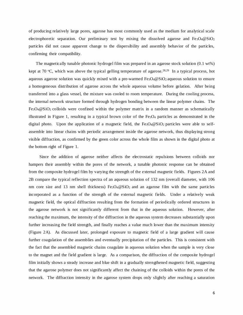
of producing relatively large pores, agarose has most commonly used as the medium for analytical scale
electrophoretic separation. Our preliminary test by mixing the dissolved agarose and Fe3O4@SiO2
particles did not cause apparent change to the dispersibility and assembly behavior of the particles,
confirming their compatibility.
The magnetically tunable photonic hydrogel film was prepared in an agarose stock solution (0.1 wt%)
kept at 70 oC, which was above the typical gelling temperature of agarose.28,29 In a typical process, hot
aqueous agarose solution was quickly mixed with a pre-warmed Fe3O4@SiO2 aqueous solution to ensure
a homogeneous distribution of agarose across the whole aqueous volume before gelation. After being
transferred into a glass vessel, the mixture was cooled to room temperature. During the cooling process,
the internal network structure formed through hydrogen bonding between the linear polymer chains. The
Fe3O4@SiO2 colloids were confined within the polymer matrix in a random manner as schematically
illustrated in Figure 1, resulting in a typical brown color of the Fe3O4 particles as demonstrated in the
digital photo. Upon the application of a magnetic field, the Fe3O4@SiO2 particles were able to self-
assemble into linear chains with periodic arrangement inside the agarose network, thus displaying strong
visible diffraction, as confirmed by the green color across the whole film as shown in the digital photo at
the bottom right of Figure 1.
Since the addition of agarose neither affects the electrostatic repulsions between colloids nor
hampers their assembly within the pores of the network, a tunable photonic response can be obtained
from the composite hydrogel film by varying the strength of the external magnetic fields. Figures 2A and
2B compare the typical reflection spectra of an aqueous solution of 132 nm (overall diameter, with 106
nm core size and 13 nm shell thickness) Fe3O4@SiO2 and an agarose film with the same particles
incorporated as a function of the strength of the external magnetic fields. Under a relatively weak
magnetic field, the optical diffraction resulting from the formation of periodically ordered structures in
the agarose network is not significantly different from that in the aqueous solution. However, after
reaching the maximum, the intensity of the diffraction in the aqueous system decreases substantially upon
further increasing the field strength, and finally reaches a value much lower than the maximum intensity
(Figure 2A). As discussed later, prolonged exposure to magnetic field of a large gradient will cause
further coagulation of the assemblies and eventually precipitation of the particles. This is consistent with
the fact that the assembled magnetic chains coagulate in aqueous solution when the sample is very close
to the magnet and the field gradient is large. As a comparison, the diffraction of the composite hydrogel
film initially shows a steady increase and blue shift in a gradually strengthened magnetic field, suggesting
that the agarose polymer does not significantly affect the chaining of the colloids within the pores of the
network. The diffraction intensity in the agarose system drops only slightly after reaching a saturation

7
value, and the peak position slightly blue-shifts with further increasing magnetic field strength (Figure
2B). The much more stable diffraction in the agarose system when subjected to strong magnetic fields
with large gradients indicate less chain coagulation in the polymer network.30 From the reflection spectra,
one can estimate an average value for the interparticle spacings using Bragg’s law, λ=2ndsinθ, as well as
the surface-to-surface distance, ds-s, by subtracting the colloid diameter.10 The Fe3O4@SiO2 colloids start
to assemble at ds-s≈106 nm in the agarose system, and reach the maximum diffraction intensity at a
spacing of ~37 nm, while the Fe3O4@SiO2 aqueous system starts to form ordered structures at ds-s≈130
nm, and achieves its maximum diffraction intensity as ds-s decreases to ~73 nm under a steadily rising
magnetic field. The respectively smaller interparticle spacing in the agarose system versus the aqueous
system demonstrates that stronger magnetic attractions in the agarose system are needed to overcome the
steric hindrance from the polymer chains in order to initiate the periodic assembly of the colloids and
achieve the most ordered structure. In both cases, ds-s does not further decrease beyond ~29 nm even
when the magnetic field is as high as ca. 4700 G with a gradient of 2590 G/cm, indicating similar
electrostatic interaction between neighboring particles in the two systems which is unaffected by the
presence of the agarose polymer chains.
Differences between the repulsive forces involved in ordering Fe3O4@SiO2 in the agarose film and
in water should be responsible for their dissimilar optical responses. Based on Derjaguin–Landau–
Verwey–Overbeek (DLVO) theory,31 electrostatic repulsion is the primary repulsive force in both systems,
in which water is the continuous phase and the colloids are highly charged. In a weak magnetic field, the
long-range electrostatic repulsion dominates the particle interactions and determines the periodicity of the
assembled structures by counteracting the induced magnetic attraction.7 Under a strong magnetic field
with a large gradient, the assembled chain-like structures in the aqueous solution are forced to coagulate
due to the magnetic packing force, which draws them toward the maximum of the local magnetic gradient,
resulting in a decrease of diffraction intensity.5 For the Fe3O4@SiO2 colloids in the agarose film,
additional repulsive forces such as steric hindrance introduced by the polymer network as well as strong
hydrogen bonding between the hydroxyl groups on the agarose network and the Fe3O4@SiO2 surface can
function as effective barriers against chain-chain coagulation.30 As a result, the ordered structures in the
agarose film can be well maintained against the magnetic packing force, as demonstrated by the
unvarying peak position and the slightly reduced intensity at short wavelengths. Even when the field is
further strengthened, the diffraction intensity remains unchanged. Comparing the two systems, the
overall maximum intensity and the tuning range of the diffraction are reduced in the agarose film due to
the relatively short assembled particle chains and the physical confinement from the polymer network.

8
The optical response of the composite hydrogel film to the changes in external magnetic field is fully
reversible and reproducible. Figure 3A shows the blue shift of the diffraction peak of a composite
hydrogel film in response to an increasing magnetic field when reducing the distance between the magnet
and the sample. The reverse process of moving the magnet away from the sample is able to red-shift the
diffraction peak back to the original position (Figure 3B). The summary in Table S1 from the supporting
information shows that the diffraction peak intensity and position can be well reproduced at a fixed
sample-magnet distance with little hysteresis in either the forward or reverse direction when moving the
magnet. The slight mismatches observed in intensities and peak positions for a few points can be ascribed
to inaccurate manual placement of the magnet, as small errors in distance will alter the magnetic field
strength applied to the colloidal sample. The photonic structures are very stable and no precipitation of
the CNCs can be observed after multiple tests of reversibility.
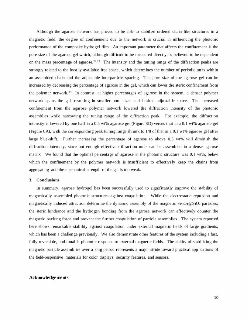
One remarkable improvement by incorporating agarose hydrogel film is the excellent long term
stability achievable in a magnetic field with a large gradient. In the agarose assisted system, the steric
hindrance and hydrogen bonding introduced by the polymer network counter the packing force and reta in
the ordered structures with stable photonic properties.32 As a demonstration, an agarose/Fe3O4@SiO2
composite hydrogel film was exposed to a magnetic field with strength of 360 G and a gradient of 115
G/cm, and the corresponding photonic performance was monitored over 12 hours. The diffraction
intensities and the peak positions were plotted according to the progression of time and summarized in
Figure 4, both of which were invariable with only slight fluctuations due to the instability of the
spectrometer. In contrast, the assembled particle chains coagulate and precipitate much more rapidly in
an aqueous system. When the same Fe3O4@SiO2 particles were dispersed in an aqueous solution, their
diffraction intensities dropped from 43 % to less than 10 % within 15 minutes under the same magnetic
field, as evidenced in Figure S1. The dramatically improved structural and photonic stability of the
assemblies in magnetic fields greatly broadens the potential of the magnetically responsive photonic
structures for practical applications.
The significantly enhanced stability of the photonic structures against chain coagulation with the
assistance of agarose gel can also be confirmed by optical microscopy study. Figure 5 shows the self-
assembly process of Fe3O4@SiO2 colloids in both agarose gel and in water, in response to an external
magnetic field with varying strength. The bright spots observed under dark field represent vertically
aligned nanochains or their aggregates, which diffract light. The microscopic images in Figures 5A-D
reveal the photonic structure assembly process in agarose gel while Figures 5F-I show the same process in
an aqueous system. Particles in both cases exhibited similar assembly behavior: when a very weak
magnetic field was vertically applied at the beginning, it was difficult to capture a clear image of the

9
particles due to their Brownian motion.5 When the magnetic field was gradually strengthened, the
movement of the particles slows down and bright red spots start to appear as chains of particles were lined
up along the magnetic field. With a further increase in the field strength, the diffraction color in both
systems turns from red to green, indicating a decrease in the interparticle separation. By comparing the
dark-field optical microscopy images in both cases, one can notice that the bright spots were not as
uniformly distributed in agarose gel as in water because the local particle concentration across the gel
were not uniform. However, if the magnet moved even closer, the bright green spots appeared thicker in
the aqueous solution, the number density gradually decreased, and the color of the bright spots changes
from green to blue (Figure 5J), indicating the formation of 3-dimensional photonic domains via the
coagulation of particle chains. But for the sample in agarose gel, the distribution of the bright spots and
their diffraction color remained unchanged in a strong magnetic field for 5 min (Figure 5E), indicating
that the assembled chain structures were stabilized by the agarose matrix against chain coagulation. No
obvious change in the assembled structures can be observed in agarose gel even after prolonging the
magnetic field application time, suggesting a great improvement in the stabilization of the photonic
structures by the agarose gel.
To further illustrate the excellent stabilization of the photonic chain structures by the agarose
matrix, we demonstrated a simple display unit by placing two composite agarose films in contact with
each other. Color contrast was achieved by using Fe3O4@SiO2 colloids of two different sizes. The
patterned film was fabricated by sequentially forming the green colored film and the orange colored film
using agarose solutions containing Fe3O4@SiO2 colloids of ~128 nm and ~142 nm, respectively. If no
magnetic field was applied, limited contrast can be observed due to the differing absorption/scattering of
the two different sized particles (Figure 6A). Interestingly, the display unit is able to reach a state without
any contrast across the film in a low magnetic field (~162 G) when both parts of agarose film diffract red
light (Figure 6B). Contrast became more evident in a stronger magnetic field: a maximum contrast
between the two parts can be realized by applying a magnetic field as strong as 570 G to achieve the
saturation of diffraction intensity for both sets of particles (Figure 6C). As a result, a film with a striking
green/orange contrast can be observed. The diffraction of the composite film was monitored in a
magnetic field of ~570 G with a gradient of ~198 G/cm over 18 hours. No obvious change could be
detected by comparing the digital photos in Figure 7: the boundary between the green and orange agarose
films remained clear and its position did not shift. The bright diffraction color in each film remained the
same without fading, indicating no effective migration of different sized particles between different
portions of the agarose matrix. This result proves again that diffusion of assembled particle chains in the
agarose film is limited, which indirectly demonstrates the effective stabilization effect of the agarose
network.
10
Although the agarose network has proved to be able to stabilize ordered chain-like structures in a
magnetic field, the degree of confinement due to the network is crucial in influencing the photonic
performance of the composite hydrogel film. An important parameter that affects the confinement is the
pore size of the agarose gel which, although difficult to be measured directly, is believed to be dependent
on the mass percentage of agarose.22,23 The intensity and the tuning range of the diffraction peaks are
strongly related to the locally available free space, which determines the number of periodic units within
an assembled chain and the adjustable interparticle spacing. The pore size of the agarose gel can be
increased by decreasing the percentage of agarose in the gel, which can lower the steric confinement from
the polymer network.33 In contrast, at higher percentages of agarose in the system, a denser polymer
network spans the gel, resulting in smaller pore sizes and limited adjustable space. The increased
confinement from the agarose polymer network lowered the diffraction intensity of the photonic
assemblies while narrowing the tuning range of the diffraction peak. For example, the diffraction
intensity is lowered by one half in a 0.5 wt% agarose gel (Figure 8D) versus that in a 0.1 wt% agarose gel
(Figure 8A), with the corresponding peak tuning range shrunk to 1/8 of that in a 0.1 wt% agarose gel after
large blue-shift. Further increasing the percentage of agarose to above 0.5 wt% will diminish the
diffraction intensity, since not enough effective diffraction units can be assembled in a dense agarose
matrix. We found that the optimal percentage of agarose in the photonic structure was 0.1 wt%, below
which the confinement by the polymer network is insufficient to effectively keep the chains from
aggregating and the mechanical strength of the gel is too weak.
3. Conclusions
In summary, agarose hydrogel has been successfully used to significantly improve the stability of
magnetically assembled photonic structures against coagulation. While the electrostatic repulsion and
magnetically induced attraction determine the dynamic assembly of the magnetic Fe3O4@SiO2 particles,
the steric hindrance and the hydrogen bonding from the agarose network can effectively counter the
magnetic packing force and prevent the further coagulation of particle assemblies. The system reported
here shows remarkable stability against coagulation under external magnetic fields of large gradients,
which has been a challenge previously. We also demonstrate other features of the system including a fast,
fully reversible, and tunable photonic response to external magnetic fields. The ability of stabilizing the
magnetic particle assemblies over a long period represents a major stride toward practical applications of
the field-responsive materials for color displays, security features, and sensors.
Acknowledgements

11
This work was partially supported by the U. S. Army Research Laboratory (Award No: W911NF-10-
1-0484) and U. S. National Science Foundation (DMR-0956081).
References
(1) Ge, J.; Yin, Y. Responsive Photonic Crystals. Angew. Chem., Int. Ed. 2011, 50, 1492.
(2) Matsubara, K.; Watanabe, M.; Takeoka, Y. A thermally adjustable multicolor photochromic hydrogel. Angew. Chem., Int. Ed. 2007, 46, 1688.
(3) Maurer, M. K.; Lednev, I. K.; Asher, S. A. Photoswitchable Spirobenzopyran- Based Photochemically Controlled Photonic Crystals. Adv. Funct. Mater. 2005, 15, 1401.
(4) Ge, J.; Hu, Y.; Yin, Y. Highly Tunable Superparamagnetic Colloidal Photonic Crystals. Angew.
Chem. Int. Ed. 2007, 46, 7428. (5) Ge, J.; Hu, Y.; Zhang, T.; Huynh, T.; Yin, Y. Self-Assembly and Field-Responsive Optical Diffractions
of Superparamagnetic Colloids. Langmuir 2008, 24, 3671. (6) Ge, J.; Yin, Y. Magnetically Responsive Colloidal Photonic Crystals. J. Mater. Chem. 2008, 18,
5041.
(7) Ge, J.; Yin, Y. Magnetically Tunable Colloidal Photonic Structures in Alkanol Solutions. Adv. Mater. 2008, 20, 3485.
(8) Hu, Y.; He, L.; Yin, Y. Charge Stabilization of Superparamagnetic Colloids for High Performance
Responsive Photonic Structures. Small 2012, 8, 3795. (9) Hu, Y.; He, L.; Yin, Y. Magnetically Responsive Photonic Nanochains. Angew. Chem., Int. Ed. 2011,
50, 3747. (10) Xu, X.; Friedman, G.; Humfeld, K. D.; Majetich, S. A.; Asher, S. A. Synthesis and Utilization of
Monodisperse Superparamagnetic Colloidal Particles for Magnetically Controllable Photonic
Crystals. Chem. Mater. 2002, 14, 1249. (11) Wang, M.; He, L.; Yin, Y. Magnetic field guided colloidal assembly. Materials Today 2013, 16, 110. (12) Zhang, Q.; Janner, M.; He, L.; Wang, M.; Hu, Y.; Lu, Y.; Yin, Y. Photonic Labyrinths: Two-
Dimensional Dynamic Magnetic Assembly and in Situ Solidification. Nano Lett. 2013, 13, 1770. (13) Malik, V.; Petukhov, A. V.; He, L.; Yin, Y.; Schmidt, M. Colloidal Crystallization and Structural
Changes in Suspensions of Silica/Magnetite Core–Shell Nanoparticles. Langmuir 2012, 28, 14777. (14) Haque, M. A.; Kurokawa, T.; Kamita, G.; Yue, Y.; Gong, J. P. Rapid and Reversible Tuning of
Structural Color of a Hydrogel over the Entire Visible Spectrum by Mechanical Stimulation. Chem.
Mater. 2011, 23, 5200. (15) Asher, S. A.; Holtz, J.; Liu, L.; Wu, Z. Self-Assembly Motif for Creating Submicron Periodic
Materials. Polymerized Crystalline Colloidal Arrays. J. Am. Chem. Soc. 1994, 116, 4997.
(16) Foulger, S. H.; Jiang, P.; Ying, Y.; Lattam, A. C.; Smith, D. W.; Ballato, J. Photonic Bandgap Composites. Adv. Mater. 2001, 13, 1898.
(17) Aront, S.; Fulmer, A.; Scott, W. E. The Agarose Double Helix and Its Function in Agarose Gel Structure. J. Mol. Biol. 1974, 90, 269.
(18) Brody, J. R.; Kern, S. E. History and principles of conductive media for standard DNA
electrophoresis. Anal. Biochem. 2004, 333, 1. (19) Djabourov, M.; Clark, A. H.; Rowlands, D. W.; Ross-Murphy, S. B. Small-angle x-ray scattering
characterization of agarose sols and gels. Macromolecules 1989, 22, 180. (20) Labropoulos, K. C.; Niesz, D. E.; Danforth, S. C.; Kevrekidis, P. G. Dynamic rheology of agar gels:
theory and experiments. Part I. Development of a rheological model. Carbohydr. Polym. 2002,
50, 393.

12
(21) Liu, Q.; Li, J.; Liu, H.; Tora, I.; Ide, M.; Lu, J.; Davis, R.; Green, D.; Landers, J. Rapid, cost-effective DNA quantification via a visually-detectable aggregation of superparamagnetic silica-magnetite
nanoparticles. Nano Research 2014, 7, 755. (22) Pernodet, N.; Maaloum, M.; Tinland, B. Pore size of agarose gels by atomic force microscopy.
Electrophoresis 1997, 18, 55. (23) Maaloum, M.; Pernodet, N.; Tinland, B. Agarose gel structure using atomic force microscopy: Gel
concentration and ionic strength effects. Electrophoresis 1998, 19, 1606.
(24) Xiong, J.; Narayanan, J.; Liu, X.; Chong, T.; Chen, S.; Chung, T. Topology Evolution and Gelation Mechanism of Agarose Gel. J. Phys. Chem. B 2005, 109, 5638.
(25) Ge, J.; Hu, Y.; Biasini, M.; Beyermann, W. P.; Yin, Y. Superparamagnetic Magnetite Colloidal
Nanocrystal Clusters. Angew. Chem. Int. Ed. 2007, 46, 4342. (26) Hu, Y.; Zhang, Q.; Goebl, J.; Zhang, T.; Yin, Y. Control over the permeation of silica nanoshells by
surface-protected etching with water. Phys. Chem. Chem. Phys. 2010, 12, 11836. (27) Workshop on Marine Algae Biotechnology: Summary Report; National Academy Press, 1986. (28) Narayanan, J.; Xiong, J.; Liu, X. Determination of agarose gel pore size: Absorbance
measurements vis a vis other techniques. JPCS 2006, 28, 83. (29) Griess, G. A.; Guiseley, K. B.; Serwer, P. The relationship of agarose gel structure to the sieving of
spheres during agarose gel electrophoresis. Biophys. J. 1993, 65, 138.
(30) Johnson, E. M.; Berk, D. A.; Jain, R. K.; Deen, W. M. Hindered diffusion in agarose gels: test of effective medium model. Biophys. J. 1996, 70, 1017.
(31) Xia, Y.; Gates, B.; Yin, Y.; Lu, Y. Monodispersed Colloidal Spheres: Old Materials with New Applications. Adv. Mater. 2000, 12, 693.
(32) Phillips, R. J.; Deen, W. M.; Brady, J. F. Hindered transport in fibrous membranes and gels: effect
of solute size and fiber configuration. J. Colloid. Interface Sci. 1990, 139, 363. (33) Yamakov, V.; Milchev, A. Diffusion of a polymer chain in porous media. Phys. Rev. E: Stat.,
Nonlinear, Soft Matter Phys. 1997, 55, 1704.
13
Figure 1. Scheme showing the assembly of superparamagnetic Fe3O4@SiO2 particles in an agarose
hydrogel film.
H

14
Figure 2. Reflection spectra of the magnetic photonic structures in magnetic fields with varying strength:
A) in water; B) in agarose gel. The field strength is varied by controlling the sample-magnet distance.
Diffraction peaks blue shift (from right to left) as the distance decreases from 4.2 to 0 cm with a step size
of 0.2 cm.

15
Figure 3. Reversible optical response of colloidal photonic crystals to an external magnetic field with
varying strength: (A) Diffraction blue-shifts as the magnet-sample distance decreases from 4.2 to 0 cm,
and (B) Diffraction red-shifts as the distance increases from 0 to 4.2 cm in step sizes of 0.2 cm in both
cases.
16
Figure 4. Plots of reflectance intensity (■) and peak position (●) versus time for a composite hydrogel
film in an external magnetic field of 360 G with a gradient of 115 G/cm.

17
Figure 5. Dark-field optical microscopy images showing the self-assembly process of Fe3O4@SiO2
colloids in agarose gel (A-E) and water (F-J) in response to varying external magnetic field strength: A,F)
141 G; B,G) 288 G; C,H) 378 G; D,E,I,J) 967 G. E, J are corresponding images to D, I after applying a
magnetic field of 967 G to the samples for 5 minutes. The scale bar corresponds to 10 μm and applies to
all images.

18
Figure 6. Digital images showing different diffraction colors of a patterned film under varying magnetic
field strengths: A) 0 G; B) 162 G; C) 570 G. The scale bar corresponds to 1 mm and applies to all images.

19
Figure 7. Digital images of the patterned photonic agarose film under a magnetic field of 570 G with a
gradient of 198 G/cm over different periods: A) 0 hours; B) 0.5 hours; C) 1 hour; D) 1.5 hours; E) 18
hours. The scale bar corresponds to 1 mm and applies to all images.

20
Figure 8. Reflection spectra of the photonic crystals in varying magnetic fields incorporated in an agarose
film with different agarose weight percentages: A) 0.1 wt%; B) 0.2 wt%; C) 0.3 wt%; D) 0.5 wt%.


![Novel Design for Photonic Crystal Ring Resonators Based ...jopn.miau.ac.ir/article_3046_01eb01affabdaa909e9328069782f311.pdf · employing photonic crystals [4]. In recent years, photonic](https://static.fdocuments.net/doc/165x107/5e7ed386707cf3599e6c8522/novel-design-for-photonic-crystal-ring-resonators-based-jopnmiauacirarticle304601eb01affabd.jpg)
