Maejo Int. J. Sci. Technol. 2019 13(03), 217-230 Maejo ...
14
Maejo Int. J. Sci. Technol. 2019, 13(03), 217-230 Maejo International Journal of Science and Technology ISSN 1905-7873 Available online at www.mijst.mju.ac.th Full Paper Tadpoles of Khao Wang frog Humerana miopus (Amphibia, Ranidae): Description of external morphology and buccal anatomy Chantip Chuaynkern 1, * , Nithina Kaewtongkum 1, 2 , Prateep Duengkae 3 and Yodchaiy Chuaynkern 1 1 Department of Biology, Faculty of Science, Khon Kaen University, Mueang Khon Kaen, Khon Kaen, 40002, Thailand 2 Sakaerat Environmental Research Station, 1 Moo 9, Udom Sab subdistrict, Wang Nam Khiao district, Nakhon Ratchasima, 30370, Thailand 3 Department of Forest Biology, Faculty of Forestry, Kasetsart University, Chatujuk, Bangkok, 10900, Thailand * Corresponding author, e-mail: [email protected] Received: 25 April 2018 / Accepted: 4 November 2019 / Published: 7 November 2019 Abstract: We describe the tadpoles of Humerana miopus (Boulenger, 1918) from Thailand (Krabi province). The collection site lies approximately 144 km west of the type locality of this species (Khao Wang Hip, Surat Thani province). The buccal features are described for the first time from photographs produced by scanning electron microscopy. Tadpoles are compared to the previous description based on specimens from Johor, peninsular Malaysia. The buccal features are also compared to those of its nearest known relative, H. humeralis from Assam. The tadpoles of H. miopus may be distinguished by their medium size (TL 38.50 mm; BL 15.33 mm), rounded body, distinct diagonal stripes on dorsum, upper labium with marginal papillae divided by a large middle gap, lower labium with three rows of papillae, with elongated marginal papillae; keratodont row formula 1:1+1/1+1:2. Keywords: Humerana miopus, morphology, buccal anatomy, tadpole, Thailand __________________________________________________________________________________ INTRODUCTION Humerana miopus (family Ranidae: Figure 1) was originally described as Rana miopus by Boulenger in 1918 [1]. The description was based on an adult female specimen collected from “Siam (Khao Wang Hip and Nakhon Sitammarat)”. The type locality “Khao Wang Hip” is presently located in the province of Nakhon Si Thammarat in southern Thailand. The holotype of this species (BMNH, by original designation) was collected in 1887 by Smith [2] and primarily identified as Rana humeralis Boulenger, 1887 (type locality: Bhamὸ, Teinzὸ,
Transcript of Maejo Int. J. Sci. Technol. 2019 13(03), 217-230 Maejo ...

Maejo Int. J. Sci. Technol. 2019, 13(03), 217-230
Maejo International Journal of Science and Technology
ISSN 1905-7873
Available online at www.mijst.mju.ac.th Full Paper
Tadpoles of Khao Wang frog Humerana miopus (Amphibia, Ranidae): Description of external morphology and buccal anatomy Chantip Chuaynkern 1, *, Nithina Kaewtongkum 1, 2, Prateep Duengkae 3 and Yodchaiy Chuaynkern 1
1 Department of Biology, Faculty of Science, Khon Kaen University, Mueang Khon Kaen, Khon Kaen, 40002, Thailand 2 Sakaerat Environmental Research Station, 1 Moo 9, Udom Sab subdistrict, Wang Nam Khiao district, Nakhon Ratchasima, 30370, Thailand 3 Department of Forest Biology, Faculty of Forestry, Kasetsart University, Chatujuk, Bangkok, 10900, Thailand * Corresponding author, e-mail: [email protected] Received: 25 April 2018 / Accepted: 4 November 2019 / Published: 7 November 2019
Abstract: We describe the tadpoles of Humerana miopus (Boulenger, 1918) from Thailand (Krabi province). The collection site lies approximately 144 km west of the type locality of this species (Khao Wang Hip, Surat Thani province). The buccal features are described for the first time from photographs produced by scanning electron microscopy. Tadpoles are compared to the previous description based on specimens from Johor, peninsular Malaysia. The buccal features are also compared to those of its nearest known relative, H. humeralis from Assam. The tadpoles of H. miopus may be distinguished by their medium size (TL 38.50 mm; BL 15.33 mm), rounded body, distinct diagonal stripes on dorsum, upper labium with marginal papillae divided by a large middle gap, lower labium with three rows of papillae, with elongated marginal papillae; keratodont row formula 1:1+1/1+1:2.
Keywords: Humerana miopus, morphology, buccal anatomy, tadpole, Thailand __________________________________________________________________________________ INTRODUCTION
Humerana miopus (family Ranidae: Figure 1) was originally described as Rana miopus by Boulenger in 1918 [1]. The description was based on an adult female specimen collected from “Siam (Khao Wang Hip and Nakhon Sitammarat)”. The type locality “Khao Wang Hip” is presently located in the province of Nakhon Si Thammarat in southern Thailand. The holotype of this species (BMNH, by original designation) was collected in 1887 by Smith [2] and primarily identified as Rana humeralis Boulenger, 1887 (type locality: Bhamὸ, Teinzὸ,

Maejo Int. J. Sci. Technol. 2019, 13(03), 217-230
218
northern Myanmar; presently Humerana humeralis). This species is known from north-eastern India, south-eastern Nepal, and eastern Bangladesh through northern Myanmar [3]. After some dramatic debate about the classification of the genus Rana [e.g. 1, 4–8], Rana miopus was placed in the genus Humerana, which contains three species [3]: Humerana humeralis (Boulenger, 1887), Humerana miopus (Boulenger, 1918) and Humerana oatesii (Boulenger, 1892).
Figure 1. Live adult Humerana miopus from Baan Thung Sung Community Forest (Khao Yai subdistrict, Ao Luek district, Krabi province). Photograph: Y. Chuaynkern (23 August 2006)
Humerana miopus is known as Khao Wang [9] or three-striped frog [10]. The species
is currently known from southern Thailand and peninsular Malaysia [3, 11], at elevations below 300 m asl [12–14] in habitat of ditches or forest pools of rainforest [11]. In Thailand the species is reported from six provinces [12, 15–19]: Krabi, Nakhon Si Thammarat, Narathiwat, Phangnga, Songkhla and Yala. The International Union for Conservation of Nature Red List of Threatened Species lists it as a species of ‘Least Concern’ due to its relatively wide distribution, tolerance of a degree of habitat modification, and presumed large population [11]. In addition to the above information on the taxonomy, distribution and conservation status of H. miopus, morphological data have been gathered based on tadpole specimens from Johor in southern peninsular Malaysia [20]. These data include a description of H. miopus tadpoles, illustration of their mouthparts and photographs of preserved specimens, but the buccal anatomy was not studied in detail. In addition, H. miopus tadpole is not consumed as food by local people. A more complete description of tadpole morphology is necessary to further our understanding of this species’ identity, biology, ecology and evolution. Although the information on the morphology of amphibian tadpoles has rapidly increased, this area is far from being fully explored and much information remains to be gathered [21–22]. Therefore, the present work aims to provide the first description of the buccal anatomy of H. miopus via scanning microscopy, and to compare tadpoles of this species collected in Thailand with previously described specimens from Malaysia.

Maejo Int. J. Sci. Technol. 2019, 13(03), 217-230
219
Figure 2. Habitat of Humerana miopus tadpoles. Photography: Y. Chuaynkern (23 August 2006)
MATERIALS AND METHODS Field Survey and Specimens
Thirty-eight Humerana miopus tadpoles were collected from Baan Tung Sung Community Forest (Figure 2), Khao Yai subdistrict, Ao Luek district, Krabi province. Tadpoles were collected during the day using a dip net and euthanised with a small amount of 70% ethanol. Specimens were preserved in a 1:1 mix of 10% formalin and 70% ethanol solutions [23]. All preserved specimens were deposited in Khon Kaen University Vertebrate Collection, Khon Kaen University (Khon Kaen province, north-eastern Thailand). Morphological Study
The developmental stages of the specimens were determined according to the guidelines proposed by Gosner [24]. Morphological terminology follows Altig and McDiarmid [25], and we used the keratodont row formula (KRF) of Dubois [26]. We measured 22 morphological characteristics (Table 1) using a digital caliper. Structures smaller than 1 mm were measured using a stereo microscope with a reticle. Abbreviations for morphological characteristics follow Altig and McDiarmid [25], Altig [27], Grosjean [28] and Wassersug [29]. These abbreviations include AL (anterior labium), BH (maximum body height), BL (body length), BW (maximum body width); ED (maximum eye diameter); IND (internarial distance), IOD (interorbital distance), LF (maximum height of lower tail fin), MTH (maximum tail height), ND (narial diameter), NP (naro-pupillar distance), ODW (oral disk width), PL (posterior labium), RN (rostro-narial distance), SD (spiracle diameter), SS (distance from tip of snout to opening of spiracle), SU (distance from tip of snout to insertion of upper tail fin), TAL (tail length), TL (total length), TMH (tail muscle height), TMW (tail muscle width) and UF (maximum height of upper tail fin). Tadpoles were identified by morphological comparison with the description
Maejo Int. J. Sci. Technol. 2019, 13(03), 217-230
220
of H. miopus tadpoles made by Leong and Lim [20]. Drawings were made with a camera lucida (Olympus SZX-DA, Olympus Co., Japan) attached to a stereo microscope (Olympus SZ9, model SZX-ILLK200, Olympus Co., Japan). Scanning Electron Microscopy (SEM)
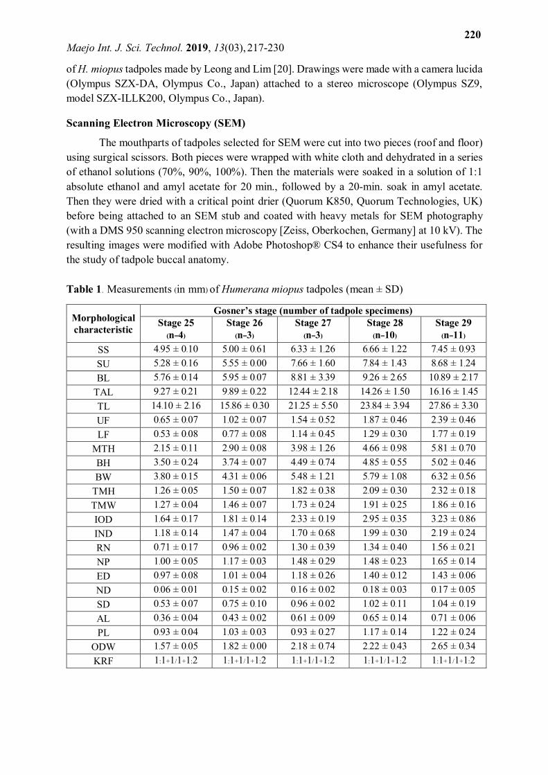
The mouthparts of tadpoles selected for SEM were cut into two pieces (roof and floor) using surgical scissors. Both pieces were wrapped with white cloth and dehydrated in a series of ethanol solutions (70%, 90%, 100%). Then the materials were soaked in a solution of 1:1 absolute ethanol and amyl acetate for 20 min., followed by a 20-min. soak in amyl acetate. Then they were dried with a critical point drier (Quorum K850, Quorum Technologies, UK) before being attached to an SEM stub and coated with heavy metals for SEM photography (with a DMS 950 scanning electron microscopy [Zeiss, Oberkochen, Germany] at 10 kV). The resulting images were modified with Adobe Photoshop® CS4 to enhance their usefulness for the study of tadpole buccal anatomy. Table 1. Measurements (in mm) of Humerana miopus tadpoles (mean ± SD)
Morphological characteristic
Gosner’s stage (number of tadpole specimens) Stage 25
(n=4) Stage 26
(n=3) Stage 27
(n=3) Stage 28 (n=10)
Stage 29 (n=11)
SS 4.95 ± 0.10 5.00 ± 0.61 6.33 ± 1.26 6.66 ± 1.22 7.45 ± 0.93 SU 5.28 ± 0.16 5.55 ± 0.00 7.66 ± 1.60 7.84 ± 1.43 8.68 ± 1.24 BL 5.76 ± 0.14 5.95 ± 0.07 8.81 ± 3.39 9.26 ± 2.65 10.89 ± 2.17
TAL 9.27 ± 0.21 9.89 ± 0.22 12.44 ± 2.18 14.26 ± 1.50 16.16 ± 1.45 TL 14.10 ± 2.16 15.86 ± 0.30 21.25 ± 5.50 23.84 ± 3.94 27.86 ± 3.30 UF 0.65 ± 0.07 1.02 ± 0.07 1.54 ± 0.52 1.87 ± 0.46 2.39 ± 0.46 LF 0.53 ± 0.08 0.77 ± 0.08 1.14 ± 0.45 1.29 ± 0.30 1.77 ± 0.19
MTH 2.15 ± 0.11 2.90 ± 0.08 3.98 ± 1.26 4.66 ± 0.98 5.81 ± 0.70 BH 3.50 ± 0.24 3.74 ± 0.07 4.49 ± 0.74 4.85 ± 0.55 5.02 ± 0.46 BW 3.80 ± 0.15 4.31 ± 0.06 5.48 ± 1.21 5.79 ± 1.08 6.32 ± 0.56
TMH 1.26 ± 0.05 1.50 ± 0.07 1.82 ± 0.38 2.09 ± 0.30 2.32 ± 0.18 TMW 1.27 ± 0.04 1.46 ± 0.07 1.73 ± 0.24 1.91 ± 0.25 1.86 ± 0.16 IOD 1.64 ± 0.17 1.81 ± 0.14 2.33 ± 0.19 2.95 ± 0.35 3.23 ± 0.86 IND 1.18 ± 0.14 1.47 ± 0.04 1.70 ± 0.68 1.99 ± 0.30 2.19 ± 0.24 RN 0.71 ± 0.17 0.96 ± 0.02 1.30 ± 0.39 1.34 ± 0.40 1.56 ± 0.21 NP 1.00 ± 0.05 1.17 ± 0.03 1.48 ± 0.29 1.48 ± 0.23 1.65 ± 0.14 ED 0.97 ± 0.08 1.01 ± 0.04 1.18 ± 0.26 1.40 ± 0.12 1.43 ± 0.06 ND 0.06 ± 0.01 0.15 ± 0.02 0.16 ± 0.02 0.18 ± 0.03 0.17 ± 0.05 SD 0.53 ± 0.07 0.75 ± 0.10 0.96 ± 0.02 1.02 ± 0.11 1.04 ± 0.19 AL 0.36 ± 0.04 0.43 ± 0.02 0.61 ± 0.09 0.65 ± 0.14 0.71 ± 0.06 PL 0.93 ± 0.04 1.03 ± 0.03 0.93 ± 0.27 1.17 ± 0.14 1.22 ± 0.24
ODW 1.57 ± 0.05 1.82 ± 0.00 2.18 ± 0.74 2.22 ± 0.43 2.65 ± 0.34 KRF 1:1+1/1+1:2 1:1+1/1+1:2 1:1+1/1+1:2 1:1+1/1+1:2 1:1+1/1+1:2

Maejo Int. J. Sci. Technol. 2019, 13(03), 217-230
221
Table 1 (continued).
RESULTS External Morphology
Material examined: Morphological description based on a single specimen (0972Y.2; Gosner’s stage 36; Figures 3 and 4) and buccopharyngeal description based on a single specimen (KKUC 00635.9; stage 34; Figures 5 and 6).
Tadpole diagnosis: Medium-sized tadpole (TL 38.50 mm; BL 15.33 mm); body rounded; body colour drab gray, tail muscle tawny olive, true cinnamon freckles all over body and tail except in gut area, distinct diagonal stripes on dorsum; upper labium with marginal papillae divided by a large middle gap, lower labium with three rows of papillae, with marginal papillae elongated; KRF 1:1+1/1+1:2.
Tadpole description: Body shape round, with rounded snout in both lateral and dorsal views (BW 8.53 mm; BH 7.16 mm). Eyes medium-sized (ED 2.09 mm), slightly bulging, not visible in ventral view, dorsolateral in position and direction. Nares very small (ND 0.18 mm), round, un-rimed, dorsal in position and direction, closer to tip of snout than to pupils (RN 2.24 mm; NP 2.12 mm). Eye and nare spacing is moderate (IND 3.18 mm; IOD 5.93 mm). Spiracle single, sinistral, medium-sized; spiracle opens on body axis at midpoint of body (SS 9.44 mm); lateral position, posterodorsal orientation; spiracle tube and spiracle opening attached to body; inner wall of spiracle presents as slight ridge; spiracle opening round. Tail muscle of moderate size (MTH 9.08 mm); somewhat strong (TMH 3.93 mm); anterior part of tail muscle of
Morphological characteristic
Gosner’s stage (number of tadpole specimens) Stage 30
(n=1) Stage 31
(n=7) Stage 34
(n=4) Stage 36
(n=1) Stage 40
(n=1) SS 7.68 8.93 ± 0.86 9.35 ± 0.32 9.44 11.21 SU 7.99 9.98 ± 1.32 10.35 ± 1.22 11.83 13.05 BL 10.03 13.28 ± 2.67 14.27 ± 2.00 15.33 15.18
TAL 18.40 19.64 ± 1.54 20.43 ± 2.35 23.70 25.51 TL 27.93 32.84 ± 3.28 35.33 ± 1.30 38.50 41.70 UF 2.37 2.82 ± 0.24 2.69 ± 0.21 4.23 3.94 LF 1.71 1.96 ± 0.24 2.10 ± 0.21 2.76 2.91
MTH 5.78 6.88 ± 0.52 6.65 ± 0.15 9.08 9.93 BH 5.34 5.97 ± 0.59 6.31 ± 0.57 7.16 7.89 BW 6.23 7.52 ± 0.70 8.12 ± 0.35 8.53 9.83
TMH 2.70 2.82 ± 0.20 3.16 ± 0.19 3.93 4.09 TMW 2.13 2.25 ± 0.13 2.24 ± 0.22 2.98 3.3 IOD 3.74 3.98 ± 0.80 4.03 ± 1.22 5.93 5.99 IND 2.16 2.49 ± 0.12 2.58 ± 0.23 3.18 2.72 RN 1.53 1.66 ± 0.20 1.67 ± 0.20 2.24 1.74 NP 1.78 1.94 ± 0.14 2.23 ± 0.16 2.12 2.3 ED 1.48 1.60 ± 0.17 1.77 ± 0.08 2.09 2.03 ND 0.14 0.18 ± 0.04 0.21 ± 0.05 0.18 0.17 SD 1.40 1.14 ± 0.32 1.20 ± 0.49 1.53 1.85 AL 0.59 0.80 ± 0.10 0.74 ± 0.05 0.85 0.8 PL 0.91 1.21 ± 0.20 1.45 ± 0.16 1.06 1.5
ODW 2.34 2.79 ± 0.39 2.96 ± 0.28 3.38 3.35 KRF 1:1+1/1+1:2 1:1+1/1+1:2 1:1+1/1+1:2 1:1+1/1+1:2 1:1+1/1+1:2

Maejo Int. J. Sci. Technol. 2019, 13(03), 217-230
222
constant height, posterior part of tail muscle gradually tapering to tip of tail. Tail fins moderately arched, height of dorsal and ventral fins equal to body height, dorsal fin slightly higher than ventral fin (UF 4.23 mm, LF 2.76 mm); dorsal fin begins near the end of the body (SU 11.83 mm); maximum height of tail fin close to the tail midpoint; posterior part of tail fin ends abruptly, acute tail tip. Vent tube broad, tube shape with oblique ending, in lateral view vent tube right wall displaced dorsally, posterior in direction. Oral disc medium-sized (ODW 3.38 mm); ventral in position and direction, laterally emarginate; upper labium presents a single row of papillae and two or three submarginal papillae in the corners of each side with large middle gap, upper labial papillae are short filiform (cone-shaped); lower labium with three rows of papillae (single row of marginal papillae and two rows of submarginal papillae), tidy arrangement, marginal papillae elongated filiform but submarginal papillae short filiform without median gap. No denticulate papillae. KRF = 1:1+1/1+1:2, keratodonts short at crown, with four to six cusps (Figure 5). Jaw sheaths medium in size, dark brown, with fine serrations; upper jaw sheath upturned curve; lower jaw sheath M-shaped.
Colouration in preservative: Body drab gray, tail muscle tawny olive, true cinnamon freckles everywhere on body and tail except in gut area, distinct diagonal stripes on dorsum in Gosner’s stages 29, 31 and 40. Tail fins translucent with light brown mottling. Oral disc and papillae transparent with small dark blue spots. Eye colour unclear.
Buccal Anatomy
Buccal roof (Figure 6A): Prenarial arena medium-sized with six medium tubercles, horizontal arrangement, middle position. A single pair of prenarial papillae, medium-sized, long filiform, rounded tip, lateral position, centre-posterior direction. Choanae medium-sized, round, internarial distance less than choanae length, oblique (45 degrees). Narial valve bulging with serrated margin at anterior wall, posterior wall thin and smooth. Single pair of narial valve projections, large filiform, with pointed tips, anterior edge serrated, free from buccal roof, lateral position, anterior direction. Postnarial arena very small and without papillae. Single pair of lateral ridge papillae, small, short filiform, rounded tips, lateral position, middle direction. Two median ridges, horizontal, bulging, with serrated edge. Buccal roof arena large in size, with an oval bulge in the middle of area, loose tubercles on oval bulge. Buccal roof arena papillae absent. Single pair of papillae on the lateral roof arena, small-short filiform, rounded tip, lateral position, middle direction, attached to buccal roof. Glandular zone presents medium tubercles. Dorsal velum with a distinctly high cushion, discontinuous, with a smooth edge.
Buccal floor (Figure 6B): Buccal floor arena large in size, round, wider than it is long, with about 30 tubercles in the posterior part, bulging cushion in posterior. Single pair of infralabial papillae, large-long filiform, serrated edge, free from buccal floor, positioned at the inside of lower beak, anterior direction. Single pair of lingual papillae, medium size, upturned-cone shape, smooth edge, middle position, attached to buccal floor. Buccal floor arena papillae include two pairs of long-medium filiform and six pairs of short-small filiform, rounded tip, smooth edge, lateral position, middle direction. Prepocket papillae absent. Buccal pockets nearly transverse, slightly curved, closer to infralabial papillae than to medial end of ventral velum. Ventral velum with medium cushion, medium tubercles, wavy smooth margin. Branchial baskets rather vertical, wider than long, three filter plates on each side, length of filter plate approximately half of buccal floor arena length, eight rows in each plate.

Maejo Int. J. Sci. Technol. 2019, 13(03), 217-230
223
Figure 3. Humerana miopus tadpole (KKUC 00635.9, Gosner’s stage 34, total length 36.20 mm) in preservative: (A) lateral view; (B) oral disc; (C) oral disc illustration. Photography and drawing: N. Kaewtongkum

Maejo Int. J. Sci. Technol. 2019, 13(03), 217-230
224
Figure 4. Drawing of a Humerana miopus tadpole (KKUC 00635.9, Gosner’s stage 34): (A) dorsal view; (B) lateral view; (C) ventral view. Drawing: N. Kaewtongkum
Figure 5. SEM photograph of Humerana miopus tadpole showing a close-up of the keratodont row

Maejo Int. J. Sci. Technol. 2019, 13(03), 217-230
225
Figure 6. SEM photographs of Humerana miopus tadpole showing buccal roof (A) and buccal floor (B) (specimen KKUC 00635.9, Gosner’s stage 34)

Maejo Int. J. Sci. Technol. 2019, 13(03), 217-230
226
DISCUSSION
The specimens upon which the tadpole morphological and buccal features described in this work are based were collected nearer to the type locality of this species than were the previously described Malaysian specimens. Baan Thung Sung Community Forest lies approximately 144 km west of the type locality, whereas the Malaysian specimens were collected from approximately 880 km south of the type locality. The external features of the Thai specimens described here are largely similar to those of the tadpoles from southern Malaysia, with two notable distinctions. Specimens from Malaysia had two or three rows of papillae on the lower labium, with the outer row being triangular in shape and the inner row conical [20]. The Thai specimens had three rows of papillae on the lower labium, and those in the outer row were distinctly long (see Figures 3B and 3C). One possible explanation for this difference is that the number of rows of marginal papillae might increase as tadpoles develop [28, 30–32]. It is also possible that differences in disc papillae may reflect adaptation to different microhabitats and food availability [33–35]. The second distinction is in colouration: Leong and Lim [20] observed three to four thin, diagonal (anterior left to posterior right) dark brown stripes evenly arranged on the dorsum in tadpoles of Gosner’s stage 34 and later. In specimens from Thailand the oblique stripes were not observed at Gosner’s stage 34 but were found in three other stages: Gosner’s stages 29 (one specimen), 31 (six specimens), and 40 (one specimen). This means that the presence of these oblique stripes may be independent of developmental stage.
The only members of the genus Humerana for which detailed descriptions of the tadpole stage exist are H. humeralis (as Hylarana humeralis; [36]) and H. miopus (as Rana miopus [20, the present work]). The tadpoles of these two species differ in several external characteristics (Table 2): diagonal stripes evenly arranged on dorsum (absent in H. humeralis versus present in H. miopus), ventral part of head and body (belly translucent gray without melanophores, branchial area and anteriorly covered with densely arranged melanophores versus dark brown spots/reticulation, rest of venter white), rows of papillae on lower labium (single row versus two to three rows in Malaysian specimens or three rows with long papillae in the outer row in Thai specimens), keratodonts (three to four cusps versus four to six cusps), shape of lower jaw sheath (V-shaped versus M-shaped), shape of body (oval versus rounded), and shape of tail (constant height in anterior part and slightly tapering in posterior part, maximum height in posterior part of tail, tail tip finely round versus moderately arched, maximum height at beginning of tail, tail tip acute).
Distinctions also exist between the buccal anatomy of Humerana humeralis [36] and H. miopus [the present work], which could be a further basis for species identification. Humerana humeralis has three to four short papillae on each side of the buccal roof arena, which are absent in H. miopus. In the prenarial arena, H. humeralis has a large M-shaped ridge, while H. miopus possesses tubercles. Humerana humeralis has a single pair of postnarial papillae; H. miopus instead has single pairs of papillae on the lateral ridge and lateral roof.
The buccal floor of Humerana humeralis is square shaped, without any tubercles. That of H. miopus is wider than it is long, and has numerous tubercles on it. There are four papillae on the buccal floor arena in H. humeralis while H. miopus has seven. There are no lingual papillae in H. humeralis; H. miopus has a single pair.

Maejo Int. J. Sci. Technol. 2019, 13(03), 217-230
227
Table 2. Comparative measurements (in mm) of Humerana tadpoles
Gosner’s stage
Humerana miopus (Thailand)
Humerana miopus [20] (Malaysia)
Humerana humeralis [36] (Assam)
n BL TL n BL TL n BL TL
21 – – – – – – 10 3.24±0.03 (3.2-3.28)
4.44±0.05 (4.40-4.50)
22 – – – – – – 10 3.13±0.01 (3.12-3.16)
4.94±0.03 (4.9-5)
23 – – – – – – 10 2.81±0.03 (2.76-2.86)
5.67±0.05 (5.6-5.7)
24 – – – – – – 10 3.1±0.03 (3.06-3.14)
7.28±0.03 (7.2-7.3)
25 4 5.76±0.14 (5.64˗5.91)
14.1±2.16 (10.87˗15.36) – – – 10 3.16±0.03
(3.1-3.2) 7.6±0.05 (7.5-7.7)
26 3 5.95±0.07 (5.91˗6.03)
15.86±0.3 (15.65˗16.07) 4 9.9-11 24.3-27.2 10 8.72±0.64
(8-9.52) 24.41±1.69 (21.6-26.4)
27 3 8.81 ± 3.39 (6.62˗12.71)
21.25± 5.5 (17.73˗27.58) 4 12.4-13 26.8-29.9 10 9.62±0.46
(8.66-9.98) 27.03±1.57 (24.1-28.6)
28 10 9.26±2.65 (7.45˗15.05)
23.84±3.94 (19.74˗31.28 2 12.8-13.3 30.4-32 8 9.13±0.28
(8.74-9.52) 26.59±1.17 (25.4-28.2)
29 4 10.89±2.17 (8.92˗13.83)
27.86±3.3 (25.64˗31.65) 1 13.7 31.6 – – –
30 1 10.03 27.93 5 13.1-15.2 31.9-34.6 – – –
31 7 13.28±2.67 (10.64˗17.19)
32.84±3.28 (27.89˗37.24) 1 14.6 35 – – –
32 – – – 1 16.1 37.6 6 13.81±0.31 (13.34-14.1)
40.04±1.35 (38.9-42.5)
33 – – – 3 14.7-15.6 34.5-37.8 10 14.15±0.61 (13-15)
41.89±1.82 (38.6-44.5)
34 4 14.27±2 (11.87˗16.35)
35.33±1.3 (33.83˗36.2) 6 15.1-17.9 35.6-41.6 10 14.3±0.53
(13-14.7) 40.65±0.56 (39.6-41.42)
35 – – – 1 15.9 36.7 10 14.42±0.56 (13.1-15.1)
42.33±1.55 (39.5-44.1)
36 1 15.33 38.5 2 17.2-17.4 40.2-40.9 10 16.56±0.61 (15.6-17.36
47.77±2.03 (44.78-50.52)
37 – – – 3 17-17.3 40-40.9 7 17.37±0.33 (16.64-17.64)
50.21±2.14 (47.72-53.48)
38 – – – 2 17.8-18.6 42.5-45.5 10 18.94±0.65 (18.2-20.1)
56.83±1.49 (55.4-59.5)
39 – – – 1 19.7 48.8 10 19.18±0.49 (18.44-19.86)
60.08±1.71 (58.1-62.7)
40 1 15.18 41.7 11 18.5-23.7 46.6-58.7 10 19.4±0.37 (18.86-20.04)
60.25±1.99 (57-62.8)
41 – – – – – – 5 20.88±0.66 (20.2-21.7)
58.27±1.34 (56.4-59.6)
42 – – – 2 19.9-20.1 42.9-43 5 21.08±0.6 (20.46-21.76)
55.56±2.4 (53.4-59.1)
43 – – – – – – 3 20.65±1.23 (19.36-21.8)
47.09±3.32 (43.4-49.8)
44 – – – – – – 3 21.7±0.73 (21.14-22.52)
35.91±5.85 (31.7-42.6)
45 – – – – – – 3 20.88±1.44 (19.34-22.2)
26.67±4.75 (21.5-30.9)
46 – – – – – – 10 21.23±1.66 (19-23.6)
21.23±1.66 (19-23.6)
KRF 1:1+1/1+1:2 (stage 25-31, 34, 36, 40)
1:1+1/1+1:2 (unknown stage)
1:1+1/1+1:2 (stage 34)

Maejo Int. J. Sci. Technol. 2019, 13(03), 217-230
228
Our knowledge of the diversity of amphibian species is rapidly growing [3]. Currently there are 7,822 known species of amphibians worldwide, approximately 189 of which are found in Thailand [3]. However, there may be far more species than this in Thailand, owing to the presence of cryptic species [37]. Studies have revealed previously unreported species diversity in Thailand [37, 38], highlighting the need for further research on amphibians, and particularly amphibian tadpoles in this area. CONCLUSIONS
Tadpoles of Humerana miopus from Thailand are largely similar to those of the same species described from Malaysia. They are of medium size (TL 38.50 mm; BL 15.33 mm), with a rounded body, distinct diagonal stripes on the dorsum, upper labium with marginal papillae divided by a large middle gap, lower labium with three rows of papillae, of which marginal papillae are elongated; KRF 1:1+1/1+1:2. Their buccal features are described here for the first time. ACKNOWLEDGEMENTS
The authors thank Khon Kaen University and Kasetsart University for facilitating this work, which was financially supported by 1) the Ministry of Science and Technology (Thailand), 2) the Faculty of Science, Khon Kaen University, 3) the Graduate School, Khon Kaen University, and 4) the International Affairs Division, Khon Kaen University. Our special thanks go to Yada Polsan (Department of Anatomy, Faculty of Medicine, Khon Kaen University) and Boonsong Kongsook (Faculty of Science, Khon Kaen University) for assistance in SEM preparation. REFERENCES 1. G. A. Boulenger, “Description of a new frog (Rana miopus) from Siam”, J. Nat. Hist. Soc.
Siam, 1918, 3, 11-12. 2. M. A. Smith, “Description of five tadpoles from Siam”, J. Nat. Hist. Soc. Siam, 1916, 2,
37-43. 3. D. R. Frost, “Amphibian species of the world: An online reference version 6.0”, 2018,
http://research.amnh.org/herpetology/amphibia/index.html (Accessed: March 2018). 4. G. A. Boulenger, “A monograph of the South Asian, Papuan, Melanesian and Australian
frogs of the genus Rana”, Rec. Indian Mus., 1920, 20, 1-226. 5. R. Bourret, “Les Batraciens de l’Indochine”, Institut Océanographique de l’Indochine,
Hanoi, 1942. 6. A. Dubois, “Miscellanea taxinomica batrachologica (I)”, Alytes, 1987, 5, 7-95. 7. A. Dubois, “Notes sur la classification des Ranidae (amphibiens anoures)”, Bull. Mens.
Soc. Linn. Lyon, 1992, 61, 305-352. 8. D. R. Frost, T. Grant, J. Faivovich, R. H. Bain, A. Haas, C. F. B. Haddad, R. O. de Sá, A.
Channing, M. Wilkinson, S. C. Donnellan, C. J. Raxworthy, J. A. Campbell, B. L. Blotto, P. Moler, R. C. Drewes, R. A. Nussbaum, J. D. Lynch, D. M. Green and W. C. Wheeler, “The amphibian tree of life”, Bull. Amer. Mus. Nat. Hist., 2006, 297, 1-370.
9. N. Frank and E. Ramus, “Complete Guide to Scientific and Common Names of Reptiles and Amphibians of the World”, N. G. Publishing Inc., Pottsville, 1995.

Maejo Int. J. Sci. Technol. 2019, 13(03), 217-230
229
10. T. Chan-ard, “A Photographic Guide to Amphibians in Thailand”, Darnsutha Press, Bangkok, 2003.
11. IUCN, “IUCN Red list of threatened species”, 2016, http://www.iucnredlist.org (Accessed: March 2018).
12. E. H. Taylor, “The amphibian fauna of Thailand”, Univ. Kansas Sci. Bull., 1962, 43, 265-599.
13. A. G. C. Grandison, “The status and relationships of some East African earless toads (Anura, Bufonidae) with a description of a new species”, Zool. Meded, 1972, 47, 30-48.
14. P. Y. Berry, “The Amphibian Fauna of Peninsular Malaysia”, Tropical Press, Kuala Lumpur, 1975.
15. J. Nabhitabhata, T. Chan-ard and Y. Chuaynkern, “Checklist of Amphibians and Reptiles in Thailand”, Office of Environmental Policy and Planning, Bangkok, 2004.
16. J. Nabhitabhata and T. Chan-ard, “Thailand Red Data: Mammals, Reptiles and Amphibians”, Office of Natural Resources and Environmental Policy and Planning, Bangkok, 2005.
17. Y. Chuaynkern and C. Chuaynkern, “A checklist of amphibians in Thailand”, J. Wildl. Thailand, 2012, 19, 163-211 (in Thai).
18. S. Karaphan, W. Chuthong, P. Sukbangnop, L. Waiprom and P. Chotiphan, “Amphibians of Hala-Bala”, Pa Phru-Pa Halabala Research Station, Narathiwat, 2013 (in Thai).
19. P. Duengkae, Y. Chuaynkern, A. Chaiyes, Y. Ponpituk and S. Siri, “Biological Resources Catalog: Amphibians of Thailand”, Biodiversity-Based Economy Development Office, Bangkok, 2016.
20. T. M. Leong and C. M. Lim, “The tadpole of Rana miopus Boulenger, 1918 from peninsular Malaysia”, Hamadryad, 2003, 27, 175-178.
21. T. M. Leong, “Status of larval identities among the Peninsular Malaysian Anura”, Herpetol. Rev., 2002, 33, 171-174.
22. I. Das and A. Haas, “Sources of larval identities for amphibians from Borneo”, Herpetol. Rev. 2005, 36, 375-382.
23. C. Inthara, Y. Chuaynkern, P. Duengkae and S. Grosjean, “The tadpole of Quasipaa fasciculispina (Inger, 1970) from southeastern Thailand, with the description of its buccal anatomy”, Alytes, 2009, 26, 86-96.
24. K. L. Gosner, “A simplified table for staging anuran embryos and larvae with notes on identification”, Herpetol., 1960, 16, 183-190.
25. R. Altig and R. W. McDiarmid, “Body plan: Development and morphology”, in “Tadpoles: The Biology of Anuran Larvae” (Ed. R. W. McDiarmid and R. Altig), Chicago University Press, Chicago, 1999, Ch.3.
26. A. Dubois, “Keratodont formulae in anuran tadpoles: Proposals for standardization”, J. Zool. Syst. Evol. Res., 1995, 33, 1-15.
27. R. Altig, “A primer for the morphology of anuran tadpoles”, Herpetol. Conserv. Biol., 2007, 2, 71-74.
28. S. Grosjean, “The choice of external morphological characters and developmental stages for tadpole based anuran taxonomy: A case study in Rana (Sylvirana) nigrovittata (Blyth, 1855) (Amphibia, Anura, Ranidae)”, Contrib. Zool., 2005, 74, 61-76.
29. R. J. Wassersug, “Oral morphology of anuran larvae: Terminology and general description”, Occas. Pap. Mus. Nat. Hist., 1976, 48, 1-23.

Maejo Int. J. Sci. Technol. 2019, 13(03), 217-230
230
30. D. A. Sánchez, “Larval development and synapomorphies for species groups of Hyloscirtus Peters, 1882 (Anura: Hylidae: Cophomantini)”, Copeia, 2010, 3, 351-363.
31. S. Grosjean, R. D. Randrianiaina, A. Strauß and M. Vences, “Sand-eating tadpoles in Madagascar: Morphology and ecology of the unique larvae of the treefrog Boophis picturatus”, Salamandra, 2011, 47, 63-76.
32. A. Terán-Valdez and J. M. Guayasamin, “The tadpole of the glassfrog Chimerella mariaelenae (Anura: Centrolenidae)”, Cienci América, 2014, 3, 17-22.
33. M. S. Khan and S. A. Mufti, “Oral disc morphology of amphibian tadpole and its functional correlates”, Pakistan J. Zool., 1994, 26, 25-30.
34. K. D. Wells, “The Ecology and Behavior of Amphibians”, University of Chicago Press, Chicago, 2010, Ch.12.
35. M. A. Anganoy-Criollo, “Tadpoles of the high-Andean Hyloxalus subpunctatus (Anura: Dendrobatidae) with description of larval variation and species distinction by larval morphology”, Pap. Avulsos Zool., 2013, 53, 211-224.
36. T. Bortamuli, S. Bordoloi, A. Ohler and S. Grosjean, “External morphology, buccopharyngeal anatomy and development rate of the tadpoles of two Asian Ranidae (Amphibia: Anura), Hylarana humeralis (Boulenger, 1887) and Hylarana leptoglossa (Cope, 1868)”, J. Nat. Hist., 2010, 44, 421-445.
37. J. A. Sheridan and B. L. Stuart, “Hidden species diversity in Sylvirana nigrovittata (Amphibia: Ranidae) highlights the importance of taxonomic revisions in biodiversity conservation”, PLoS ONE, 2018, 13, e0192766.https://doi.org/10.1371/journal.pone. 0192766.
38. C. Suwannapoom, M. Sumontha, J. Tunprasert, T. Ruangsuwan, P. Pawangkhanant, D. V. Korost and N. A. Poyarkov, Jr., “A striking new genus and species of cave-dwelling frog (Amphibia: Anura: Microhylidae: Asterophryinae) from Thailand”, PeerJ., 2018, 6, e4422.https://doi.org/10.7717/peerj.4422.
© 2019 by Maejo University, San Sai, Chiang Mai, 50290 Thailand. Reproduction is permitted
for noncommercial purposes.