
Mads Friberg - UiO
38
Oxytocin and first impressions Mads Friberg Hovedoppgave i psykologi ved Psykologisk Institutt Det samfunnsvitenskaplige fakultet Veileder: Bruno Laeng
Transcript of Mads Friberg - UiO

Oxytocin and first impressions
Mads Friberg
Hovedoppgave i psykologi ved Psykologisk Institutt Det samfunnsvitenskaplige fakultet
Veileder: Bruno Laeng

II

III

IV
Oxytocin and first impressions

V
© Mads Friberg
2012
Oxytocin and first impressions
Mads Friberg
http://www.duo.uio.no/
VI
Abstract Subtle facial expressions may cause “core impressions” of other people, i.e. a feeling of like
or dislike witch is affected by facial cues that is not explicitly and consciously recognized. In
the present investigation, we were interested in how the neuropeptide oxytocin affects
recognition of these subtle facial expressions. Participants received oxytocin or placebo, and
viewed static and dynamic “hybrid” faces that showed a facial expression (happiness, anger,
fear, sadness) only in the lowest spatial frequency (1–6 cycles/image), which was blended
with the same face’s neutral expression in the rest of the bandwidth (7–128 cycles/image).
Two tasks was used as measures, a core impression task where participants was asked to rate
“hybrid” images indicating friendliness, and a emotional labeling task, where participants was
asked to choose the emotion they believed the “hybrid” images presented. We expected rates
of friendliness to be higher after oxytocin administration versus placebo, especially for the
hybrids containing low passed happy expressions. Further, we expected a higher “hit rate” on
the emotional labeling task after oxytocin administration. Contrary to the hypothesis, ratings
of friendliness did not increase after oxytocin administration. In the labeling task, oxytocin
did not increase hit rate of emotional expressions in the lowest spatial frequency. Future
implications are discussed.
VII
Introduction
Core impressions, that is, feelings without our own direct access, define a set of
associations towards a phenomena witch is implicit and unconscious, assumed to affect
behavior without being directly available for conscious experience. However, feelings that is
not explicitly and consciously experienced is a debated topic in psychology and neuroscience,
initiating a philosophical debate about whether such feelings can exist at all (Berridge &
Winkielman, 2003). Put short, this debate has been concerned with the question of what could
be said to constitute a feeling. If a feeling is not felt, as implied by the notion of unconscious
feelings, is it really a feeling? For the present purpose, a pragmatic definition proposed by
Berridge & Winkielman (2003) is used, saying that emotions are something that causes
affects, and particularly involves a like or dislike towards something. Extending this notion of
like and dislike to a behavioral disposition, emotions may be theoretically distinguished by
their unique motivational properties (Izard, 1978). That is, emotions are understood to guide
behaviors by motivating a person to take specific adaptive actions, such as to avoid danger
and seek safety or pursue adaptive goals such as romantic relationships. The overarching
question in the present study may be formulated as: what mechanisms are involved when we
form first impressions of unfamiliar others? Based on advances in knowledge generated by
the interception between biology and psychology, the present experiment seeks to investigate
a possible relationship between the neuropeptide oxytocin and “core impressions”.
Background
First impressions
From faces, first impressions are shown to form very fast (Fox, 2002). As little as 100
ms exposure to unfamiliar faces provides sufficient information in order to elicit trait
inferences (Willis & Todorov, 2006) and evolutionarily important inferences such as threat
can be made even after shorter exposures (Todorov, Baron, & Oosterhof, 2008). Interestingly,
even subtle resemblance in neutral faces to expressions that signal whether a person should be
avoided (anger) or approached (happiness) serves as the basis of valence evaluation (Todorov,
Said, Engell, & Oosterhof, 2008). Based on what could be called a “core impression”, first
impression may happen with very little involvement of conscious evaluations and may
operate more or less on a sub-conscious, automatic level (Fox, 2002; Laeng et al., 2010), and
VIII
further, these first impressions can be impressively stable over time (Ballew & Todorov,
2007; Bar, Neta, & Linz, 2006; Willis & Todorov, 2006).
In one of the first systematic attempts to understand trait judgments from faces, Secord (1958)
suggested that first impressions are based on misattribution of momentary states to enduring
attributes (Secord, 1958). Accessible facial cues (e.g. smile) can be generalized to stable
dispositions (e.g. friendly). This overgeneralization of momentary social cues to more stable
impressions may account for rapid and efficient, but not necessarily accurate trait judgments
from facial expressions. One explanation for this phenomenon may be evolutionary; to be
able to engage in adaptive behavior, such as approaching a possible lover or avoiding a foe,
people need to make situational choices based on the available information at hand.
Importantly, emotional expressions convey information about other people’s internal states,
and are highly potent cues about what a person thinks and potential actions in a given
situation (Todorov, 2011). Thus, first impressions may serve as a form of heuristics providing
a coarse framework that guides social cognition, based on an evolutionary developed ability
to detect structural features in a persons` face that can at a level that is better than chance (i.e.,
on the basis of a “kernel of truth”) give away information about the person’s emotional state
and traits.
Detection of subtle emotional stimuli
To be able to detect subtle resemblances to emotional expressions, the human
perceptual system should be highly sensitive to expressions of emotionality (and
subsequently; intentionality). Indeed, the processing of emotional-laden stimuli are prioritized
in the brain and leads to stronger activation in visual processing regions (Pessoa, Kastner, &
Ungerleider, 2002). For example, emotional expressions has been shown to be given
perceptual priority over neutral expressions at the very early stages of information processing
using visual search tasks (Sato & Yoshikawa, 2010). Further, in a series of four studies, Bar et
al. (2006) showed that first impressions from neutral faces perceived to be threatening could
happen as fast as 39 ms (Bar et al., 2006). They argued that, to form first impressions when
the stimulus is available for such a short duration, the brain needs to rely on whatever visual
information is available very early. Crucially, some structural features of the human face may
be detectable from a rather limited range within the low spatial frequencies of visual
information (i.e. more or less diffuse visual signals with poor contrast). Spatial frequency is a
characteristic of luminance variations across space, where low frequency content provides
IX
information about the global, holistic aspects of a stimulus, and high frequency content
provides information about local features (Goffaux & Rossion, 2006). Low spatial
frequencies are known to be extracted more rapidly than the other higher spatial frequencies
and, furthermore, to involve neural circuitry implicated in processes related to threat
perception; i.e. the amygdala (Vuilleumier, Armony, Driver, & Dolan, 2003).
Processing of emotional expressions activates a particularly strong activation in the
amygdala and the face-orientated fusiform gyrus in the visual cortex (Vuilleumier & Pourtois,
2007). However, Vuilleumier et al. (2003) has shown using fMRI that the human amygdala,
witch receive visual input from magnocelluar channels through the dorsal parietal stream and
connected sub-cortical regions such as superior colliculus and pulvinar, is essentially “blind”
to most of the visible spatial frequency spectrum except the lowest (<6 cycles/image). In
contrast, the fusiform cortex in the temporal lobe was engaged more by high-passed spatial
frequency images than by low-pass spatial frequency images; the latter evoking only a very
weak response in fusiform cortex (Winston, Vuilleumier & Dolan, 2003). In a classical
neuropsychological account, LeDoux` (1996) hypothesized that there exist two neural
networks for emotional processing, working more or less in parallel. Originally developed
from neurophysiologic research on the auditory system in rats, such a dual neural network is
hypothesized to also exist for the human visual system. That is, a “low route” compromising a
set of subcortical nuclei is theorized to be involved in the earliest stages of emotional
processing, involving structures like amygdala, thalamus and superior colliculus, and a “high
route” compromising the ventral visual stream, bypassing the amygdala and projects more
directly to the cortical visual areas, including fysiform gyrus. The dual road theory may be
supported by Krolak-Salmon et al. (2004), who investigated patients who where being
evaluated for neurosurgery to alleviate a seizure disorder, recording electrical potentials from
the amygdala and visual association cortex through electrodes that had been implanted
directly on to the surface of these brain areas. They presented the people with photographs of
faces showing neutral expressions or expressions of fear, disgust or happiness. They found
that fearful faces produced the largest response and notably, that the amygdala showed
activity before the visual cortex did (Krolak-Salmon, Henaff, Vighetto, Bertrand, &
Mauguiere, 2004). The dual road theory is further supported by the phenomena blindsight,
that is, some people with blindness caused by damage to the visual cortex (e.g. from a stroke)
can recognize facial expressions even though they have no conscious awareness of looking at
a persons face (de Gelder, Vroomen, Pourtois, & Weiskrantz, 1999). Finally, the pattern of
X
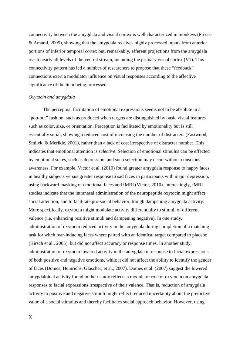
connectivity between the amygdala and visual cortex is well characterized in monkeys (Freese
& Amaral, 2005), showing that the amygdala receives highly processed inputs from anterior
portions of inferior temporal cortex but, remarkably, efferent projections from the amygdala
reach nearly all levels of the ventral stream, including the primary visual cortex (V1). This
connectivity pattern has led a number of researchers to propose that these “feedback”
connections exert a modulator influence on visual responses according to the affective
significance of the item being processed.
Oxytocin and amygdala
The perceptual facilitation of emotional expressions seems not to be absolute in a
“pop-out” fashion, such as produced when targets are distinguished by basic visual features
such as color, size, or orientation. Perception is facilitated by emotionality but is still
essentially serial, showing a reduced cost of increasing the number of distracters (Eastwood,
Smilek, & Merikle, 2001), rather than a lack of cost irrespective of distracter number. This
indicates that emotional attention is selective. Selection of emotional stimulus can be effected
by emotional states, such as depression, and such selection may occur without conscious
awareness. For example, Victor et al. (2010) found greater amygdala response to happy faces
in healthy subjects versus greater response to sad faces in participants with major depression,
using backward masking of emotional faces and fMRI (Victor, 2010). Interestingly, fMRI
studies indicate that the intranasal administration of the neuropeptide oxytocin might affect
social attention, and to facilitate pro-social behavior, trough dampening amygdala activity.
More specifically, oxytocin might modulate activity differentially to stimuli of different
valence (i.e. enhancing positive stimuli and dampening negative). In one study,
administration of oxytocin reduced activity in the amygdala during completion of a matching
task for witch fear-inducing faces where paired with an identical target compared to placebo
(Kirsch et al., 2005), but did not affect accuracy or response times. In another study,
administration of oxytocin lowered activity in the amygdala in response to facial expressions
of both positive and negative emotions, while it did not affect the ability to identify the gender
of faces (Domes, Heinrichs, Glascher, et al., 2007). Domes et al. (2007) suggest the lowered
amygdaloidal activity found in their study reflects a modulator role of oxytocin on amygdala
responses to facial expressions irrespective of their valence. That is, reduction of amygdala
activity to positive and negative stimuli might reflect reduced uncertainty about the predictive
value of a social stimulus and thereby facilitates social approach behavior. However, using
XI
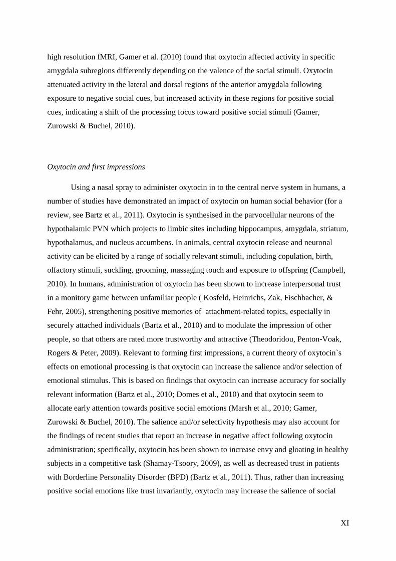
high resolution fMRI, Gamer et al. (2010) found that oxytocin affected activity in specific
amygdala subregions differently depending on the valence of the social stimuli. Oxytocin
attenuated activity in the lateral and dorsal regions of the anterior amygdala following
exposure to negative social cues, but increased activity in these regions for positive social
cues, indicating a shift of the processing focus toward positive social stimuli (Gamer,
Zurowski & Buchel, 2010).
Oxytocin and first impressions
Using a nasal spray to administer oxytocin in to the central nerve system in humans, a
number of studies have demonstrated an impact of oxytocin on human social behavior (for a
review, see Bartz et al., 2011). Oxytocin is synthesised in the parvocellular neurons of the
hypothalamic PVN which projects to limbic sites including hippocampus, amygdala, striatum,
hypothalamus, and nucleus accumbens. In animals, central oxytocin release and neuronal
activity can be elicited by a range of socially relevant stimuli, including copulation, birth,
olfactory stimuli, suckling, grooming, massaging touch and exposure to offspring (Campbell,
2010). In humans, administration of oxytocin has been shown to increase interpersonal trust
in a monitory game between unfamiliar people ( Kosfeld, Heinrichs, Zak, Fischbacher, &
Fehr, 2005), strengthening positive memories of attachment-related topics, especially in
securely attached individuals (Bartz et al., 2010) and to modulate the impression of other
people, so that others are rated more trustworthy and attractive (Theodoridou, Penton-Voak,
Rogers & Peter, 2009). Relevant to forming first impressions, a current theory of oxytocin`s
effects on emotional processing is that oxytocin can increase the salience and/or selection of
emotional stimulus. This is based on findings that oxytocin can increase accuracy for socially
relevant information (Bartz et al., 2010; Domes et al., 2010) and that oxytocin seem to
allocate early attention towards positive social emotions (Marsh et al., 2010; Gamer,
Zurowski & Buchel, 2010). The salience and/or selectivity hypothesis may also account for
the findings of recent studies that report an increase in negative affect following oxytocin
administration; specifically, oxytocin has been shown to increase envy and gloating in healthy
subjects in a competitive task (Shamay-Tsoory, 2009), as well as decreased trust in patients
with Borderline Personality Disorder (BPD) (Bartz et al., 2011). Thus, rather than increasing
positive social emotions like trust invariantly, oxytocin may increase the salience of social
XII
cues and, therefore, may trigger a range of emotions and behaviors both positive and negative
involved in regulating social interactions.
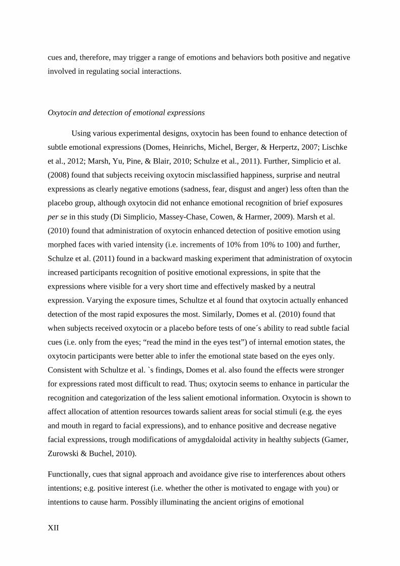
Oxytocin and detection of emotional expressions
Using various experimental designs, oxytocin has been found to enhance detection of
subtle emotional expressions (Domes, Heinrichs, Michel, Berger, & Herpertz, 2007; Lischke
et al., 2012; Marsh, Yu, Pine, & Blair, 2010; Schulze et al., 2011). Further, Simplicio et al.
(2008) found that subjects receiving oxytocin misclassified happiness, surprise and neutral
expressions as clearly negative emotions (sadness, fear, disgust and anger) less often than the
placebo group, although oxytocin did not enhance emotional recognition of brief exposures
per se in this study (Di Simplicio, Massey-Chase, Cowen, & Harmer, 2009). Marsh et al.
(2010) found that administration of oxytocin enhanced detection of positive emotion using
morphed faces with varied intensity (i.e. increments of 10% from 10% to 100) and further,
Schulze et al. (2011) found in a backward masking experiment that administration of oxytocin
increased participants recognition of positive emotional expressions, in spite that the
expressions where visible for a very short time and effectively masked by a neutral
expression. Varying the exposure times, Schultze et al found that oxytocin actually enhanced
detection of the most rapid exposures the most. Similarly, Domes et al. (2010) found that
when subjects received oxytocin or a placebo before tests of one´s ability to read subtle facial
cues (i.e. only from the eyes; “read the mind in the eyes test”) of internal emotion states, the
oxytocin participants were better able to infer the emotional state based on the eyes only.
Consistent with Schultze et al. `s findings, Domes et al. also found the effects were stronger
for expressions rated most difficult to read. Thus; oxytocin seems to enhance in particular the
recognition and categorization of the less salient emotional information. Oxytocin is shown to
affect allocation of attention resources towards salient areas for social stimuli (e.g. the eyes
and mouth in regard to facial expressions), and to enhance positive and decrease negative
facial expressions, trough modifications of amygdaloidal activity in healthy subjects (Gamer,
Zurowski & Buchel, 2010).
Functionally, cues that signal approach and avoidance give rise to interferences about others
intentions; e.g. positive interest (i.e. whether the other is motivated to engage with you) or
intentions to cause harm. Possibly illuminating the ancient origins of emotional
XIII
communication, studies have shown that implicit core feelings of like and dislike towards
some object or person can be induced experimentally, i.e. that an unconscious emotional
activation affects observed physical and verbal behavior, and without the persons` knowledge
of the influence (Beggearian, 2003; Laeng, 2010). Based on the notion that very first
impressions are overgeneralizations of subtle emotional expressions, we find it interesting that
oxytocin is shown to both enhance detection of subtle emotional cues, and to modulate the
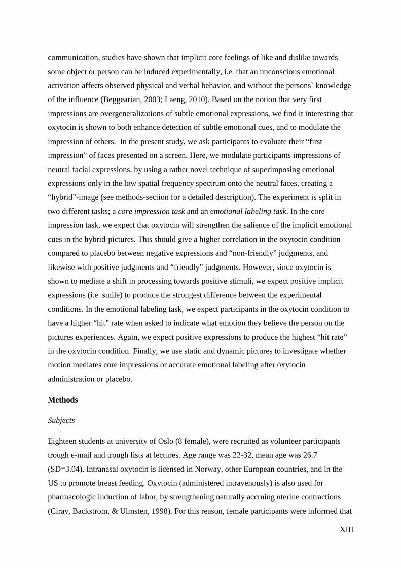
impression of others. In the present study, we ask participants to evaluate their “first
impression” of faces presented on a screen. Here, we modulate participants impressions of
neutral facial expressions, by using a rather novel technique of superimposing emotional
expressions only in the low spatial frequency spectrum onto the neutral faces, creating a
“hybrid”-image (see methods-section for a detailed description). The experiment is split in
two different tasks; a core impression task and an emotional labeling task. In the core
impression task, we expect that oxytocin will strengthen the salience of the implicit emotional
cues in the hybrid-pictures. This should give a higher correlation in the oxytocin condition
compared to placebo between negative expressions and “non-friendly” judgments, and
likewise with positive judgments and “friendly” judgments. However, since oxytocin is
shown to mediate a shift in processing towards positive stimuli, we expect positive implicit
expressions (i.e. smile) to produce the strongest difference between the experimental
conditions. In the emotional labeling task, we expect participants in the oxytocin condition to
have a higher “hit” rate when asked to indicate what emotion they believe the person on the
pictures experiences. Again, we expect positive expressions to produce the highest “hit rate”
in the oxytocin condition. Finally, we use static and dynamic pictures to investigate whether
motion mediates core impressions or accurate emotional labeling after oxytocin
administration or placebo.
Methods
Subjects
Eighteen students at university of Oslo (8 female), were recruited as volunteer participants
trough e-mail and trough lists at lectures. Age range was 22-32, mean age was 26.7
(SD=3.04). Intranasal oxytocin is licensed in Norway, other European countries, and in the
US to promote breast feeding. Oxytocin (administered intravenously) is also used for
pharmacologic induction of labor, by strengthening naturally accruing uterine contractions
(Ciray, Backstrom, & Ulmsten, 1998). For this reason, female participants were informed that
XIV
they could not participate if pregnant, and the experimenter explicitly repeated question about
pregnancy upon arrival in the laboratory. A number of studies have described the use of single
doses of intranasal oxytocin in healthy male and female volunteers in experimental
investigations (e.g. see Guastella, Mitchell, & Dadds, 2008; Guastella, Mitchell, & Mathews,
2008; Heinrichs, Baumgartner, Kirschbaum, & Ehlert, 2003; Kirsch et al., 2005; Kosfeld,
Heinrichs, Zak, Fischbacher, & Fehr, 2005; Petrovic, Kalisch, Singer, & Dolan, 2008;
Savaskan, Ehrhardt, Schulz, Walter, & Schachinger, 2008; Theodoridou, Rowe, Penton-Voak,
& Rogers). These studies have not reported any significant adverse effects with intranasal
doses up to 60IU (see Heinrichs et al., 2003). Here, we used 32 IU (four “puffs” in each nasal)
of oxytocin or normal saline (placebo) by nasal inhaler.
Participants received information about oxytocin and the tasks the participants were asked to
perform, at the beginning of the experiment. This information was given in written text,
which the participants could bring home from the experiment. The participants were informed
in text and orally that all data will be stored anonymously, and that they are free to leave the
experiment at all times without any form of consequence. The participants could leave their e-
mail address in order to receive the results when the study was over. All participants gave
written consent to the study procedures; witch had previously been approved by the regional
ethics comity.
Experimenter
The experiment was conducted by a student at the professional clinical program of
psychology, attending the last 12th semester of this education.
Stimuli
A total of 158 “hybrid” images, including both static and dynamic presentations, were used.
These static images were previously generated by Laeng et al. (2010) based on the close-up
gray scale photos from the Karolinska Directed Emotional Faces database (Karolinska
Hospital, Stockholm, Sweden, 1998). All of the selected photos showed full frontal or straight
views of the head. The present material consisted of 60 images, 6 female and 6 male models,
showing angry, happy, sad, fearful and neutral facial expressions. Dynamic presentations
were then produced from the static hybrids, including an emotionally neutral “identity
hybrid”, producing a total of 158 images. Hybrid images are images that show the emotional
expression (i.e. sad or happy) only in the lowest spatial frequencies and a neutral expression
XV
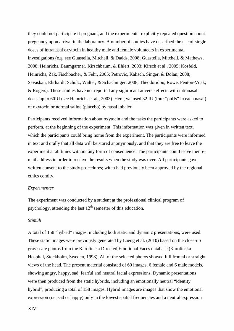
in the rest of the bandwidth. Using a filtering technique originally developed by Schyns and
Oliva (Schyns & Oliva, 1999), each image is filtered using a low pass cut-off of six
cycles/image to produce the low spatial frequency versions (1–6 cycles/image); whereas a
high pass cut-off of 7 cycles was used on the neutral expression pictures to obtain the high
spatial frequency images (7–128 cycles/image). The neutral high pass version of each model’s
face is then blended with each low pass version of the same face, to obtain five final images
of each face, four containing a different emotion (anger, fear, happiness, and sadness) that
appeared only in the low spatial frequencies and one reconstituting the original broadband
neutral expression of the same face (see Figure 1 for an illustration of the steps used in
generating a test image).
The resolution of the computer screen was 1440x900 and the dimension of the images
117x117 mm. The distance between the computer screen and the participants eyes where
measured to 55 cm to ensure a visual angle of 6°. Each image was presented for 8,000 ms.
Stimulus presentations were controlled by SMI Experimental Center software (version 3.0),
which also stored each key press.
Figure 1. An example of the editing procedure used to obtain a hybrid expressive face: Images A and B is
separate photographs of the same actress assuming a “happy” and a “neutral” expression, respectively. Image C
is the low-passed version (_6 cycles/image) of Image A, whereas Image D is the high-passed version (_7
cycles/image) of Image B. Image E is the hybrid picture or a combination of Images C and D with a happy
XVI
expression embedded exclusively in the lowest spatial frequencies. Modulated from the Karolinska Directed
Emotional Faces—KDEF (CD-ROM), by D. Lundqvist, A. Flykt, & A. O¨ hman, 1998, Stockholm, Sweden:
Department of Clinical Neuroscience, Psychology section, Karolinska Institutet. Image and description adopted
from Laeng et al. (2010).
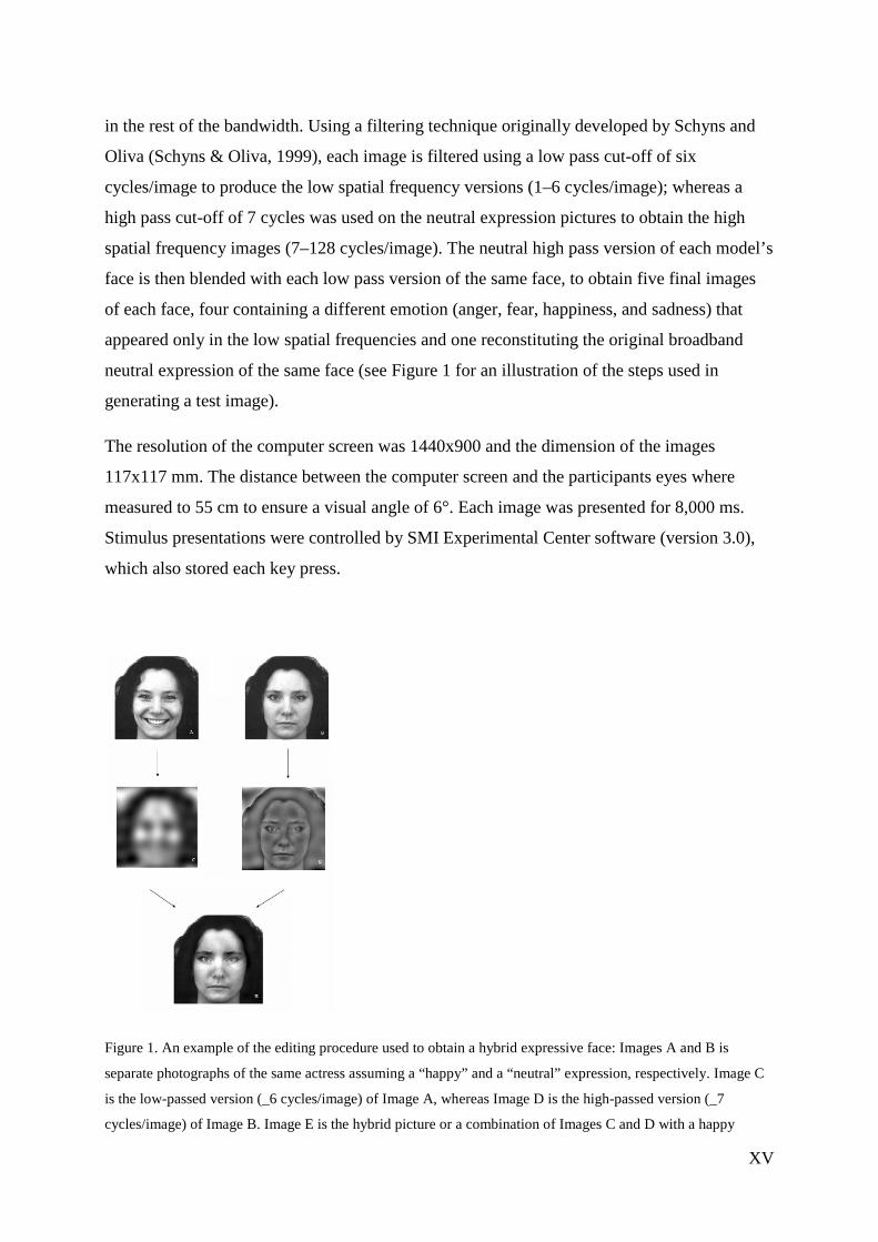
Figure 2. An example of one model`s face with the original and hybrid expressions. The leftmost face shows the
broadband “neutral” expression whereas the top row shows (from left to right) the broadband afraid, angry,
happy and sad expressions. The bottom row shows (from left to right) the hybrids for afraid, angry, happy and
sad expressions. Each image was shown at the size of 6° of visual angle. Image and description adopted from
Laeng et al. (2010).
Dynamic images
Dynamic “hybrid” images were generated using Morpheus software (version 3.6). The
broadband neutral image (non-hybrid) was blended with hybrid version of the same identity,
dynamically shifting back and fourth from 100% broadband to 100% hybrid picture within
1,000 ms at a rate of 15 frames per second, in a total duration of 8,000 ms. Thus, the hybrid
image was presented at 100% four times during the total duration.
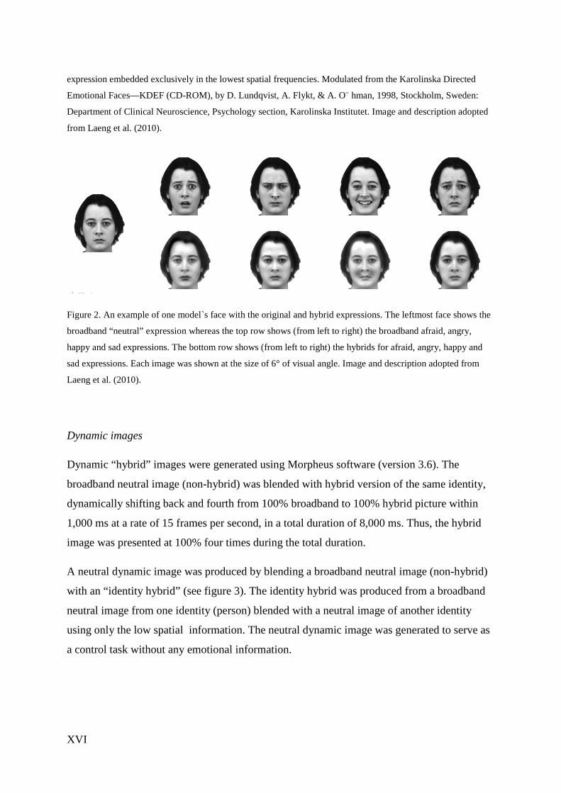
A neutral dynamic image was produced by blending a broadband neutral image (non-hybrid)
with an “identity hybrid” (see figure 3). The identity hybrid was produced from a broadband
neutral image from one identity (person) blended with a neutral image of another identity
using only the low spatial information. The neutral dynamic image was generated to serve as
a control task without any emotional information.
XVII
1. 2.
3. 4.
Figure 3. Image 1 showing the broadband version of one identity witch was filtered so that only low spatial
spectrum information could be blended with another neutral identity (image 2). Image 3 and 4 showing the
images that was morphed to a dynamically changing “identity hybrid”.
Procedure
This investigation was conducted as a double-blind, placebo-controlled, within-subjects
design. Thus each subject was tested with both oxytocin and placebo on two different
occasions (with at least 1 week between trials). The participants received either oxytocin or
placebo 30 minutes before testing started.
Arriving at the test site, participants self-administered oxytocin or placebo with a nasal
spray. The different nasal sprays containing either oxytocin or placebo were coded with white
or blue cap by a supervisor, hence neither the experimenter nor the participant knew if he/she
received oxytocin or placebo. The color of the cap was recorded and the participant received
the other nasal spray at the second trial. The participants received the blue and white nasal
sprays in a counterbalanced order. Since the effect of intranasal administration is stable
between ~30 and 90 minutes post-administration, all experimental testing occurred within this
time window. The participants were told that they could wait at the test site or come back
after 30 minutes, but were asked to not drink coffee or other possible stimulating food or
drinks. The tasks took approximately 40 minutes to complete.
The participants were told that the experiment was split in two parts (core impression task and
emotional labelling task), and told about the first part before the experiment started and the
second part before this part started.
XVIII
Core impression task
The stimulus set contained 78 pictures (first half of the total set) showing static and dynamic
“hybrid” pictures with emotional expressions from the low spatial spectrum only (happy,
angry, sad, fear) blended with a broadband neutral picture of the same person. The
participants were told that they should indicate the “first impressions” of the images probed
by a question on the screen after each image (“How friendly do you think this person is?”).
Answers was predefined as 1 = most friendly, 2 = friendly, 3 = neutral, 4 = unfriendly and 5 =
most unfriendly. Participants were asked to have the “friendly/unfriendly” dimension in mind
when viewing the images, and to respond when probed with the question using their “gut
feeling”. They were told that it was no right or wrong answers and that what we were
interested in for was their particular impression of the images` friendliness.
Emotional labelling task
The stimulus set contained 78 pictures (second half of the total set). The participants were told
that they should indicate the emotion felt by the person on the images, probed by a question
on the screen after each image (“Do you think this person is...?”). Answers was predefined as
1 = happy, 2 = sad, 3 = neutral, 4 = angry and 5 = afraid.
Results
Core impression task
Static images
We calculated descriptive statistics for each participant, obtaining mean ratings for each low
passed expression and then performed a repeated-measures ANOVA on mean ratings as the
dependent variable, with expressions (neutrality, happy, anger, fear and sadness) as the
within-subject variable and sex (female, male) and drug condition (oxytocin, placebo) as
between-subjects factors. The ANOVA revealed a main effect of expressions, F(1,4) = 27.82
p = .000. No significant difference in participants scores after oxytocin administration versus
placebo was observed, F(1,4) = .58 p = .45. No significant differences between sex and rating
of expressions was observed F(1,4) = .60 p = .66 or interaction sex x oxytocin x expressions
F(1,4) = .89 p = .479).
XIX
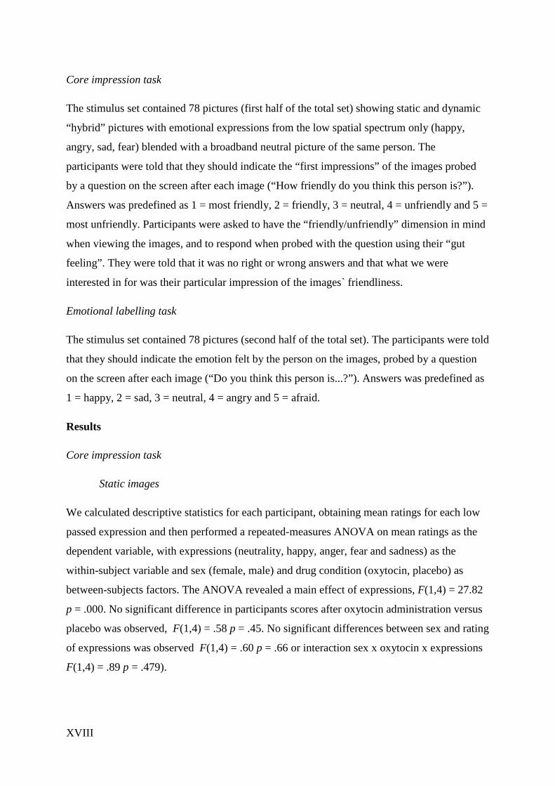
Happy and angry static pictures produced the expected tendency towards
“friendly/unfriendly” ratings, respectively. For fear, responses were distributed equally at
neutral, friendly and unfriendly. For sad pictures, the opposite of the expected “unfriendly”
tendency was observed, in fact, sad pictures showed a strong tendency to be judged as
“friendly” (56.9% after oxytocin administration and 65.5% after receiving placebo). Pairwise
comparisons of mean ratings on each low passed expressions revealed that mean ratings on
angry and sad expressions differed significantly from ratings of the other expressions (p <
.000).
Figure 4. Ratings on static low passed emotional expressions.
XX
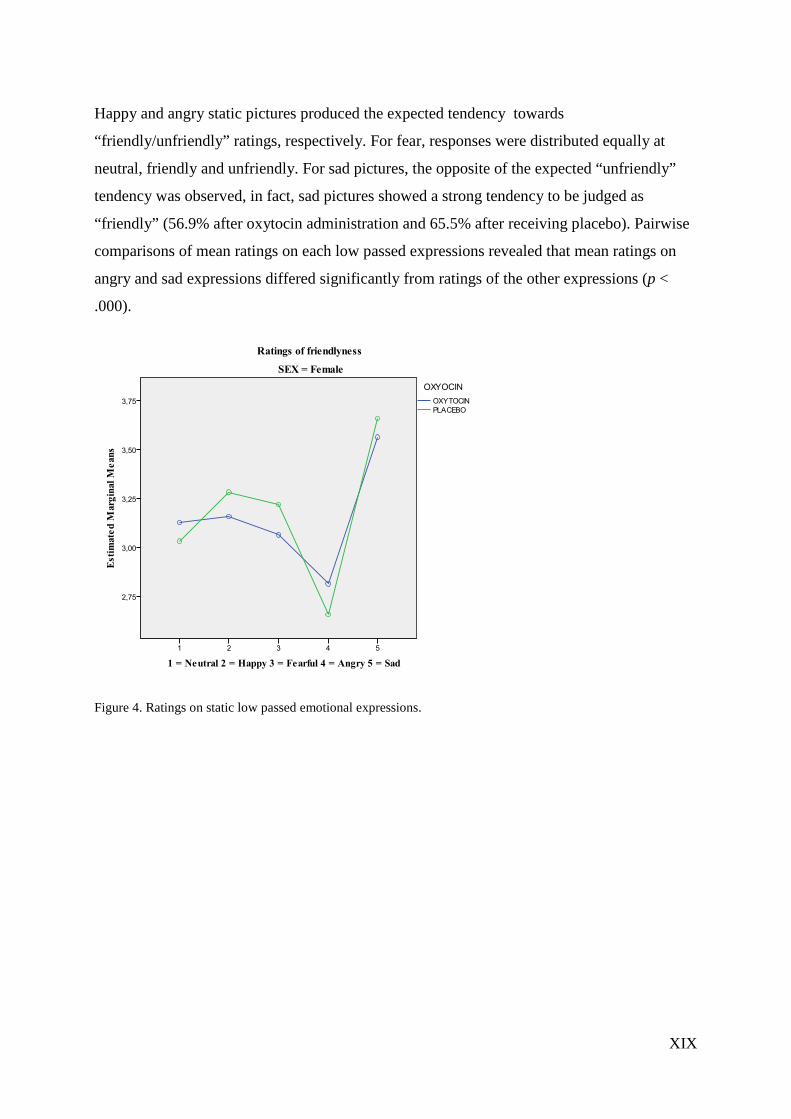
Figure 5. Ratings on static low passed emotional expressions.
Dynamic images
The ANOVA revealed a main effect of expressions, F(1,4) = 8,41 p < .000. The low passed
dynamic expressions showing positive (happy) or negative (angry, fearful, sad) emotional
expressions produced “first impressions” congruent with the emotional valence in the
expressions (i.e. “friendly” for happy, “unfriendly” for angry, fearful and sad) for the pictures
with happy, angry, afraid and sad expressions. The neutral images (with a different identity
dynamically shifting in the low spatial spectrum) actually produced a strong “unfriendly”
response after both oxytocin (40.3% neutral, 55.6% unfriendly, 4.2% friendly) and placebo
administration (43.1% neutral, 44.4% unfriendly, 12.5% friendly).
No significant difference in participants scores after oxytocin administration versus placebo
was observed, F(1,4) = .58 p = .45. There was a significant interaction between expressions x
sex F(1,4) = 3.83 p = .13, but no significant interaction expressions x oxytocin x sex F(1,4) =
.58 p = .67.
Univariate tests using sex as the independent variable revealed that ratings on two expressions
showed significantly effect of sex. Females rated the neutral dynamic hybrid less friendly than
males (females M=2,31, males M=2,62 F(1,4) = .41 p = .050). Males rated the fearful

XXI
dynamic hybrids less friendly than females (females M=2,81, males M=2,37 F(1,4) = 1,70 p
= .014). However non-significant, it may be worth mentioning is that although administration
of oxytocin caused no difference in friendliness ratings for the happy dynamic expression in
females F(1,4) = .00 p = 1.0, this was the ratings closest to a significant difference after
oxytocin administration in males F(1,4) = 1,86 p = .18.
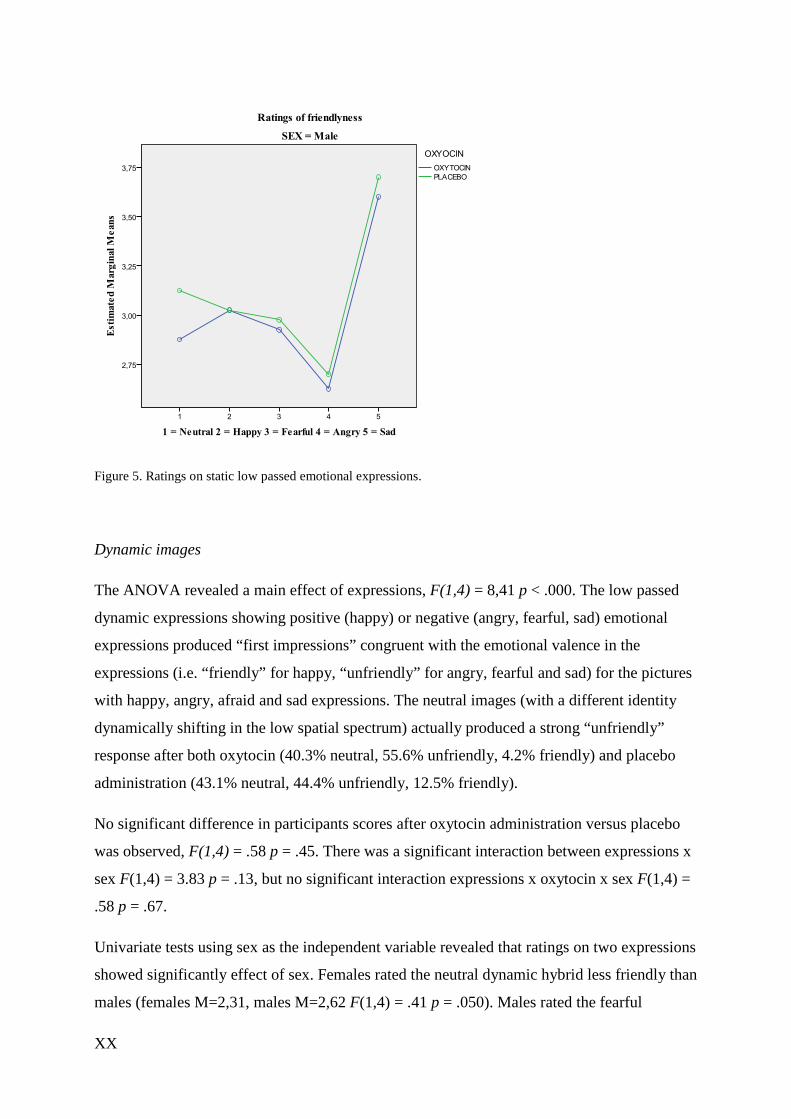
Figure 6. Ratings on dynamic low passed expressions.
XXII
Figure 7. Ratings on dynamic low passed expressions.
Emotional labeling task
Static images
To investigate accuracy in labeling the low passed emotional expressions, we performed a
chi-square analysis obtaining expected and observed scores for each emotional expression.
Contrary to our predictions, no statistical difference between participants after oxytocin
administration versus placebo was found for in the labeling task for low passed static
expressions: Neutral expressions c2(4, N = 144) = 1.61, p=.80, Happy expressions c2(4, N =
144) = .42, p=.98, Angry expressions c2(4, N = 144) = 3.33, p=.50, Fearful expressions
c2(4, N = 144) = .49, p=.97, Sad expressions c2(4, N = 144) = .75, p=.94.
Dynamic images
A significant difference after oxytocin administration versus placebo was found responses to
low passed fearful dynamic expressions c2(4, N = 144) = 10.04, p = .040 (figure 6), and low
passed angry dynamic expressions c2(4, N = 144) = 9.80, p = .044 (figure 7).
XXIII
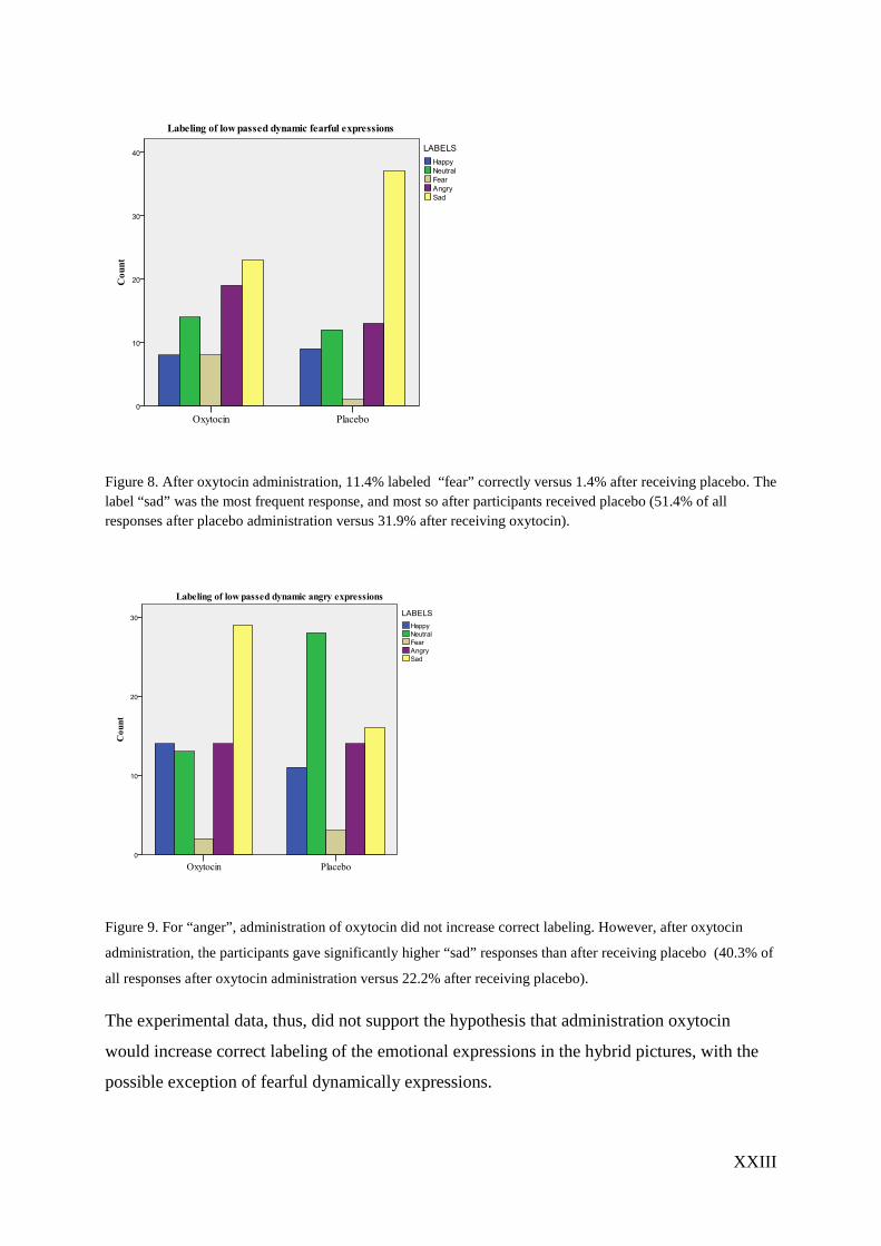
Figure 8. After oxytocin administration, 11.4% labeled “fear” correctly versus 1.4% after receiving placebo. The label “sad” was the most frequent response, and most so after participants received placebo (51.4% of all responses after placebo administration versus 31.9% after receiving oxytocin).
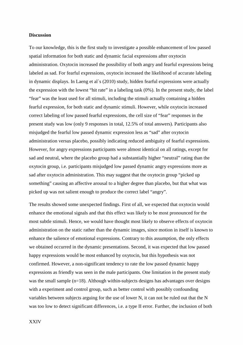
Figure 9. For “anger”, administration of oxytocin did not increase correct labeling. However, after oxytocin
administration, the participants gave significantly higher “sad” responses than after receiving placebo (40.3% of
all responses after oxytocin administration versus 22.2% after receiving placebo).
The experimental data, thus, did not support the hypothesis that administration oxytocin
would increase correct labeling of the emotional expressions in the hybrid pictures, with the
possible exception of fearful dynamically expressions.
XXIV
Discussion
To our knowledge, this is the first study to investigate a possible enhancement of low passed
spatial information for both static and dynamic facial expressions after oxytocin
administration. Oxytocin increased the possibility of both angry and fearful expressions being
labeled as sad. For fearful expressions, oxytocin increased the likelihood of accurate labeling
in dynamic displays. In Laeng et al`s (2010) study, hidden fearful expressions were actually
the expression with the lowest “hit rate” in a labeling task (0%). In the present study, the label
“fear” was the least used for all stimuli, including the stimuli actually containing a hidden
fearful expression, for both static and dynamic stimuli. However, while oxytocin increased
correct labeling of low passed fearful expressions, the cell size of “fear” responses in the
present study was low (only 9 responses in total, 12.5% of total answers). Participants also
misjudged the fearful low passed dynamic expression less as “sad” after oxytocin
administration versus placebo, possibly indicating reduced ambiguity of fearful expressions.
However, for angry expressions participants were almost identical on all ratings, except for
sad and neutral, where the placebo group had a substantially higher “neutral” rating than the
oxytocin group, i.e. participants misjudged low passed dynamic angry expressions more as
sad after oxytocin administration. This may suggest that the oxytocin group “picked up
something” causing an affective arousal to a higher degree than placebo, but that what was
picked up was not salient enough to produce the correct label “angry”.
The results showed some unexpected findings. First of all, we expected that oxytocin would
enhance the emotional signals and that this effect was likely to be most pronounced for the
most subtle stimuli. Hence, we would have thought most likely to observe effects of oxytocin
administration on the static rather than the dynamic images, since motion in itself is known to
enhance the salience of emotional expressions. Contrary to this assumption, the only effects
we obtained occurred in the dynamic presentations. Second, it was expected that low passed
happy expressions would be most enhanced by oxytocin, but this hypothesis was not
confirmed. However, a non-significant tendency to rate the low passed dynamic happy
expressions as friendly was seen in the male participants. One limitation in the present study
was the small sample (n=18). Although within-subjects designs has advantages over designs
with a experiment and control group, such as better control with possibly confounding
variables between subjects arguing for the use of lower N, it can not be ruled out that the N
was too low to detect significant differences, i.e. a type II error. Further, the inclusion of both
XXV
male and female participants may demand more control of confounding interactions with
oxytocin in females. Future studies could follow up on oxytocin`s effect on labeling emotions
based on low passed expressions, perhaps using forced choice tasks (without neutrality)
labeling the low passed expressions to investigate a threshold in regard to when and how
oxytocin enhances the perception of low passed angry expressions. Further, future studies
could include higher N to investigate the observed non-significant effect of oxytocin on
friendliness ratings of low passed dynamic happy expressions in males.

XXVI
References
Ballew, C. C., & Todorov, A. (2007). Predicting political elections from rapid and
unreflective face judgments. Proceedings of the National Academy of Sciences of the
United States of America, 104(46), 17948-17953.
Bar, M., Neta, M., & Linz, H. (2006). Very first impressions. Emotion, 6(2), 269-278.
Bartz, J., Simeon, D., Hamilton, H., Kim, S., Crystal, S., Braun, A., Hollander, E. (2011).
Oxytocin can hinder trust and cooperation in borderline personality disorder. Social
Cognitive and Affectective Neuroscience, 6(5), 556-563.
Bartz, J. A., Zaki, J., Ochsner, K. N., Bolger, N., Kolevzon, A., Ludwig, N., & Lydon, J. E.
(2010). Effects of oxytocin on recollections of maternal care and closeness. PNAS
Proceedings of the National Academy of Sciences of the United States of America,
107(50), pp.
Berridge, K., & Winkielman, P. (2003). What is an unconscious emotion?(The case for
unconscious "liking"). Cognition & Emotion, 17(2), 181-211.
Campbell, A. (2010). Oxytocin and human social behavior. Personal and Social Psychological Reviewv, 14(3), 281-295.
Ciray, H. N., Backstrom, T., & Ulmsten, U. (1998). Ineffectiveness of oxytocin on
intercellular communication between term pregnant human myometrial cells before
labor. American Journal of Obstetrics and Gynecology, 178(4), 855-861.
de Gelder, B., Vroomen, J., Pourtois, G., & Weiskrantz, L. (1999). Non-conscious recognition
of affect in the absence of striate cortex. Neuroreport, 10(18), 3759-3763.
Di Simplicio, M., Massey-Chase, R., Cowen, P. J., & Harmer, C. J. (2009). Oxytocin
enhances processing of positive versus negative emotional information in healthy male
volunteers.
Journal of Psychopharmacoly, 23(3), 241-248.
Domes, G., Heinrichs, M., Glascher, J., Buchel, C., Braus, D. F., & Herpertz, S. C. (2007).
Oxytocin attenuates amygdala responses to emotional faces regardless of
valence.Biolical Psychiatry, 62(10), 1187-1190.
Domes, G., Heinrichs, M., Michel, A., Berger, C., & Herpertz, S. C. (2007). Oxytocin
improves "mind-reading" in humans. Biological Psychiatry, 61(6), 731-733.
XXVII
Eastwood, J. D., Smilek, D., & Merikle, P. M. (2001). Differential attentional guidance by
unattended faces expressing positive and negative emotion. Perception &
Psychophysics, 63(6), 1004-1013.
Fox, E. (2002). Processing emotional facial expressions: The role of anxity and awereness.
Cognitive, affective & Behavioral neuroscience, 2(1), 52-63.
Freese, J. L., & Amaral, D. G. (2005). The organization of projections from the amygdala to
visual cortical areas TE and V1 in the macaque monkey. Journal of Comparative
Neurology, 486(4), 295-317.
Gamer M., Z., B; Büchela C. (2010). Different amygdala subregions mediate valencerelated
and attentional effects of oxytocin in humans. PNAS, 107(20), 9400–9405.
Goffaux, V., & Rossion, B. (2006). Faces are "spatial" - Holistic face perception is supported
by low spatial frequencies. Journal of Experimental Psychology-Human Perception
and Performance, 32(4), 1023-1039.
Guastella, A. J., Mitchell, P. B., & Dadds, M. R. (2008). Oxytocin Increases Gaze to the Eye
Region of Human Faces. Biological Psychiatry, 63(1), 3-5.
Guastella, A. J., Mitchell, P. B., & Mathews, F. (2008). Oxytocin Enhances the Encoding of
Positive Social Memories in Humans. Biological Psychiatry, 64(3), 256-258.
Heinrichs, M., Baumgartner, T., Kirschbaum, C., & Ehlert, U. (2003). Social support and
oxytocin interact to suppress cortisol and subjective responses to psychosocial stress.
Biological Psychiatry, 54(12), 1389-1398.
Izard, C. E. (1978). Emotions as motivations: An evolutionary-developmental perspective. .
Nebraska Symposium on Motivation, 26, 163-200.
Kirsch, P., Esslinger, C., Chen, Q., Mier, D., Lis, S., Siddhanti, S., . . . Meyer-Lindenberg, A.
(2005). Oxytocin Modulates Neural Circuitry for Social Cognition and Fear in
Humans. Journal of Neuroscience, 25(49), 11489-11493.
Kosfeld, M., Heinrichs, M., Zak, P. J., Fischbacher, U., & Fehr, E. (2005). Oxytocin increases
trust in humans. Nature, 435(7042), 673-676.
Kosfeld, M., Heinrichs, M., Zak, P. J., Fischbacher, U., & Fehr, E. (2005). Oxytocin increases
trust in humans. Nature, 435(7042), 673-676.
Krolak-Salmon, P., Henaff, M. A., Vighetto, A., Bertrand, O., & Mauguiere, F. (2004). Early
amygdala reaction to fear spreading in occipital, temporal, and frontal cortex: a depth
electrode ERP study in human. Neuron, 42(4), 665-676.
XXVIII
Laeng, B., Profeti, I., Saether, L., Adolfsdottir, S., Lundervold, A. J., Vangberg, T., . . .
Waterloo, K. (2010). Invisible expressions evoke core impressions. Emotion, 10(4),
573-586.
Lischke, A., Berger, C., Prehn, K., Heinrichs, M., Herpertz, S. C., & Domes, G. (2012).
Intranasal oxytocin enhances emotion recognition from dynamic facial expressions
and leaves eye-gaze unaffected. Psychoneuroendocrinology, 37(4), 475-481.
Marsh, A. A., Yu, H. H., Pine, D. S., & Blair, R. J. (2010). Oxytocin improves specific
recognition of positive facial expressions. Psychopharmacology, 209(3), 225-232.
Pessoa, L., Kastner, S., & Ungerleider, L. G. (2002). Attentional control of the processing of
neutral and emotional stimuli. Cognitive Brain Research, 15(1), 31-45.
Petrovic, P., Kalisch, R., Singer, T., & Dolan, R. J. (2008). Oxytocin Attenuates Affective
Evaluations of Conditioned Faces and Amygdala Activity. Journal of Neuroscience,
28(26), 6607-6615.
Sato, W., & Yoshikawa, S. (2010). Detection of emotional facial expressions and anti-
expressions. Visual Cognition, 18(3), 369-388.
Savaskan, E., Ehrhardt, R., Schulz, A., Walter, M., & Schachinger, H. (2008). Post-learning
intranasal oxytocin modulates human memory for facial identity.
Psychoneuroendocrinology, 33(3), 368-374.
Schulze, L., Lischke, A., Greif, J., Herpertz, S. C., Heinrichs, M., & Domes, G. (2011).
Oxytocin increases recognition of masked emotional faces.
Psychoneuroendocrinology, 36(9), 1378-1382.
Schyns, P. G., & Oliva, A. (1999). Dr. Angry and Mr. Smile: when categorization flexibly
modifies the perception of faces in rapid visual presentations. Cognition, 69(3), 243-
265.
Secord, P. F. (1958). Facial features and inference processes in interpersonal perception. . In
R. a. P. Tagiuri, L. (Ed.), Person Perception and Interpersonal Behavior (pp. 300–
315): Stanford University Press.
Shamay-Tsoory, S. G. F., Meytal; Dvash, Jonathan; Harari, Hagai; Perach-Bloom, Nufar;
Levkovitz, Yechiel. (2009). Intranasal administration of oxytocin increases envy and
schadenfreude (gloating). Biological Psychiatry, 66(9), 864-870.
XXIX
Theodoridou, A. R., Angela C; Penton-Voak, Ian S; Rogers, Peter J. (2009). Oxytocin and
social perception: Oxytocin increases perceived facial trustworthiness and
attractiveness. Hormones and Behavior, 56(1), 128-132.
Todorov, A., Baron, S. G., & Oosterhof, N. N. (2008). Evaluating face trustworthiness: A
model based approach. Social Cognitive and Affective Neuroscience, 3(2), pp.
Todorov A, S., CP, Penton-Voak IS, Rogers PJ. (2011). Task-invariant brain responses to the
social value of faces. Journal of cognitive neuroscience, 23(10), 2766-2781.
Todorov, A., Said, C. P., Engell, A. D., & Oosterhof, N. N. (2008). Understanding evaluation
of faces on social dimensions. Trends in Cognitive Sciences, 12(12), 455-460.
Victor TA, F. M., Fromm SJ, Ohman A, Drevets WC. . (2010). Relationships between
amygdala responses to masked faces and mood states and treatment in major
depressive disorder. Archives of General Psychiatry, 67(11), 1128-1138.
Vuilleumier, P., & Pourtois, G. (2007). Distributed and interactive brain mechanisms during
emotion face perception: evidence from functional neuroimaging. Neuropsychologia,
45(1), 174-194.
Willis, J., & Todorov, A. (2006). First impressions: Making up your mind after a 100-ms
exposure to a face. Psychological Science, 17(7), 592-598.

XXX

XXXI

XXXII

XXXIII


1
1 [Overskrift]

2
Register

3
Litteraturliste [Følg instruksene ditt fakultet/institutt har for skriving av litteraturlister]

4
Vedlegg