M Priyanthi Kumarasinghe - AGPSagps.org.au/resources/AGM_Stuff/2016_AGM_presentations/AGPS...
40
“Barrett’s Oesophagus: When it comes to the Pathologist” M Priyanthi Kumarasinghe Pathologist, PathWest, QEII Medical Centre Clinical Professor, University of Western Australia, Perth, WA [email protected]
Transcript of M Priyanthi Kumarasinghe - AGPSagps.org.au/resources/AGM_Stuff/2016_AGM_presentations/AGPS...
“Barrett’s Oesophagus:
When it comes to the Pathologist”
M Priyanthi Kumarasinghe
Pathologist, PathWest, QEII Medical
Centre
Clinical Professor, University of Western
Australia, Perth, WA
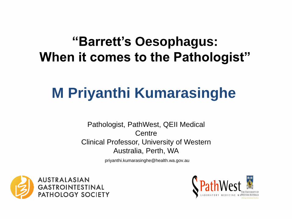
Specimen handling • Mucosa upward, pinned on a cork
board/similar firm base by the endoscopist
• Pinning (immediate) - Margins do not roll - Preserve the tissue size, shape, and orientation - Avoid overstretching: tears of the mucosa
• Tumour morphology: provided by the
interventional endoscopist (Paris classification)
Specimen handling
• Surgical margins must be appropriately inked
• Single specimen: may be oriented using the designation of O (oral) and A (anal) or P (proximal) and D (distal) marked on the board - ink appropriately to assess designated lateral & deep margins
• Multiple or piecemeal resection (long segment of Barrett)- orientation is often difficult - assessment of lateral margins unhelpful
• Best fixed for at least 12 hours in formalin

Specimen dissection:
• Entire specimen: cut into 2-3 mm (not < 2 mm) parallel slices from end to end
• Record/photograph
• Circumferential (lateral) surgical margins: “en face” or perpendicular sections, depending on the size of the specimen & proximity of the lesion/s
• Not more than 4 slices in one block
•ENDOSCOPIC RESECTION (ER) OF THE OESOPHAGUS AND GASTRO-OESOPHAGEAL JUNCTION STRUCTURED REPORTING PROTOCOL. 1st Edition 2013. © RCPA
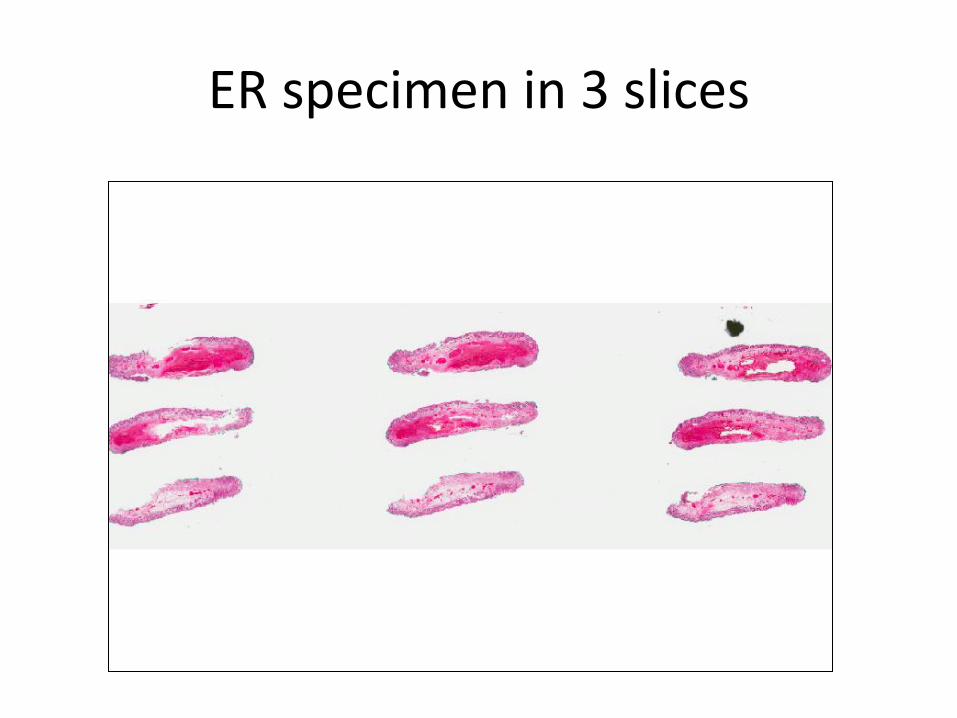
ER specimen in 3 slices

ER sections • Mucosa
• MM
• SM (often not the entire depth)

ER: Therapy of choice for IEN and (visible) T1a lesions
T1b
T1a
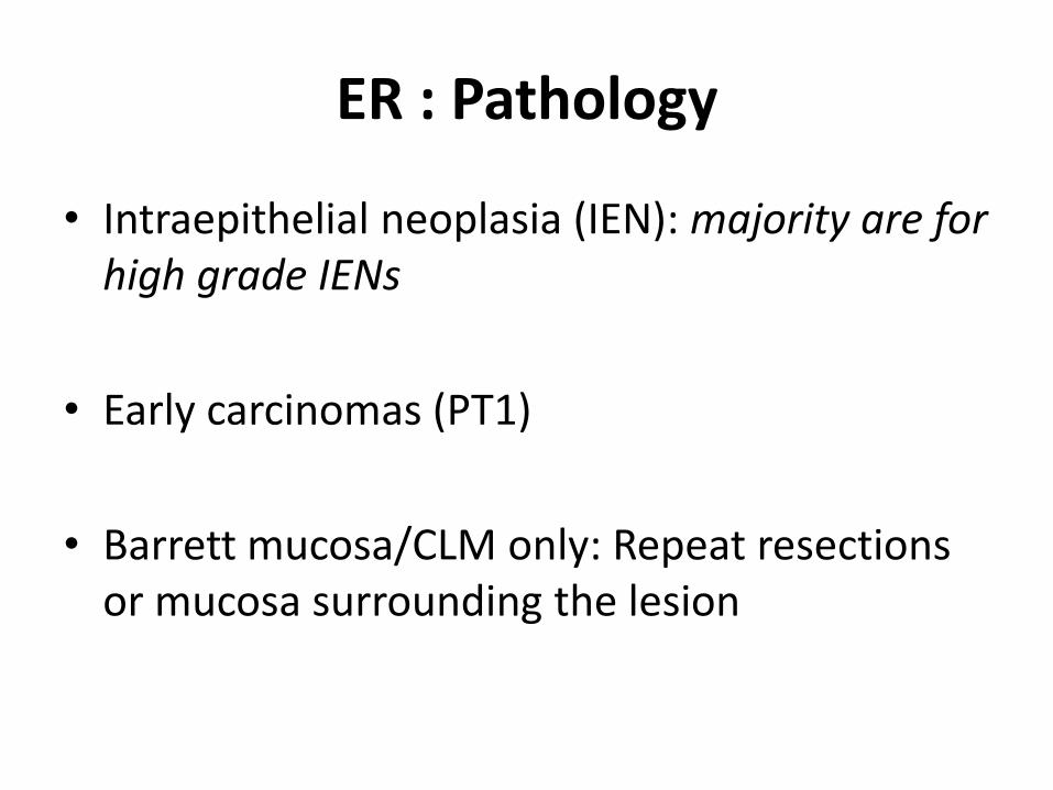
ER : Pathology
• Intraepithelial neoplasia (IEN): majority are for high grade IENs
• Early carcinomas (PT1)
• Barrett mucosa/CLM only: Repeat resections or mucosa surrounding the lesion
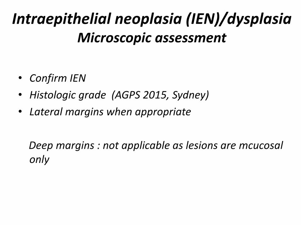
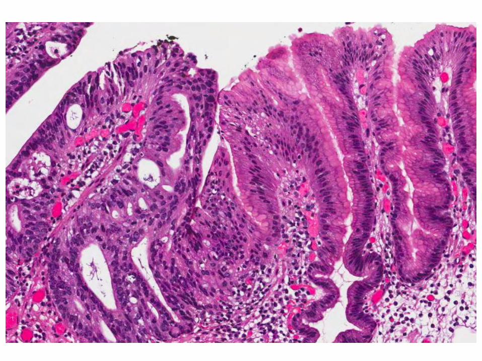
Intraepithelial neoplasia (IEN)/dysplasia Microscopic assessment
• Confirm IEN
• Histologic grade (AGPS 2015, Sydney)
• Lateral margins when appropriate
Deep margins : not applicable as lesions are mcucosal only

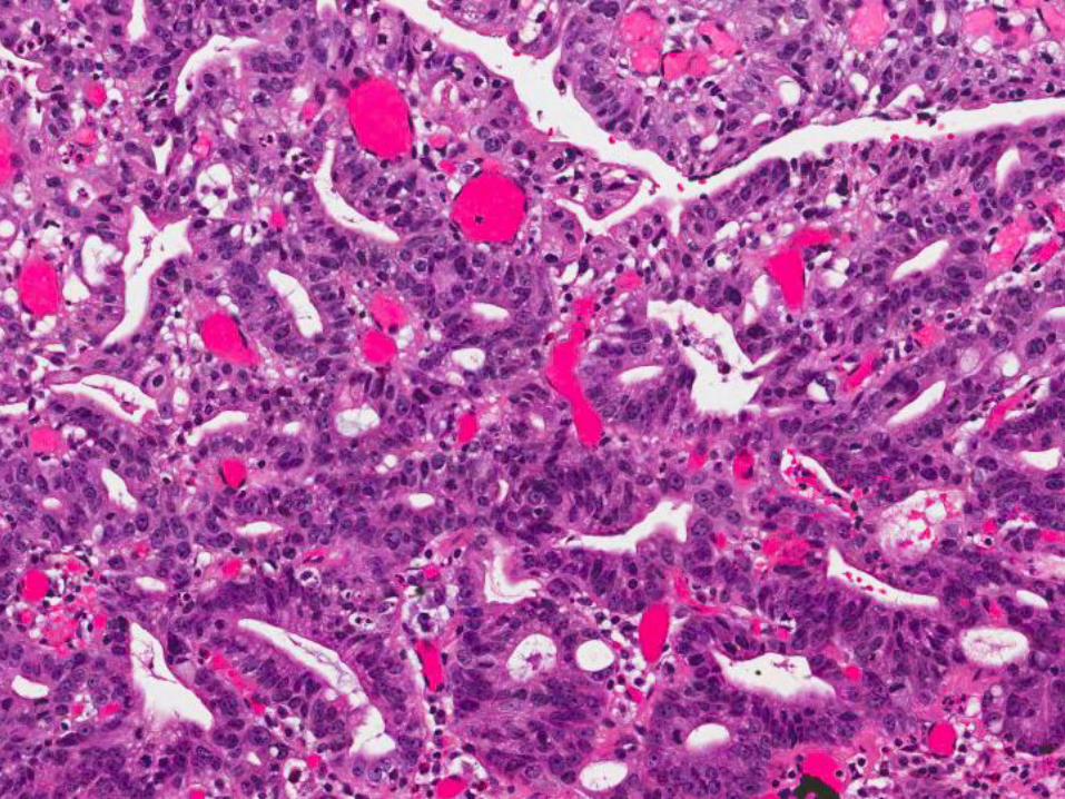


Invasive carcinoma Microscopic assessment
• Confirmation of invasive carcinoma: invasion into lamina
propria or beyond • Depth of invasion • Degree of differentiation • Presence or absence of lymphovascular invasion • Margin status These features dictate further management • tumour budding/size

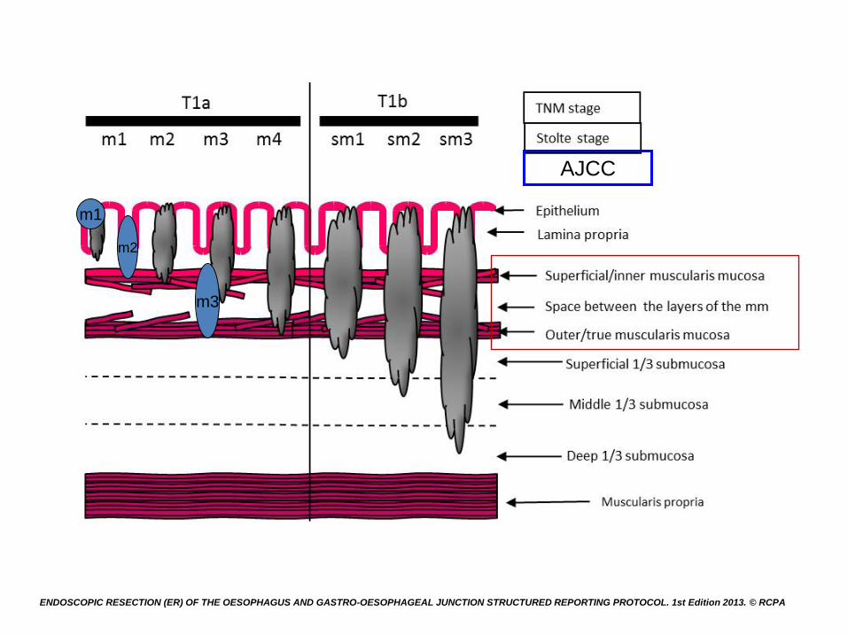
T1 carcinoma (AJCC)
• T1a – Invade lamina propria or muscularis mucosae
• T1b - Invade submucosa
T1a
T1b


Muscularis mucosae in Barrett mucosa: Duplicated and distorted

mucosa
muscularis mucosae
submucosa
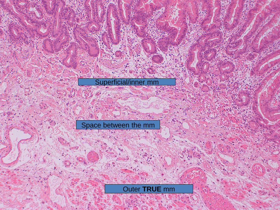
Superficial/inner mm
Space between the mm
Outer TRUE mm

AJCC
m1
m2
m3
ENDOSCOPIC RESECTION (ER) OF THE OESOPHAGUS AND GASTRO-OESOPHAGEAL JUNCTION STRUCTURED REPORTING PROTOCOL. 1st Edition 2013. © RCPA
AJCC
m1
m2
m3
ENDOSCOPIC RESECTION (ER) OF THE OESOPHAGUS AND GASTRO-OESOPHAGEAL JUNCTION STRUCTURED REPORTING PROTOCOL. 1st Edition 2013. © RCPA
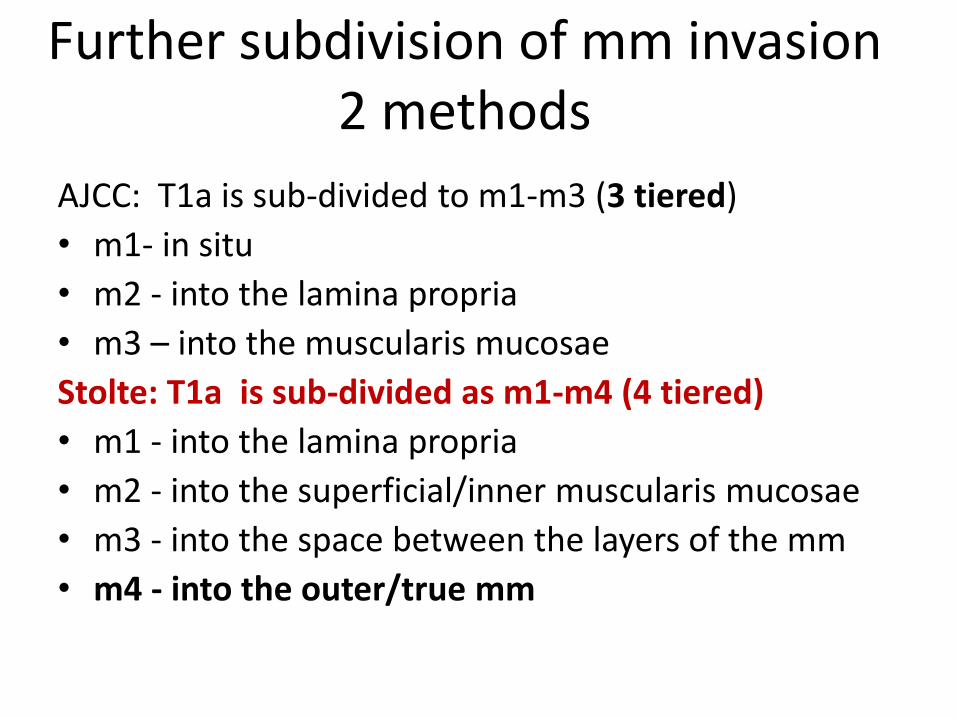
Further subdivision of mm invasion 2 methods
AJCC: T1a is sub-divided to m1-m3 (3 tiered)
• m1- in situ
• m2 - into the lamina propria
• m3 – into the muscularis mucosae
Stolte: T1a is sub-divided as m1-m4 (4 tiered)
• m1 - into the lamina propria
• m2 - into the superficial/inner muscularis mucosae
• m3 - into the space between the layers of the mm
• m4 - into the outer/true mm
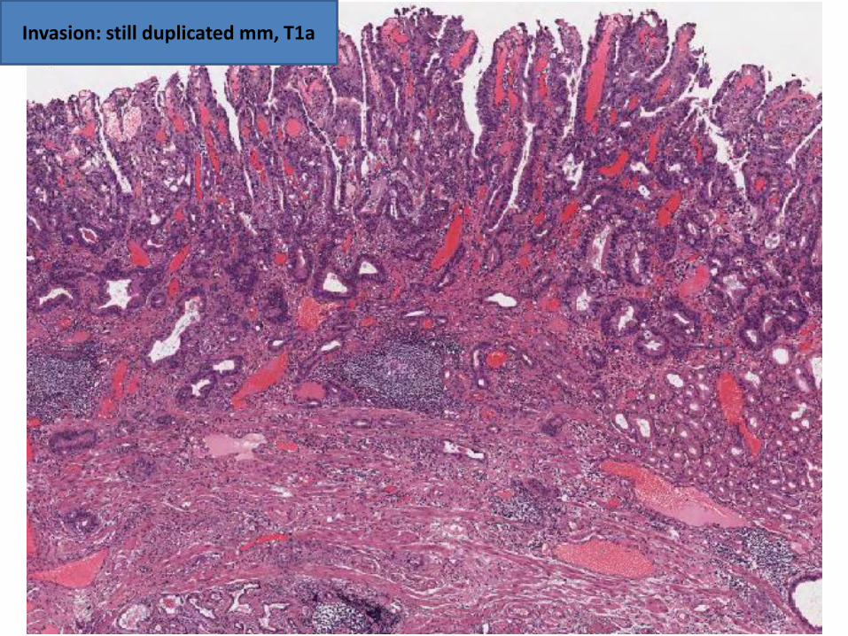
Invasion: still duplicated mm, T1a
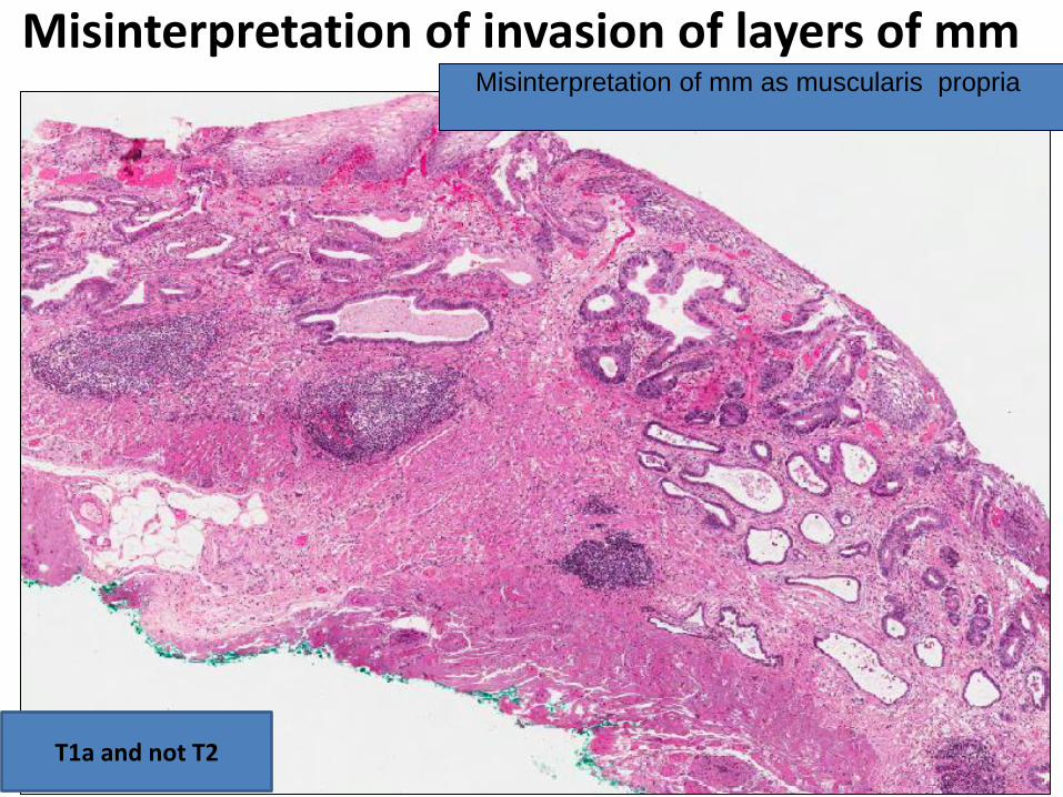
Misinterpretation of invasion of layers of mm Misinterpretation of mm as muscularis propria
T1a and not T2

Misinterpretation of mm as muscularis propria!
T1 and not T2


T1b- SM1
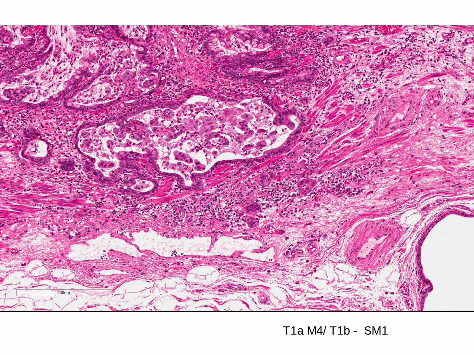
T1a M4/ T1b - SM1

T1a- Stolte M3,
AJCC M3

T1b SM 2-3
desmoplasia
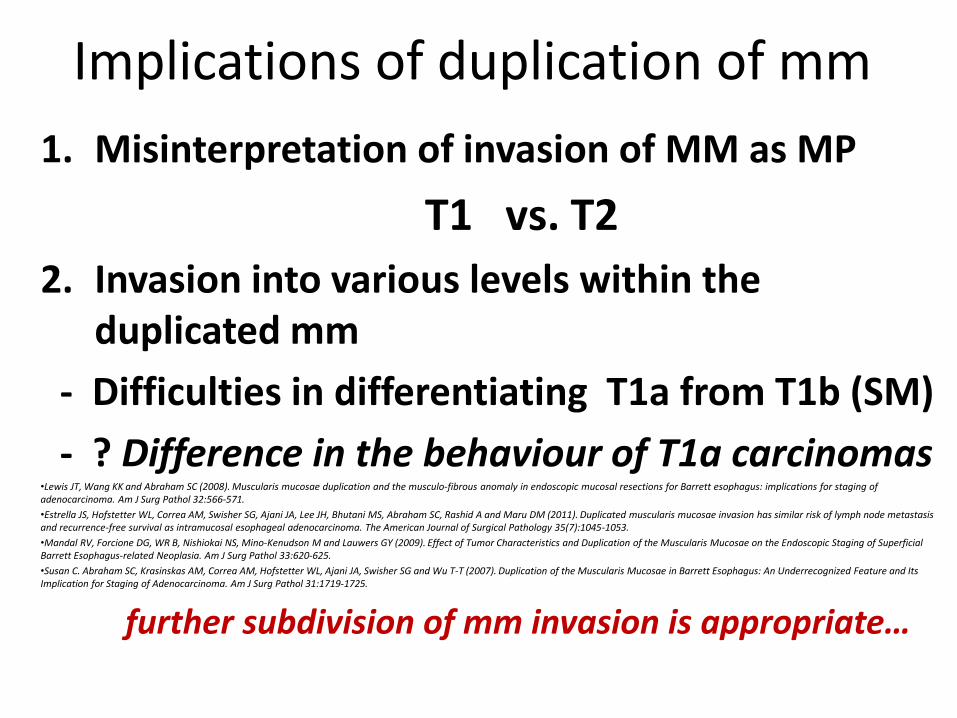
Implications of duplication of mm
1. Misinterpretation of invasion of MM as MP
T1 vs. T2
2. Invasion into various levels within the duplicated mm
- Difficulties in differentiating T1a from T1b (SM)
- ? Difference in the behaviour of T1a carcinomas •Lewis JT, Wang KK and Abraham SC (2008). Muscularis mucosae duplication and the musculo-fibrous anomaly in endoscopic mucosal resections for Barrett esophagus: implications for staging of adenocarcinoma. Am J Surg Pathol 32:566-571.
•Estrella JS, Hofstetter WL, Correa AM, Swisher SG, Ajani JA, Lee JH, Bhutani MS, Abraham SC, Rashid A and Maru DM (2011). Duplicated muscularis mucosae invasion has similar risk of lymph node metastasis and recurrence-free survival as intramucosal esophageal adenocarcinoma. The American Journal of Surgical Pathology 35(7):1045-1053.
•Mandal RV, Forcione DG, WR B, Nishiokai NS, Mino-Kenudson M and Lauwers GY (2009). Effect of Tumor Characteristics and Duplication of the Muscularis Mucosae on the Endoscopic Staging of Superficial Barrett Esophagus-related Neoplasia. Am J Surg Pathol 33:620-625.
•Susan C. Abraham SC, Krasinskas AM, Correa AM, Hofstetter WL, Ajani JA, Swisher SG and Wu T-T (2007). Duplication of the Muscularis Mucosae in Barrett Esophagus: An Underrecognized Feature and Its Implication for Staging of Adenocarcinoma. Am J Surg Pathol 31:1719-1725.
further subdivision of mm invasion is appropriate…
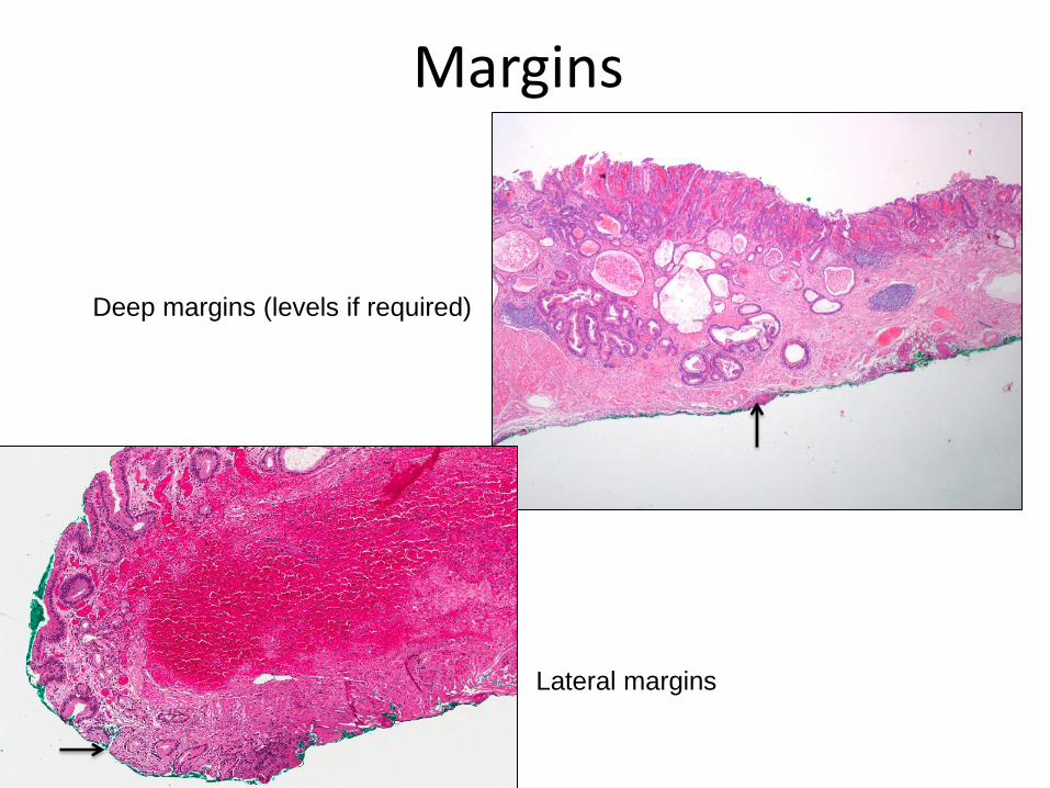
Margins
Lateral margins
Deep margins (levels if required)

Lympho-vascular invasion
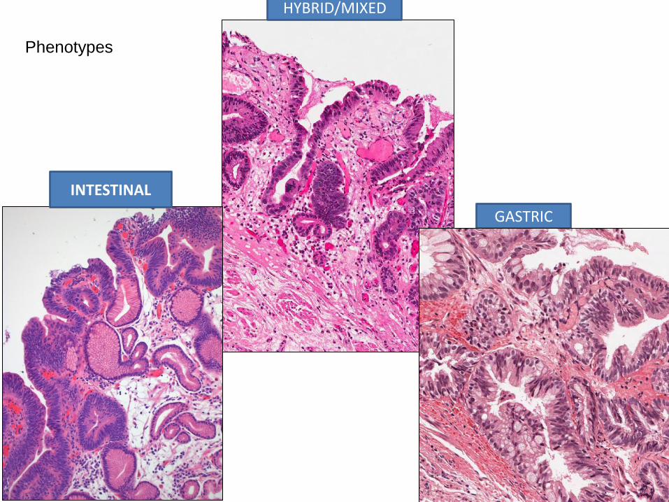
INTESTINAL
HYBRID/MIXED
GASTRIC
Phenotypes
xxx

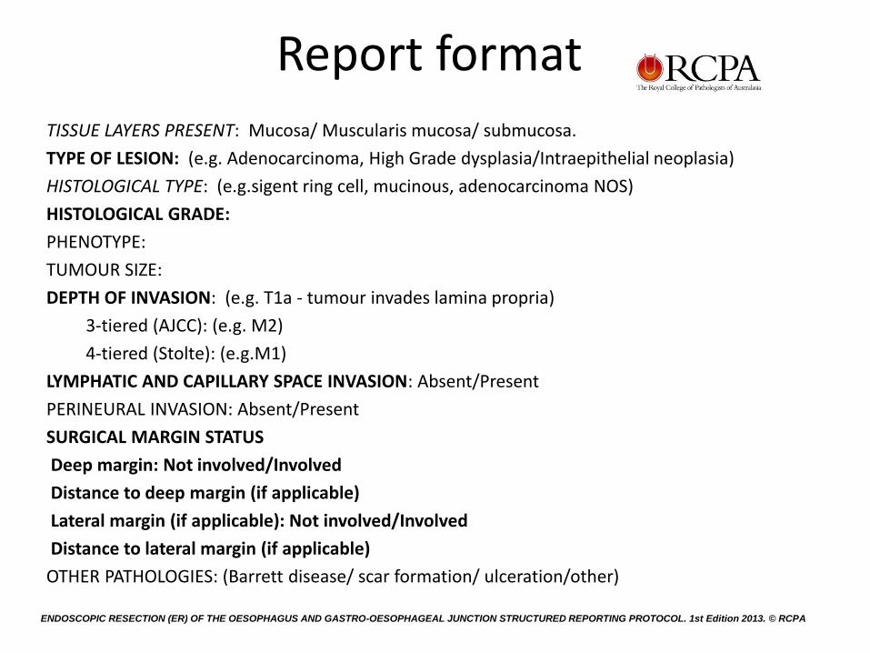
Report format TISSUE LAYERS PRESENT: Mucosa/ Muscularis mucosa/ submucosa.
TYPE OF LESION: (e.g. Adenocarcinoma, High Grade dysplasia/Intraepithelial neoplasia)
HISTOLOGICAL TYPE: (e.g.sigent ring cell, mucinous, adenocarcinoma NOS)
HISTOLOGICAL GRADE:
PHENOTYPE:
TUMOUR SIZE:
DEPTH OF INVASION: (e.g. T1a - tumour invades lamina propria)
3-tiered (AJCC): (e.g. M2)
4-tiered (Stolte): (e.g.M1)
LYMPHATIC AND CAPILLARY SPACE INVASION: Absent/Present
PERINEURAL INVASION: Absent/Present
SURGICAL MARGIN STATUS
Deep margin: Not involved/Involved
Distance to deep margin (if applicable)
Lateral margin (if applicable): Not involved/Involved
Distance to lateral margin (if applicable)
OTHER PATHOLOGIES: (Barrett disease/ scar formation/ ulceration/other)
ENDOSCOPIC RESECTION (ER) OF THE OESOPHAGUS AND GASTRO-OESOPHAGEAL JUNCTION STRUCTURED REPORTING PROTOCOL. 1st Edition 2013. © RCPA
ENDOSCOPIC RESECTION (ER) OF THE OESOPHAGUS AND GASTRO-
OESOPHAGEAL JUNCTION STRUCTURED REPORTING PROTOCOL. 1st Edition
2013. © RCPA

END