Lymphotoxin, but Not TNF, Is Required for Prion Invasion of Lymph
15
Zurich Open Repository and Archive University of Zurich Main Library Strickhofstrasse 39 CH-8057 Zurich www.zora.uzh.ch Year: 2012 Lymphotoxin, but Not TNF, Is Required for Prion Invasion of Lymph Nodes O’Connor, Tracy ; Frei, Nathalie ; Sponarova, Jana ; Schwarz, Petra ; Heikenwalder, Mathias ; Aguzzi, Adriano Abstract: Neuroinvasion and subsequent destruction of the central nervous system by prions are typically preceded by a colonization phase in lymphoid organs. An important compartment harboring prions in lymphoid tissue is the follicular dendritic cell (FDC), which requires both tumor necrosis factor receptor 1 (TNFR1) and lymphotoxin receptor (LTR) signaling for maintenance. However, prions are still detected in TNFR1(-/-) lymph nodes despite the absence of mature FDCs. Here we show that TNFR1- independent prion accumulation in lymph nodes depends on LTR signaling. Loss of LTR signaling, but not of TNFR1, was concurrent with the dedifferentiation of high endothelial venules (HEVs) required for lymphocyte entry into lymph nodes. Using luminescent conjugated polymers for histochemical PrP(Sc) detection, we identified PrP(Sc) deposits associated with HEVs in TNFR1(-/-) lymph nodes. Hence, prions may enter lymph nodes by HEVs and accumulate or replicate in the absence of mature FDCs. DOI: https://doi.org/10.1371/journal.ppat.1002867 Posted at the Zurich Open Repository and Archive, University of Zurich ZORA URL: https://doi.org/10.5167/uzh-64522 Journal Article The following work is licensed under a Creative Commons: Attribution 4.0 International (CC BY 4.0) License. Originally published at: O’Connor, Tracy; Frei, Nathalie; Sponarova, Jana; Schwarz, Petra; Heikenwalder, Mathias; Aguzzi, Adriano (2012). Lymphotoxin, but Not TNF, Is Required for Prion Invasion of Lymph Nodes. PLoS Pathogens, 8(8):e1002867. DOI: https://doi.org/10.1371/journal.ppat.1002867
Transcript of Lymphotoxin, but Not TNF, Is Required for Prion Invasion of Lymph

Zurich Open Repository andArchiveUniversity of ZurichMain LibraryStrickhofstrasse 39CH-8057 Zurichwww.zora.uzh.ch
Year: 2012
Lymphotoxin, but Not TNF, Is Required for Prion Invasion of Lymph Nodes
O’Connor, Tracy ; Frei, Nathalie ; Sponarova, Jana ; Schwarz, Petra ; Heikenwalder, Mathias ; Aguzzi,Adriano
Abstract: Neuroinvasion and subsequent destruction of the central nervous system by prions are typicallypreceded by a colonization phase in lymphoid organs. An important compartment harboring prions inlymphoid tissue is the follicular dendritic cell (FDC), which requires both tumor necrosis factor receptor1 (TNFR1) and lymphotoxin � receptor (LT�R) signaling for maintenance. However, prions are stilldetected in TNFR1(-/-) lymph nodes despite the absence of mature FDCs. Here we show that TNFR1-independent prion accumulation in lymph nodes depends on LT�R signaling. Loss of LT�R signaling, butnot of TNFR1, was concurrent with the dedifferentiation of high endothelial venules (HEVs) required forlymphocyte entry into lymph nodes. Using luminescent conjugated polymers for histochemical PrP(Sc)detection, we identified PrP(Sc) deposits associated with HEVs in TNFR1(-/-) lymph nodes. Hence,prions may enter lymph nodes by HEVs and accumulate or replicate in the absence of mature FDCs.
DOI: https://doi.org/10.1371/journal.ppat.1002867
Posted at the Zurich Open Repository and Archive, University of ZurichZORA URL: https://doi.org/10.5167/uzh-64522Journal Article
The following work is licensed under a Creative Commons: Attribution 4.0 International (CC BY 4.0)License.
Originally published at:O’Connor, Tracy; Frei, Nathalie; Sponarova, Jana; Schwarz, Petra; Heikenwalder, Mathias; Aguzzi,Adriano (2012). Lymphotoxin, but Not TNF, Is Required for Prion Invasion of Lymph Nodes. PLoSPathogens, 8(8):e1002867.DOI: https://doi.org/10.1371/journal.ppat.1002867

Lymphotoxin, but Not TNF, Is Required for Prion Invasionof Lymph NodesTracy O’Connor*, Nathalie Frei, Jana Sponarova, Petra Schwarz, Mathias Heikenwalder, Adriano Aguzzi*
Institute of Neuropathology, University Hospital of Zurich, Zurich, Switzerland
Abstract
Neuroinvasion and subsequent destruction of the central nervous system by prions are typically preceded by a colonizationphase in lymphoid organs. An important compartment harboring prions in lymphoid tissue is the follicular dendritic cell(FDC), which requires both tumor necrosis factor receptor 1 (TNFR1) and lymphotoxin b receptor (LTbR) signaling formaintenance. However, prions are still detected in TNFR12/2 lymph nodes despite the absence of mature FDCs. Here weshow that TNFR1-independent prion accumulation in lymph nodes depends on LTbR signaling. Loss of LTbR signaling, butnot of TNFR1, was concurrent with the dedifferentiation of high endothelial venules (HEVs) required for lymphocyte entryinto lymph nodes. Using luminescent conjugated polymers for histochemical PrPSc detection, we identified PrPSc depositsassociated with HEVs in TNFR12/2 lymph nodes. Hence, prions may enter lymph nodes by HEVs and accumulate or replicatein the absence of mature FDCs.
Citation: O’Connor T, Frei N, Sponarova J, Schwarz P, Heikenwalder M, et al. (2012) Lymphotoxin, but Not TNF, Is Required for Prion Invasion of LymphNodes. PLoS Pathog 8(8): e1002867. doi:10.1371/journal.ppat.1002867
Editor: Neil A. Mabbott, University of Edinburgh, United Kingdom
Received March 19, 2012; Accepted July 5, 2012; Published August 9, 2012
Copyright: � 2012 O’Connor et al. This is an open-access article distributed under the terms of the Creative Commons Attribution License, which permitsunrestricted use, distribution, and reproduction in any medium, provided the original author and source are credited.
Funding: A.A. is the recipient of a European Research Council Advanced Investigator Award. M.H. is a recipient of a European Research Council StartingInvestigator Award. T.O. is supported by a fellowship from the University of Zurich. J.S. is supported by a FEBS long-term fellowship. This work was funded bySwiss National Science Foundation grant # 3100A0-111917 to A.A. The funders had no role in study design, data collection and analysis, decision to publish, orpreparation of the manuscript.
Competing Interests: The authors have declared that no competing interests exist.
* E-mail: [email protected] (AA); TracyLynn.O’[email protected] (TO)
Introduction
Prions are unusual infectious agents thought to be comprised
solely of an abnormally folded, aggregated isoform (PrPSc) of the
endogenous cellular prion protein (PrPC) [1]. Deposition of PrPSc
aggregates, vacuolation, and neuronal loss in brain tissue are histo-
pathological hallmarks of a group of neurological disorders collec-
tively known as prion diseases or transmissible spongiform enceph-
alopathies (TSEs), including scrapie in sheep, bovine spongiform
encephalopathy (BSE) in bovids, chronic wasting disease (CWD) in
cervids, and Creutzfeldt-Jakob disease (CJD) in humans [2].
Although TSEs seem to selectively damage the central nervous
system (CNS), peripheral prion exposure leads to the accumulation of
prions and PrPSc in secondary lymphoid organs (SLOs) long be-
fore neurological symptoms appear [3,4,5,6,7,8], and it is largely
from these extraneural sites that prions transmigrate to the peripheral
nervous system (PNS) and finally the CNS [9,10,11]. Extraneural
prion accumulation is thought to occur primarily within stromal cells
found in germinal centers of lymphoid follicles known as follicular
dendritic cells (FDCs) [4,12,13,14,15,16,17]. Maintenance of mature
FDC networks depends on signaling through FDC-expressed lym-
photoxin b receptor (LTbR) and tumor necrosis factor receptor 1
(TNFR1) by B cell-derived tumor necrosis factor (TNF) and lym-
photoxins (LT) a and b [18,19,20,21,22,23]. Accordingly, ablation of
B cells, and hence loss of LTa/b and TNFa ligands, antagonizes
prion deposition in secondary lymphoid organs [24,25], and intra-
peritoneal (i.p.) injection of mice with TNFR1 or LTbR blocking
antibodies prior to peripheral prion inoculation causes transient de-
differentiation of FDCs, reduces splenic prion accumulation, and
delays prion neuroinvasion [26,27,28,29].
However, extraneural prion accumulation in SLOs is not strictly
dependent on the presence of mature FDCs. Although prion titers
remain below detection in spleens of i.p.-inoculated TNFR12/2 and
TNFa2/2 mice, PrPSc levels and prion infectivity in TNFR12/2 and
TNFa2/2 lymph nodes are only marginally reduced compared to
TNFR12/2, LTbR2/2, LTa2/2, or LTb2/2 spleens [30,31].
Furthermore, TNFR12/2 and TNFa2/2 mice succumb to terminal
disease upon i.p. prion inoculation at a noticeably higher rate than
lymphotoxin signaling-deficient mice, indicating that prions are still
effectively transmitted to the CNS in the absence of TNFR1
signaling. Since TNFR12/2 lymph nodes are devoid of detectable
mature FDCs, this implies that other undefined cell types may also
be required for prions to colonize SLOs. However, lymph nodes are
either absent or profoundly disrupted in LTbR2/2, LTa2/2, and
LTb2/2 mice compared to TNFR12/2 and TNFa2/2, making it
difficult to formally conclude that LTbR signaling is specifically re-
quired for prion accumulation in lymph nodes while TNFR1 is not.
To determine whether continuous LTbR signaling is required for
prions to accumulate in TNFR12/2 lymph nodes, we investigated
the ability of prions to colonize SLOs of prion-infected TNFR12/2
mice treated with an LTbR-Ig blocking antibody.
Results
Prion accumulation in TNFR12/2 lymph nodes is LTbRsignaling-dependent
We previously showed that mice devoid of TNF signaling accu-
mulate prion infectivity and PrPSc in lymph nodes but not in spleen,
in contrast to LT signaling-deficient mice which do not accumulate
prions in either spleen or lymph nodes [31]. To determine whether
PLoS Pathogens | www.plospathogens.org 1 August 2012 | Volume 8 | Issue 8 | e1002867
prion accumulation in TNFR12/2 lymph nodes was dependent on
continuous LTbR signaling, we administered weekly 100 mg intra-
peritoneal (i.p.) injections of an LTbR immunoglobulin fusion
protein (LTbR-Ig) or control pooled human immunogloblulin (Ig)
to wild-type (WT) and TNFR12/2 mice to achieve sustained inhi-
bition of LTbR signaling [32]. One week following the initial
LTbR-Ig or control Ig injection, mice were inoculated intraperi-
toneally (i.p.) with 6 log LD50 RML6 prions. At 60 days post-
injection (d.p.i.), spleens and mesenteric lymph nodes (mLNs) from
these mice were assessed for accumulation of prion infectivity and
PrPSc. To confirm that LTbR-Ig treatment effectively inhibited
LTbR signaling, we analyzed follicular dendritic cell marker 1
(FDCM1) immunoreactivity in spleens from WT or TNFR12/2
mice treated with either control Ig or LTbR-Ig. FDCM1 immuno-
reactivity was absent in spleens and mLNs from TNFR12/2-Ig,
WT-LTbR-Ig, and TNFR12/2-LTbR-Ig spleens in contrast to
WT-Ig, indicating that LTbR-dependent FDCs had de-differenti-
ated in response to LTbR-Ig treatment (Fig. 1A–J). In addition, we
analyzed transcriptional targets of the LTbR pathway in spleens
from WT or TNFR12/2 mice treated with either control Ig or
LTbR-Ig. As expected, levels of both NFkB2 (p100) and CXCL13
mRNA were reduced in TNFR12/2-Ig, WT-LTbR-Ig, and
TNFR12/2-LTbR-Ig spleens compared to WT-Ig (Fig. 1K & L).
Next, to determine the effect of inhibited LTbR signaling on
prion accumulation in SLOs, we compared the pattern of PrPSc
deposition in spleens and mLNs from prion-infected TNFR12/2-Ig,
WT-LTbR-Ig, and TNFR12/2-LTbR-Ig mice to WT-Ig mice by
histoblotting. As expected, TNFR12/2-Ig, WT-LTbR-Ig, and
TNFR12/2-LTbR-Ig spleens accumulated less PrPSc than WT-Ig
spleens (Fig. 2A–D), though chronic LTbR-Ig administration
seemed less effective at preventing PrPSc deposition in WT spleens
than genetic ablation of TNFR1 (compare Fig. 2B with 2C). Since
the 60 day treatment period approaches the limit of effective LTbR-
Ig inhibition, this most likely reflects partial FDC re-maturation in
WT SLOs near the end of the experiment (J. Browning; personal
communication). Regardless, the ability of LTbR-Ig to block prion
replication in WT SLOs is well-established [26,27]. Consistent with
our previous studies, mLNs from WT and TNFR12/2-Ig-treated
mice contained similar numbers of PrPSc deposits (Fig. 2E & F),
whereas PK-resistant PrP deposits in WT-LTbR-Ig mLNs were less
numerous than in WT-Ig or TNFR12/2-Ig mLNs (Compare
Fig. 2G with Fig. 2E & F). Background PrP immunoreactivity in
PK-digested histoblots from non-infected wild-type spleens was
negligible (Supp. Fig. S1).
Of note, TNFR12/2-LTbR-Ig mLNs were devoid of PrPSc im-
munoreactivity (Fig. 2H), demonstrating that prion accumulation
in lymph nodes in the absence of TNFR1 is dependent on LTbR
signaling. To confirm this result quantitatively, we analyzed prion
infectivity in mLNs from Ig-treated versus LTbR-Ig-treated WT
and TNFR12/2 mice using the scrapie cell assay (SCA; [33,34,35]).
Consistent with the corresponding histoblots from these mice,
LTbR-Ig treatment decreased prion infectivity in WT and
TNFR12/2 mLNs by 2–3 log tissue culture infectious (TCI) units
per gram of tissue compared to Ig treatment (Fig. 2I).
To determine the effect of LTbR-Ig treatment on PrPC
expression in SLOs, which might explain the differential ability
of TNFR12/2-Ig versus TNFR12/2-LTbR-Ig mLNs to accumu-
late prions, we measured Prnp mRNA levels in spleens and lymph
nodes from all treatment groups by quantitative PCR. Prnp mRNA
levels were reduced in WT-LTbR-Ig, TNFR12/2-Ig, and
TNFR12/2-LTbR-Ig spleens compared to WT-Ig (Fig. 2J), most
likely reflecting de-differentiation of FDCs, the primary PrPC-
expressing cell type in spleen. In contrast, no differences in Prnp
mRNA expression were found in mLNs from mice of different
treatment groups. Of particular note, no difference in Prnp mRNA
expression was found between TNFR12/2-Ig and TNFR12/2-
LTbR-Ig mLNs (Fig. 2K). Taken together, our data indicate that
TNFR12/2 lymph nodes accumulate prions in the absence of
mature FDCs yet in an LTbR-dependent manner. Moreover,
inhibited prion deposition in mLNs upon loss of LTbR signaling
seems to be unrelated to local Prnp levels.
MadCam1 expression correlates with prion infectivity inmesenteric lymph nodes
We reasoned that prion accumulation in TNFR12/2 lymph
nodes might rely on a putative LTbR signaling-dependent,
TNFR1 signaling-independent cell present in lymph nodes but
not in spleens. In order to identify such a cell, we screened spleens
and mLNs from WT-Ig, WT-LTbR-Ig, TNFR12/2-Ig, and
TNFR12/2-LTbR-Ig mice for a variety of hematopoietic and stro-
mal cell markers by immunohistochemistry (IHC) whose expres-
sion correlated with prion replication ability. As previously noted,
the pattern of FDCM1 immunoreactivity was consistent with PrPSc
deposition in spleen, but not in mLNs (Fig. 1A–J). Likewise, sev-
eral other markers for B cells (B220), T cells (CD3), marginal zone
macrophages (MOMA-1), monocytes (F4/80), and stromal cells
(CD21/35, C4, VCAM1, ICAM1) revealed no staining patterns
consistent with an involvement in splenic or lymph nodal prion
accumulation (Supp. Figs S2A–H & S3A–H). However, we
identified one stromal cell marker that exhibited a staining pattern
consistent with prion deposition in both spleen and mLNs:
mucosal addressin cell adhesion molecule 1 (MadCam1 [36]).
MadCam1 weakly stained FDC networks of both WT-Ig spleen
(Fig. 3A & E) and lymph nodes (Fig. 4A), as well as the marginal
sinus (MS) of spleens from WT-Ig mice (Fig. 3A). Although FDC
and MS-associated MadCam1 immunoreactivity was absent upon
loss of TNFR1 or LTbR signaling in both spleen (Fig. 3B–E) and
lymph nodes (Fig. 4 B–D), MadCam1 immunoreactivity persisted
in TNFR12/2-Ig mLNs (Fig. 3G & J; 4B). Of note, MadCam1
immunoreactivity was completely absent in mLNs from LTbR-Ig-
treated WT and TNFR12/2 mice (Fig. 3I–J & 4C–D). Fur-
thermore, analysis of MadCam1 mRNA expression by Real Time
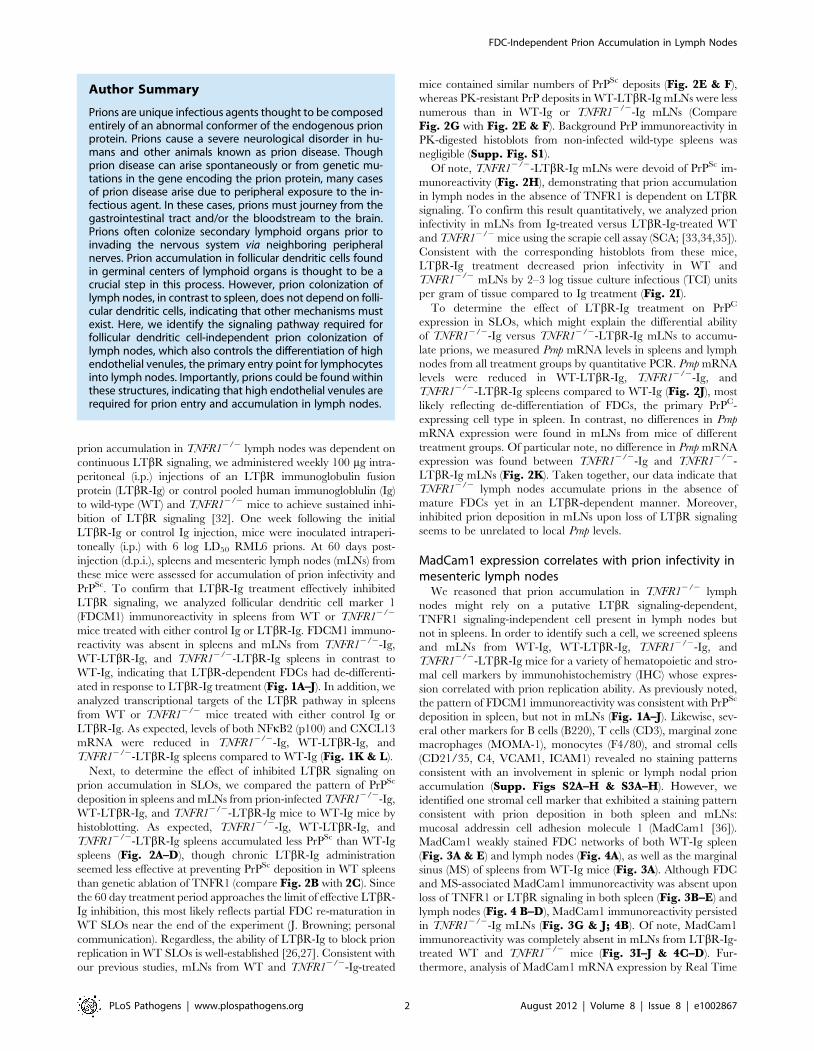
Author Summary
Prions are unique infectious agents thought to be composedentirely of an abnormal conformer of the endogenous prionprotein. Prions cause a severe neurological disorder in hu-mans and other animals known as prion disease. Thoughprion disease can arise spontaneously or from genetic mu-tations in the gene encoding the prion protein, many casesof prion disease arise due to peripheral exposure to the in-fectious agent. In these cases, prions must journey from thegastrointestinal tract and/or the bloodstream to the brain.Prions often colonize secondary lymphoid organs prior toinvading the nervous system via neighboring peripheralnerves. Prion accumulation in follicular dendritic cells foundin germinal centers of lymphoid organs is thought to be acrucial step in this process. However, prion colonization oflymph nodes, in contrast to spleen, does not depend on folli-cular dendritic cells, indicating that other mechanisms mustexist. Here, we identify the signaling pathway required forfollicular dendritic cell-independent prion colonization oflymph nodes, which also controls the differentiation of highendothelial venules, the primary entry point for lymphocytesinto lymph nodes. Importantly, prions could be found withinthese structures, indicating that high endothelial venules arerequired for prion entry and accumulation in lymph nodes.
FDC-Independent Prion Accumulation in Lymph Nodes
PLoS Pathogens | www.plospathogens.org 2 August 2012 | Volume 8 | Issue 8 | e1002867

Figure 1. Repeated LTbR-Ig administration chronically downregulates LTbR signaling and de-differentiates FDC networks in C57BL/6 and TNFR12/2 spleens and lymph nodes. Frozen sections from spleens (A–D) and mesenteric lymph nodes (E–H) of C57BL/6 (WT) Ig-treated(A & E), C57BL/6 (WT) LTbR-Ig-treated (C & G), TNFR12/2 Ig-treated (B & F), or TNFR12/2 LTbR-Ig-treated (D & H) mice were analyzed byimmunohistochemistry and developed with alkaline phosphatase for follicular dendritic cell marker 1 (FDCM1). The total number of FDCM1-positive(FDCM1+; black) or FDCM1-negative (FDCM12; white) lymphoid follicles were scored for spleens (I) and mesenteric lymph nodes (mLNs; J) andexpressed as a percentage of total follicles in each treatment group. FDCM1+ FDC networks were visible in 82% of WT Ig-treated spleen follicles (A; I)and 94% of WT-Ig lymph node follicles (E; J), whereas FDCM1+ FDCs were absent (0% of follicles) in spleens and lymph nodes from mice lackingTNFR1 and/or LTbR signaling (B–D; F–H; I–J). Total mRNA was isolated from spleens of mice from the indicated treatment groups and analyzed forexpression of LTbR signaling targets CXCL13 (Mean 6 S.E.M.: WT-Ig = 102.68611.56, WT-LTbR-Ig = 18.0562.30, TNFR12/2-Ig = 19.5661.63, andTNFR12/2-LTbR-Ig = 15.1460.15) (K) and NFkB2 (Mean 6 S.E.M.: WT-Ig = 103.10614.65, WT-LTbR-Ig = 33.3062.10, TNFR12/2-Ig = 46.0467.53, andTNFR12/2-LTbR-Ig = 33.3961.18) (L) by Real Time PCR. Both CXCL13 and NFkB2 mRNA levels were reduced in all treatment groups relative to WT-Ig.doi:10.1371/journal.ppat.1002867.g001
FDC-Independent Prion Accumulation in Lymph Nodes
PLoS Pathogens | www.plospathogens.org 3 August 2012 | Volume 8 | Issue 8 | e1002867

Figure 2. PrPSc accumulation in TNFR12/2 lymph nodes requires LTbR signaling independent of Prnp expression. C57BL/6 (WT) orTNFR12/2 mice inoculated i.p. with 6 log LD50 RML6 and treated weekly with control Ig or LTbR-Ig were sacrificed at 60 d.p.i. Histoblots wereperformed on frozen sections from spleens (SPL; A–D) or mesenteric lymph nodes (mLN; E–H) from mice in each treatment group to visualize PrPSc
deposition. Whole organs are shown in left panels, and corresponding higher resolution images for each treatment group are shown in right panels.Note that lack of TNFR1 signaling can prevent PrPSc accumulation in spleen (B) but not lymph node (F). However, blocking LTbR signaling can preventPrPSc accumulation in TNFR12/2 lymph nodes (compare F and H). Prion infectivity titers in mLN homogenates from individual TNFR12/2 Ig-treatedand LTbR-Ig treated mice were measured using the scrapie cell assay (I). Whereas TNFR12/2-Ig mLNs all harbored $6.1 log TCI units/g tissue, prioninfectivity in TNFR12/2-LTbR-Ig mLNs was reduced by at least 2.5 log TCI units/g tissue. Total mRNA was isolated from spleens (J) or mesentericlymph nodes (mLN; K) of mice from the indicated treatment groups and analyzed for Prnp expression by Real Time PCR (Mean 6 S.E.M.: Spleen – WT-Ig = 100.6966.74, WT-LTbR-Ig = 46.0264.30, TNFR12/2-Ig = 48.9460.55, and TNFR12/2-LTbR-Ig = 50.1160.55; mLN – WT-Ig = 106.05622.54, WT-LTbR-Ig = 101.69612.88, TNFR12/2-Ig = 56.4363.13, and TNFR12/2-LTbR-Ig = 72.3566.65). Whereas spleens from WT-LTbR-Ig, TNFR12/2-Ig, and TNFR12/2-LTbR-Ig mice all showed decreases in Prnp expression relative to WT-Ig spleens (J), no differences could be found in Prnp expression in mLNs frommice in any treatment group (K).doi:10.1371/journal.ppat.1002867.g002
FDC-Independent Prion Accumulation in Lymph Nodes
PLoS Pathogens | www.plospathogens.org 4 August 2012 | Volume 8 | Issue 8 | e1002867

Figure 3. MadCam1 immunoreactivity in lymphoid tissue correlates with prion deposition. Formalin-fixed cryosections from spleens (SPL;A–D) or mesenteric lymph nodes (mLN; F–I) of C57BL/6 (WT) Ig-treated (A & F), TNFR12/2 Ig-treated (B & G), C57BL/6 (WT) LTbR-Ig-treated (C & H), orTNFR12/2 LTbR-Ig-treated (D & I) mice were immunostained with an antibody against the stromal cell marker, mucosal addressin cell adhesionmolecule 1 (MadCam1), and visualized with alkaline phosphatase. (E) The total number of MadCam1-positive (MadCam1+; black) or MadCam1-negative (MadCam1-; white) lymphoid follicles were scored for spleens and expressed as a percentage of total follicles in each treatment group. (J)The total number of MadCam1-postive (MadCam1+) structures per mesenteric lymph node (mLN) was counted and averaged for each treatmentgroup. WT-Ig mLNs contained 47.5614 MadCam1+ structures, WT-LTbR-Ig = 9.563, TNFR12/2-Ig = 74619, and TNFR12/2-LTbR-Ig = 0. MadCam1immunoreactivity in WT-Ig spleens was localized to the marginal sinus (A; black arrow) and 55% (E) of germinal centers (A; white arrow). This staining
FDC-Independent Prion Accumulation in Lymph Nodes
PLoS Pathogens | www.plospathogens.org 5 August 2012 | Volume 8 | Issue 8 | e1002867

PCR in mLNs quantitatively corroborated the MadCam1 IHC
findings: MadCam1 mRNA levels were equally reduced in spleens
from WT-LTbR-Ig, TNFR12/2-Ig, and TNFR12/2-LTbR-Ig
mice, compared to WT-Ig (Fig. 4E). In contrast, MadCam1
mRNA levels were intermediately reduced in TNFR12/2-Ig mLNs
compared to WT-Ig, and a further reduction in MadCam1 mRNA
levels was observed in TNFR12/2-LTbR-Ig mLNs compared to
TNFR12/2-Ig (Fig. 4F). Thus far, our data suggested that the
presence of a MadCam1-expressing cell was associated with
accumulation of prions in lymph nodes but not in spleen.
pattern was absent (0%; E) in the spleens of mice from all other treatment groups (B,C & D). In contrast, MadCam1 immunoreactivity in WT-Ig mLNswas largely found in thick vessels (F). MadCam1 immunoreactivity was retained in TNFR12/2-Ig mLNs (G) but absent in mLNs from mice treated withLTbR-Ig (H–J). Size bars: Left panels = 200 mm; Right panels = 100 mm.doi:10.1371/journal.ppat.1002867.g003
Figure 4. Vessel-associated MadCam1 expression in lymph nodes is preserved in the absence of TNFR1 signaling. Frozen sectionsfrom mesenteric lymph nodes (mLN) of C57BL/6 (WT) Ig-treated (A), TNFR12/2 Ig-treated (B), C57BL/6 (WT) LTbR-Ig-treated (C), or TNFR12/2 LTbR-Ig-treated (D) mice were analyzed by immunofluorescence with MadCam1 antibody. In WT-Ig mLNs (A), MadCam1 robustly stained thick vessels (yellowarrow), while germinal centers were weakly MadCam1-positive (white arrow). In TNFR12/2-Ig mLNs (B), MadCam1-positive germinal centers wereabsent, while vessel-associated MadCam1 staining persisted. In contrast, both WT-LTbR-Ig and TNFR12/2-LTbR-Ig mLNs were MadCam1-negative (C &D). Size bars = 100 mm. Total mRNA was isolated from spleens (E) or mesenteric lymph nodes (F) of C57BL/6 (WT) Ig-treated, C57BL/6 (WT) LTbR-Ig-treated, TNFR12/2 Ig-treated, or TNFR12/2 LTbR-Ig-treated mice and analyzed for MadCam1 expression by Real Time PCR (Mean 6 S.E.M.: Spleen –WT-Ig = 104.21616.56, WT-LTbR-Ig = 29.9861.28, TNFR12/2-Ig = 30.4162.56, and TNFR12/2-LTbR-Ig = 27.9563.77; mLN – WT-Ig = 100.8968.06, WT-LTbR-Ig = 45.4066.75, TNFR12/2-Ig = 49.4260.63, and TNFR12/2-LTbR-Ig = 24.7967.25). MadCam1 expression was reduced in spleens of WT-LTbR-Ig-treated, TNFR12/2 Ig-treated, and TNFR12/2 LTbR-Ig-treated mice compared to WT-Ig. MadCam1 expression in WT-LTbR-Ig and TNFR12/2 Ig mLNswas reduced compared to WT-Ig, and MadCam1 expression in TNFR12/2 LTbR-Ig mLNs was reduced compared to TNFR12/2-Ig.doi:10.1371/journal.ppat.1002867.g004
FDC-Independent Prion Accumulation in Lymph Nodes
PLoS Pathogens | www.plospathogens.org 6 August 2012 | Volume 8 | Issue 8 | e1002867

MadCam1-positive structures in TNFR12/2 lymph nodesare PrPC-expressing HEVs
MadCam1 immunoreactivity in TNFR12/2-Ig mLNs was re-
stricted to thick vessels (Fig. 3G & 4B) that were absent from
TNFR12/2-LTbR-Ig mLNs (Fig. 3I & Fig. 4D), as well as iso-
type controls (Supp. Fig. S4B), and were morphologically distinct
from MadCam1-positive FDC networks found in WT-Ig spleens
(Fig. 3A) and mLNs (Fig. 4A). These MadCam1-positive struc-
tures were morphologically consistent with high endothelial ve-
nules (HEVs) responsible for controlling lymphocyte entry into
LNs, Peyer’s patches (PPs), and other SLOs, with the exception of
spleen [37]. Co-localization of Madcam1-positive vessels with
peripheral node addressin (PNAd; a.k.a. MECA-79), a specific
marker of HEVs [38], confirmed that these structures in
TNFR12/2-Ig mLNs were HEVs (Fig. 5A–F), whereas Mad-
Cam1-positive FDC networks in WT-Ig mLNs (Fig. 5A–C) and
isotype controls (Supp. Fig. S4A) were PNAd-negative. To gather
evidence that HEVs could be potential sites of prion replication, we
analyzed PrPC immunoreactivity in HEVs of WT-Ig mLNs. Co-
immunofluorescent (co-IF) staining of WT-Ig mLNs with Mad-
Cam1 and PrPC antibodies confirmed that PrPC immunoreactivity
was present in HEVs (Fig. 5G–I) and absent from isotype controls
(Supp. Fig. S4C). Thus, HEVs fulfill at least one requirement of a
prion replicating tissue - PrPC expression.
PrPSc localizes to HEVs in TNFR12/2 lymph nodesTo determine whether prions were localized to HEVs in mLNs,
we performed co-IF with PrPC and MadCam1 antibodies in prion-
infected TNFR12/2-Ig mLNs. Prion-infected TNFR12/2-Ig
mLNs contained areas of intense PrP-positive deposits which were
not detectable in non-infected TNFR12/2-Ig mLNs (data not
shown; [31]). Of note, many of these PrPC-positive areas localized
to MadCam1-postive vessels in prion-infected TNFR12/2-Ig
mLNs, indicating that HEVs are probable sites of PrPSc
localization in infected lymph nodes (Fig. 6A–C). However, PrP
immunostaining of prion-infected tissue cannot reliably distinguish
between PrPSc deposits and sites of high PrPC expression, and
HEVs expressed relatively high levels of PrPC in uninfected mLNs
(Fig. 5G–I).
To develop an independent method of distinguishing PrPSc
from PrPC in lymphoid organs, we tested the ability of a series of
fluorescent amyloid-binding dyes known as luminescent conjugat-
ed polymers (LCPs [39,40,41,42]) to stain PrPSc deposits in spleen
and lymph node. LCPs were previously shown to recognize PrPSc
in brain [43]. One LCP, pentameric formic thiophene acetic acid
(p-FTAA; [44]), stained PrP-positive FDC networks of prion-
infected spleens from WT mice (Supp. Fig. S5). In contrast, no
immunofluorescence could be detected with p-FTAA in spleens
and lymph nodes from uninfected mice (Supp. Fig. S6). Using
this method, points of PrPSc/MadCam1 co-localization could be
reliably identified in prion-infected TNFR12/2 lymph nodes,
which again indicated that a proportion of PrPSc localizes to HEVs
in TNFR12/2 mLNs (Fig. 6D–F). However, at lower magnifica-
tion we also noted that a number of PrPSc deposits were located
outside of HEVs (Fig. 6G–I). To confirm this observation and to
analyze the tissue-wide distribution of PrPSc deposits relative to
HEVs in prion-infected TNFR12/2-Ig mLNs, we performed
histoblot co-stains with PNAd antibody, which is highly immuno-
reactive to HEVs and can be visualized even after proteinase K
digestion (Supp. Fig. S7). This analysis confirmed that some
PrPSc deposits were localized to HEVs in TNFR12/2-Ig mLNs.
However, PrPSc deposits were also present outside of HEVs
(Fig. 6J–K), which is consistent with our previous study showing
that strong PrP immunoreactivity in prion-infected TNFR12/2
mLNs can also be found in other cell types (i.e. macrophages [31]).
Since HEVs could potentially serve as entry portals for prion-
harboring lymphocytes, and it was recently reported that neigh-
boring dendritic cells (DCs) are responsible for HEV differentia-
tion [45], we also performed PrP co-immunofluorescence on
prion-infected TNFR12/2-Ig mLNs using the DC marker, CD11c,
to determine whether DCs in the vicinity of HEVs might also
contain PrPSc. However, no overlap of PrP and CD11c immu-
noreactivity in prion-infected TNFR12/2-Ig mLNs was identified
(Supp Fig. S8).
Discussion
The means by which prions evade the immune system’s nu-
merous defense mechanisms and finally transmigrate to, and se-
lectively damage, neurons of the CNS has been the subject of
scientific scrutiny for two decades. A number of studies have im-
plicated FDCs in the germinal centers of SLOs as the primary
reservoirs of prions prior to neuroinvasion [4,12,13,14,15,16,17].
Yet the ability of TNFR12/2 mLNs to accumulate prions with a
minimal loss of infectivity compared to WT mLNs presents an
apparent paradox, since FDC maintenance depends on TNFR1
signaling [21,22,23].
Here we have established that lymph nodal prion accumulation
in the absence of TNFR1 signaling is LTbR signaling-dependent.
Crucially, transient loss of LTbR signaling was sufficient to block
TNFR1-independent prion accumulation in lymph nodes, indi-
cating that prion accumulation in lymph nodes specifically
requires LTbR signaling and is not simply prevented by general
developmental defects or architectural disruptions caused by lack
of LTbR signaling in LTb2/2 lymph nodes.
We previously showed that intense PrP immunoreactivity was
localized to macrophages in prion-infected TNFR12/2 mLNs [31],
and others have reported that PK-resistant PrP is localized to
macrophages in spleens with PrPC-deficient FDCs [46], indicating
that macrophages serve as alternative sites of prion accumulation in
the absence of PrPC-expressing FDCs. How this phenomenon was
mechanistically linked to LTbR signaling and the pattern of prion
accumulation in lymph nodes was initially unclear, since most
macrophage populations were preserved in the absence of both
TNFR1 and LTbR signaling [31]. Here, we discovered that loss of
LTbR signaling in mLNs was also correlated with the dedifferen-
tiation of HEVs – the primary point of entry for lymphocytes into
lymph nodes and a likely determinant in the ability of prions to
colonize lymph nodes.
Consistent with the pattern of prion accumulation in spleens
and lymph nodes of mice lacking TNF and/or LT signaling com-
ponents, HEVs exist in lymph nodes and other SLOs but not
spleens [37], and the maintenance of HEV architecture is TNFR1
signaling-independent yet LTbR signaling-dependent [47]. More-
over, we identified sites of PrPSc-HEV overlap in TNFR12/2-Ig
mLNs, indicating that HEVs might replicate prions and/or serve
as points of entry for prions or prion-harboring lymphocytes.
FDC-deficient mice can succumb to scrapie even in the absence of
detectable splenic prion titers [24]. In light of our current results,
this phenomenon is most likely explained by HEV-dependent
entry and accumulation of prions in lymph nodes and other HEV-
containing SLOs, such as Peyer’s patches.
HEVs can also form ectopically in certain chronic inflammatory
conditions. Prion accumulation at sites of chronic inflammation has
previously been observed, but in most cases this could be attributed
to local formation of FDC networks [48,49,50]. However, we also
previously reported LTbR-dependent prion colonization of gran-
ulomas in the absence of FDC markers [51]. Since ectopic HEV
FDC-Independent Prion Accumulation in Lymph Nodes
PLoS Pathogens | www.plospathogens.org 7 August 2012 | Volume 8 | Issue 8 | e1002867

Figure 5. MadCam1-positive vessels in mesenteric lymph nodes are prion protein-expressing high endothelial venules. Co-immunofluorescent confocal microscopy was performed on frozen sections from WT-Ig mesenteric lymph nodes with the high endothelial venule(HEV)-specific marker, peripheral node addressin (PNAd; green; A & D) and MadCam1 (red; B & E). Merged image shown in C & F. MadCam1-positivevessels co-localized with PNAd (A,B & C; yellow arrow). Note that MadCam1-positive germinal centers were PNAd-negative (B & C; white arrow).Standard co-immunofluorescence was performed on frozen sections from WT-Ig mesenteric lymph nodes with MadCam1 (red; G) and an antibodydirected against the C-terminus of the prion protein (PrP; green; H; POM1; [62]). MadCam1-positive HEVs were also PrP-positive (I). Size bars in A–C = 150 mm. Size bars in D–F = 50 mm. Size bars in G–I = 20 mm.doi:10.1371/journal.ppat.1002867.g005
FDC-Independent Prion Accumulation in Lymph Nodes
PLoS Pathogens | www.plospathogens.org 8 August 2012 | Volume 8 | Issue 8 | e1002867
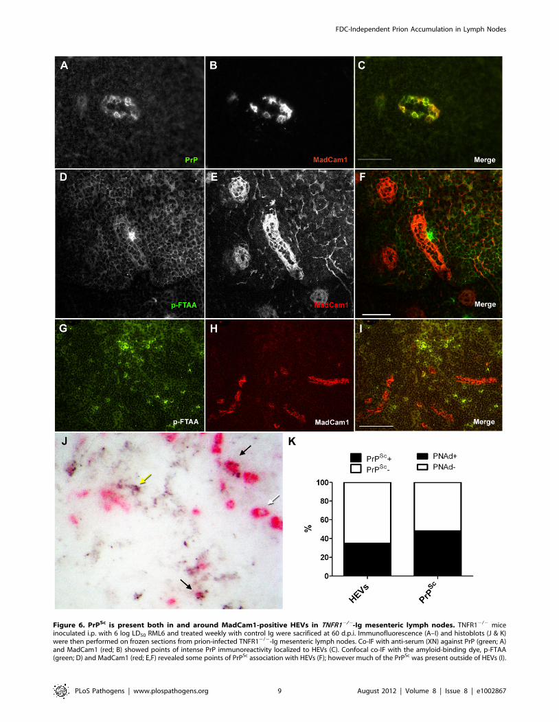
Figure 6. PrPSc is present both in and around MadCam1-positive HEVs in TNFR12/2-Ig mesenteric lymph nodes. TNFR12/2 miceinoculated i.p. with 6 log LD50 RML6 and treated weekly with control Ig were sacrificed at 60 d.p.i. Immunofluorescence (A–I) and histoblots (J & K)were then performed on frozen sections from prion-infected TNFR12/2-Ig mesenteric lymph nodes. Co-IF with anti-serum (XN) against PrP (green; A)and MadCam1 (red; B) showed points of intense PrP immunoreactivity localized to HEVs (C). Confocal co-IF with the amyloid-binding dye, p-FTAA(green; D) and MadCam1 (red; E,F) revealed some points of PrPSc association with HEVs (F); however much of the PrPSc was present outside of HEVs (I).
FDC-Independent Prion Accumulation in Lymph Nodes
PLoS Pathogens | www.plospathogens.org 9 August 2012 | Volume 8 | Issue 8 | e1002867

formation without any evidence of FDC networks has previously
been reported in certain inflammatory conditions [52], ectopic HEV
formation might be a source of prion replication and/or uptake in
granulomas, as well.
Of note, LTa/b signaling to HEVs differs from LTa/b signaling
to FDCs. Though HEVs express LTbR [53], HEVs persist in the
absence of B cells [47], in contrast to FDCs which rely on B cells to
provide LTa/b ligand [19,20]. This indicates that the LTa/b signal
to HEVs emanates from another cell type. Until recently the cell
type providing LTa/b to HEVs was not known; however a recent
publication reports that dendritic cells (DCs) may be the source of
LTa/b signaling to HEVs [45]. If HEV differentiation is indeed B
cell-independent, this may explain why prion neuroinvasion can
occur in the absence of B cells in some cases, since LTbR signaling
to HEVs and hence the ability of prions to enter and accumulate in
lymph nodes would be preserved [24,54].
Do prions actually replicate in TNFR12/2 lymph nodes, or do
they simply accumulate? Accumulation seems likely, as the only cell
type in SLOs known to replicate prions are FDCs, and prion
replication in macrophage populations was not observed [46]. In
any case, experimental evidence suggests that prion accumulation in
SLOs is sufficient for prions to invade the CNS [55,56,57].
Moreover, deletion of complement factors dramatically impairs
prion accumulation in SLOs and subsequent neuroinvasion without
affecting PrPC levels [58], indicating that the ability of SLOs to
physically capture prions is indeed critical for the development of
downstream pathology.
Curiously, we previously showed that PrPC expression is required
in either the stromal or the hematopoietic compartment for lymph
nodes to accumulate prion infectivity [31], which implies that prion
replication can occur in both compartments in lymph nodes.
However, an alternative explanation is that a hematopoietic cell is
required for delivery of prions to a ‘‘trapping’’ cell within the lymph
node, and PrPC expression on the hematopoietic cell mediates
efficient uptake of PrPSc in the bloodstream. Hence, HEVs may
facilitate the selective uptake and accumulation of prions or prion-
containing lymphocytes into lymph nodes, rather than serving as
sites of bona fide prion replication. Based on both current and past
findings, it is likely that the ‘‘trapping’’ cells in lymph nodes are
endothelial cells in HEVs (HEVECs), and the hematopoietic de-
livery cells are macrophages. Once prions or prion-harboring cells
have successfully invaded mLNs through HEVs, they may be
transported via conduits to FDCs [59] under normal conditions.
However, in the absence of mature FDCs, prions may remain in or
be transferred to macrophages.
Materials and Methods
Animals, treatments & ethical statementTNFR12/2 mice carrying a targeted deletion of exons 2, 3, and
part of exon 4 of the tumor necrosis factor receptor 1 (TNFR1)
open reading frame [60] were maintained on a C57BL/6 back-
ground in-house. C57BL/6 wild-type control mice were pur-
chased from Harlan Laboratories and bred in-house. Murine
LTbR-murine IgG1 (LTbR-Ig) and MOPC21 mouse immuno-
globulin control were obtained from Biogen Idec. For analysis of
prion-infected tissues (Figs 2A–I, 6 & Supp. Figs S5, S7 & S8),
TNFR12/2 or C57BL/6 mice were injected intraperitoneally (i.p.)
with 100 mg LTbR-Ig or MOPC21 (n = 3–4/group). One week
later, mice were inoculated i.p. with 100 mL 6 log LD50 Rocky
Mountain Laboratory mouse-adapted prion strain [61], passage 6
(RML6). Mice were then boostered weekly with 100 mg LTbR-Ig
or MOPC21 until 60 days post-inoculation (d.p.i), at which point
mice were sacrificed. For analysis of uninfected tissue (Figs 1, 2J–
K, 3, 4, 5 & Supp. Figs S1, S2, S3, S4 & S6), TNFR12/2 or
C57BL/6 mice received 2 weekly injections of LTbR-Ig or
MOPC21 and were sacrificed 3 weeks following the initial
injection (n = 3–4/group). Spleens and lymph nodes from prion-
infected and uninfected mice were either flash frozen for scrapie
cell assays and gene expression analysis or frozen in 1x Hank’s
balanced salt solution for immunohistochemistry. Tissues were
stored at 280uC until analysis. All animal experiments were
carried out in strict accordance with the rules and regulations for
the Protection of Animal Rights (Tierschutzverordnung) of the
Swiss Bundesamt fur Veterinarwesen and were pre-approved by
the Animal Welfare Committee of the Canton of Zurich. Animal
permit # 130/2008.
RML inoculumCD1 wild-type mice were inoculated i.c. with 30 mL 1% (w/v)
RML-5-infected brain homogenate and sacrificed at terminal
stage disease. Brains were flash frozen in liquid nitrogen and stored
at 280uC. Brains were then homogenized in sterile 0.32 M
sucrose in 1x PBS (20% w/v) using a Ribolyser (Hybaid, Catalys).
Subsequent dilutions of inoculum were performed in sterile 5%
(w/v) bovine serum albumin (BSA) in 1x PBS. For titer deter-
minations, serial dilutions of RML6 were inoculated i.c. into indi-
cator mice, and LD50 was defined as the dilution that induced a
50% attack rate.
Real time PCRFrozen spleens and lymph nodes were homogenized in Qiazol
(10% w/v) using a TissueLyser, and total mRNA was isolated using
an RNeasy Mini kit, according to the manufacturer’s instructions
(Qiagen). 1 mg mRNA from each sample was used for first-strand
cDNA synthesis using random hexamers from a Superscript II kit
(Invitrogen). ,100 ng cDNA, 500 nM primers, and 1x Faststart
SYBR Green reaction mixture (Roche) in 25 mL reaction volumes
were used for Real Time PCR amplification of target sequences
from spleen and lymph nodes normalized to GAPDH using an ABI
7900HT (Applied Biosystems). (See Table 1 for list of primer
sequences.) Reactions from each tissue were performed in triplicate
and averaged. Samples with a triplicate variability exceeding 10%
were eliminated from further analysis. Amplification data was
analyzed using the relative quantification method (RQ) with WT-Ig
samples serving as the calibrator values. RQ values were calculated
and averaged for each sample. Average values depicted in the
graphs represent the mean value of single spleens or lymph nodes
from each individual mouse from each treatment group. N = 2–4
mice per group, depending on the specific tissue and treatment
group.
Histoblots10 mM frozen sections were transferred to nitrocellulose pre-
soaked in 1x Tris-buffered saline with Tween 20 (TBST; 50 mM
Histoblots pre-stained with PNAd antibody and developed with AP (pink; J) also revealed some prion-infected HEVs (black arrows), some non-infected HEVs (white arrow), and some PrPSc deposits that were not HEV-associated (yellow arrow). (K) Total numbers of PNAd-positive HEVs inhistoblot co-stains were counted and scored as PrPSc-positive (PrPSc+; black) or PrPSc-negative (PrPSc+; white), and total PrPSc deposits were countedand scored as PNAd-positive (PNAd+; black) or PNAd-negative (PNAd; white). 35% of HEVs were PrPSc-positive, and 58% of PrPSc deposits were PNAd-positive. Size bars in A–F = 50 mm. Size bars in G–I = 100 mm.doi:10.1371/journal.ppat.1002867.g006
FDC-Independent Prion Accumulation in Lymph Nodes
PLoS Pathogens | www.plospathogens.org 10 August 2012 | Volume 8 | Issue 8 | e1002867

Tris-HCl, pH = 7.8, 150 mM NaCl, 0.1% Tween 20) and air-dried.
Membranes were washed in 1x TBST for 1 hr. and then digested
with proteinase K (Roche, 20 mg/mL) diluted in digestion buffer
(10 mM Tris-HCl, pH = 7.8, 100 mM NaCl, 0.1% (v/v) Brij 35) at
37uC for 4 hrs. Membranes were then washed in TBST, incubated
in denaturing solution (10 mM Tris-HCl, pH = 7.8, 3 M guanidine
thiocyanate) for 10 min, washed in 1x TBST, blocked in 5% dried
milk in TBST for 1 hr, and then probed with 0.1 mg/mL POM1
[62] diluted in 1% milk in 1x TBST overnight at 4uC. Membranes
were then washed in 1x TBST, blocked in 1% milk in 1x TBST,
and then incubated with 1 mg/mL alkaline phosphatase (AP)-
conjugated goat anti-mouse secondary antibody (Dako) for 1 hr.
Membranes were then washed in 1x TBST, 10 min. in B3
(100 mM Tris, 100 mM NaCl, 100 mM MgCl2, pH = 9.5), and
then developed for 40 min. with BCIP/NBT (Roche). Histoblot
sections were then washed in distilled water, dried, and imaged
using an Olympus SZX12 stereomicroscope. Uninfected spleens
from wild-type mice served as negative controls for background PrP
immunoreactivity (Supp. Fig. S1).
Scrapie cell assay in endpoint formatScrapie-susceptible neuroblastoma cells (subclone N2aPK1,
[33]) were incubated with uninfected brain homogenate, defined
titers of RML6-infected brain homogenate, or 1023 to 1026 dilu-
tions of mesenteric lymph node homogenate from WT-Ig, WT-
LTbR-Ig, TNFR12/2-Ig, or TNFR12/2-LTbR-Ig for 3 days.
Infected N2aPK1 cells were passaged 1:3 three times every 2 days,
and then 1:10 four times every 3 days. After reaching confluence,
26104 cells from each well were filtered onto the membrane of an
ELISPOT plate (Millipore; MultiScreenHTS filter plates with
Immobilon-P PVDF membrane) and denatured with 0.5 mg/mL
proteinase K (PK). Individual prion-infected cells were immuno-
detected with POM1. Wells were scored positive if the spot
number exceeded mean background values, determined as three
times the standard deviation of the uninfected control. In this
experiment, an ELISPOT membrane with $3 PrPSc+ colonies was
regarded as infected. From the proportion of negative to total
wells, the number of tissue culture infectious units per mL was
calculated with the Poisson equation. In two independent exper-
iments, a 1028 dilution of a standard inoculum (brain homogenate
from a terminally scrapie-sick mouse) yielded 11/24 or 12/24
positive wells, corresponding to a titer of ,8.3 log tissue culture
infectivity (TCI) units/g of brain tissue for the initial inoculum.
The sensitivity threshold was calculated to be 2.8 log TCI units/g
of brain tissue.
Immunohistochemistry7 mM frozen sections on glass coverslips (Thermofisher) were
dried for several hours at room temp, fixed in 4% formalin for
2 min, 50% acetone for 2 min, 100% acetone for 2 min, and 50%
acetone for 2 min. Sections were then washed in 1x PBS, then 1x
PBS+ 0.05–0.1% Tween 20, and blocked for 1 hr. in SuperBlock
(Pierce). Sections were then incubated overnight at 4uC in primary
antibody (see Table 2 for antibodies and dilutions) diluted in 1:10
SuperBlock. For vessel stains, isotype controls (rat IgM for PNAd,
Pharmingen # 553941; rat IgG2a for MadCam1, eBioscience #14-4321; and mouse IgG1 for PrP, Sigma # 15381) were per-
formed using equivalent concentrations to the corresponding pri-
mary antibodies (Supp. Fig. S4). Sections were then washed in 1x
PBS, followed by 1x PBST, then incubated with 0.2–0.4 mg/mL
Alexa Fluor secondary antibody (Invitrogen) diluted in 1x PBST
(for immunofluorescence [IF]) or 5.3 mg/mL unconjugated goat
anti-rat secondary antibody (Caltag Laboratories) or goat anti-
mouse (Jackson Immunoresearch) diluted in 1:10 SuperBlock for
1 hr. at room temperature (for light microscopy [LM]). Sections
were then washed in 1x PBS followed by 1x PBST and incubated
in 7.5 mg/mL AP-conjugated donkey anti-goat tertiary antibody
(Jackson Immunoresearch) in 1x PBST for 1 hr for LM. Sections
were then washed and developed with Fast Red (Sigma) staining
kit and counter-stained with hematoxylin & eosin (H&E) for LM.
Sections were then mounted (fluorescent mounting medium for IF
or aqueous mounting medium for LM; Dako) and coverslipped.
Sections were imaged using an Olympus BX61TRF fluorescent
microscope, a Zeiss Axiophot light microscope, or a Leica SP5
confocal microscope (where indicated).
Image quantificationFDCM1: Boundaries of lymphoid follicles were identified by
H&E counterstaining in LM images of AP-developed spleens and
Table 2. Antibodies used for immunohistochemistry andimmunofluorescence.
Antibody Product No. Source Working Conc.
MadCam1 553805 BD Pharmingen 20 mg/mL
POM1 mousemonoclonal
N/A Polymenidou, 2008 1 mg/mL
PrP (XN) rabbitantiserum
N/A* Montrasio, 2000 1:1000
FDCM1 551320 BD Pharmingen 6.25 mg/mL
CD21/35 553817 BD Pharmingen 5 mg/mL
F4/80 MCA497R Serotec 10 mg/mL
B220 553084 BD Pharmingen 2.5 mg/mL
FITC-MOMA-1 MCA947F Serotec 2 mg/mL
C4 Abcam ab11863 1 mg/mL
VCAM1 MCA1229 Serotec 10 mg/mL
ICAM1 MCA818 Serotec 5 mg/mL
PE-CD11c 553802 BD Pharmingen 4 mg/mL
PNAd 553863 BD Pharmingen 5 mg/mL
CD3 supernatant RM-9107-S1 Thermoscientific 1:50
*Immunogen = full-length recombinant mouse PrPC.doi:10.1371/journal.ppat.1002867.t002
Table 1. Primer sequences used for Real Time PCR analysis.
Transcript Primer Sequence
mCXCL13 F – 59 - TGGCCAGCTGCCTCTCTC – 39
R – 59 - TTGAAATCACTCCAGAACACCTACA - 3
mNFkB2 F – 59 - GCA GAG AATGAGGAGCCTCTGTG – 39
R – 59 - GCCTCGGAAGTTTCTTTGGGTAT – 39
mPrnp F – 59 - GCC AGT GGA TCA GTA CAG CA – 39
R – 59 – ATCCCACGATCAGGAAGATG – 39
mMadCam1 F – 59 - CCT ACA TCC TGA CCT CAT CAA GT –39
R – 59 - AGA GCT CAG AGT CCT AGG GCT AA –39
mGAPDH F – 59 – CCACCCCAGCAAGGAGACT – 39
R – 59 – GAAATTGTGAGGGAGATGCT – 39
doi:10.1371/journal.ppat.1002867.t001
FDC-Independent Prion Accumulation in Lymph Nodes
PLoS Pathogens | www.plospathogens.org 11 August 2012 | Volume 8 | Issue 8 | e1002867

lymph nodes immunostained with FDCM1. Follicles containing
FDCM1-stained germinal centers were classified as ‘‘FDCM1-
positive,’’ whereas follicles devoid of FDCM1 staining were classi-
fied as ‘‘FDCM1-negative.’’ The total number of FDCM1-postive
or negative follicles per treatment group was expressed as a per-
centage of the total follicles. A total of 55 follicles were scored for
WT-Ig spleens, 39 for WT-LTbR-Ig spleens, 111 for TNFR12/2-
Ig spleens, 27 for TNFR12/2-LTbR-Ig spleens, 19 for WT-Ig
mLNs, 10 for WT-LTbR-Ig mLNs, 13 for TNFR12/2-Ig mLNs,
and 10 for TNFR12/2-LTbR-Ig mLNs. MadCam1: Boundaries
of lymphoid follicles were identified by H&E counterstaining in
LM images of AP-developed spleens immunostained with
MadCam1. Follicles containing MadCam1-stained germinal
centers were classified as ‘‘MadCam1-positive,’’ whereas follicles
devoid of MadCam1 staining were classified as ‘‘MadCam1-ne-
gative.’’ The total number of MadCam1-postive or negative
follicles per treatment group was expressed as a percentage of the
total follicles. For mLNs, the total number of MadCam1-positive
vessels was scored per organ and averaged. A total of 80 follicles
were scored for WT-Ig spleens, 37 for WT-LTbR-Ig spleens, 137
for TNFR12/2-Ig spleens, 39 for TNFR12/2-LTbR-Ig spleens,
n = 2 mLNs for all treatment groups. PNAd/PrPSc co-stains:
Total numbers of HEVs and PrPSc deposits were counted on
PNAd pre-stained histoblots from TNFR12/2-Ig mLNs. HEVs
that overlapped with PrPSc deposits were scored as ‘‘PrPSc-
postive,’’ whereas HEVs with no PrPSc were scored as ‘‘PrPSc-
negative.’’ Likewise, PrPSc deposits that overlapped with HEVs
were scored as ‘‘PNAd-positive,’’ whereas PrPSc deposits that were
not associated with HEVs were scored as ‘‘PNAd-negative.’’ For
HEVs, values represent the percentage of total HEVs that were
either PrPSc-positive (black) or negative (white). For PrPSc deposits,
values represent the percentage of total PrPSc deposits that were
associated with HEVs (black) or not (white). A total of 82 HEVs
and 112 PrPSc deposits were scored.
Pentameric formic thiophene acetic acid (pFTAA) co-staining
7 mM frozen sections on glass coverslips were dried and then
fixed in pre-chilled 100% acetone or ethanol at 220uC for 10 min.
Sections were dried for 1 min., re-hydrated in 1x PBS for 10 min.,
blocked in SuperBlock, and then incubated in 20 mg/mL Mad-
Cam1 or 6.25 mg/mL FDCM1 in 1x PBS overnight at 4uC. Sec-
tions were then washed in 1x PBS and incubated with 0.2–2 mg/mL
Alexa Fluor 594-conjugated goat anti-rat secondary antibody for
1 hr. Sections were then washed in 1x PBS and incubated with
30 mM pentameric formic thiophene acetic acid (p-FTAA; [44]) in
1x PBS for 30 min. Sections were then washed in 1x PBS, mounted
with fluorescent mounting medium (Dako), coverslipped, and
imaged using an Olympus BX61TRF fluorescent microscope.
Sections from uninfected WT spleens were used as negative controls
for non-specific pFTAA staining (Supp. Fig. S6).
Histoblot co-stainsTNFR12/2-Ig mesenteric lymph node sections (10 mm) on
nitrocellulose were soaked in 1x TBST for 1 hr., blocked in 5%
(w/v) milk in 1x TBST for 1 hr., and then probed with 0.5 mg/mL
PNAd antibody diluted in 1% (w/v) milk in 1x TBST overnight at
4uC. Histoblots were then washed in 1x TBST and probed with
0.5 mg/mL AP-conjugated goat anti-rat (Biosource # ARI3405)
secondary antibody in 1% (w/v) milk in 1x TBST. Histoblots were
then washed in 1x TBST and developed with Fast Red (Sigma) for
20 min. Histoblots were then washed in 1x TBST, digested with
20 mg/mL PK for 4 hrs at 37uC, washed and further processed as
described above for standard histoblots.
Supporting Information
Figure S1 Low basal PrP immunoreactivity in histoblotsfrom spleens of uninfected wild-type mice. Histoblots were
performed on frozen sections from uninfected C57BL/6 (WT)
spleens to determine the background level of PrPSc staining in
tissue devoid of PrPSc. (A) Whole organ. (B) High resolution image.
(TIF)
Figure S2 Common stromal markers in lymphoidorgans do not correlate with prion deposition. Frozen
sections from spleens (A, C, E & G) and mesenteric lymph nodes
(B, D, F & H) of C57BL/6 (WT) Ig-treated, C57BL/6 (WT)
LTbR-Ig-treated, TNFR12/2 Ig-treated, or TNFR12/2 LTbR-
Ig-treated mice were analyzed by immunohistochemistry and
developed with alkaline phosphatase (A & B) or immunofluores-
cence (C–H) for CD21/35 (A & B), complement factor C4 (C &
D), vascular cell adhesion molecule 1 (VCAM1; E & F), and
intercellular adhesion molecule 1 (ICAM1; G & H). Size bars in A
& C = 200 mm; C–H = 100 mm.
(TIF)
Figure S3 Common hematopoietic markers in lym-phoid organs do not correlate with prion deposition.Frozen sections from spleens (A, C, E & G) and mesenteric lymph
nodes (B, D, F & H) from C57BL/6 (WT) Ig-treated, C57BL/6
(WT) LTbR-Ig-treated, TNFR12/2 Ig-treated, or TNFR12/2
LTbR-Ig-treated mice were analyzed by immunohistochemistry
and developed with alkaline phosphatase (A–D) or immunofluo-
rescence (E–H) for macrophages (F4/80; A & B), B-cells (B-cells; C
& D), metallophilic macrophages (MOMA-1; E & F), and T-cells
(CD3; G & H). Size bars in A & C = 100 mm; B & D = 200 mm; E–
H = 100 mm.
(TIF)
Figure S4 Neither rat nor mouse isotype controlsimmunoreact with mesenteric lymph node vessels orfollicles. Frozen sections from mesenteric lymph nodes of
C57BL/6 (WT) mice were immunostained with rat IgM (A), rat
IgG2a (B), or mouse IgG1 (C) isotype controls and developed with
alkaline phosphatase. No specific staining of vessels or follicles was
observed. Size bars = 200 mm.
(TIF)
Figure S5 Pentameric formic thiophene acetic aciddetects prion-infected FDC networks in spleens. Frozen
sections from spleens of prion-infected C57BL/6 mice were
analyzed by standard (A–C; G–I) or confocal (D–F; J–L)
immunofluorescence with follicular dendritic cell marker 1
(FDCM1; orange; A & red; D) or prion protein antibody POM1
(PrP; red; G & J) and pentameric formic thiophene acetic acid (p-
FTAA; green; B, E, H & K). p-FTAA co-localizes with PrP-
positive (I & L; overlay) FDC networks (C & F; overlay) of prion-
infected mice. Size bars in C & I = 100 mm; size bar in F = 60 mM;
size bar in L = 40 mm.
(TIF)
Figure S6 Pentameric formic thiophene acetic acid doesnot detect PrP-positive FDC networks in uninfectedspleens. Frozen sections from spleens of uninfected C57BL/6
mice were analyzed by standard immunofluorescence with
follicular dendritic cell marker 1 (FDCM1; red; A) or prion
protein antibody POM1 (PrP; red; D) and pentameric formic
thiophene acetic acid (p-FTAA; green; B & E). No p-FTAA
staining was detected in PrPC-positive (C; overlay) FDC networks
(F; overlay) of uninfected mice. Size bars = 100 mm.
(TIF)
FDC-Independent Prion Accumulation in Lymph Nodes
PLoS Pathogens | www.plospathogens.org 12 August 2012 | Volume 8 | Issue 8 | e1002867

Figure S7 Tissue-wide analysis of PNAd pre-stainedhistoblots from prion-infected TNFR12/2-Ig mLNs showthat a proportion of PrPSc deposits co-localize to HEVs.Histoblots of mesenteric lymph nodes from TNFR12/2 mice in-
oculated i.p. with 6 log LD50 RML6, treated weekly with control Ig,
and sacrificed at 60 d.p.i. were pre-stained with PNAd antibody,
developed with alkaline phosphatase (pink), digested with PK,
probed with POM1, and developed with BCIP/NBT (black). Low
resolution images of histoblots revealed some prion-infected HEVs,
some non-infected HEVs, and some PrPSc deposits that were not
HEV-associated.
(TIF)
Figure S8 No co-localization between dendritic cellsand PrP in prion-infected TNFR12/2-Ig lymph nodes.Frozen sections from prion-infected TNFR12/2-Ig lymph nodes
were analyzed by immunofluorescence with POM1 (PrP; green;
A), peripheral node addressin (PNAd; blue; B), and a dendritic cell
marker (CD11c; red; C). No co-localization between PrP and
CD11c was identified (D; overlay). Size bar = 100 mm.
(TIF)
Acknowledgments
The authors thank Birgit Riepl for performing scrapie cell assays and
Mirzet Delic for maintenance of transgenic lines. LTbR-Ig and MOPC21
control immunoglobulins were a generous gift from Jeffrey Browning at
Biogen Idec.
Author Contributions
Conceived and designed the experiments: TO NF JS PS MH AA.
Performed the experiments: TO NF MH. Analyzed the data: TO NF MH.
Contributed reagents/materials/analysis tools: AA. Wrote the paper: TO
MH AA.
References
1. Prusiner SB (1982) Novel proteinaceous infectious particles cause scrapie.
Science 216: 136–144.
2. Aguzzi A, Calella AM (2009) Prions: protein aggregation and infectious diseases.Physiol Rev 89: 1105–1152.
3. Bruce ME, McConnell I, Will RG, Ironside JW (2001) Detection of variant
Creutzfeldt-Jakob disease infectivity in extraneural tissues. Lancet 358: 208–209.
4. Clarke MC, Haig DA (1971) Multiplication of scrapie agent in mouse spleen.Res Vet Sci 12: 195–197.
5. Dickinson AG, Fraser H (1972) Scrapie: effect of Dh gene on incubation period
of extraneurally injected agent. Heredity (Edinb) 29: 91–93.
6. Fraser H, Dickinson AG (1970) Pathogenesis of scrapie in the mouse: the role ofthe spleen. Nature 226: 462–463.
7. Glatzel M, Abela E, Maissen M, Aguzzi A (2003) Extraneural pathologic prion
protein in sporadic Creutzfeldt-Jakob disease. N Engl J Med 349: 1812–1820.
8. Mabbott NA, MacPherson GG (2006) Prions and their lethal journey to thebrain. Nat Rev Microbiol 4: 201–211.
9. Glatzel M, Heppner FL, Albers KM, Aguzzi A (2001) Sympathetic innervation
of lymphoreticular organs is rate limiting for prion neuroinvasion. Neuron 31:
25–34.
10. Blattler T, Brandner S, Raeber AJ, Klein MA, Voigtlander T, et al. (1997) PrP-
expressing tissue required for transfer of scrapie infectivity from spleen to brain.
Nature 389: 69–73.
11. Prinz M, Heikenwalder M, Junt T, Schwarz P, Glatzel M, et al. (2003) Positioningof follicular dendritic cells within the spleen controls prion neuroinvasion. Nature
425: 957–962.
12. Clarke MC, Kimberlin RH (1984) Pathogenesis of mouse scrapie: distribution ofagent in the pulp and stroma of infected spleens. Vet Microbiol 9: 215–225.
13. Fraser H, Farquhar CF (1987) Ionising radiation has no influence on scrapie
incubation period in mice. Vet Microbiol 13: 211–223.
14. Kitamoto T, Muramoto T, Mohri S, Doh ura K, Tateishi J (1991) Abnormalisoform of prion protein accumulates in follicular dendritic cells in mice with
Creutzfeldt-Jakob disease. J Virol 65: 6292–6295.
15. Aguzzi A, Krautler NJ (2010) Characterizing follicular dendritic cells: A progressreport. Eur J Immunol 40: 2134–2138.
16. Brown KL, Stewart K, Ritchie DL, Mabbott NA, Williams A, et al. (1999)
Scrapie replication in lymphoid tissues depends on prion protein- expressingfollicular dendritic cells. Nat Med 5: 1308–1312.
17. Montrasio F, Cozzio A, Flechsig E, Rossi D, Klein MA, et al. (2001) B lymphocyte-
restricted expression of prion protein does not enable prion replication in prion
protein knockout mice. Proc Natl Acad Sci U S A 98: 4034–4037.
18. De Togni P, Goellner J, Ruddle NH, Streeter PR, Fick A, et al. (1994) Abnormal
development of peripheral lymphoid organs in mice deficient in lymphotoxin.
Science 264: 703–707.
19. Futterer A, Mink K, Luz A, Kosco-Vilbois MH, Pfeffer K (1998) The lymphotoxinbeta receptor controls organogenesis and affinity maturation in peripheral lym-
phoid tissues. Immunity 9: 59–70.
20. Endres R, Alimzhanov MB, Plitz T, Futterer A, Kosco-Vilbois MH, et al. (1999)Mature follicular dendritic cell networks depend on expression of lymphotoxin
beta receptor by radioresistant stromal cells and of lymphotoxin beta and tumornecrosis factor by B cells. J Exp Med 189: 159–168.
21. Pasparakis M, Alexopoulou L, Episkopou V, Kollias G (1996) Immune and
inflammatory responses in TNF alpha-deficient mice: a critical requirement for
TNF alpha in the formation of primary B cell follicles, follicular dendritic cellnetworks and germinal centers, and in the maturation of the humoral immune
response. J Exp Med 184: 1397–1411.
22. Le Hir M, Bluethmann H, Kosco-Vilbois MH, Muller M, di Padova F, et al.(1995) Tumor necrosis factor receptor-1 signaling is required for differentiation
of follicular dendritic cells, germinal center formation, and full antibodyresponses. J Inflamm 47: 76–80.
23. Le Hir M, Bluethmann H, Kosco-Vilbois MH, Muller M, di Padova F, et al.
(1996) Differentiation of follicular dendritic cells and full antibody responses
require tumor necrosis factor receptor-1 signaling. J Exp Med 183: 2367–2372.
24. Klein MA, Frigg R, Flechsig E, Raeber AJ, Kalinke U, et al. (1997) A crucial
role for B cells in neuroinvasive scrapie. Nature 390: 687–690.
25. Klein MA, Frigg R, Raeber AJ, Flechsig E, Hegyi I, et al. (1998) PrP expressionin B lymphocytes is not required for prion neuroinvasion. Nat Med 4: 1429–
1433.
26. Mabbott NA, Mackay F, Minns F, Bruce ME (2000) Temporary inactivation of
follicular dendritic cells delays neuroinvasion of scrapie. Nat Med 6: 719–720.
27. Montrasio F, Frigg R, Glatzel M, Klein MA, Mackay F, et al. (2000) Impaired
prion replication in spleens of mice lacking functional follicular dendritic cells.Science 288: 1257–1259.
28. Mabbott NA, McGovern G, Jeffrey M, Bruce ME (2002) Temporary blockadeof the tumor necrosis factor receptor signaling pathway impedes the spread of
scrapie to the brain. J Virol 76: 5131–5139.
29. Mabbott NA, Young J, McConnell I, Bruce ME (2003) Follicular dendritic cell
dedifferentiation by treatment with an inhibitor of the lymphotoxin pathwaydramatically reduces scrapie susceptibility. J Virol 77: 6845–6854.
30. Oldstone MB, Race R, Thomas D, Lewicki H, Homann D, et al. (2002)
Lymphotoxin-alpha- and lymphotoxin-beta-deficient mice differ in susceptibility
to scrapie: evidence against dendritic cell involvement in neuroinvasion. J Virol76: 4357–4363.
31. Prinz M, Montrasio F, Klein MA, Schwarz P, Priller J, et al. (2002) Lymph
nodal prion replication and neuroinvasion in mice devoid of follicular dendritic
cells. Proc Natl Acad Sci U S A 99: 919–924.
32. Crowe PD, VanArsdale TL, Walter BN, Dahms KM, Ware CF (1994) Productionof lymphotoxin (LT alpha) and a soluble dimeric form of its receptor using the
baculovirus expression system. J Immunol Methods 168: 79–89.
33. Klohn PC, Stoltze L, Flechsig E, Enari M, Weissmann C (2003) A quantitative,
highly sensitive cell-based infectivity assay for mouse scrapie prions. Proc Natl
Acad Sci U S A 100: 11666–11671.
34. Mahal SP, Baker CA, Demczyk CA, Smith EW, Julius C, et al. (2007) Prion straindiscrimination in cell culture: The Cell Panel Assay. Proc Natl Acad Sci U S A
104: 20908–20913.
35. Mahal SP, Demczyk CA, Smith EW, Jr., Klohn PC, Weissmann C (2008)
Assaying prions in cell culture: the standard scrapie cell assay (SSCA) and thescrapie cell assay in end point format (SCEPA). Methods Mol Biol 459: 49–68.
36. Streeter PR, Berg EL, Rouse BT, Bargatze RF, Butcher EC (1988) A tissue-specific endothelial cell molecule involved in lymphocyte homing. Nature 331:
41–46.
37. Miyasaka M, Tanaka T (2004) Lymphocyte trafficking across high endothelial
venules: dogmas and enigmas. Nat Rev Immunol 4: 360–370.
38. Streeter PR, Rouse BT, Butcher EC (1988) Immunohistologic and functional
characterization of a vascular addressin involved in lymphocyte homing intoperipheral lymph nodes. J Cell Biol 107: 1853–1862.
39. Nilsson KP, Ikenberg K, Aslund A, Fransson S, Konradsson P, et al. (2010)
Structural typing of systemic amyloidoses by luminescent-conjugated polymer
spectroscopy. Am J Pathol 176: 563–574.
40. Nilsson KPR, Herland A, Hammarstrom P, Inganas O (2005) Conjugatedpolyelectrolytes: conformation-sensitive optical probes for detection of amyloid
fibril formation. Biochemistry 44: 3718–3724.
41. Herland A, Nilsson KP, Olsson JD, Hammarstrom P, Konradsson P, et al.
(2005) Synthesis of a regioregular zwitterionic conjugated oligoelectrolyte, usableas an optical probe for detection of amyloid fibril formation at acidic pH. J Am
Chem Soc 127: 2317–2323.
42. Nilsson KPR, Hammarstrom P, Ahlgren F, Herland A, Schnell EA, et al. (2006)
Conjugated polyelectrolytes-conformation-sensitive optical probes for staining
and characterization of amyloid deposits. Chembiochem 7: 1096–1104.
FDC-Independent Prion Accumulation in Lymph Nodes
PLoS Pathogens | www.plospathogens.org 13 August 2012 | Volume 8 | Issue 8 | e1002867

43. Sigurdson CJ, Nilsson KP, Hornemann S, Manco G, Polymenidou M, et al.
(2007) Prion strain discrimination using luminescent conjugated polymers. NatMethods 4: 1023–1030.
44. Aslund A, Sigurdson CJ, Klingstedt T, Grathwohl S, Bolmont T, et al. (2009)
Novel Pentameric Thiophene Derivatives for in Vitro and in Vivo OpticalImaging of a Plethora of Protein Aggregates in Cerebral Amyloidoses. ACS
Chem Biol 4: 673–684.45. Moussion C, Girard JP (2011) Dendritic cells control lymphocyte entry to lymph
nodes through high endothelial venules. Nature 479: 542–546.
46. McCulloch L, Brown KL, Bradford BM, Hopkins J, Bailey M, et al. (2011)Follicular Dendritic Cell-Specific Prion Protein (PrP) Expression Alone Is
Sufficient to Sustain Prion Infection in the Spleen. PLoS Pathog 7: e1002402.47. Browning JL, Allaire N, Ngam-Ek A, Notidis E, Hunt J, et al. (2005) Lym-
photoxin-beta receptor signaling is required for the homeostatic control of HEVdifferentiation and function. Immunity 23: 539–550.
48. Heikenwalder M, Zeller N, Seeger H, Prinz M, Klohn PC, et al. (2005) Chronic
lymphocytic inflammation specifies the organ tropism of prions. Science 307:1107–1110.
49. Hamir AN, Kunkle RA, Miller JM, Hall SM (2006) Abnormal prion protein inectopic lymphoid tissue in a kidney of an asymptomatic white-tailed deer ex-
perimentally inoculated with the agent of chronic wasting disease. Vet Pathol 43:
367–369.50. Ligios C, Sigurdson CJ, Santucciu C, Carcassola G, Manco G, et al. (2005) PrPSc in
mammary glands of sheep affected by scrapie and mastitis. Nat Med 11: 1137–1138.51. Heikenwalder M, Kurrer MO, Margalith I, Kranich J, Zeller N, et al. (2008)
Lymphotoxin-dependent prion replication in inflammatory stromal cells ofgranulomas. Immunity 29: 998–1008.
52. Rangel-Moreno J, Carragher DM, de la Luz Garcia-Hernandez M, Hwang JY,
Kusser K, et al. (2011) Induction of BALT in the absence of IL-17. Nat Immunol 13: 2.
53. Drayton DL, Bonizzi G, Ying X, Liao S, Karin M, et al. (2004) I kappa B kinase
complex alpha kinase activity controls chemokine and high endothelial venule
gene expression in lymph nodes and nasal-associated lymphoid tissue. J Immunol
173: 6161–6168.
54. Aucouturier P, Geissmann F, Damotte D, Saborio GP, Meeker HC, et al. (2001)
Infected splenic dendritic cells are sufficient for prion transmission to the CNS in
mouse scrapie. J Clin Invest 108: 703–708.
55. Race RE, Priola SA, Bessen RA, Ernst D, Dockter J, et al. (1995) Neuron-
specific expression of a hamster prion protein minigene in transgenic mice
induces susceptibility to hamster scrapie agent. Neuron 15: 1183–1191.
56. Race R, Oldstone M, Chesebro B (2000) Entry versus blockade of brain in-
fection following oral or intraperitoneal scrapie administration: role of prion
protein expression in peripheral nerves and spleen. J Virol 74: 828–833.
57. Crozet C, Lezmi S, Flamant F, Samarut J, Baron T, et al. (2007) Peripheral
circulation of the prion infectious agent in transgenic mice expressing the ovine
prion protein gene in neurons only. J Infect Dis 195: 997–1006.
58. Klein MA, Kaeser PS, Schwarz P, Weyd H, Xenarios I, et al. (2001) Complement
facilitates early prion pathogenesis. Nat Med 7: 488–492.
59. Cyster JG (2010) B cell follicles and antigen encounters of the third kind. Nat
Immunol 11: 989–996.
60. Rothe J, Lesslauer W, Lotscher H, Lang Y, Koebel P, et al. (1993) Mice lacking
the tumour necrosis factor receptor 1 are resistant to TNF-mediated toxicity but
highly susceptible to infection by Listeria monocytogenes. Nature 364: 798–802.
61. Chandler RL (1961) Encephalopathy in mice produced by inoculation with
scrapie brain material. Lancet 1: 1378–1379.
62. Polymenidou M, Moos R, Scott M, Sigurdson C, Shi YZ, et al. (2008) The POM
monoclonals: a comprehensive set of antibodies to non-overlapping prion protein
epitopes. PLoS ONE 3: e3872.
FDC-Independent Prion Accumulation in Lymph Nodes
PLoS Pathogens | www.plospathogens.org 14 August 2012 | Volume 8 | Issue 8 | e1002867