
Lung Remodeling After Pulmonary Exposure of Mice to … 5.1/PS5.1-1… · Lung Remodeling After...
32
Lung Remodeling After Pulmonary Exposure of Mice to Cerium oxide Nanoparticles - Role of Autophagy Balasubramanayam Annangi [email protected] 10 th Nov. 2016 7th to 10th Nov. 2016 Minatec-Grenoble, France.
Transcript of Lung Remodeling After Pulmonary Exposure of Mice to … 5.1/PS5.1-1… · Lung Remodeling After...
Lung Remodeling After Pulmonary Exposure of Mice to Cerium oxide Nanoparticles - Role
of Autophagy
Balasubramanayam Annangi
10th Nov. 2016
7th to 10th Nov. 2016 Minatec-Grenoble, France.
NPs can cause lung fibrosis• Carbon nanotubes (CNTs) could cause progressive fibrotic response in the alveolar tissues of mice
lungs (Shvedova et al. 2008, Mercer et al. 2011)
• Nickel NPs are implicated in exaggerated lung and airway remodeling in mice (Glista-Baker et al. 2014)
• Crystalline silica NPs could cause silicotic nodules with collagen fibers and dust-laden macrophagessurrounding the mature collagen (Fujimura, 2000)
• CeO2 NPs would induce inflammation, air/blood barrier damage, and phospholipidosis withenlarged alveolar macrophages leading to lung fibrosis (Ma et al. 2011, 2012, 2014)
Introduction
Unanswered questions:• Where does fibrotic lung remodelling occur? (Bronchial and/or Alveolar)• What are the underlying mechanisms?
2
Lung Fibrosis: Airway walls and bronchial thickening, irregular scars composed of
dense collagen fibers, fibroblastic proliferation and cystically remodeled airspaces (Araya et al.
2008, 2013)
Defective Autophagy has a role to play in idiopathic pulmonary fibrosis (Mi et al. 2011, Patel et al. 2012, Araya et al. 2013)

Autophagy: potential mechanism for fibrosis?
Autophagy: Turnover of unnecessary or dysfunctionalcellular components
Induction, Autophagosome formation, Fusion and Degradation
Cohignac et al. 2014
Autophagy in fibrosis
3

Hypothesis
1) To characterize the pulmonary fibrosis induced by exposure ofmice to CeO2NPs
2) To evaluate the role of autophagy in the fibrotic response toCeO2NPs
Objectives
Pulmonary exposureto CeO2NPs Leads to lung fibrosis via
defective autophagicpathway
4

Nanoparticles used: CeO2NPs, (99.9% purity, Size range 15-30nm, spherical)
Exposure Protocol:
Methods
Diesel fuel catalysts to reduce the emission of particulate matter in diesel
Inhaled DEP
Generates CeO2 NPs(Hebb 1998)
Diesel exhaust
Causes lung diseases
5

6
CeO2NPs induce lung fibrosis in mice
Alveolar and brocheolar thickening or inflammation observed in mice exposed to nanoceria after 1 week and 90days of exposure
Control 50µg
1 week
Control 50µg
24h
Results:
(n=6)
Control 50µg
90 days

90 days
exposure
α-SMA and expression of TGF-β1in lung sections of mice exposed to CeO2NPs
Control 50 µg
α-SMA TGFβ1
Control 50 µg
(n=6)
• An increase in α-SMA and TGF-β1expression expression observed
7
CeO2NPs induce lung fibrosis in mice
IHC

8
Induction of autophagy in GFP-LC3 mice exposed to CeO2NPs
Control Nanoceria 50 µg
1 week
exposure
Control Nanoceria 50 µg
24h
exposure
Control Nanoceria 50µg
90 days
exposure
CeO2NPs activate autophagy in macrophages a evidenced by
co-localisation of LC3 and LAMP1
LC3 seems to be accumulated in macrophages in vivo
(n=6)
Role of autophagy in macropahes?

9
Atg5: an early marker of autophagy
What if Atg5 is floxed in macrophages?
Conditional knockout of Atg5 gene in myeloid
lineage
Lacks Atg5 activity in Macrophages
Defectiveautophagy in Macrophages
Implicated in CeO2NPs-induced
lung fibrosis?

10
Mice exposed to CeO2NPs
• Alvelolar thickening or diffused inflammation in Wild type mice exposed to CeO2NPs
• Atg5-/- mice are protected from CeO2NPs inducedalveloar thickening
HE staining
(n=5)
Wild
type
atg5-/-
Control 50 µg
28 days exposure
Control 50 µg
Wild
type
atg5-/-
• Bronchial thickening in both wild type and atg5-/- mice exposed to CeO2NPs
• Bronchial inflammation characterized by macrophages inflitration in atg5-/- mice

11
• Type 1 collagen deposition in alveloli of wild type mice exposed to CeO2NPs
• No Type 1 collagen deposition in alveoli occured in atg5-/-
mice exposed to CeO2 NPs
28 days exposure
Picro sirius red staining
(n=5)
Wild
type
atg5-/-
Control 50 µg
Mice exposed to CeO2NPs
Control 50 µg
Wild
type
atg5-/-
• Type 1 collagen deposition in bronchi of wild type mice treated with CeO2NPs
• Type 1 collagen bundles in bronchi of atg5-/- treated with CeO2NPs
Wild type atg5-/-
28 days exposure
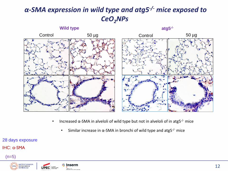
α-SMA expression in wild type and atg5-/- mice exposed to CeO2NPs
Control 50 µg Control 50 µg
• Increased α-SMA in alveloli of wild type but not in alveloli of in atg5-/- mice
• Similar increase in α-SMA in bronchi of wild type and atg5-/- mice
IHC: α-SMA
12
(n=5)

TGF-β1expression in Wild type and atg5-/- mice exposed to CeO2NPs
Control 50 µg Control 50 µg
• Expression of TGF-β1 in alveloli and bronchi in wild type mice noticed
• Atg5-/- mice are protected from CeO2NPs-induced accumulation of TGF-β1 in alveoli but no protective effect in bronchi
13
Wild type atg5-/-
28 days exposure
IHC:TGF-β1
(n=5)

14
Alveoli Bronchiole
Mice exposed to CeO2NPs
Fibrotic markers Wild type atg5-/-
Thickening/Inflammation
↑↑↑↔
TypeI collagen↑↑↑ ↔
TGFβ1↑↑↑ ↔
αSMA↑↑↑ ↔
Summary
Mice exposed to CeO2NPs
Fibrotic markers Wild type atg5-/-
Thickening/Inflammation
↑↑↑↑↑
TypeI collagen↑↑↑ ↑↑↑
TGFβ1↑ ↑
αSMA↑↑↑ ↑↑
Lack of ATG5 gene in myeloid lineage seems to be protective in alveoli but not in bronchi of atg5-/- over wild type mice
Autophagy may possibly play a dual role in CeO2NPs-induced lung fibrosis

Tél. : +33 (0)1 49 81 37 70Fax. : +33 (0)1 49 81 39 00
INSERM U955Hôpital Henri MondorFaculté de Médecine de Créteil8, rue du Général Sarrail94000 CréteilFrance
Jorge Boczkowski – Director, IMRBSophie Lanone – Head Team 4, Stéphane Tchankouo-NguetcheuMarie-Laure Franco-Montoya Philippe CaramelleArnaud TiendrebeogoBenjamin EvenShamila VibhushanEmmanuel PaulAudrey Ridoux

16

17

18

19
Future studies1. Characterization of alveolar modifications:
• Quantification of histological modification and markers like Type collagen1, alpha SMA, TGF beta1,
elastin,
• To study inflammatory infiltration by macrophages markers
2. Characterization of bronchial modifications:
• Quantification of histological modifications and expression of fibrotic markers
3. Luminex will be done on BALF samples of 24h, 1week and 90 days exposures
4. Mechanisms of pulmonary fibrosis in vitro:
• Isolation of bronchial and parenchymal fibroblasts from mice lungs (in progress)
• Exposure to NPs
• Myofibroblasts analysis: α- Sma, collagen, migration and proliferation
5. Characterization and role of autophagy: In vitro
• Expression of LC3, p62 and LAMP1 in fibroblasts treated with nanoceria
• Exposing the fibroblasts with supernatants of macrophages treated with nanoceria
• Co-culture of the fibroblasts with marcophages, exposing to nanoceria
6. Analyses of lung sections from WT and atg5-/- mice exposed to nanoceria for 90
days (sections are ready)
• HES, IHC for alphaSMA, TGF beta1, collagen Type III, IV etc, Picro Sirius Red staining for Type 1
collagen etc

20

21
Nature Reviews Cancer 12, 401-410 2012
p62 is still subject to
autophagy in cells
experiencing cellular
stress
Autophagy-defective cells and tissues, the
autophagy substrate p62 is not degraded

22
LI ET AL. 2014, ACS Nano 8 (10) 10280–10292
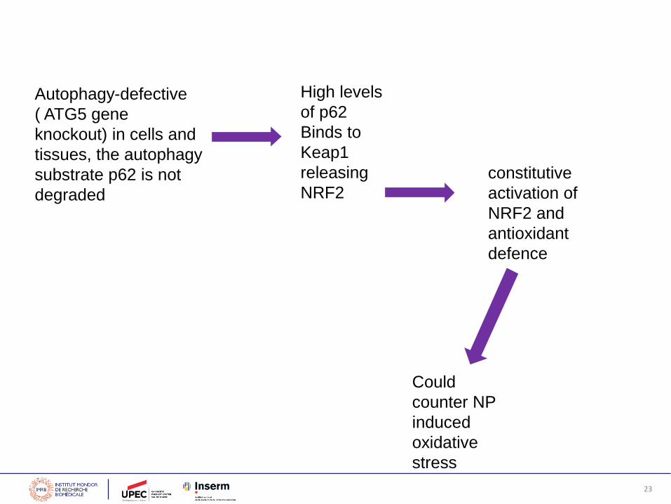
High levels
of p62
Binds to
Keap1
releasing
NRF2
constitutive
activation of
NRF2 and
antioxidant
defence
Could
counter NP
induced
oxidative
stress
Autophagy-defective
( ATG5 gene
knockout) in cells and
tissues, the autophagy
substrate p62 is not
degraded
23

Tél. : +33 (0)1 49 81 37 70Fax. : +33 (0)1 49 81 39 00
INSERM U955Hôpital Henri MondorFaculté de Médecine de Créteil8, rue du Général Sarrail94000 CréteilFrance
Jorge Boczkowski – Director, IMRBSophie Lanone – Head Team 4, Stéphane Tchankouo-NguetcheuMarie-Laure Franco-Montoya Philippe CaramelArnaud TiendrebeogoBenjamin EvenShamila VibhushanEmmanuel PaulAudrey Ridoux

25
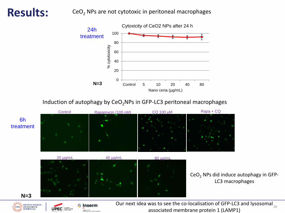
CeO2 NPs are not cytotoxic in peritoneal macrophages
0
20
40
60
80
100
Control 5 10 20 40 80
% c
yto
toxic
ity
Nano ceria (µg/mL)
Cytoxicity of CeO2 NPs after 24 h24h
treatment
N=3
CeO2 NPs did induce autophagy in GFP-LC3 macrophages
Our next idea was to see the co-localisation of GFP-LC3 and lysosomalassociated membrane protein 1 (LAMP1)
Control
20 µg/mL 40 µg/mL 80 µg/mL
CQ 100 µMRapamycin (100 nM)
6h
treatment
N=3
Induction of autophagy by CeO2NPs in GFP-LC3 peritoneal macrophages
Results:
Rapa + CQ
26

27N=1
Control
10 µg/mL
5 µg/mL
Merged (green, red)LAMP1 (Red )LC3 (green)
CeO2
NPs
6h
treatment
28
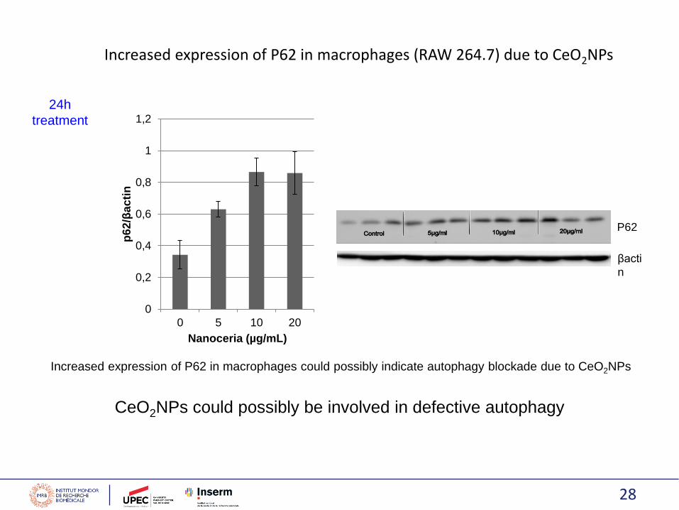
Increased expression of P62 in macrophages (RAW 264.7) due to CeO2NPs
Increased expression of P62 in macrophages could possibly indicate autophagy blockade due to CeO2NPs
P62
βacti
n
0
0,2
0,4
0,6
0,8
1
1,2
0 5 10 20
p6
2/β
ac
tin
Nanoceria (µg/mL)
24h
treatment
CeO2NPs could possibly be involved in defective autophagy

29

30
NPs interact and inihibitchaperones activity?

31
The projected human pulmonary dose for inhalation of CeO2 in diesel exhaust from engines using a CeO2fuel additive is 0.09 mg/kg body weight for 8 h (Health Effects Institute [HEI] 2001). CeO2 is insoluble particle,and studies have shown that the clearance of CeO2 from the lung may take 20 years or more (Pairon et al.1994).As a diesel exhaust product, it is likely that the potential exposure (occupational or environmental) to CeO2 iscontinuous and the lung burden is cumulative. Assuming a person has been exposed to the projected dosefor 40 years with 8 h working day, the total lung burden of CeO2 will be 936 mg/kg (0.09 mg/kg.d 5 d/week52 week/year 40 years = 936 mg/kg).
Usually, conversion from rodents to humans includes a safety factor of 10-fold.
Therefore, to assess the potential toxicological consequence of CeO2 NPs we used 50µg well with the range .

32