Lung function in patients with tracheobronchopathia ... · Recurrent maxillary sinusitis or ozaena...
4
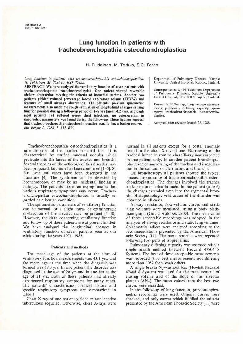
Eur Resplr J 1988, 1' 632-635 Lung function in patients with tracheobronchopathia osteochondroplastica H. Tukiainen, M. Tor kko, E.O. Terho Lung function in patients with tracheobronchopathia osteochondroplastica. H. Tukiainen, M. Torkko, E.O. Terho. Department of Pulmonary Diseases, Kuopio University Central Hospital, Kuopio, Finland ABSTRACT: We have analysed the ventilatory function of seven patients wi th tracheobronchopathia osteochondroplastica. One patient showed reversible airflow obstruction meet ing the criteria of bronchial asthma. Another two patients yiel ded reduced percentage forced expiratory volume ( FEV%) and features of small airways obstruction. The pat ients' previous spirometric measurements also made the rough estimation of longitudinal changes in lung function possi bl e during a follow-up period of 1- 8 yrs (mean 4.2 yrs). Although most patients had suffered severe chest infections, no deteriorat ion in spirometric parameters was found during the follo w-up. These findings suggest that tracheobronchopathia osteochondroplastica usuall y has a benign course. Eur Respir J., 1988, I, 632- 635. Cor respondence: Dr. H. Tukiainen, Department of Pulmonary Diseases, Kuopio University Central Hospital, SF-71800 Siilinjarvi, Finland. Keywords: Follow-up; lung volume measure- ments; pulmonary diffusing capacity; spiro- metry; tracheobronchopathia osteochondro· plastica. Accepted aft.er revision March 22, 1988. Tracheobronchopathia osteochondroplastica is a rare disorder of the tracheobronchial tree. It is characterized by ossified mucosal nodules which protrude into the lumen of the trachea and bronchi. Several theories on the aetiology of this disorder have been proposed, but none has been confirmed [1 - 3]. So far, over 300 cases have been described in the literature [4]. The syndrome can be detected by bronchoscopy, or may be an incidental finding at autopsy. The patients are often asymptomatic, but various respiratory symptoms may occur. Tracheo- bronchopathia osteochondroplastica is usually re- garded as a benign condition. The spirometric parameters of ventilatory function can be normal, or a slight intra- or extrathoracic obstruction of the airways may be present [4- 10]. However, the data concerning ventilatory function and follow-up of these patients are at present limited. We have analysed the longitudinal changes in ventilatory function of seven patients seen at our clinic during the years 1971 - 1985. P atients and methods The mean age of the patients at the time of ventilatory function measurements was 43.1 yrs, and the mean age at the time when the diagnosis was formed was 39.3 yrs. In one patient the disorder was diagnosed at the age of 20 yrs and in another at the age of 21 yrs. Both of these patients had already experienced respiratory symptoms for many years. The patients' characteristics, medical history and specific respiratory symptoms are summarized in table l. Chest X-ray of one patient yielded minor inactive tuberculous sequelae. Otherwise, chest X-rays were normal in all patients except for a costal anomaly found in the chest X-ray of one. Narrowing of the tracheal lumen in routine chest X-ray was suspected in one patient only. In another patient bronchogra- phy revealed narr owing of the trachea and irregulari- ties in the contour of the trachea and bronchi On bronchoscopy all patients showed the typical mucosal appearance of tracheobronchopathia osteo- chondroplastica. The changes involved the trachea and/or main or lobar bronchi. In one patient (case 6) the changes extended even into the segmental bron- chi. Histopathologic verification of the disorder was obtained in a ll cases. Airway resistance, flow-volume curves and static lung volumes were measured, using a body pleth- ysmograph (Gould Autobox 2800). The mean value of three acceptable recordings was adopted in the analyses of airway resistance and static lung volumes. Spirometric indices were analysed according to the recommendations presented by the American Thor- acic Society [ ll]. The measurements were repeated following two puffs of isoprenaline. Pulmonary diffusing capacity was assessed with a single breath method (Hewlett Packard 47804 S System). The best of three acceptable measurements was recorded (two best measurements not differing more than I 0% from each ot her). A single breath N 2 -washout test (Hewlett Packard 47804 S System) was used for the measurement of closing volume and of the slope of the alveolar plateau (LlN 2 ). The mean values from the best two curves were recorded. In the fo ll ow-up of lung function, previ ous spiro- metric recordings were used. Original curves were checked, and only curves which fulfilled the criteria presented by the American Thoracic Society (I I] were
-
Upload
nguyenphuc -
Category
Documents
-
view
218 -
download
0
Transcript of Lung function in patients with tracheobronchopathia ... · Recurrent maxillary sinusitis or ozaena...
Eur Resplr J 1988, 1' 632-635
Lung function in patients with tracheobronchopathia osteochondroplastica
H. Tukiainen, M. Torkko, E.O. Terho
Lung function in patients with tracheobronchopathia osteochondroplastica. H. Tukiainen, M. Torkko, E.O. Terho.
Department of Pulmonary Diseases, Kuopio University Central Hospital, Kuopio, Finland
ABSTRACT: We have analysed the ventilatory function of seven patients with tracheobronchopathia osteochondroplastica. One patient showed reversible airflow obstruction meeting the cri teria of bronchial asthma. Another two patients yielded reduced percentage forced expiratory volume (FEV%) and features of small airways obstruction. The patients' previous spirometric measurements also made the rough estimation of longitudinal changes in lung function possible during a follow-up period of 1- 8 yrs (mean 4.2 yrs). Although most patients had suffered severe chest infections, no deterioration in spirometric parameters was found during the follow-up. These findings suggest that tracheobronchopathia osteochondroplastica usually has a benign course. Eur Respir J., 1988, I, 632- 635.
Correspondence: Dr. H. Tukiainen, Department of Pulmonary Diseases, Kuopio University Central Hospital, SF-71800 Siilinjarvi, Finland.
Keywords: Follow-up; lung volume measurements; pulmonary diffusing capacity; spirometry; tracheobronchopathia osteochondro· plastica.
Accepted aft.er revision March 22, 1988.
Tracheobronchopathia osteochondroplastica is a rare disorder of the tracheobronchial tree. I t is characterized by ossified mucosal nodules which protrude into the lumen of the trachea and bronchi. Several theories on the aetiology of this disorder have been proposed, but none has been confirmed [1 - 3]. So far, over 300 cases have been described in the literature [4]. The syndrome can be detected by bronchoscopy, or may be an incidental finding at autopsy. The patients are often asymptomatic, but various respiratory symptoms may occur. Tracheobronchopathia osteochondroplastica is usually regarded as a benign condition.
The spirometric parameters of ventilatory function can be normal, or a slight intra- or extrathoracic obstruction of the airways may be present [4- 10]. However, the data concerning ventilatory function and follow-up of these patients are at present limited. We have analysed the longitudinal changes in ventilatory function of seven patients seen at our clinic during the years 1971- 1985.
P atients and methods
The mean age of the patients at the time of ventilatory function measurements was 43.1 yrs, and the mean age at the time when the diagnosis was formed was 39.3 yrs. In one patient the disorder was diagnosed at the age of 20 yrs and in another at the age of 21 yrs. Both of these patients had already experienced respiratory symptoms for many years. The patients' characteristics, medical history and specific respiratory symptoms are summarized in table l.
Chest X-ray of one patient yielded minor inactive tuberculous sequelae. Otherwise, chest X-rays were
normal in all patients except for a costal anomaly found in the chest X-ray of one. Narrowing of the tracheal lumen in routine chest X-ray was suspected in one patient only. In another patient bronchography revealed narrowing of the trachea and irregularities in the contour of the trachea and bronchi
On bronchoscopy all patients showed the typical mucosal appearance of tracheobronchopathia osteochondroplastica. The changes involved the trachea and/or main or lobar bronchi. In one patient (case 6) the changes extended even into the segmental bronchi. Histopathologic verification of the disorder was obtained in all cases.
Airway resistance, flow-volume curves and static lung volumes were measured, using a body plethysmograph (Gould Autobox 2800). The mean value of three acceptable recordings was adopted in the analyses of airway resistance and static lung volumes. Spirometric indices were analysed according to the recommendations presented by the American Thoracic Society [ ll]. The measurements were repeated following two puffs of isoprenaline.
Pulmonary diffusing capacity was assessed with a single breath method (Hewlett Packard 47804 S System). The best of three acceptable measurements was recorded (two best measurements not differing more than I 0% from each other).
A single breath N 2-washout test (Hewlett Packard 47804 S System) was used for the measurement of closing volume and of the slope of the alveolar plateau (LlN2). The mean values from the best two curves were recorded.
In the fo llow-up of lung function, previous spirometric recordings were used. Original curves were checked, and only curves which fulfilled the criteria presented by the American Thoracic Society (I I] were

LUNG FUNCTION IN TRACHEOBRONCHOPATHIA OSTEOCHONDROPLAST ICA
Table 1. - Clinical data of seven patients with tracheobronchopathia osteochondroplastica
Male/female Mean age, yrs (at the time of the srudy) Mean age, yrs (at the time of making the diagnosis) Smoker Ex-smoker Non-smoker Recurrent maxillary sinusitis or ozaena Chronic rhinitis Recurrent cough or expectoration Haemoptysis Pneumonia Dyspnoea Positive skin prick tests* Bronchial hyperreactivity (methacholine inhalation challenge) Blood eosinophilia (>5%) Histologic verification
4(3 43.1 (range 23- 59) 39.3 (range20-58)
0 1 6 4 (3 operated) 6 6 3 5 4 2/5 0/5
0 7
*At least two positive reactions (wheal diameter 8x3 mm) to common allergens.
accepted. The follow-up period ranged from I .0-8.5 yrs (mean 4.2 yrs). The apparatus used in the first and final spirometry was not necessari ly the same.
Results
The lung function measurements yielded quite normal results (table 2). One patient (case 6) had significant airAow obstruction which involved both large and small ::tirways. On bronchoscopy, this patient had extensive changes or lracheobronchopathia osteochondroplaslica extending as far as the segmental bronchi. This patient had, however, also shown reversible airways obstruction on several occasions, and so fulfilled the criteria for bronchial asthma [12]. Two other patients (cases 2 and 5) also displayed a reduced percentage forced expiratory volume (FEV%) and features of small airways obstruction. However, in these patients the airway resistances were within normal limits as well as the indices of inspiratory flow-volume curves. In other patients there was no evidence of significant impairment of ventilatory function. The inspiratory flowvolume curves were also normal, thus excluding any significant extrathoracic airways obstruction. Following isoprenaline inhalation. a bronchodilating effect was observed only in the patient who had previously shown reversible airways obstruction. Pulmonary diffusing capacity was normal in all patients.
The retrospective comparison of spirometric recordtngs (forced expiratory volume in one second (FEY 1 ), forced vital capacity (FYC) and F EY%) revealed no deterioration during the follow-up (table 3), except for the patient with reversible airways obstruction. Her spirometric parameters at the final assessment were lower than they had been 8.5 yrs previously.
('(! g Vi ('(!
a. e "0 c 0 .c () 0 (!)
Vi 0 ('(!
:c n; Cl. 0
..c g e
..0 g
..c
~ ..c ·~ VI
c .£! n; Cl. c ~ (!) VI
.S c
.Q ts c :::> -
...-......-......-... ........... ,-... ........... .....tO\('I"')V')f""""(.-t
00 0\ "'" !"- \0 00 '-" '-"' ....-4 .._, .._, ...._.. ._,
\0 r----or--o ,.... ll"l"'"t---<"> qr-;<X!q"<<;q"<<;q~~~...t~r-: ll"l\0.-<lf"l<"lt--"'"NNON<"l,....O
\0 0\ ....
* -: * f{l"7 -: A... t{l
* ~ ~ "':"~: :!!:.:!!:.:!!:.:!!:.!.~-~~~~~
633

634 H. TUKIAINEN, M. TORKKO, E.O. TERHO
Table 3. _ Retrospective comparison of lung function (FEV1
and FVC) of seven patients with tracheobronchopathia osteochondroplastica.
First assessment FEY
1 l FYC
Casei 4.19 5.84 Case2 2.95 4.59 Case3 4.89 5.36 Casc4 3 .75 4.50 CaseS 3.30 5.30 Case6 2.65 3.85 Case 7 3.25 3.50
Mean 3.57 4.71 ±sD ±JJ.77 ±0.85
Second assessment FEV1 l FYC
4.67 3.40 5.00 3.77 3.16 1.73 3.31
3.58 ±1.08
6.30 5.07 5.45 4.67 4.89 2.62 4.08
4.73 ±1.15
Time interval yrs
0.8 2.3 3.6 5.3 0.8 8.4 8.4
4.2
FEY1
: forced expiratory volwne in one second; FVC: forced vital capacity.
Discussion
The present results are in accordan~ with the v~ew that progressive deterioration of venhl~tory functwn is not typical for trachcobronchopathta oste~cho?droplastica. Previously, a significant worsenmg m ventilatory function during an 8-month follow-up of one patient has been reported by ALROY et al. [I 0]._ In contrast, NIEROP er al. [9] found no progresstve deterioration in lung function of one patient over an 8 yr follow-up. The mucosal appearance check~~ by bronchoscopy also remained unchanged. In addttton, no obvious change was observed in bronchial pathology of another patient whose bronchoscopy was repeated with a I yr interval _[13). ROSE et al. [1_4) h~ve also reported a case in whtch the endoscoptc vtew remained unchanged over a 6 yr follow-up.
Chest infections seem to be common in these patients [6- 8, 15], and recurrent pneumonia was characteristic in our subjects. However, the occurrence of respiratory infections did not seem to lead to progressive deterioration of lung function. In t~is respect, tracheobronchopathia ~steochondroplastt~a seems to resemble the producttve type of chrome bronchitis in its benign course [16].
One patient in our study showed reversible ~irAow obstruction. At the moment of control, the sptrometric values of this patient may have been low by chance, due to spontaneously changing obstruction of simultaneous asthma. The study group was too small for further conclusions about the relationships between tracheobronchopathia osteochondroplastica and asthma or reversible bronchial obstruction. Features of minor expiratory obstruction were also observed in another two patients. This finding is consistent with previous observations that slight expiratory and/or inspiratory obstruction may occur in some patients. ll could be expected that the bony nodules of the tracheal and bronchial mucosa might cause airflow limitation. On the other hand, it may be assumed that the bronchial tree with bony formations is rather sti lT thus opposing tendencies for an intrathoracic ;irway collapse. The normal diffusing
capacity in our study excludes any significant abnormality of the gas exchanging surfaces of the. lu~g.
Tracheobronchopathia osteochondroplast1ca tS not usually diagnosed before the age of 50 yrs. In our study there were two young patients. There are also some other reports about the condition in young people ( 13, 15, 17]. This also favours the conc~pt that the disorder has a benign course and at least m some patients it may have an early, perhaps even congeni-tal, onset. . . .
There may exist geographical drfferences m ~he occurrence of this disorder. For example, accordmg to the British literature, it seems to be extremely rare [13). [n Finland, in addition to our material, HARMA et al. [15] have presented thirty pat~ents. The~e regional differences may. ~ due to d_tfferences 111
diagnostic practice, but 1t IS also poss1ble that the differences in prevalence are real and due to the different genetic background of populations.
Our analysis of follow-up data _was bas~d _on previous spirometric reco~dings. Admttted~y this kmd of analysis may be subJect to severe btas due to possible changes in spirometric methodology. In fact, the spirometric apparatus used in this study was not necessarily the same in the first and ~he final assessment. In addition, the number of pattcnts was rather small. Taking into account these limitations, an approximate estimation ~f changes in s~irometric parameters is, however, posstble. No essenttal ch~nge was found , which, together with the few prev10us findings, speaks in favour of a benign. nature for tracheobronchopathia osteochondroplasttca.
References
1. Aschoff L. - Obcr trachcopathia oslcochondroplastica. Verh Dtsch Ges Pathol, 1910. 14, 125- 126. 2. Dalgaard JB. - Trachcopathia chondro-osteoplastica: A ~se elucidating the problems concerning development and OSSificatiOn of clastic cartilage. Acta Pathol Microbial Scand, 1947, 24, 118- 134. . 3. Saku1a A. - Tracheobronchopathia osteoplastica. Its relationship to primary tracheobronchial amyloidosis. Thomx, 1968, 23, 105- 110.
LUNG FUNCTION IN TRACHEOBRONCHOPATH IA OSTEOCHONDROPLASTICA 635
4. Nagy I, Fricke G , Ouch J, Weis E. - Tracheobroncbopathia osteochondroplastica - Computertomografic als sinnvollc Ergiinzung cndoskopischer und radiologischer Diagnostik. Prax Klin Pneuma/, 1985, 39. 176-179. 5. Bergeron D, Cormier Y, Desmeules M. - Tracheobronchopathia osteochondroplastica. Am Rev Respir Dis, 1976, 114,803- 806. 6. Castclla J, Puzo C, Cornudella R, Curell R. Tarres J. - Tracheobronchopathia osteochondroplastica. Respiration, 1981, 42, 129- 134. 7. Lundgreo R, Stjernberg NL. - Tracheobronchopathia osteochondroplastica. A clinical bronchoscopic and spirometric study. Chest, 1980, 80, 706- 709. 8. Primer G. - Tracheobronchopathia osteochondroplastica. Prax Klin Pneuma/, 1979, 33, 1060-1063. 9. Van Nierop MA, Wagenaar SS, van den Bosch JM, Westermann CJ. - Tracheobronchopathia osteochondroplastica. Report of four cases. Eur J Respir Dis, 1983, 64, 129-133. 10. Alroy GG, Lichtig C, Kaftori JK. - Tracheobronchopathia osteochondroplastica: end stage of primary amyloidosis? Chest, 1972, 61' 465- 468. 11. American Thoracic Society. ATS statement. - Snowbird workshop on standardization of spirometry. Am Rev Respir Dis, 1979, 119, 831 - 838. 12. Meneely GR, Renzetti AD, Steele JD, Wyatt JP, Harris HW. - Chronic bronchitis, asthma and pulmonary emphysema. Am Rev Respir Dis, 1962, 85, 762- 768. 13. Clee MD, Anderson JM, Johnston RN. - Clinical aspects of trachcobronchopathia osteochondroplastica. Br J Dis Chest, 1983, 77, 308-314. 14. Rose Y, Roucou Y, Roujeau J, Fromentin JC.- Mctaplasie, ossifiante de la muqueuse tracheo-bronchique et ozene. Rev Fr Mal Respir, 1974, 2, 637- 645.
15. Hiirmii RA, Suurkari S. - Tracheopathia chondro-osteoplastica. A clinical study of thirty cases. Acta Otolaryngo/, 1977, 84, 118- 123. 16. Fletcher C, Peto R, Tinker C, Speizer FE. - In: The natural history of chronic bronchitis and emphysema. An eight-year study of early chronic obstructive lung disease in working men in London. Oxford University Press, Oxford, 1976. 17. Jonard P, Mairesse M. - Tracheo-bronchopathie osteoplastique. Revue de la littcmturc ::1 propos d'une obs.:rvation chez un sujet jeune. Lille Ml!d, 1980, 25, 519- 521. 18. Viljanen A. - Reference values for spirom<:lric, pulmonary diffusing capacity and body plethysmographic studies. Smll(/ J Clin Invest, 1982, 42 (Suppl. 159). 19. Buist AS, Ghezzo H, Anthonisen NR, Cherniack RM, Ducie S. Macklem PT. Manfreda J, Martin RR, McCarthy D, Ross BB.Relationship between the single-breath N2 test and age, sex, and smoking habit in three North American cities. Am Rev Respir Dis, 1979, 120, 305- 318.
RESUME: Nous avons etudie la fonction ventilatoire de 7 patients atteints de trachcobronchopathie osteochondroplastique. Chez l'un d' entre eux, on a releve une obstruction reversible des debits aeriens rencontrant les criteres de l'asthme bronchique. Deux autres ratients avuicnt une diminution du VEMS et des signes d'obstruction des pctitcs voies aeriennes. Les mesures spirometriques unterieurcs. realisees chcz ces patients, ont permis d'etudier les modifications longitudinales de la fonction pulmonaire au cours d'un follow-up d'un a I!Uit ans (moycnne 4.2). Quoique la plupart des patients aient eu des infections bronchiques scvcres. ron n'a pas trouve de deti~rioration des parametrcs spiromettiques pendant le suivi. Ces constatations suggerent que la tracheobronchopathie ostcochondroplastique a habituellement un decours benin.