Lumbar Spine - Mechanical Diagnosis Therapy
178
THE LUMBAR SPINE MECHANICAL DIAGNOSIS AND THERAPY R. A. McKenzie, O.B.E., F.e.S.p., F.N.Z.S.P. (HaN), DIP. M.T. SPINAL PUBLICATIONS 1981 ]3",'8 ..)HI.".;; 7/.. JJ 16 I
-
Upload
jimitkapadia -
Category
Documents
-
view
5.617 -
download
1
Transcript of Lumbar Spine - Mechanical Diagnosis Therapy

THE LUMBAR SPINE
MECHANICAL DIAGNOSIS ANDTHERAPY
R. A. McKenzie, O.B.E., F.e.S.p., F.N.Z.S.P. (HaN), DIP. M.T.
SPINAL PUBLICATIONS1981
]3",'8 U"i~";/fll £tl,~..)HI.".;; 7/.. JJ 16I
physio4all
Rectangle
physio4all
Rectangle
SPINAL PUBLICATIONS NEW ZEALAND LIMITEDPO. BOX 93, WAlKANAE, WELLINGTON, NEW ZEALAND
FIRST PRINTED 1981REPRINTED 1981REPRINTED 1982
REPRINTED 1983 (TWICE)REPRINTED 1984REPRINTED 1985REPRINTED 1986REPRINTED 1987REPRINTED 1989REPRINTED 1990REPRINTED 1991REPRINTED 1992REPRINTED 1993REPRINTED 1995REPRINTED 1997REPRINTED 1998
© R. A. McKENZIE, 1981
ISBN 0 473 00064 4
No part of this publication may be reproduced or transmitted in any form orby any means electronic or mechanical, including photocopy, recording or anyinformation storage retrieval system without permission in writing from thepublisher.

RD
To my wifeJoy.
physio4all
Rectangle
physio4all
Rectangle

AcknowledgmentsIt is no coincidence that many of the statements in this book have a familiarityabout them which traces back to the work of Dr James Cyriax. His influence haspervaded all mechanical spinal therapy, and his place as the father oforthopaedic medicine is unchallenged. I gratefully acknowledge the inspirationobtained from Cyriax which enabled me to develop some of his ideas, notnecessarily in his fashion but, I am sure, in a manner of which he will approve.
My warmest thanks must go to Dr John Ebbetts who, early in my career,arranged for the international exposure of my treatment method for acutelumbar scoliosis.
I am most grateful to the doctors of Wellington, New Zealand, who havesupported me over the past twenty-eight years. Without their confidence andencouragement the development of these concepts and treatment methodswould not have been possible.
I thank Dr Arthur White of San Francisco for contributing the Foreword tothis book and for giving me the opportunity to demonstrate to him, theeffectiveness of the methods described within.
To Dr Edward Miller, Professor of Orthopaedics at University of CincinattiMedical Center, to the physical therapists, physicians and orthopaedic surgeonsof the Kaiser Permanente Medical Centres of California, I am greatly indebtedfor support and backing that has led to wide acceptance of my ideas throughoutthe United States.
My former assistant Glen Pendergrast must be held responsible for meputting pen to paper, as he casually enquired who would know my workproperly if I were run over by a bus. I appreciate his stimulation and support.
Finally, I sincerely thank my present assistant Paula Van Wijmen for thecompilation of the information presented in this book. She is one of the veryfew people who, besides myself, fully understand these concepts. Without herknowledge, loyalty and perserverance this book would still be a notebook.
R. McKENZIE

Foreword
I am very pleased to present a foreword and commentary to this book by MrMcKenzie. It is a refreshing publication which I favour for several reasons.
First, the underlying thesis for this book is that the great majority ofdisabilities of the lumbar spine are mechanical, and thus can be treated in amechanical manner. More important than that, once the principles areunderstood most of the mechanical treatments can be accomplished by thepatient himself. In fact, the very goal of treatment as proposed by Mr McKenzieis to place the patient on a self care programme so that he will not require thecare of therapists or medical clinicians. The mechanical abnormalities andpostural variations are incurable, but at least if appropriate principles areunderstood, the majority of disability related to these can be avoided.
The second reason I favour this publication is its unpretentious flavour. Theauthor recognises that physicians and surgeons seldom are interested or adept atthe mechanical approach to physical disability. This is an area where therapiststruly are adept, and by experience and training remain interested in the subject.On the other hand, there is very little scientific basis for our currentunderstanding of the principles of care of the spine. Thus, to place a treatmentprogramme on a level of scientific security which should be the ideal of medicaltreatment, is inappropriate. The author takes the position that the principlesadvocated in this text work on the majority of people, and the rationale for theirsuccess can only be presumed on a logical progression of our current level ofknowledge. The programme is safe, inexpensive and indeed, may bescientifically accurate. Certainly the concepts proposed are as reliable and secureas any other conceptual framework used today in our understanding of chronicback disability.
I suppose the reason I really enjoyed the book most is that the author hastaken a new tack in the analysis and treatment of the problem. He has shown anappropriate disrespect for "accepted principles". Only by the constantquestioning of previously accepted truths will progress be made toward morecomplete understanding. Certainly many of the accepted principles of the past,such as the avoidance of lumbar extension, have not diminished the problem ofchronic back disability which currently fills the offices of clinicians around theworld. Lets try this new approach which has as rational a base as any of ourtreatments for clinical syndromes. It certainly is more rational than the use ofvarious modalities such as ultrasound and various pharmacologic agents whichtry to solve the problem of local mechanical problems by systemic alterations innervous system response. Anything that costs less, is not health threatening,
IX

x Foreword
and fixes most of the problems deserves a respected place in the medicaltreatment programmes. I am pleased that Mr McKenzie has chosen to staywithin the mainstream of medicine and allied health professionals rather than toescape the scrutiny of cautious peers which has allowed many a flimsy fad tocome and go. I expect the programme outlined by Mr McKenzie to grow insignificance and acceptance as more people try it and benefit from it.
Vert Mooney, M.D.
Robin McKenzie has had a great influence on my treatment of low back pain. Itwas quite by accident that he and I lectured at the same low back pain seminarseveral years ago. Even though we had differing philosophies in regard to theetiology and treatment of low back pain, there was so much in common betweenus that I began a thorough scientific study of his techniques. I have found thattwenty percent of my patient population are much more responsive to hismethods than to any of the methods I have used in the past. My patientpopulation is more of a chronic multi-operated back population and hasgenerally speaking, resisted all previous therapies. In populations where thepatients are seen early in their disease with acute injuries, I feel that fifty toeighty percent would be rapidly relieved by the techniques described in thisbook.
It is all too easy for those of us in the clinical profession to shun newtechniques which do not fit our model of how medicine should be practised. Iwas fortunate enough to be introduced to Robin McKenzie during a period ofprofessional growth in which I felt a need for a better understanding of thediagnosis and treatment of low back pain. The understanding of the McKenzietechniques has allowed me to improve the diagnosis and treatment of low backpain in my orthopaedic practice.
Information contained in the pages of this book works. I cannot substantiate,nor do I believe anyone can substantiate the exact reasons why it works. RobinMcKenzie has some very appealing explanations for why his methods work. Ionly know for certain that they do work in a very busy orthopaedic and physicaltherapy spine centre in San Francisco. . I
I believe that this volume is one of the more important contributions to theliterature on the treatment of low back pain and we will continue attempting toscientifically prove how well the McKenzie method works and the reason for itsworking. In addition to all of the above considerations, this book is enjoyable, asis the author.
Arthur H. White, M.D.San Francisco.

ContentsAcknowledgments viiForeword ixIntroduction xvii
CHAPTER 1 - Definition and Selection 1Epidemiology 1Self-limitation 2Definition and selection 2
CHAPTER 2 - Predisposing and Precipitating Factors 4Predisposing factors 4
Sitting posture 4Loss of extension 5Frequency of flexion 6
Precipitating factors 7Movements 7~~ 7
CHAPTER 3 - The Cause of Pain 9The nociceptive receptor system 9
Mechanism of pain production 10Chemical cause of pain 10Mechanical cause of pain 10Trauma as a cause of pain 11
Causes of mechanical deformation 12Postural stresses 12Abnormal forces 13A popular misconception 13
CHAPTER 4 - The Intervertebral Disc 15Structure 15Pressure within the disc 16Nuclear movement 16Tangential stress 17Disc damage and repair 18The disc and pain 20The centralisation phenomenon 22Differentiation between disc degeneration and frank protrusion 22

xii Contents
CHAPTER 5 - Diagnosis 24The therapist's responsibility 24Diagnostic difficulties 24The three syndromes 25
The postural syndrome 25The dysfunction syndrome 25The derangement syndrome 25
CHAPTER 6 - The History 27Interrogation 27
CHAPTER 7 - The ExaminationPosture sittingPosture standing
Reduced or accentuated lordosisLateral shiftLeg length discrepancy
Examination of movementFlexionDeviation in flexionExtensionSide gliding
Movements in relation to painRepeated movements
Flexion in standing compared with flexion in lyingExtension in standing compared with extension in lying
The test movementsFlexion in standingExtension in standingSide gliding in standingFlexion in lyingExtension in lying
Other examination proceduresConclusions
Postural correctionThe extension principleThe flexion principle
CHAPTER 8 - TechniqueThe procedures and their effectsTable of procedures
Procedure 1 - Lying prone2 - Lying prone in extension
3434353535363636373838394041424242434445454647474747
495153
5456

Contents
3 - Extension in lying4 - Extension in lying with belt fixation5 - Sustained extension6 - Extension in standing7 - Extension mobilisation8 - Extension manipulation9 - Rotation mobilisation in extension
10 - Rotation manipulation in extension11 - Sustained rotation/mobilisation in flexion12 - Rotation manipulation in flexion13 - Flexion in lying14 - Flexion in standing15 - Flexion in step standing16 - Correction of lateral shift17 - Self-correction of lateral shift
CHAPTER 9 - The Postural SyndromeDefinition
HistoryExaminationClinical examplePostures involved
Treatment of the postural syndromeCorrection of the sitting postureTo obtain the correct sitting postureTo maintain the correct sitting postureThe lumbar roll as sitting supportCorrection of the standing postureCorrection of the lying postureThe lumbar roll as lying supportConclusionConsequences of postural neglect
Typical treatment progression - the postural syndrome
CHAPTER 10 - The DYsfunction SyndromeDefinition
HistoryExaminationThe test movementsClinical example
Treatment of the dysfunction syndromeTreatment of extension dysfunction
ExercisesSpecial techniques
Treatment of flexion dysfunction
xiii
575960626365666769707274757678
8181818181838585868788909191929393
95959697989999
102102103104
xiv Contents
Recovery of pure flexion loss 104Exercises 104Special techniques 105Treatment of flexion with deviation 105
Treatment of side gliding dysfunction - correction of secondarylateral shift 106
Typical treatment progression - the dysfunction syndrome 107
CHAPTER 11 - The Derangement Syndrome 109Definition 110
Acute lumbar kyphosis 111Acute lumbar scoliosis 111History 112Clinical examples 112Examination 114The test movements 115Clinical examples 118Straight-leg-raising 118
Treatment of the derangement syndrome 119Table of derangements 120
CHAPTER 12 - The Derangements and Their Treatment 122Derangement One 122
Treatment 122Reduction of Derangement 122Maintenance of reduction 123Recovery of full flexion 127Prevention of recurrence 128
Typical treatment progression - Derangement One 129
Derangement Two 131Treatment 134
Derangement Three 137Treatment 137
Derangement Four 139Treatment 144
Derangement Five 146Intermittent sciatica 146Treatment 147
Derangement Six 148Constant sciatica 148Treatment 149Stretching of adherent nerve root 149

Contents
Derangement SevenTreatment
CHAPTER 13 - ProphylaxisProlonged sittingActivities involving prolonged stoopingLiftingRecurrence
CHAPTER 14 - Contra-IndicationsBedrestSupportsSurgery
References
xv
151152
153153153153154
159160160160
163
IntroductionThe treatment of low back pain remains as controversial today as it was fiftyyears ago. Over the years the medical profession used a wide range oftreatments, such as heat or cold, rest or exercise, flexion or extension,mobilisation or immobilisation, manipulation or traction. Nearly always drugswere prescribed, even when the disturbance proved purely mechanical in origin.Amazingly, most of the patients recovered, very often in spite of treatmentrather than because of it.
It is not surprising that the first half of this century found osteopaths andchiropractors in the forefront of those dispensing the most satisfactorytreatments, at least as far as patients were concerned. Mechanical disturbancesof the articular system are best treated by mechanical means. It is for this reasonthat unorthodox operators were more successful than the medical profession,because only few medical practitioners were capable of applying mechanicaltreatment.
We have to thank osteopathy and chiropractic for providing the stimulusrequired to move medicine in the right direction. Beginning with JamesMennell and James Cyriax that momentum is gathering strength and, atpresent, understanding of the mechanics of low back pain is emanating fromclinics and laboratories throughout the world.
There has been a trend in manipulative therapy to meekly follow in thefootsteps of osteopaths and chiropractors. This is unlikely to produce significantand rapid improvement in spinal therapy. Many of the procedures andtechniques presented by the early manipulators for treatment of mechanicalspinal pain are valid, but only when applied to patients with certain well definedsigns and symptoms. Manipulative procedures, already suspect in many cases,have a definite but limited application, and it is now clear which patients benefitfrom manipulation and which do not. Furthermore, it is likely that as ourunderstanding improves, patients will require less rather than moremanipulative procedures.
There has been a tendency among exponents of mobilisation andmanipulation to overclaim and exaggerate the benefits to be derived from itsadministration. This trend must cease if we wish scientific development ofmechanical therapy to proceed.
Most treatments appear to be directed at pain relief for the present episode.Episodic relief by therapy of any kind makes the patient dependent on thattherapy and thereafter he will seek a quick answer for what is essentially a lifelong problem. Whenever his back pain recurs he must attend a physician,manipulative therapist, chiropractor or osteopath. I believe that treatment
xvii
xviii The Lumbar Spine
dependency is undesirable and should be avoided where possible. Therefore, inaddition to whatever treatment is necessary for present symptoms the patientshould be taught to become self-reliant and independent of therapists in themanagement offuture low back pain.
It is often thought that all patients presenting for treatment must have apathological condition. I suggest to those peering down a microscope to find apathological cause for mechanical joint pain, and to those seeking to provide awonder drug for low back pain that, in its simplest form, low back pain starts forthe same reason as pain arises in the forefinger when it is bent backwards farenough to stimulate the free nerve endings of periarticular structures. Nopathology needs to exist, and no chemical treatment will cure this form ofmechanical pain.
In this book I hope to explain clearly that there is a time when the low backshould be extended and a time when it should be flexed; that there arecircumstances when both procedures should be applied; and, perhaps mostimportant of all, that it is now possible to identify in advance those patients whowill respond to manipulation.
I have developed a simple classification: mechanical pain may develop frompostural stresses; it may be the result of joint derangement or it may be causedby dysfunction. In patients with pure postural pain no pathology is present, andall that is required in their treatment is adequate explanation of the cause of painand instruction in self-treatment by postural correction. In the majority ofpatients with joint derangement and dysfunction the pathological condition canbe successfully influenced by mechanical therapy using the patient's ownmovements. A method of self-treatment and self-manipulation of the articularsystem is now available to patients who have enough understanding to learnand carry out the required procedures. In this book I intend to explain why selftreatment procedures are preferable to procedures that must be applied by atherapist.
Successful application of the self-treatment concept depends on the correctselection of patients. Throughout this book the assumption has been made thatpatients are referred for therapy by their doctor. Indeed, we require our medicalpractitioners to provide us with the appropriate patients - that is, patients whodo not suffer from serious pathology (mechanical or other) and who have hadall but mechanical causes of low back pain excluded. I hope that this book willenable the clinician to select from the people with low back pain those who mayrespond to mechanical treatment, and to place them in the three relativelysimple categories. Once the appropriate syndrome is exposed the application ofsimple and inexpensive procedures, usually through the patient's own efforts,may ,effect cure. I believe that we can now identify those patients who maybecome self-reliant (seventy percent) and those who will always require someassistance from a therapist (thirty percent).
It is not necessary, indeed it is impossible, for the majority of doctors to keepabreast with modern developments in mechanical therapy. Consequently, notmany doctors have the knowledge to prescribe specific treatments for patients
Introduction XIX
with low back pain. As it is the responsibility of the therapist to maintain hisknowledge and techniques in the field of mechanical therapy at the highestpossible level, he is best able to define which type of treatment is most suitablefor a particular mechanical problem. Thus, the choice of treatment lies with thetherapist.
With this book I present a new concept of diagnosis for the whole musculoskeletal system. The procedures I developed for the lumbar spine to arrive atappropriate conclusions regarding diagnosis and treatment, may also be appliedsuccessfully to the thoracic and cervical spine, and indeed to all peripheral jointsand their surrounding soft tissues. Irrespective of the presenting pathology, theprinciples of diagnosis and treatment remain the same.
Having developed a method for treating low back' pain that appears to workvery well, it is incumbent on me to offer an explanation suggesting why theseprocedures are effective. I have hypothesised using mans present knowledge ofthe structure function and behaviour of the lumbar intervertebral discs. Noproof absolute exists to substantiate this hypothesis but I believe it to besomewhere near the truth. Only time and much research will reveal the truenature of the mechanism responsible for the production of recurrent low backpain, but the influence of these procedures on that mechanism will be just aseffective in fifty years time as they are today.
I have chosen to ignore many well accepted and established procedures, forafter twenty seven years of clinical experience I must conclude that at best someof these are ineffective and at worst, injurious. We must constantly search forbetter methods to relieve pain and restore function and we will not progress ifwe repeat the procedures that have become established. Only by departing fromthe old pathway will we discover the new. It is my belief that we have merelyscratched the surface and are in the embryonic stages of developing therapy forthe musculo-skeletal system.

CHAPTER 1
Definition and SelectionEpidemiology
The frequency of back pain is such that in the United States alone there areseven million people off work because of it at anyone time.! It is impossible totell how many of these people suffer from low back pain but as it causes morelost time in industry than any other problem, the number must be impressive.
Figures obtained from the United Kingdom show that 1.1 million people agedfifteen and over consult their general practitioner in anyone year for low backpain, and 13.2 million working days are lost because of low back pain.2
Elsewhere is reported that back pain accounts for sixty-three percent of sicknessabsence in manual workers currently employed and probably causes the loss ofmore than fifteen million man-days per annum. J
Low back pain is the commonest cause of occupational disability in industrialsocieties and, with headache, is the most frequent variety of pain with whichgeneral practitioners have to contend.4 From an extensive study5 it appears thatsignificant low back pain begins at the age of about thirty-five. The same studyreveals that of the total number of people examined thirty-five percent wouldget sciatica, and ninety percent would become recurrent.
Low back pain is not necessarily a consequence of degenerative processes formany patients with recurring low back pain have no evidence of degenerativechanges, and many people who do have degenerative radiological changes haveno back pain. It is clearly stated6 that there is no obvious relationship betweendegenerative changes and low back pain. Furthermore, x-rays of well knownweight lifters show no relationship between radiological changes and the abilityto perform heavy work.5
The occupational incidence oflow back pain is described by Nachernson: 7
"Low back pain occurs with about the same frequency in people withsedentary occupations as in those doing heavy labour, although the latterhave a higher incidence of absence from work because they are unable towork with their complaint".It follows that a common denominator must exist in the production of low
back pain. If physical exertion is not a predominant factor, there must be someinherent faults in our lifestyle to cause such a widespread problem. The greatmajority of patients with low back pain state that they have increased pain whilesitting or on arising from sitting. It is my belief that almost all low back pain is
1

2 The Lumbar Spine
aggravated and perpetuated, if not caused, by poor sitting postures in bothsedentary and manual workers.
The enormous cost incurred by a society bearing the responsibility for care,treatment and rehabilitation of people with low back pain is overshadowed bythe problems of human disability and suffering, experienced not only by thosedirectly affected but also by their immediate families.
Self-limitationAll practitioners within and without the orthodox medical field are assured ofexcellent results in the treatment of many disorders, including low back pain,because of the self-limiting nature of many human ills. Statistics have shownthat fourty-four percent of patients with low back pain are better in one week,eighty-six percent within one month, and ninety-two percent within twomonths.s So, the manipulator who continues to adjust the spine for about eightweeks is assured of a ninety-two percent success rate. An equally good resultcan be obtained by applying a heat lamp for a similar period of time, or by doingprecisely nothing.
If the problems surrounding low back pain were as simple as that, therewould be little need for clinicians and therapists to devote so much time to itstreatment. However, the difficulties do not lie in treating a particular episode oflow back pain but more in the prevention of future episodes. From my ownfigures it appears that about sixty-two percent of patients attending for treatmenthave had episodic pain on at least three occasions in the past five years.
Although self-limiting, low back pain will often recur and the recurrencestend to become progressively more severe with each successive attack. If we areto succeed in reducing the incidence of recurrent low back pain, we must aimour treatments at patient education and teaching of prophylactic methods. It ismy practise to teach patients to stop their own pain, and this can only be donewhen that pain is present. Therefore, treatment must be implemented during anattack of low back pain rather than after it has subsided. A patient who has nopain at present cannot be taught effectively to stop pain when it next appears.For this reason a good case exists for treatment early in an episode of low backpain, despite the automatic recovery that can generally be expected.
Definition and selectionMost likely to respond to my treatment methods are patients suffering from lowback pain, which is defined by Nachemson7 as follows:
"Acute, sub-acute or chronic low back pain, which is characterised by either aslowly or a suddenly occurring rather sharp pain with or without radiationover the buttocks or slightly down the leg, and concomitant restriction ofmotion. When subsiding to the chronic type, the pain will be a little lesssevere and continue for more than two months."
Often these patients describe recurrent symptoms, and a recurrent episodichistory is a common feature of the low back pain syndrome.

Definition and Selection 3
In addition to patients with low back pain as described above it is important toinclude the patients who have intermittent sciatica without neurological deficit,for many of these can successfully be treated as well. However, theintermittency must be a truly intermittent phenomenon - that is, there must betimes in the day when the patient feels neither sciatic pain nor paraesthesiae.
I exclude the patients in whom no position or movement can be found toreduce or centralise pain patterns. Patients who have truly constant severesciatica with neurological deficit are, in my opinion, unsuited to any mechanicalprocedure other than perhaps traction applied while on bed rest. This is not tosuggest they will remain unsuitable. Reassessment at the end of a week or two isprobably appropriate.
It may well be that a patient without sciatica has such intense pain in the backthat he cannot be treated immediately. After one to two days of bedrest in thecorrect position he must be reassessed and may be found suitable for treatment.
I rely on my referring medical practitioners to exclude all patients with seriousand unsuitable pathologies, but occasionally a patient with low back pain of nonmechanical origin will slip through the sieve. We must be aware that this mayhappen and, provided we use mechanical diagnostic procedures and carefullyassess the patients response to treatment we will always detect mistakes of thisnature.
CHAPTER 2
Predisposing and PrecipitatingFactors
PREDISPOSING FACTORS
Sitting postureThere are three predisposing factors in the etiology of low back pain thatovershadow most others. The first and most important factor is the sittingposture. A good sitting posture maintains the spinal curves normally present inthe erect standing position. Postures which reduce or accentuate the normalcurves enough to place the ligamentous structures under full stretch willeventually be productive of pain. Such postures are referred to as poor sittingpostures.
A poor sitting posture may produce back pain in itself without any additionalother strains of living.9,l0 We have all seen patients who entered an airliner, acar, or even a common lounge chair in a perfectly healthy and painfree stateonly to emerge hours later crippled with pain and unable to walk upright.
Alternatively, a poor sitting posture will frequently enhance and alwaysperpetuate the problems in patients suffering from low back pain. By far thegreat majority of patients complain of an increase in pain while sitting or onrising from sitting. On examination of thousands of patients, many of them inEurope and North America, the same picture emerges: those people who aredeveloping low back pain problems nearly always have a poor sitting posture.
As Wyke4 has said, once a person has been sitting in a chair for more than afew minutes the lumbar spine assumes the fully flexed position. In this positionthe musculature is relaxed and the weight bearing strains are absorbed by theligamentous structures. Try the following experiment yourself: sit relaxed inany chair and think of nothing in particular; after ten minutes deliberately try toproduce more flexion in the low back; very little will happen. Without yourealising it your spine has fallen into full flexion. Relaxed sitting for any lengthof time places the lumbar spine in a fully stretched position. This will becomepainful, if maintained for a prolonged period.
By sitting in this manner we are repeatedly doing to our low back somethingwe would not permit to happen in any extremity joint. We do not hold ourwrist, ankle, knee or shoulder in a fully stretched position until or after it hasbecome painful. Instead, when the stress exceeds a certain limit the position of
4
Predisposing and Precipitating Factors 5
the limb is automatically changed from the fully stretched position. A similarbut less effective mechanism applies to the low back in sitting: when pain ariseswhile sitting we merely change from one position of full stretch to another.
In general, relaxed sitting tends to become a poor sitting posture. It is difficultto avoid stress on the lumbar spine in relaxed sitting unless special instructionsare followed. There is little hope of curing low back pain as long as our patientsare permitted to sit incorrectly.
Andersson et al. ll have demonstrated how in sitting the intradiscal pressureincreases as the spine moves into kyphosis, and decreases as it moves intolordosis. Clinically, patients often describe that during sitting their painincreases with movement towards kyphosis and decreases with movementtowards lordosis. In these instances there is a correlation between intradiscalpressure and pain patterns which may well incriminate the intervertebral disc asbeing responsible for, or at least contributing to, the production of low backpain.
Environmental factors may contribute greatly to the etiology of low back paindue to sitting. Working platforms which are not adjusted to individualrequirements, and poorly designed seating for domestic, commercial andtransportation purposes will promote poor sitting postures. Expensiveanthropometric and ergonomic stUdies, aimed at improving office furniture,have failed to produce the desired result in respect of adequate support andcomfort for the low back. A re-design of furniture may be necessary, based onthe concepts of efficient working positions in sitting. Unless pressure can bebrought to bear on manufacturers of seating furniture, poorly designed chairswill continue to add to the misery of patients with low back pain. In themeantime we should educate our young and re-educate the rest of thecommunity regarding the correct sitting posture, for even the best designedchairs will be used incorrectly unless the user understands what is the correctposition.
Postural factors other than sitting may predispose to low back pain. Somesleeping positions and work-related postures may be potentially damaging andwill under certain circumstances cause or perpetuate low back pain. Such factorsare not discussed here. However, they should be kept in mind and dealt withindividually as they present themselves.
Loss of extension rangeThe second factor predisposing to the production of low back pain and itsrecurrence is the loss oflumbar extension. Studies in 197212 and 197910 indicatedthat respectively seventy-five and eighty-six percent of patients with low backpain had a loss of extension. A reduced range of extension influences theposture in sitting, standing and walking.
As a result of poor postural habits especially in affluent· societies mangradually loses the ability to perform certain movements. From postural causesalone the lumbar spine undergoes adaptive changes and from my ownobservations it appears that few adults reach thirty years of age and maintain

6 The Lumbar Spine
normal extension movements. The loss is reversible (with effort) up to fifty orsixty years of age in many patients.
If low back pain has been evident in the patients previous history, there isnearly always some modest restriction of lumbar extension which will improveif the appropriate exercising is commenced. I believe in these patients the loss ofextension is caused by bad posture or lack of adequate movement at the timethat repair mechanisms were operative. As healing occurs, adaptive shorteningof scar tissue prevents movement and unless the patient is adequately advised,the scarring will form with the spine held in the slightly flexed relaxed position,for few patients rest with the spine extended. Again it is the movement towardsextension that remains limited.
A reduced range of extension acquired in either manner described, rarelyrecovers spontaneously to the full. Unless the patient takes specific measures toregain it, extension remains reduced and the ability to sit with a lordosis isimpaired or 10st. 1O It is not generally recognised that it is impossible to sit andmaintain a lordosis without an adequate extension range. Patients who, forsome reason or other, have to sit with a flattened lumbar spine, are condemnedto sit with a raised intradiscal pressure as well as a taut posterior annular wall.
Reduced extension is not only an impediment to the adoption of good sittingpostures, it is also a major obstruction to obtaining the fully upright posture instanding. A reduced extension range will produce full stretch positionsprematurely during prolonged and relaxed standing, and once sufficient stress ispresent, pain will arise.
As the loss of extension increases, the patient will be forced to walk slightlystooped. The maintenance of the slightly flexed posture creates a constant stresson the nucleus and posterior annular wall. Under normal circumstances thisstress is relieved by moving into extension. However, as extension is no longerpossible lasting relief cannot be obtained. Eventually adaptive changes willextend to all periarticular structures including the apophyseal joints.
Frequency of flexion
The third predisposing factor to low back pain is the frequency of flexion. Whenone examines the lifestyle of western cultures in the twentieth century, it is nothard to understand why man is losing his ability to freely extend the spine. Hewakes in the morning, stoops over a wash basin and sits to have breakfast; all sofar in flexion. He then travels to work by bus, train or car; he works bentforwards either in sitting or in standing; almost the whole day is spent inflexion. He sits travelling home, and again for his meal; then he may work inthe garden or watch television for the evening, remaining flexed for most of thetime. He sleeps in a flexed position nearly the whole night, awaking the nextmorning to repeat the cycle. It can safely be claimed that the spine is constantlybeing flexed to the maximum, but is rarely extended to the maximum.
When evaluating these predisposing factors it appears beneficial torecommend that patients with low back pain should extend the lumbar spinefrom time to time and under certain circumstances yet to be defined. This will
Predisposing and Precipitating Factors 7
theoretically reduce the stress on the posterior annular wall and simultaneouslycause the fluid nucleus to move anteriorly - that is, away from the site of mostprotrusions and extrusions. l3, 14 Moreover, patients should sit with the lumbarspine supported in some extension as in this position the intradiscal pressure isreduced. I I, 15 The sensible use of extension to overcome the disadvantages ofprolonged flexion seems to be a simple and logical step towards reducing someof the predisposing factors involved in low back pain and its recurrence. Herewe have the beginning of a prophylactic concept.
Insufficient understanding of the mechanics involved in the production oflow back pain has led some people to condemn extension of the spine and thosewho advocate it. If a Higher Authority had decided that extension is undesirableor harmful, the facet joints in the spine would have been placed accordingly! Inthe absence of such an indication it appears impertinent for man to place suchrestrictions on the use of the human frame, which after all has evolved overmillions of years to become the wonderful, dynamic, mechanically bewilderingand self-repairing marvel that it is, It managed to do this, I must add, withoutthe benefits of medical specialisation.
PRECIPITATING FACTORSThe predisposing factors for low back pain and its recurrence are mostly relatedto positions and the short and long term consequences of maintaining them.Movement and activity may precipitate low back pain and therefore contributeto its incidence and recurrence.
MovementsIt is often the unexpected and unguarded movement that causes a suddenepisode of low back pain. This may occur during work related activities, be itdomestic or occupational, and in sports and recreational activities - forexample, squash, tennis, golf, football and gymnastics. Whatever the situation,when any of the predisposing factors are present very little is required toprecipitate a sudden onset of low back pain, and the exciting strain may be anevent as trivial as stooping momentarily.12 When attempting to reduce thefrequency of low back pain episodes, it is necessary to examine and advise eachpatient individually regarding the precipitating circumstances involved in hisparticular case.
LiftingLifting produces a strain which is often a precipitating factor, especially whenheavy, prolonged and repeated lifting are involved. The risks of incurring lowback pain are greater when the weight of the load to be lifted increases, andwhen lifting is performed by untrained and unfit people.9
Nachemsonl6 describes the effects on the intradiscal pressure when certainpositions are adopted while weights are held in the hands. Lifting from theforward bent position is one of the most stressful activities: when a certain

8 The Lumbar Spine
weight is lifted with the back bent and the knees straight, the intradiscalpressure rises up to five times compared with that present when standing erect;however, when the same weight is lifted with the back straight and the kneesbent there is a marked reduction in intradiscal pressure.
The coach of the Canadian Olympic weight lifting team explained to me thathis team members were instructed to lift with a hollow in the low back. This hesaid prevented low back problems among the weight lifting fraternity.
Correct lifting techniques do have an effect on pain brought about by lifting.The more the lordosis is maintained while lifting, the less discomfort will beexperienced. When appropriate, correct lifting should be taught as a prophylacticmeasure.

CHAPTER 3
The Cause of PainTHE NOCICEPTIVE RECEPTOR SYSTEM
Most tissues in the body possess a system of nerve endings which, beingparticularly sensitive to tissue dysfunction, may be referred to as nociceptivereceptors.4 The free nerve endings of the nociceptive system provide the meansby which we are made aware of pain.
Wyke4 describes the distribution of the nociceptive receptor system in thelumbar area: it is found in the skin and subcutaneous tissue; throughout thefibrous capsule of all the synovial apophyseal joints and sacro-iliac joints; in thelongitudinal ligaments, the flaval and interspinous ligaments and sacro-iliacligaments; in the periosteum covering the vertebral bodies and arches, and inthe fascia, aponeuroses and tendons attached thereto; and also in the spinal duramater, including the dural sleeves surrounding the nerve roots.
The nociceptive innervation of the spinal ligaments varies from one ligamentto another. The system is found to be most dense in the posterior longitudinalligament, less in the anterior longitudinal ligament and sacro-iliac ligaments,and least in the flaval and interspinous ligaments.4 This would suggest, that theposterior longitudinal ligament is more sensitive than the anterior ligament, andthat the flaval and interspinous ligaments are least sensitive of all. Irrespective ofthis suggestion it is significant that the ligament which is most richly endowedwith free nerve endings, is situated immediately adjacent to the only vitalstructures in the area - that is, the nerve roots and the spinal cord.
Although there is disagreement on the topic, Wyke states that theintervetebral disc contains no free nerve endings either in the nucleus or in theannulus. However, there are some nerve endings in the fibro-adipose tissue, thatbinds the posterior longitudinal ligament to the posterior portion of the annulus.Wyke4 states:
"The only place where a nociceptive receptor system is directly related to theintervertebral discs is at the point where the discs (through the annulusfibrosus) are attached to the posterior longitudinal ligament; and the receptorsystem is not, in fact, in the disc itself, but in the surrounding connectivetissue that links the disc to the posterior longitudinal ligament."The wide distribution of the nociceptive receptor system in the lumbar area
would make it difficult to devise testing procedures which selectively stressindividual components of the spinal segments.
9

10 The Lumbar Spine
Mechanism of pain productionAgain I must quote Wyke4 who states that there are only two possible causes ofpain. The following concept is most fundamental and essential in theunderstanding of the mechanism of pain production:
"In normal circumstances this receptor system (that is, the nociceptivereceptor system - Author's addition) is relatively (although not entirely)inactive; but its afferent activity is markedly enhanced when its constituentunmyelinated fibres are depolarised by the application of mechanical forces tothe containing tissues that sufficiently stress, deform or damage (Author'sitalics) it (as with pressure, distraction, distension, abrasion, contusion orlaceration) or by their exposure to the presence in the surrounding tissue fluidof sufficient concentration of irritating chemical substances that are releasedfrom traumatised, inflamed, necrosing or metabolically abnormal (andespecially ischaemic) tissues."
Chemical cause of painPain is produced by chemical irritation as soon as the concentration of chemicalsubstances is sufficient to irritate free nerve endings in the involved soft tissues.This is of lesser interest to us as it encompasses either inflammatory or infectiveprocesses, such as active rheumatoid arthritis, ankylosing spondylitis,tuberculous and other bacterial infections. However, it also occurs in the firstten to twenty days following trauma. This will be discussed later.
Mechanical cause of painPain is produced by the application of mechanical forces as soon as themechanical deformation of structures containing the nociceptive receptorsystem is sufficient to irritate free nerve endings. It is not necessary to actuallydamage tissues containing the free nerve endings in order to provoke pain. Painwill also be produced by the application of forces sufficient to stress or deformthe ligamentous and capsular structures. Pain will disappear when theapplication of that force is terminated, and this often occurs by a mere change ofposition. A good example is the pain, incurred during prolonged sitting whichdisappears on standing up.
Another simple example of mechanical articular pain is readily at hand. Bendyour left forefinger backwards, using your right forefinger to apply overpressure.Keep applying this pressure until the nociceptive receptor system indicates itsenhanced active state by the arrival of pain. This is simple mechanicaldeformation of pain sensitive structures. If you bend the finger backwardsfurther, the intensity of the pain will increase; and if you maintain the painfulposition longer, the pain will become more diffuse, widespread and difficult todefine. Thus, pain alters with increasing and prolonged mechanicaldeformation. If you now slowly return the finger to its normal resting position,the pain will disappear. This example has one significant implication: the fingeris obviously being moved in the wrong direction as the pain increases, and inthe correct direction as the pain decreases.
The Cause of Pain 11
When the finger is used as an example the mechanism of pain production iseasy to understand. But the same idea applied to the spine is more difficult toaccept. In the spine the same mechanism is involved, but there are morestructures which may give rise to pain and the mechanics are more complicated.
Let us return to the forefinger once more. Bend the finger backwards untilyou feel pain and then release it suddenly. The pain ceases at once. What wasthe pathology in the finger at the moment the pain appeared? What is thepathology now that the stress is released? Of course, the answer is that nopathology need exist at all under these circumstances. The sensation of paindoes not depend on the existence of pathology. The example cited above is oneof the most common causes of articular pain in man. The intermittent pain wasproduced by mechanical forces sufficient to stress or deform the nociceptivereceptor system; the activity of the system was merely enhanced by theapplication of the stress, and as soon as the stress was withdrawn the activityreturned to its normal rest level. Intermittent low back pain is frequently causedin this manner. No chemical treatment will rectify or prevent pain arising frommechanical deformation. When intermittent mechanical pain is the mainpresenting symptom, drugs should never be the treatment of choice, except inthe presence of extreme pain.
Trauma as a cause of painPain due to trauma is produced by a combination of mechanical deformationand chemical irritation. Initially, mechanical deformation causes damage to softtissues, and pain of mechanical origin will be felt. In most instances this is asharp pain. When in the lumbar spine mechanical deformation is severe enoughto traumatise soft tissues, it is usually the result of an external force - forexample, a fall from a ladder, a motor vehicle accident, a sudden unexpectedstep from the pavement, or a kick in the back during football.
Shortly after injury, chemical substances accumulate in the damaged tissues.As soon as the concentration of these chemical irritants is sufficient to enhancethe activity of the nociceptive receptor system in the surrounding tissues, painwill be felt. In most instances pain of chemical origin will be experienced as apersistent- discomfort or dull aching as long as the chemicals are present insufficient quantities. In addition, the chemical irritants excite the nociceptivereceptor system in such a way that the application of relatively minor stressescauses pain which under normal circumstances would not occur. Thus, at thisstage there is a constant pain, possibly a mild aching only, which may beenhanced but will never reduce or cease due to positioning or movement.
The reaction of the body to trauma is to institute processes of repair, and theapplication of mechanical treatment should not be so vigorous as to delayhealing. I believe that strenuous mechanical therapy in the presence of constantchemical pain merely delays recovery, and if the condition appears to improvewhile such treatment is given, improvement must take place in spite of it.
Over a period of five to twenty days healing occurs slowly and develops withthe passage of time. Relative immobilisation of the damaged structures allows

12 The Lumbar Spine
scarring to take place, and as scarring increases the concentration of chemicalirritants decreases. In the later stages of healing when movements areperformed more willingly, dysfunction caused by contraction and adaptiveshortening of scar tissue will be exposed. Thus, after two to three weeks theconstant pain due to chemical irritation will have disappeared and is replaced byintermittent pain felt when adaptively shortened tissues are stretched.
Causes of mechanical deformationMechanical deformation is caused by mechanical stress which, when applied tosoft tissues, will lead to pain under certain circumstances. The followingsituations are possible:
- Normal stress applied to normal tissue will not immediately produce pain.- Abnormal stress applied to normal tissue may produce pain without
causing damage. This occurs in pure postural pain. Postural stresses,although normal when applied for short periods, become abnormal whensustained for long periods. Abnormal stress applied to normal tissue andresulting in damage will produce pain. This occurs in trauma.
- Normal stress applied to abnormal tissue will produce pain. The normalstress of the end range of movement, although painless in normal tissues,becomes painful in the presence of tissue abnormalities, especially inadaptive shortening.
- Abnormal stress applied to abnormal tissue will produce pain - forexample, prolonged bending or sitting applied to adaptively shortened scartissue may readily distort the tissue and be productive of pain. Reversal ofthe stressful position will result in reduction of pain. A similar stressapplied to normal tissue is much less likely to cause pain.
Mechanical stresses sufficient to cause pain are usually created either bypostural distortion or by abnormal forces applied to the stationary or movingbody.
Postural stresses:
These are, according to Wyke,4 by far the most often encountered and theirimportance is generally underestimated:
"Thus, although the fact has been demonstrated repeatedly over the pasttwenty-five years, it is still not sufficiently appreciated by the generality ofdoctors that much of the static postural support for the lower spine in theerect, sitting and fully flexed positions of the body is provided by the passiveelastic tension of the ligaments and aponeuroses attached thereto, rather thanby neurologically-engendered motor unit activity in the paravertebralmusculature. As these connective tissues are richly innervated by nociceptivenerve endings, it will be clear that backache is readily produced from thesetissues when they are subjected to abnormal mechanical stresses (as byprolonged standing, especially while wearing high heeled shoes; bypersistantly distorted postures in occupational circumstances, or as a result of

The Cause of Pain 13
structural abnormalities of the vertebral column; or by attempts to lift orsupport heavy weights), or when their elasticity is decreased (as it inevitablyis with advancing age, or as a result of hormonal changes)".
When a relaxed position is assumed for more than a few minutes, the muscularcontrol required to hold the individual in that particular position diminishes, thebody sags and the support is derived from the ligaments. Essentially the musclesrelax slowly in order to relieve themselves of the burden of opposing gravity orany other forces at work. In the fully relaxed position, muscular activity stopsand the stresses are transferred to the ligaments. The inherent elastic property ofthe ligaments is sufficient to support most positions with almost nil activity fromthe surrounding musculature. The ligaments are bearing nearly the entire load,which in the low back consists of the weight of the body above the levelconcerned. This process is a gradual one, occurring unconsciously over severalminutes and varying in time for each individual.
The positions which most commonly stress the low back are the variousforms of flexion. When the ligaments are positionally loaded, a constantmechanical stress is being applied to them. In situations of prolonged flexion theposterior ligamentous structures are likely to become elongated andoverstretched,43 which may cause sufficient stress to trigger and fire thenociceptive receptor mechanism. Prolonged stooping, bending and sitting placethe low back into prolonged full flexion and are recognised as circumstanceswhich often enhance low pack pain. Andersson et al. 11 have described howthe myoelectric activity of the back muscles reduces once the ligaments areproviding the support in sitting. We can assume safely that this is also the casein prolonged stooping and bending.
It is clear that purely postural or positional mechanisms may produce pain.Thus, frequently low back pain is caused or enhanced by overstretching ofligamentous structures brought about by positions of prolonged flexion. It is myopinion, that all low back pain includes elements of postural stress. Withoutremoving these postural stresses the low back pain patient is doomed toperpetuate his suffering. The importance of the factors, described by Wyke,4 hasnot been understood fully by the medical and physiotherapy professions. Thesefactors can nearly all be dealt with by example and education.
Abnormal forces:
Abnormal forces during movement are the cause of most other mechanical backpains. The abnormal forces are most commonly found when heavy loads aremanually controlled or when comparatively light weights are handled in greatnumbers and frequency. Activities involving sudden unexpected mov~ments,
such as football, cricket, tennis, athletics, and gymnastics, may sometimes causeenough mechanical stresses to produce low back pain. /
A popular misconception
Patients with low back pain commonly complain that their symptoms are worseas a direct result of certain activities. On questioning a patient he will state that

14 The Lumbar Spine
his low back has become painful, because he played tennis or football or due tosome other activity. He was symptom free prior to and during the exertion, andthe pain commenced following the exertion. The immediate and generallyaccepted conclusion is that the activity was in some way harmful to the patient,and the actual exertion has produced significant pain.
When pain is experienced not during but following exertion, it is not oftenthe exertion itself that is responsible for the pain. Mechanical deformation musthave occurred in the period following the activity. In the majority of cases theposition adopted by the patient while relaxing after activity is responsible for theonset of low back pain. It must be emphasised that mechanical deformationproduced during and as a result of activity will be apparent during theperformance of the activity. The patient may not necessarily feel pain at thetime, but usually he will be aware of some damaging sensation.
CHAPTER 4
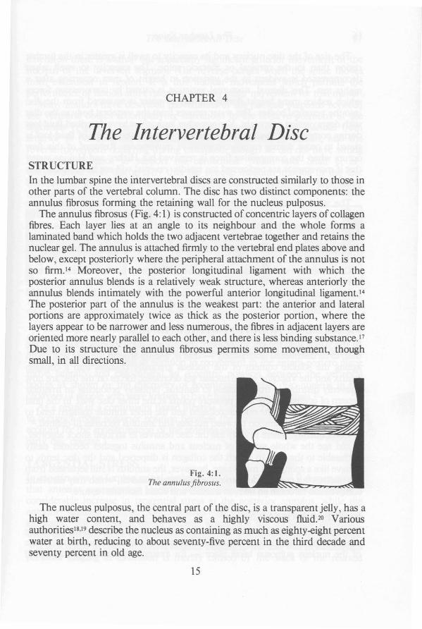
The Intervertebral DiscSTRUCTUREIn the lumbar spine the intervertebral discs are constructed similarly to those inother parts of the vertebral column. The disc has two distinct components: theannulus fibrosus forming the retaining wall for the nucleus pulposus.
The annulus fibrosus (Fig. 4: 1) is constructed of concentric layers of collagenfibres. Each layer lies at an angle to its neighbour and the whole forms alaminated band which holds the two adjacent vertebrae together and retains thenuclear gel. The annulus is attached firmly to the vertebral end plates above andbelow, except posteriorly where the peripheral attachment of the annulus is notso firm. 14 Moreover, the posterior longitudinal ligament with which theposterior annulus blends is a relatively weak structure, whereas anteriorly theannulus blends intimately with the powerful anterior longitudinal ligamenL14The posterior part of the annulus is the weakest part: the anterior and lateralportions are approximately twice as thick as the posterior portion, where thelayers appear to be narrower and less numerous, the fibres in adjacent layers areoriented more nearly parallel to each other, and there is less binding substance.J7Due to its structure the annulus fibrosus permits some movement, thoughsmall, in all directions.
Fig. 4:1.The annulus fibrosus.
The nucleus pulposus, the central part of the disc, is a transparent jelly, has ahigh water content, and behaves as a highly viscous fluid. 20 Variousauthorities18•19 describe the nucleus as containing as much as eighty-eight percentwater at birth, reducing to about seventy-five percent in the third decade andseventy percent in old age.
15
16 The Lumbar Spine
The size of the disc nucleus and its capacity to swell is greater in the lumbarregion than in the cervical or thoracic spine. The capacity to swell whendecompressed is evident in the variation in height of man occurring after anights rest. This diurnal, nocturnal variation is caused by compression forceswhich reduce mans height during the day as water is squeezed from the discinto the vertebral bodies. The water returns from the vertebral body to the discwith degravitation overnight. Various other authorities have described fluid lossduring compressive loading of the discs.21 ,43 Up to five percent of fluid loss isstated to occur during certain compressive movements. Reversal of this flowoccurs when the compressive force is removed but Hickey and Hukins state43
that if movements are performed too rapidly, reversal will not be complete. It ispossible then that repetition of a particular movement may cause a progressiveloss of fluid resulting in reduced bulk.
The water content of the annulus fibrosus changes less dramatically fromseventy-eight percent at birth to seventy percent in middle and old age, so thatin ageing the nuclear fluid content reduces to that of the annulus. Perhaps, whenthe viscosity of nucleus and annulus reaches an equilibrium, internalderangement is less likely to occur. This could account for the decreasedincidence of low back pain from age fifty onwards.
Of all the intervertebral discs the lumbar discs are by far the thickest and bearthe greatest loading and stresses. Even when slightly degenerated they behavehydrostatically7 - that is, the pressure within the disc is equally distributed inall directions of the intervertebral compartment.
PRESSURE DISTRIBUTION WITHIN THE DISCIn the young disc the gel structure of the nucleus allows forces, placed on thedisc, to be distributed isotropically - that is, evenly around the disc wall. Withageing the soluble content of the nucleus gradually changes into a collagenmatrix and the viscosity of the nuclear gel decreases; forces on the disc are nowunevenly distributed from nucleus to annulus, probably producing an irregularpattern of comparatively high pressure points at the inner disc wall. In the finalstages of disc ageing the nuclear collagen and the inner annular collagen tend tocoalesce and the separation between nucleus and annulus becomes ill-defined.20
Consequently, where in early life the disc behaves as an ideal shock absorber,in old age the whole system of nucleus and annulus together becomes easilypermeable to the fluid in which the collagen is dispersed and the disc tends tobehave like a sponge. In middle age, however, the annulus is still separated fromthe nucleus but contains a matrix of precipitated material, which may distributecompression forces in an uneven manner and could facilitate rupture. 20
NUCLEAR MOVEMENTThe centre of the lumbar disc nucleus is usually found posterior to the geometriccentre of the vertebral body. During movements of the spine a positional changeof the nucleus pulposus takes place - for example, from full flexion to full

The Intervertebral Disc 17
extension there is a small but apparently significant anterior movement of thenucleus of the involved segment. The reverse occurs when the spine movesfrom extension to flexion. It is this nuclear movement which permits theperformance of flexion and extension, and any other movement for that matter.
Many authors have described movements of the nucleus pulposus betweenthe vertebral bodies accompanying alterations in the relative positions of thesegments (Fig. 4:2). Armstrong14 described movement of the nucleus fromanterior to posterior occurring during the performance of flexion, and thereverse movement occurring during extension, though he did not have stronglaboratory evidence to support his contention. Much later Shah et al 13
demonstrated with discography that the opaque medium injected in the discmoves in a similar way during offset compression loading tests simulatingflexion and extension.
Fig.4:2a.The nucleus pulposus withthe spine in neutral position.
Fig.4:2b.The nucleus pulposus withthe spine in extension.
Fig.4:2c.The nucleus pulposus withthe spine in flexion.
Following laboratory experiments with silastic placed between the vertebralbodies Farfan21 concluded that evidence acquired this way suggests an ability ofthe nucleus to move away from the site where compressive forces are applied.The nucleus therefore can in addition to movement in the antero-postero planemove laterally and can inhabit an eccentric position between the vertebralbodies as shown by the discovery of eccentrically placed disc nucleus in thecadavers of people who were known to have had idiopathic scoliosis.21
As a result of a continually flexed lifestyle I believe the nucleus may migrateto occupy a more posterior position between the vertebral bodies. (This wouldaccount for the approximation of the anterior vertebral margins said to occur inearly disc disease.)
TANGENTIAL STRESSIn experiments on lumbar spinal sections of cadavers Shah et al. 13 demonstratedthat anterior compression loading of the disc simulating flexion, causes aconsiderable increase in tangential stress at the posterior annulus, while theanterior annulus bulges. On posterior compression loading simulatingextension, the tangential stress reduces posteriorly but increases anteriorly,while the annular bulge disappears anteriorly but appears posteriorly (Fig. 4:3).
It seems that in these situations anterior bulging of the disc wall in flexion andposterior bulging in extension is merely caused by the slack of the relaxed

18 The Lumbar Spine
annulus.43 The bulge is under reduced tangential stress and the nucleus hasmoved away from the bulge. It is unlikely that nuclear material will be extrudedunder these circumstances.
Fig.4:3a.Compression loading of thedisc.
Fig.4:3b. Fig.4:3c.Posterior compression loading Anterior compression loadingof the disc. Tangential of the disc. Tangential stressstress increased anteriorly - increased posteriorly-decreased posteriorly. decreased anteriorly.Annulus relaxes posteriorly. Annulus relaxes anteriorly.
I have come to conclude that, with an intact annular wall, a bulge appearingin the posterior annulus on extension is normal. In extension the posteriorannulus is not under tangential stress and, with the hydrostatic mechanismintact, the nucleus must move anteriorly. It is unlikely that annular tearing willoccur under these circumstances.
A bulge appearing in the posterior wall on flexion when the annular wall isdamaged may be a threat, as it indicates a weakening posterior annulus. Thistime the bulge is under increased tangential stress and the nucleus has movedposteriorly. Radial fissuring may occur and nuclear material may occupy thisspace thus further distending the annulus (Fig. 4:4a).Fig. 4:4. Development ofdisc protrusion.
a. Radial fissure. b. Annular bulge. c. Nuclear protrusion.
DISC DAMAGE AND REPAIRThe evidence suggesting that in the lumbar spine the intervertebral disc is acommon source of back pain is overwhelming7, 14,22,24,25 The most convincingsigns are the gross kyphosis and scoliosis accompanying severe sciatica.Following laminectomy the patients deformity and sciatica are usually
The Intervertebral Disc 19
significantly improved. The inference that a disturbance within the disc isresponsible for these signs is inescapable. And it is likely that patients who showsimilar signs but who do not have sciatica, have a similar though lesserdisturbance within the disc.
The cause of damage to the disc is still uncertain but it seems unlikely thatcompression is a significant factor. 14.43 Tension however is considered by variousauthorities to be a significant factor in the production of damaging stressesespecially those affecting the posterior annulus. Brown and co-workers applied asmall constant axial load and a repetitive forward bending motion of fivedegrees and the lumbar discs showed signs of failure after only 200 cycles ofbending and completely failed after 1000 cycles. Hickey and Hukins43 foundthat bending is particularly damaging because it concentrates stress on a limitednumber of collagen fibres and if overstretching exceeds four percent irreversibledamage occurs.
Markolf47 describes the spine as being twenty-five to thirty percent less stiffin the flexed position and it can be properly assumed that in this position it isless able to withstand stress.
From my own clinical observations I conclude that the lumbar disc is mostcommonly damaged in flexed positions especially where flexion is sustained.This usually gives rise to symmetrically distributed pain patterns. Should anytorsion or asymetrical stress be applied in addition, the symptoms tend to appearasymetrically. This is manifest by the patients description clinically of the painfirst appearing in the back near the mid line and moving laterally andperipherally with the subsequent imposition of torsion.
From the reviewed literature it would appear reasonable to assume thatfollowing sustained and repeated flexion stresses the nucleus is forcedposteriorly. This coincides with a raising of the intradiscal pressure andincreased tangential stress on the postero-lateral disc wall. This happens to bethe weakest part of the annular wall, because in this area the annulus has theleast radius, is thinner and is least firmly attached to the bone. 14, 17,23 Should theflexed position be maintained, stress will eventually fatigue the posteriorannulus and overcome its inherent strength and should overstretching exceedfour percent irreversible damage will result.43
The raised intradiscal pressure against the now damaged annulus coupledwith the posterior movement of nuclear fluids forces these fluids through thelattice of the weakened collagen and the fibres begin to part. The wideningfissure permits nuclear gel to enter the tear accelerating damage and causingseparation at the end plate. Should the process continue a significant posterioraccumulation of nuclear gel occurs as more and more nucleus is forced downthe fissure and eventually bulging occurs at the outer annulus (Fig.4:4b).Should the tearing be centrally situated the patient exhibits a kyphotic deformityand if the tear extends then posterolaterally the patient will exhibit a scolioticdeformity. When the annular wall is sufficiently weakened by fissuring,extrusion of nuclear material may occur (Fig. 4:4c). The disc has now lost itshydrostatic mechanism and on attempting extension the nucleus is unable to

20 The Lumbar Spine
move anteriorly. The ability to extend is seriously impaired, for anyapproximation of the posterior vertebral rims results in increased pressure onthe extrusion itself. This explains why patients with an accomplished protrusionoften present with a flattened lumbar spine, and any attempt to extend the lowback results in enhancement of low back pain and sciatica.
Farfan26 has stated that a disc protrusion commences with a tear in theannulus, starting off at the bony vertebral end plate. Tearing must extend to acertacn degree before fragmentation occurs, allowing the annulus to give way.This is nearly always associated with the development of a radial tear whichpermits the nucleus to force an increase in annular bulging or a widening of thefissure.
Once the disc is damaged by this type of derangement, the natural healingprocesses will be initiated. Through exposure at the vertebral end plate thevascular tissue of the vertebral body comes in contact with the avascular disc,invading it and removing all the tissue that does not have a blood supply. Scartissue is now laid down in the inner annulus and nucleus. 26
Contraction of the invading scar results in the formation of an inelasticstructure within the elastic disc. In this way dysfunction develops, causing a lossof mobility in the segment involved. When sufficient stres~ is applied to thelumbar spine, the scarred areas tend to fragment and tear and the cycle repeatsitself.
If we are to prevent the development of dysfunction in the disc followingderangement or protrusion, we must provide early movement in our treatmentto ensure the formation of an extensible scar within the elastic structure of thedisc.
THE DISC AND PAINAlthough the disc does not contain actual nerve endings, it may cause pain invarious ways. Severe midline backache may be caused by direct mechanicalirritation of the nerve endings in the posterior longitudinal ligament and fibroadipose tissue, binding the ligament to the annulus. Similarly, pain in or close tothe midline of the back may be caused by pressure on the anterior dura mater orits sleeve-like extensions in the intervertebral foramen. These situations occurin central posterior and postero-lateral protrusions of the nucleus pulposus4
(Fig. 4:5).Regarding postero-lateral herniation of the nucleus pulposus of a lumbar
intervertebral disc Wyke4 has stated:"As such a protrusion develops it impinges initially on the sinuvertebralnerve, in which it not only interrupts mechano-receptor afferent activity butmay also irritate the contained nociceptive afferent fibres and thereby giverise to pain in the lower back in the absence of sciatica. Should the protrusiondevelop further, it begins to impinge on the related dorsal nerve roots (andtheir containing dural sleeves), as a result of which the backache becomesmore severe and more widely distributed, and to it are added sensory changes(paraesthesiae and numbness) and pain experienced in the distribution of thesciatic nerve" (Fig. 4:6).

The Intervertebral Disc 21
Fig. 4:5. (below)Posterior or postera-Iateral herniation ofthe nucleus pulposus.
Fig. 4:6. Postera-Iateral herniation of the nucleuspulposus with nerve root compression.
It can be clearly seen that as this type of lesion develops and worsens, initiallythe pain is felt in the midline of the back. It progressively increases in intensityand spreads across the back into the buttock and thigh, and as the climax isreached the pain appears in the lower limb. The further from the midline thepain is felt, the greater is the derangement (Fig. 4:7).
Fig. 4:7. Pain pattern of a developing lower lumbar disc lesion. When the centralisationphenomenon occurs a reversal of this pattern will be observed.

22 The Lumbar Spine
THE CE TRALISATIO PHE OMENON
In 1959 I noticed in a retrospective observation of case histories that patientswho responded rapidly to treatment experienced a centralisation of pain asimprovement took place. I called this the 'centralisation phenomenon'.
I would define this phenomenon as the situation in which pain arising fromthe spine and felt laterally from the midline or distally, is reduced andtransferred to a more central or near midline position when certain movementsare performed. It is permissable for pain to increase centrally provided there is areduction in the lateral or distal pain.
Centralisation of symptoms only occurs in the derangement syndrome. Thesignificance of the centralisation phenomenon is that in derangement themovement which causes centralisation will, if repeated, reduce thederangement. The phenomenon is not applicable to the dysfunction syndromeand, of course, will not occur in patients with postural problems.
I believe that the centralisation phenomenon is merely the reversal of thedevelopment of pain in progressive disc lesions which is described by variousauthors.4• 24 As the sequence of symptoms from onset is perfectly logical, so isthe reversal of symptoms during centralisation. When the protrusion reduces insize, it releases first the nerve root and then the dura mater, which results in acessation of pain and paraesthesiae below the knee followed by a reduction inthigh pain. At this stage the pain should be felt mainly in the buttock or centrallumbar area.
The typical pattern of pain produced by a developing lower lumbar disc lesionis generally acknowledged. However, it is not always recognised thatcentralisation as recovery takes place applies to derangement situations and is anindication that reduction of derangement is occurring. Pain of a radiating orreferred nature will reduce distally and may simultaneously increase proximally,when the involved joints are moved in the correct direction - that is, reducingthe derangement. Thus the pain appears to reverse the order in which itcommenced.
The centralisation phenomenon can also be observed in unilateral orsymmetrical pain felt solely in the spine. In this case the pain moves from acrossthe low back to a central midline location, and on further reduction of thederangement pain is replaced by an aching or merely a stiffness in the center ofthe back.
The phenomenon occurs when appropriate movements are performed in thecervical and thoracic spine in the presence of derangement, and is just asreliable.
DIFFERENTIATION BETWEE
DISC DEGENERATION AND FRANK PROTRUSIONPart 27 has made some observations by discography which have an importantclinical implication. He states:
The Intervertebral Disc 23
"When there is difficulty in differentiating disc degeneration with annulusrupture from frank nuclear protrusion, flexion and extension films can behelpful. In disc degeneration, forward flexion opens the posterior disc widely?the annulus fibrosus and posterior longitudinal ligament become tightlystretched and the contrast medium tends to disperse uniformly. This is ahelpful indication of the true state of affairs in those patients where thecontrast appears localised in the neutral or extended positions. On the otherhand, when frank nuclear prolapse is present, the opaque medium remainscontained in a localised area irrespective of the position of the spine."The assessment of the effects of certain chosen movements - the test
movements - on the pain will enable us to make a clinical diagnosis. In franknuclear protrusion the hydrostatic mechanism of the disc is impaired, and theposition of the nucleus cannot be influenced by movement and positioning.Clinically, the patient's symptoms will not reduce as a result of the testmovements, and such a patient is not likely to benefit from mechanicaltreatment utilising movements and position.
On the other hand, where the disc merely shows degeneration with annularfissuring and no protrusion exists, the hydrostatic mechanism is still intact andthe position or shape of the nucleus can be influenced by movement andpositioning. Clinically, there will be a change in intensity or site of the patient'ssymptoms as a result of the test movements, and such a patient can be treatedsuccessfully with mechanical procedures utilising movement and positioning.
If disturbance of the normal intervertebral disc mechanism is a causativefactor in low back pain it conveniently explains the behaviour of pain,movement and deformity found in patients with the derangement syndrome.
In the postural syndrome, by my definition, no pathology will be found.In the dysfunction syndrome pathology affecting muscles, ligaments, disc,
apophyseal joints and fascias may be found separately or together.

CHAPTER 5
DiagnosisTHE THERAPIST'S RESPONSIBILITY
The therapist is part of the team involved in the treatment and rehabilitation ofpatients suffering low back pain. In some countries manipulative therapists areprimary contact practitioners. Consequently, their diagnostic skills have greatlyimproved, enabling them to define which mechanical conditions can be helpedby mechanical therapy and to separate these conditions from the nonmechanical lesions which have no place in the therapy clinic.
However, differential diagnosis is really not within the scope of manipulativetherapy. It is my view that differential diagnosing by medical practitioners isnecessary to exclude serious and unsuitable pathologies from being referred formechanical therapy. In making diagnoses the manipulative therapist shouldconfine himself to musculo-skeletal mechanical lesions. Specialised in this field,he is usually able to make far more accurate diagnoses than most medicalpractitioners. As the manipulative therapy profession gains internationalrespect, we may soon see the day that this specialisation becomes generallyaccepted.
DIAGNOSTIC DIFFICULTIES
In the low back mechanical diagnosis is extremely difficult. As yet, no meanshave been devised which enable us to selectively stress individual structures andidentify the source of many pains. As Nachemson7 states, there is only onecondition which allows a fairly confident diagnosis to be made:
"the patient with sciatica caused by sequestration from the disc whichimpinges on a nerve root. Such patients, though, represent only a smallproportion of those who have low back pain problems, and constitute at mostonly a few percent."
This means that perhaps as many as ninety percent of patients cannot bediagnosed in a very specific manner. Various authorities28• 29 have stated that inmany instances it is impossible to define the exact pathological basis for lowback pain and, consequently, to achieve a precise diagnosis. Nachemson hassaid: 30
"No one in the world knows the real cause of back pain and I am noexception. "
24

Diagnosis 25
When authorities such as these clearly state that the problems surroundingspecific diagnosis of low back pain are insurmountable, it seems that the timehas come to alter the rules of the game. Instead of aiming for a specific diagnosisbased on a particular pathology, we must apply an alternative system ofassessment. This can be used until further development of our knowledge anddiagnostic procedures enables us to become more specific.
In order to analyse mechanical low back pain and categorise the symptoms anew approach is necessary. I believe we have a means of overcoming the presentdiagnostic impasse. If mechanical pain is caused by mechanical deformation ofsoft tissues containing nociceptive receptors, we must confine our diagnosiswithin this framework.
THE THREE SYNDROMESAll spinal pain of mechanical origin can be classified in one of the followingsyndromes:
The postural syndrome:This is caused by mechanical deformation of soft tissues as a result of posturalstresses. Maintenance of certain postures or positions which place some softtissues under prolonged stress, will eventually be productive of pain. Thus, thepostural syndrome is characterised by intermittent pain brought on by particularpostures or positions, and usually some time must pass before the pain becomesa~parent. The pain ceases only with a change of position or after posturalcorrection.
The dysfunction syndrome:This is caused by mechanical deformation of soft tissues affected by adaptiveshortening. Adaptive shortening may occur for a variety of reasons which willbe discussed later. It leads to a loss of movement in certain directions and causespain to be produced before normal full range of movement is achieved. Thus,the dysfunction syndrome is characterised by intermittent pain and a partial lossof movement. The pain is brought on as soon as shortened structures arestressed by end positioning or end movement and ceases almost immediatelywhen the stress is released.
The derangement syndrome:This is caused by mechanical deformation of soft tissues as a result of internalderangement. Alteration of the position of the fluid nucleus within the disc, andpossibly the surrounding annulus, causes a disturbance in the normal restingposition of the two vertebrae enclosing the disc involved. Various forms anddegrees of internal derangement are possible, and each presents a somewhatdifferent set of signs and symptoms. These will be discussed later. Thus, thederangement syndrome is usually characterised by constant pain, butintermittent pain may occur depending on the size and location of the
26 The Lumbar Spine
derangement. There is a partial loss of movement, some movements being fullrange and others partially or completely blocked. This causes the deformities inkyphosis and scoliosis so typical of the syndrome in the acute stage.
The three syndromes presented are totally different from each other, andeach syndrome must be treated as an entity on its own, requiring specialprocedures which are often unsuitable for the other syndromes. In order toidentify which syndrome is present in a particular patient a history must beestablished and an examination must be performed. The method of historytaking and examination is discussed in the following chapters.

CHAPTER 6
The .HistoryTaking an accurate history is the most important part of the initial consultationwhen one is dealing with any medical or surgical problem. Unfortunately, whenthe mechanical lesion is involved there is still lack of understanding regardingthe nature of the questions that should be asked, the reasons for asking them,and the conclusions to be drawn from the answers.
I will set out step by step the stages that should be developed in history taking,and the questions that should be asked at each stage. Practitioners will alreadyhave their own method of history taking, and I do not suggest at all that theyshould alter their routine. However, I believe that the following questions mustbe included, if one is to reach a conclusion following the examination of patientswith mechanical low back pain.
INTERROGATION
As well as the usual questions regarding name, age and address, one shouldenquire as to the occupation of the patient, in particular his position at workwhich provides us with the most important and relevant information. Managersare not always sitting down as we tend to believe, and postmen are not alwayswalking.
Where is the present pain being felt?We need to know all the details about the location of the pain, because this willgive us some indication of the level and extent of the lesion and the severity ofthe condition. If there are any associated symptoms such as anaesthesia,paraesthesiae and numbness, their location must be noted as well. Referred painindicates that derangement is likely.
Because the location of the pain can change rapidly and dramatically, we mustfind out if the pain has been present on the same site or sites since the onset.
At this stage we must determine whether the symptoms are central, bilateralor unilateral in origin, as this is an important factor in the classification of thepatient and consequently in his treatment. Bilateral symptoms indicate a centralorigin, whereas central symptoms cannot arise from a unilateral structure. 24
How long has the pain been present?It is important to find out whether we are dealing with an acute, a subacute or
27
28 The Lumbar Spine
chronic condition. In recurrent low back pain we are not interested in an answerbased on the length of time since the first attack; at this stage of the examinationwe want to know how long the present episode has been evident.
If the symptoms have been present for any length of time, as is often the case,we must find out whether the patient feels that his condition is improving,stationary or worsening.
The length of time that the condition has been present may assist us todetermine the stability of healing following a disc prolapse. It may also indicatethe development of dysfunction which is likely to occur following trauma orderangement. The longer the symptoms have been present, the greater are thechances that adaptive changes have taken place.
The length of time that the patient has had symptoms can also guide us indeciding how vigorous we can be with our examining procedures. If a patienthas had his symptoms for several months and has been able to work all thistime, he will probably have placed more stress on the joints at fault than we arelikely to do during the examination, which means that we can be fairly vigorous.If on the other hand the patient had a sudden onset of pain within the past twoweeks, we could be dealing with a derangement situation and may well increasethe degree of derangement with out test procedures which, if applied toovigorously, may significantly worsen the condition of the patient. Generallyspeaking, if the condition has been present only for a few days to two or threeweeks, we must take great care in handling and exercising the patient; but if thepresent pain has been evident for months, we can be rather vigorous with ourprocedures.
How did the pain commence?
Basically we want to find out if there was an apparent or no apparent reason forthe onset of the pain. Most of the histories commonly state that the painappeared for no apparent reason. Two of every three patients fall into thiscategory, and only one patient will recognise a causative strain.
If pain has arisen for an apparent reason a recognisable strain has caused thesymptoms - for example, an accident, a sports injury, lifting. If the patient wasinvolved in an accident - for example, if he were struck by a bus, - he mayhave sustained multiple injuries and the mechanics of none of these would beclear. This type of patient must be treated gently and with caution.
If pain has arisen for no apparent reason, a derangement or dysfunction hasdeveloped during the course of normal living usually without the application ofany external force. Strains arising in this fashion can under most circumstancesbe avoided by modification of the patient's daily living habits or movements.However, if the pain commenced for no apparent reason and is gradually andinsidiously worsening, we may well suspect serious pathology, particularly if thepatient feels or looks unwell at the time of interrogation. It is always better tosuspect the worst and be wrong, than to overlook the worst and be wrong.
Careful evaluation of the patient's information regarding the onset of hissymptoms is necessary in order to avoid faulty conclusions. There are situations

The History 29
in which the patient thinks that his pain commenced for no apparent reason,whereas we may recognise a causative strain; alternatively the patient maywrongfully relate the onset to certain activities in an attempt to find a cause forhis low back pain, which in fact appeared for quite different reasons.
Is the pain constant or intermittent?This is the most important question we must ask patients with low back pain. Ifin patients referred for mechanical therapy the pain is found to be constant, itusually is produced by constant mechanical deformation. However, we mustkeep in mind that constant pain can also be caused by chemical irritation.Intermittent pain is always produced by mechanical deformation.
Constant chemical pain:
This is present as long as chemical irritants are present in sufficient quantitiesand occurs in inflammatory and infective disorders and in the first ten to twentydays following trauma. Chemical pain following trauma reduces steadily ashealing takes place. Chemical irritants do not appear and disappear during thecourse of the day. Therefore, pain of chemical origin is always constant, andpatients who describe periods in the day when no pain is present must haveintermittent pain which is of mechanical and not of chemical origin.
Mechanical stresses that would normally be painless, can become painfulwhere chemical irritation has raised the threshold of excitation of thenociceptive receptors. Thus, movements may superimpose mechanical forces onan existing chemical pain and enhance it, but they will never reduce or abolishchemical pain. This is significant in the differentiation between chemical andmechanical pain.
Low back pain caused by chemical irritation is comparatively easy to identify,as the pain is usually constant and no mechanical means can be found tosignificantly reduce it. Five days of treatment and observation should besufficient to arrive at this conclusion.
Constant and intermittent mechanical pain:
This is present as long as mechanical stresses are sufficient to cause mechanicaldeformation. This type of pain will frequently reduce or even disappear whenmovements or positions are adopted that sufficiently reduce the mechanicalstresses. On the other hand, the mechanical stresses may as easily be increased·by movements and positions. Constant mechanical pain will vary significantly in'intensity but never disappears, whereas intermittent mechanical pain appearsand disappears according to circumstances.
Constant pain must be truly constant - that is, there is no time in the daywhen pain or aching is not present. Pain must be classified as intermittent even ifthere is only half an hour in the day when the patient feels completely painfree.In that half hour there is no mechanical deformation present, and we mustexamine the circumstances in which the patient is painfree and utilise this

30 The Lumbar Spine
information for treatment purposes. It is much simpler to treat a patient who hasintermittent pain than one whose pain is constant and in whom mechanicaldeformation is present under all circumstances.
It is my observation that about seventy percent oflow back pain patients haveintermittent pain, and the remaining thirty percent have truly constant pain.The majority of patients with constant mechanical pain are likely to belong tothe derangement category. Derangement alters the tension in the structuresabout the segment involved, increasing mechanical deformation in some tissuesand decreasing it in others. The constant increase in tension produces constantpain which continues, until the tension is decreased by reduction of thederangement or adaptive lengthening of surrounding tissues.
Intermittent pain is relatively easy to treat, because if there is one hour in theday when no mechanical deformation is present, it is possible to graduallyextend that painfree time period. Constant pain is rather a different problem andis more difficult to treat. The percentage of patients failing to respond totreatment is higher in the constant pain group than in the intermittent paingroup. Generally speaking, the derangement syndrome can be associated withconstant pain, whereas the postural and dysfunctional syndromes arecharacterised by intermittent pain.
What makes the pain worse? and What makes the pain better?We must specifically ask about sitting, standing, walking, lying, and activitieswhich involve stooping or prolonged stooping. In these positions the jointmechanics of the lumbar spine are relatively well understood, and therefore wewill be able to determine which situations increase and which decreasemechanical deformation. We must carefully record any position or activityreported to reduce or relieve the pain, as we will utilise this information in ourinitial treatment.
Sitting:
In relaxed or prolonged sitting the lumbar spine falls into full flexion, the effectsof which are described in detail elsewhere (Chapter 2). If a patient tells us thatsitting increases his symptoms, we know that sustained flexion causesmechanical deformation of his lumbar spine. But if a patient finds relief insitting, flexion actually reduces the mechanical deformation.
Standing:
Standing, expecially relaxed standing, places the lower lumbar spine in full endrange extension which means that certain structures are on full stretch. If apatient tells us that he is worse in relaxed standing, sustained maximumextension produces mechanical deformation of his lower lumbar spine. But if apatient finds relief in relaxed standing, sustained maximum extension reducesthe mechanical deformation.
The History 31
Walking:
Walking accentuates extension - that is, it further increases the lordosis of thelumbar spine as the hind leg by its backward movement brings the pelvis intoforward inclination. If walking produces or increases pain, extension mustproduce or increase mechanical deformation of the lower lumbar spine. But ifthe patient has no pain in walking, extension is reducing the mechanicaldeformation.
Lying:
There are basically three positions which may be adopted while lying: prone,supine and side lying. The many variations of these, caused by different legpositions, make evaluation of the effects of lying rather difficult. Apart from thelying position itself, the effects on the lumbar spine depend on the nature of thesurface on which one lies - usually the mattress and its supporting base which may be firm and unyielding, or soft and giving.
The effects of the three basic positions on the lumbar spine can besummarised as follows:
(a) In lying supine on a firm surface the lumbar spine falls into extension,whereas on a soft surface the degree of extension is decreased and insome cases flexion may be produced.
(b) In lying prone on a firm as well as a soft surface the lumbar spine isalways placed near or at full end range of extension.
(c) In side lying the lumbar spine is brought into side gliding towards theside one is lying on, more so when on a soft surface than on a hardsurface.
Activities which involve bending:
In bending or prolonged bending the lumbar spine falls into full flexion andadded to this are gravitational stresses. If, for example, gardening produces pain,sustained flexion must produce mechanical deformation of the lumbar spine.But if the pain is reduced while gardening, sustained flexion stops themechanical deformation.
Patients who have had pain for a long time, may have difficulty indetermining what makes their pain better or worse. They are no longer able toobserve objectively their own pain patterns because of the length of time thepain has been present. It is necessary to spend extra time to extract detailedinformation regarding the pain behaviour, because without this we cannotproceed to an adequate conclusion and appropriate treatment.
Occasionally a patient will tell us that there is no position or movement whichaffects the pain. In this case the information obtained from the history isinsufficient, and during the examination we must try to produce a change in thepatient's symptoms by utilising extremes of movement or sustained positions.
32 The Lumbar Spine
Have there been previous episodes of low back pain?We should enquire about the nature of any similar or other low back painepisodes, the time span over which they occurred, and their frequency. At thisstage we should also find out about previous treatments and their results.Episodic history indicates derangement. Dysfunction is likely to developinsidiously after each episode and will now coexist but be masked by the presentdisturbance. Its presence will be revealed after resolution of pain resulting fromthe derangement.
Further questionspain on cough/sneeze?disturbed sleep?pain on arising in the morning?recent X-Rays? - results?on medication at present?on steroids, in past or at present?general health? - recent weight loss?major surgery or accident, recently or previously?saddle anaesthesia? - bladder control?
Information gained from these questions may complete the picture of thecondition we are dealing with. The reasons for asking these questions areobvious and straight forward, and they will not be discussed in detail.
Although the referring medical practitioner will almost certainly haveexcluded any serious or unsuitable pathology, we must remain alert for itspresence.
Fig. 6:1. (opposite.) Lumbar spine assessment sheet.
physio4all
Rectangle
physio4all
Rectangle

LUMBAR SPINE ASSESSMENT
at onset .Present for .Commenced as a result of .
No apparent reason. DConstant/I ntermittent.When worse - bending, sitting or rising from, standing, walking, lying.Other .When better _ bending, sitting or rising from, standing, walking, lying.Other .Disturbed sleep - Yes No Cough/sneeze +ve -vePrevious history .
and treatment .X-Rays ..General health Meds/Steroids ..Recent surgery Accident .Gait Bladder .
Date of birth ..Occupation .
Date .Name. .Address .
HISTORYSymptoms now ..
EXAMINATIONPosture sitting ..Posture standing ..Lateral shift (R) or (L) or nil
Lordosis reduced/accentuated ..Leg length .
MOVEMENT LOSSFlexion: Major. Moderate. Minimal.Extension: Major. Moderate. Minimal.Side Gliding: (R) or (L) ..
Deviation (R) or (L) .Deviation (R) or (L) .
TEST MOVEMENTSFIS................................................................................. SG IS (R) ..
REP. FIS SGIS (L) .EIS REP. SGIS (R) .
REP. EIS REP. SGIS (L) .FIL ..
REP. FI L ..EI L ..
REP. EI L .Neurological .Hip joints ?.~I~ ..Conclusion: Posture Dysfunction Derangement
PRINCIPLE OF TREATMENT 1I3",'11 U"/w,..Hl/ £,l,."..Alie....l, 7/.. 3316'
physio4all
Rectangle
physio4all
Rectangle

CHAPTER 7
The ExaminationHaving digested the information supplied by the referring doctor, extracted asmuch relevant information as possible from the patient, and checked theradiologist's report, we may proceed to the examination proper.
If the patient is able to do so, we should make him sit on a straight backedchair while taking his history. During this time he will reveal the true nature ofhis sitting posture. When the patient rises to undress after the interrogation weshould observe the way he rises from sitting, his gait, the way he moves, andany deformity that may be obvious.
We will record the following:
I. POSTURE SITTINGIf the patient has been sitting during history taking, we already have a goodimpression of his posture (Fig. 7: 1). We now ask him to sit on the edge of theexamination table with his back unsupported. In the majority of cases thepatient will sit slouched with a flexed lumbar spine. Some patients are moreaware of the relationship between their posture and pain. They have discoveredthat they can control their sitting pain by sitting upright and may sit very wellon first observation. Unfortunately, these patients are few and far between.
Fig. 7:1.Relaxed sitting posture.
34

The Examination
II. POSTURE STANDINGWe examine in particular the following features:
35
1. Reduced or accentuated lordosisThe most common postural fault to be observed in the standing position is theflattened lumbar spine or reduced lordosis (Fig. 7:2a). Some patients have adeformity in kyphosis.
Another common deformity or departure from the 'norm' is the patientexhibiting an accentuated lumbar lordosis (Fig. 7:2b). Though much lessCOmmon than the flattened spine this is clearly a separate category and shouldbe treated accordingly.
Fig.7:2a.Reduced lumbar lordosis.
Fig.7:2b. Fig.7:2c.Accentuated lumbar lordosis. Lateral shift to the right.
2. Lateral shiftA departure from the midline causing a lumbar scoliosis or lateral shift isevident in about fifty-two percent of patients. 10 There are many reasons for thelumbar spine to depart even slightly from the midline: the anatomicalconfiguration of the joint surfaces may dictate this; a congenital anomaly may bepresent; there may be some remote mechanical cause; and an alteration in theposition of the disc nucleus may be responsible.
The lateral shift is sometimes barely discernable, and great care must be takento ensure that the deformity is not overlooked (Fig. 7:2c). This is importantbecause some movements, especially extension, produce pain when performedin the presence of a lateral shift, whereas if there was no shift they would be
36 The Lumbar Spine
painfree. We must never fail to recognise a minor lateral shift and its possiblerole in the production of symptoms.
I have chosen to describe a right lateral shift - or a lateral shift to the rightas the situation which exists, when the vetebra above has rotated and laterallyflexed to the right in relation to the vertebra below, carrying the trunk with it.Thus the top half of the patient's body has moved to his right in relation to thebottom half.
3. Leg length discrepancyIf a leg length discrepancy is encountered, we must investigate the relevance ofthe discrepancy to the patient's symptoms. When in the history there is norecord of pain being enhanced by standing and walking, the difference in leglength is not likely to be relevant. But when pain is produced by standing orwalking, we should increase the length of the short leg to bring about symmetryand await the effect of this on the patient's symptoms. When relevant, leglength adjustment causes a fairly rapid change in the symptoms experiencedduring standing and walking. This change should become apparent within a fewdays at the most.
There are various methods of measuring the leg length, but the only trulyreliable way to detect minor differences in leg lengths is by taking an x-ray ofthe pelvis and both full legs with the patient in standing. However, it is doubtfulthat minor leg length discrepancies cause significant low back pain.
At this stage, with the patient still standing, it may be advisable to quickly testthe integrity of the conduction of the lower lumbar and upper sacral nerve roots.Therefore we ask the patient to walk on his toes and then on his heels. In case ofdifficulty or when in doubt about the outcome a more detailed neurologicalexamination must be performed.
III. EXAMINATION OF MOVEMENT
Here we are interested in observing the quality of the movement itself - that is,the range of movement and the movement pathway. We will determine if thereis a movement loss and if deviation from the normal movement path takesplace. The word 'pain' should not be mentioned until we are ready to assess theeffects of the movements on pain.
The patient should be standing with his feet about thirty centimeters apartand only one movement will be performed in the direction to be evaluated. Weexamine:
1. FlexionThis is the first movement to be examined, because in patients with dysfunctionor derangement the flexion movement provides us with the most relevantinformation regarding the nature and the degree of the disturbance. Thestanding patient is asked to run the hands down the front of both legs, movingas far as possible into flexed standing, followed immediately by returning toneutral standing (Fig. 7:3).

The Examination 37
Fig. 7:3.Examination ofmovement - flexion.
Any loss of flexion should be noted. Loss of flexion manifests itself in one oftwo ways: either the end range of flexion is limited, or a deviation from thenormal pathway of flexion has developed. In some patients with a severe loss offlexion end range, the lumbar lordosis is still present after the patient has bentforward as far as possible, in others the lumbar spine merely remains flat. Anyasymmetrical impediment to flexion may cause the spine to take the path ofleast resistance, resulting in a deviation from the sagittal pathway. By far themajority of patients with a flexion loss divert from the sagittal plane duringflexion and deviate to one side or the other of the midline. This may occur in anarc-type of movement, the flexion commencing and ending in midlinepositions; or, once movement is commenced, it may divert from the midlineand increase its departure for as long as flexion is continued.
Deviation in Flexion
In my experience there are three clearly defined and separate causes fordeviation in flexion. The mechanism is different in each case and the treatmentmust be varied accordingly. Thus deviation in flexion may be due to:
(a) Derangement within the vertebral joint. In this situation the alteredposition of the fluid nucleus compels a deviant flexion pathway.Generally, the deviation in flexion occurs away from the painful side aslong as there is no sciatic nerve root irritation. In some patients thedeviation is variable and will occur one day to the left and the next day tothe right.
(b) Dysfunction within the vertebral joint. This develops following repair ofdamage after derangement. In this situation the consequent scarring byfibrous connective tissue prevents flexion in the sagittal plane and thedeviation may take place towards or away from the painful side, but isnever variable.
(c) Dysfunction external to the vertebral joint. This exists in the presence ofan entrapped or adherent sciatic nerve root. In this situation the root isno longer able to lengthen adequately and allow flexion to occur in thesagittal plane. It will now act as an anchor and pull the patient during
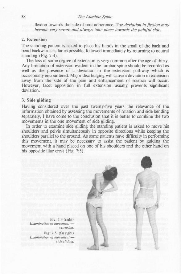
38 The Lumbar Spine
flexion towards the side of root adherence. The deviation in flexion maybecome very severe and always take place towards the painful side.
2. ExtensionThe standing patient is asked to place his hands in the small of the back andbend backwards as far as possible, followed immediately by returning to neutralstanding (Fig. 7:4).
The loss of some degree of extension is very common after the age of thirty.Any limitation of extension evident in the lumbar spine should be recorded aswell as the presence of a deviation in the extension pathway which isoccasionally encountered. Major disc bulging will cause a deviation in extensionaway from the side of the pain and enhancement of sciatica will occur.However, facet apposition in full extension usually prevents significantdeviation.
3. Side glidingHaving considered over the past twenty-five years the relevance of theinformation obtained by assessing the movements of rotation and side bendingseparately, I have come to the conclusion that it is better to combine the twomovements in the one movement of side gliding.
In order to examine side gliding the standing patient is asked to move hisshoulders and pelvis simultaneously in opposite directions while keeping theshoulders parallel to the ground. As some patients have difficulty in performingthis movement, it may be necessary to assist the patient by guiding themovement with a hand placed on one of his shoulders and the other hand onhis oppositic iliac crest (Fig. 7:5).
Fig. 7:4 (right)Examination ofmovement
extension.
Fig. 7:5. (far right)Examination ofmovement
side gliding.

The Examination 39
The side gliding movement is frequently unilaterally impaired. When thepatient has a lateral shift, there is always some unilateral loss of side gliding. Inthis situation the movement is restricted or completely blocked in the directionopposite to the lateral shift.
IV. MOVEMENTS IN RELATION TO PAINAfter examining the lumbar spine in relation to function, we must nowinvestigate the effects of various movements on the pain. Let us assume thatpain is produced by mechanical deformation as described by Wyke.4 Asdiscussed before, stresses applied to soft tissues will under certain circumstancesbe productive of pain. Any attempt to force normal movement (application ofabnormal stress) in a joint with a visibly impaired function (abnormal tissue),must result in the production or enhancement of pain.
In order to stress the joints in a controlled manner and avoid exacerbation Ihave devised a sequence of test movements, the mechanics of which arerelatively well understood and the effects of which can be controlled. Byapplying the test movements during the examination we will enhance painunder some circumstances and reduce it in others. Information gained bydeliberately stressing the joints is vital and enables us to select and categorisepatients into three groups - that is, patients with pain arising from postural,dysfunctional or derangemental causes.
The test movements are first performed in standing and then in lying. Whenperformed in lying they must be done in such a way that the effect on thelumbar spine is a passive stretch, and any form of active movement produced bythe muscles surrounding the lumbar spine should be avoided. In this way wecan achieve a better end range stress than with active movements.
When the test movements are performed in standing a normal stress is appliedto normal or abnormal tissue; in the former case no pain will be produced; in thelatter case pain will be produced or increased if the test movements enhance themechanical deformation, but pain will be decreased or abolished if the testmovements reduce the mechanical deformation. When the test movements areperformed in lying a passive stress is added by the patient and an abnormal stressis applied to normal or abnormal tissue; in the former case no pain should result,because the stress is not excessive and is only applied momentarily; in the lattercase pain will be produced or increased if the test movements enhance themechanical deformation, but pain will be decreased or abolished if the testmovements reduce the mechanical deformation.
If we are to relate movements to pain, the test movements must be performedin such a way that they produce a change in the patient's symptoms. Thischange may be brought about in various ways: if prior to movement pain ispresent, the test movement may increase or reduce its intensity; it may alter thesite of the pain by centralisation or by abolishing one pain and introducinganother one. If prior to movement no pain is present, the test movement mayproduce the pain complained of.
If there is no change in the patient's symptoms during or immediatelyfollowing the test movements, the joints have not been stressed adequately and
40 The Lumbar Spine
the process should be repeated more vigorously. It may also be that the pain isnot of mechanical origin, because mechanical pain must be and always isaffected by movement or position. Or, alternatively, the lumbar spine is notcausing the problems and other areas should be investigated.
REPEATED MOVEME TSWhen assessing the results of the test movements it is important to examine theeffects of repeating them. Repeated movements are vital in the examination ofspinal segments when disc pathology is suspected and it is necessary todetermine if a derangement situation exists. It is my belief that with movementof the vertebral column the nucleus can alter its shape, and with sustainedpositions or repeated movements it will eventually alter its position. Clinicallythis manifests itself in the derangement syndrome by a change in intensity orsite of the symptoms. A decrease or centralisation of pain is absolutely reliablein indicating which movement should be chosen to reduce mechanicaldeformation. I have learnt to rely implicitly on centralisation as the mostimportant clinical guide to establish the correct direction of movement whichwill reduce derangement. An increase or peripheralisation of pain is just asreliable in indicating which movement should be avoided because it enhancesmechanical deformation. If there is an increase or peripheralisation of painwhen performing any technique or movement, a worsening of the condition ofthe patient is likely. There is one exception to this rule: the patient with anadherent nerve root in chronic sciatica. There are special tests which easilyidentify this condition, and these will be discussed later.
In derangement the performance ofrepeated movements in the direction whichincreases accumulation of nuclear material will result in a progressivelyincreasing derangement and increasing or peripheralising pain. The performanceof repeated movements in the opposite direction will result in a reduction of thederangement and reduction or centralisation of pain. Thus, repeatedmovements are diagnostic in derangement pathologies.
In dysfunction the performance of repeated movements in the direction whichstretches adaptively shortened structures will produce pain at the end range ofmovement, but repetition does not make the patient progressively worse. When hereturns to the neutral position the pain will disappear. Thus, repeatedmovements are diagnostic in dysfunction as well.
Patients with the postural syndrome will not experience pain with any of thetest movements or their repetition. These patients must be positioned in order tohave their pain reproduced.
Apart from exposing the derangement and dysfunction syndromes, repeatedmovements are essential in determining whether the timing is appropriate tocommence stretching procedures following trauma and derangement. Whenrepeated movements, applied to painful structures, produce less and less painwith each repetition these structures should be exercised. On the other hand,when more and more pain is experienced with each repetition exercising is notindicated and more time should be allowed for the condition to heal. This
The Examination 41
fundamental response ofpain sensitive structures to stress must be applied to softtissue lesions throughout the musculo-skeletal system in order to determinewhether a passive or an active treatment approach should be developed.
The movements that I have chosen as test movements are the ones of flexion,extension and side gliding. In my experience these movements will induce orreduce mechanical deformation in the lumbar spine quicker and moreeffectively than any other movement. Generally speaking, the movements thatproduce the greatest amount of mechanical deformation and therefore pain can,when reversed or modified, be used to have the greatest effect on the reductionof that mechanical deformation and pain. For example, rotation is rarelycomplained of as producing significant pain and therefore neither enhances norreduces mechanical deformation significantly. On the other hand, flexion andextension are commonly stated to be the most painful movements and arepotentially the most useful movements for treatment purposes.
Different effects are produced when the tests movements are performed instanding compared with lying. This requires further discussion for both flexionand extension.
Flexion in standing compared with flexion in lying
In the test movements flexion of the lumbar spine is examined in two ways: instanding by bending the trunk forwards; and in supine lying by using the handsto passively bend the knees onto the chest. Apart from the obvious differenceachieved by removal of gravitational stress in flexion in lying, there are twomajor points to note:
In flexion in lying the flexion takes place from below upwards, the L5-S1 jointmoving first followed by flexion in turn of each successively higher segment. Onthe other hand, in flexion in standing the flexion occurs from abovedownwards.
A better flexion stretch is obtained, especially at L5-S1, by the passivelyperformed flexion in lying, and patients with flexion dysfunction describe astretch pain in flexion in lying which they may not experience in flexion instanding. Frequently in flexion in lying, pain is produced immediately when themovement commences and the pain increases as the degree of flexion increases.Thus, in flexion in lying pain is produced as soon as the L5-S1 segment (andperhaps L4-L5) is placed under full stretch which occurs immediately flexion isinitiated. In flexion in standing the pain will only be experienced at the end ofthe movement, because only when flexion in standing is almost full are the L4L5-S1 segments stretched to the full.
The effects on the sciatic nerve roots are different in flexion in standing andflexion in lying. In flexion in standing the sciatic nerve is fully lengthened andplaced on full stretch, producing effects identical to those obtained in straightleg-raising tests. Flexion in lying, when performed with simultaneous hip andknee flexion as described, has no such effect and root adherance or root tensioncan not be identified in this manner.
The production or enhancement of sciatic pain by flexion in standing may becaused by a bulging disc or an adherent root. However, production or

42 The Lumbar Spine
enhancement of sciatic pain by flexion in lying can only be caused by a bulgingdisc. We have now at our disposal a simple test to differentiate between discbulging and root adherence. No one should persist with the performance offlexion in lying in the presence of increasing referred pain. Such perserverancecan be rewarded by the production of a severe disc lesion.
Extension in standing compared with extension in lyingIn the test movements extension of the lumbar spine is examined in two ways:in standing by bending the trunk backwards; and in prone lying by passivelyraising the trunk, using the arms instead of the back muscles and at the sametime keeping the pelvis down. Both manoeuvres cause extension of the lumbarspine from above downwards. There are two major points to note:
The gravitational forces applied to the joints are different in extension in lyingand extension in standing. In extension in lying the weight of pelvis andabdomen causes an increase of extension range in the joints of the low back.The force exerted is almost perpendicular to the plane of the body and has amaximal mechanical effect. In extension in standing the gravitational forces acton the joints of the low back at an angle of up to forty five degrees fromperpendicular and are therefore less efficient. The greatest extension stretch thata person can apply to his own back is by performing extension in lying. Theextension range and stretch are never as complete in extension in standing.
In extension in standing the compressive forces appear sometimes sufficientto prevent full end range movement. This would indicate that somederangements are too large to be reduced in the presence of compressive forcesin standing. However, reduction of the same derangements becomes possible inthe prone lying position, when the vertical compressive forces are removed.
I believe that there must be other factors, unexplained as yet, whichcontribute to the purely mechanical effects of exercises performed in lying andin standing. Gravitational or compressive forces alone do not account for thenature of the differences which can be observed clinically.
THE TEST MOVEMENTSAll patients should perform the test movements except when they are in suchsevere pain that it is intolerable to do so. This only occurs in major derangementsituations, and it may be necessary to place such patients on bed rest in order tofacilitate reduction of the derangement by removing gravitational stresses. Aftera period of twenty-four to forty-eight hours in bed, positioned so that reductionmay be enhanced, re-assessment should follow.
Flexion in standingThe patient, standing with his feet about thirty centimeters apart, is asked to runhis hands down the front of both legs as far as range and pain allow (Fig. 7:6).He then immediately returns to neutral standing. With the hands actually placedon the legs the more acute or insecure patients will feel safer in performing

The Examination 43
flexion. Flexion in standing is performed once and its effects on the pain arerecorded. The movement should then be repeated up to ten times and theeffects of repetition recorded. We must ensure that maximum possible stretch isobtained during the last few movements.
For example: Flexion in standing - produces (R) buttock pain at endrange.Repeated flexion in standing - worsens (R) buttock painand produces (R) calf pain.
Fig. 7:6. (right)Test movements -flexion in
standing.
Fig. 7:7. (far right)Test movements - extension in
standing.
Extension in standing
The patient, standing with the hands in the small of the back to act as a fulcrum,is asked to bend backwards as far as possible (Fig. 7:7). He then immediatelyreturns to netural standing. The test movement is performed once and its effectson the pain are recorded. The movement should then be repeated up to tentimes and the effects of repetition recorded. We must ensure that maximumpossible stretch is obtained during the last few movements.
For example: Extension in standing - produces central back pain.Repeated extenstion in standing - reduces central backpain.
If extension in standing increases or peripheralises the pain, we must considerthe possibility of the presenceuf a lateral shift. If a lateral shift is present and isrelevant to the patient's symptoms, the performance of any of the testmovements with the shift uncorrected will always increase mechanicaldeformation and thus enhance pain. Extension in standing following shiftcorrection will reduce the derangement and thus decrease the pain.
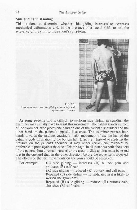
44 The Lumbar Spine
Side gliding in standing
This is done to determine whether side gliding increases' or decreasesmechanical deformation and, in the presence of a lateral shift, to test therelevance of the shift to the patient's symptoms.
Fig. 7:8.Test movements -side gliding in standing with
operator-assistance.
As some patients find it difficult to perform side gliding in standing theexaminer may initially have to assist this movement. The patient stands in frontof the examiner, who places one hand on one of the patient's shoulders and theother hand on the patient's opposite iliac crest. The examiner presses bothhands towards the midline, causing a major movement of the top half of thepatient's body in relation to the bottom half (Fig. 7:8). Instead of applying thepressure on the patient's shoulder, it may under certain circumstances bepreferable to press against the side of his rib cage. In all instances both shouldersof the patient should remain parallel to the ground. Side gliding must be testedfirst in the one and then in the other direction, before the sequence is repeated.The effects of the test movements on the pain should be recorded.
For example: (L) side gliding - increases (R) buttock pain andproduces (R) calf pain.(R) side gliding - reduced (R) buttock and calf pain.Repeated (L) side gliding - not indicated as it is likely toworsen the symptoms.Repeated (R) side gliding - reduces (R) buttock pain,abolishes (R) calf pain.

The Examination 45
When side gliding is found to be painful and blocked usually a lateral shift ispresent and in the treatment therapist assistance may be required.
When side gliding is found to be painful but not blocked, a lateral shift isunlikely to be present and in the treatment, therapist assistance is usuallyunnecessary.
When assessing the relevance of a lateral shift, we must determine whetherthe shift results from the same mechanical distrubance which has produced thepatient's pain. The shift is considered to be relevant when the movement of sidegliding alters the site or intensity of the pain. If no pain change occurs during theperformance of side gliding to the one side or the other, the scoliosis cannot beconsidered part of the mechanical problem causing pain.
Flexion in lyingThe patient, lying supine with the knees flexed and the feet flat on the couch, isasked to bend the knees onto the chest. Clasping the knees with his hands heapplies a firm pressure to produce maximum possible lumbar flexion (Fig. 7:9).The legs are then lowered to the starting position. The effects of the first testmovement on the pain are recorded. The movement should then be repeated upto ten times, ensuring that the maximum possible stretch is obtained during thelast few movements. The effects of repetition are recorded.
For example: Flexion in lying - increases pain (R) L5.Repeated flexion in lying - worsens pain (R) L5 andproduces (R) buttock pain.
Fig. 7:9.Test movements -flexion in
lying.
~
~"'-''''''''''''"""",,,~...:..JJ''''-'''''''''-~·''''''''-4'lr~ ....~~.........,~~
Extension in lyingThe patient, lying prone with the hands directly under the shoulders as in thetraditional press-up position, is asked to raise the top half of his body bystraightening the arms, at the same time keeping the thighs and legs on thecouch (Fig. 7: 10). If the pelvis lifts from the couch as the arms are straightened,we must make sure that the low back is allowed to sag as much as possible. Thepatient then returns to the starting position. The effects of this movement onthe pain are recorded. The movement should then be repeated up to ten times,

46 The Lumbar Spine
ensuring that the maximum possible stretch is obtained during the last fewmovements. Again, the effects of repetition are recorded.
For example: Extension in lying - reduces (L) buttock pain.Repeated extension in lying - abolishes (L) buttock painand produces central low back pain.
Fig. 7:10.Test movements - extension in
lying.
The test movements should be performed in a certain sequence. Theexaminer should not deviate from this method, unless it is obvious that topersist will inflict unnecessary pain on the patient.
In abbreviated form the sequence of test movements is as follows:FIS (flexion in standing)
Rep FIS (repeated flexion in standing)EIS (extension in standing)
Rep EIS (repeated extension in standing)SGIS (right and left side gliding in standing)
Rep SGIS (repeated right and left side gliding in standing)FIL (flexion in lying)
Rep FIL (repeated flexion in lying)ElL (extension in lying)
Rep ElL (repeated extension in lying)
V. OTHER EXAMINAnON PROCEDURESAt this stage it is appropriate to test the mobility of the hip joints and theintegrity of the sacro-iliac joints, if these are thought to contribute to theproblems of the patient. Should passive stretching of the various structuresabout the hip reproduce the symptoms complained of, this joint must beconsidered a possible source of the pain. The sacro-iliac joints, although notcommonly the cause of low back pain, turn out to be involved often enough tomake a fool of those failing to check them. In my opinion the tests described byCyriax31 are adequate to determine if the symptoms are arising from these joints.(Testing of sacro-iliac structures by using bony prominences as land marks andattributing pain to asymmetries so located is dishonest.)

The Examination 47
A more detailed neurological examination may be required, if there is theslightest suggestion of nerve root or spinal cord signs. In these cases thepatient's symptoms will be felt in the lower limb. Further neurologicalexamination should include testing of reflexes, muscle strength and sensation.
VI. CONCLUSIONSThe correlation of information from history, examination and test movementswill indicate whether the patient is suffering from the postural, derangementalor dysfunctional syndrome. This differentiation is essential, as it indicateswhich principle of treatment should be used.
Postural correctionIn the postural syndrome postural correction is the only treatment required.
The extension principleIn posterior derangement the extension principle should be applied whenextension reduces mechanical deformation. Thus in the treatment we aremaking use of those movements which centralised, decreased or stopped thepain during the examination. The patient should receive mechanical therapybased on the principle of extension. This includes: passive extension, extensionmobilisation, extension manipulation, extension in standing, extension in lying,and sustained extension.
Patients with an acute lumbar scoliosis or a lateral shift are placed in theextension principle category once correction of the lateral deformity has beenachieved.
In dYsfunction, however, the extension principle should be applied whenextension produces mechanical deformation. Thus in the treatment we aremaking use of those movements which actually produced the pain during theexamination. For treatment of dysfunction to be successful it is imperative thatsome pain be experienced, especially in the initial stages when adaptivelyshortened tissues must be stretched enough to assist them to regain theiroriginal length and elasticity. Pain thus produced during treatment of extensiondysfunction must always be across the back and near the midline, andsignificant buttock pain should not occur. The treatment procedures are basedon the principle of extension (see above).
The flexion principleIn anterior derangement the flexion principle should be applied when flexionreduces mechanical deformation. Again, in the treatment we are making use ofthose movements which centralised, decreased or stopped the pain during theexamination. The treatment consists of procedures based on the principle offlexion. This includes: flexion mobilisation, flexion manipulation, flexion inlying, and flexion in standing.
In dYsfunction, however, the flexion principle should be applied when flexionproduces mechanical deformation, and in the treatment we use those
48 The Lumbar Spine
movements which actually produced the pain during the examination. Somepain must be experienced in treatment of flexion dysfunction to be effective andtreatment consists of procedures utilising flexion (see above).
Generally speaking:
in the treatment of derangement we must choose the movement that relievesthe pain, as this movement decreases the mechanical deformation by reducingthe derangement.
but:
in the treatment of dysfunction we must always choose the movement thatproduces the pain, as this movement will gradually stretch and lengthencontracted soft tissues, eventually reducing mechanical deformation.
About seventy-five to eighty percent of all the patients with low back pain willrespond to the extension and flexion programmes. These patients have a verygood chance of becoming independent of therapists. They will be able toperform exercises to relieve themselves of pain without requiring techniquesperformed by specialist therapists. The remaining twenty to twenty-five percentof patients will require special techniques and manipulative procedures. Theexperienced practitioner will be able to identify these patients with a trial of testprocedures carried out over a twenty-four hour period.
Hopefully at this stage we will be able to categorise patients into theappropriate syndrome and adopt a principle of treatment. We are now ready toteach the patient how to correct the posture, restore lost function and, wherepossible, reduce derangement himself.
I will proceed with a discussion of the procedures which may be used intreatment, as these will be frequently referred to in chapters to follow. Then Iwill consider the three syndromes and their respective treatment approachesindividually.

CHAPTER 8
TechniqueThere are many differing philosophies and concepts surrounding the practise ofspinal manipulation and its effects on the pathologies which may exist in thespine. To satisfy all these philosophies an equal number of institutions hasdeveloped, teaching those wishing to learn. No matter what school presents itscase or which philosophy is adhered to, all manipulative specialists claim to havea high success rate. They all use techniques which vary in nature, applicationand intent; they proclaim that their own methods are superior to those used byothers; and yet, somehow they all obtain uniformly good results. Self-limitationof low back pain plays, of course, a significant role in this happy situation. Apartfrom this there are definite benefits which are obtained quickly by usingmanipulative techniques. .
Throughout the years I have practised many forms of mobilisation andmanipulation, including osteopathic and chiropractic techniques and thosetaught by Cyriax. I have come to believe that it is immaterial whichmanipulative school or philosophy one upholds for, no matter what particularmanipulations are used, the results of technique appear to be the same. Thegeneral procedures of Cyriax are just as effective as the finely developed specificprocedures of the osteopaths and chiropractors.
It is now well documented32• 33 that asymmetries in the facet joints and otherdevelopmental anomalies occur as regularly in the lumbar region as in the otherareas of the spine, and are present in up to fifty-two percent of the population.Where palpable movement restrictions and departures from the typical exist, itis impossible to state that these are either the cause of the patient's presentsymptoms or are likely to cause symptoms in the future. To conclude thatpalpable anomalies should be mobilised is no longer a tenable suggestion. Onemay well be dealing with perfectly 'normal' and asymptomatic asymmetricallumbar articulations.
Diagnosis by palpation is relevent only when associated with the increase anddecrease of mechanical deformation. Those who claim to 'feel' restrictions bypalpation and simultaneously fail to reproduce the patient's symptoms, are onlyfooling themselves and their patients. Enthusiasts searching for sacro-iliacpathology are frequently misled by this philosophy. The production of pain at acertain level by palpation and passive movement is not enough to justifytreatment at that level. The pain produced must be the one that has forcedconsultation - not a new pain induced by the palpation.
49

50 The Lumbar Spine
Various manipulative authorities maintain that mobilisation or manipulationof a hypermobile spinal segment should be avoided at all times. This is excellentadvice when it applies to pathological hypermobilities as fractures andspondylolisthesis. However, a hypermobile joint in itself is considered by someto cause mechanical pain more readily than joints with a normal or decreasedmobility. I must emphasise that a hypermobile joint becomes painful in thesame manner as any other joint. When it is placed on full stretch for a longenough period or when the stretch is severe enough pain will be felt. It sohappens that in the hypermobile segment a greater range of movement must beaccomplished before full stretch is achieved. Thus, hypermobility is not in itselfa painful state.
The basic misunderstanding that passive movements applied to ahypermobile joint are harmful, has led to the systematic development oftechniques of specific manipulation which spare the hypermobile joints andaffect only the adjacent hypomobile joints. It has also brought about unjustifiedcondemnation of those who advocate general instead of specific manipulation.
Many people have criticised and rejected the techniques of Cyriax which areconsidered to be unselective, non-specific and rather coarse in their application.However, at this time hundreds of Cyriax-trained doctors and physiotherapistssuccessfully apply his methods in many parts of the world and it appears that noharm is caused by his approach. If general manipulation were damaging indeed,then by now there would be enough evidence to support this contention. I amsure that the antagonists of Cyriax would not have allowed any compromisingsituation to pass unnoticed.
UnfortunatelY, it appears that some manipulative schools attempt to enhancethe quality and mystique of their skills by increasing the complexity of theirtechniques. They create the impression that to master the art and science ofmanipulation one must acquire very complex skills; these are possessed andtaught only by an elite group, and if one wishes to enjoy the highest reputationin manipulation one must join this elite group; the only way to do this is to trainat an exclusive establishment. This continuing trend is a threat to the scientificdevelopment of mechanical therapy.
The procedures which I have chosen for the treatment of low back pain aretotally unsophisticated and non-specific. In general, the techniques affect manysegments and localisation is not attempted. However, these procedures have animmediate cause and effect on the patient's symptoms. The reasons put forthfor their effectiveness may prove to be wrong in the future, but the effectivenessitself will not change: if patients are selected as suggested and the techniques areapplied as suggested, the procedures will be as effective fifty years from now asthey are today.
It is one of the main theses of this book that patients can be taught to manage,treat and control their own low back pain. In order to achieve this it is necessaryto depart from the traditional methods of mechanical treatment, whereby thetherapist does something to the patient to bring about change. In that case thepatient attributes his recovery, rightly or wrongly, to what was done to him and
Technique 51
for all future episodes of low back pain he commits himself to the care of thetherapist. By avoiding the use of therapist-technique in the initial stages oftreatment and substituting patient-technique, the patient will recognise that hisrecovery is clearly the result of his own efforts. Only few patients fail to assumeresponsibility for active participation in their treatment.
At present a great deal of research and academic discussion is taking place inattempts to establish the scientific validation of manipulation and explain itseffects. 34 The most obvious and most important effect of mobilisation andmanipulation is the increase of range of movement at any joint to which thetechniques are applied. This may be caused by a change in the position of aninternal structure, or by such an alteration in an adjacent structure that a morenormal function is possible than existed prior to the application of thetechnique. The increase in range of movement as obtained by manipulation andmobilisation can also be achieved by exercises when performed in a certain way.
It is my belief that an exercise becomes a mobilisation when performed with acertain frequency and in such a way that a rhythmical passive stretch is created.And in a similar manner a passive mobilisation can become a manipulation. Thesuggestion put forth is that mobilisation and manipulation are nothing morethan extended exercises, and an exercise can therefore become a mobilisation ormanipulation. Therapy based on self-mobilisation and self-manipulation hasarrived and it is possible to teach patients to practise mobilisation andmanipulation of their own spine.
Provided there is adequate instruction and careful explanation regarding theaims of treatment, the self-treatment concept can be applied successfully to mostlow back pain patients - that is, to all the patients with the postural syndrome,nearly all with the dysfunction syndrome, and about seventy-five percent of thepatients with the derangement syndrome. Thus, twenty-five to thirty percent ofpatients with low back pain will not recover on the exercise programme alone,and need additional techniques of either mobilisation or manipulation whichmust be applied by a specialist therapist. The only equipment required to treatpatients is an adjustable treatment table, the height of which can be varied andwhich has an end section of about two feet that can be inclined up or down.
THE PROCEDURES AND THEIR EFFECTSAfter much investigation to determine the optimum number of movementsnecessary to effect stretching of shortened tissues and alteration in the positionof the fluid nucleus, I have come to conclude that usually any significant changewill occur within ten to fifteen repetitions of the procedure and no benefit will beobtained by exceeding this number. Therefore, exercises are performed in seriesof ten to fifteen excursions each. The number of times in the day that a series ofexercises must be done varies according to the syndrome to be treated, theeffects to be obtained, and the capabilities of the patient involved.
Unless stated otherwise, exercises will be performed with an almostcontinuous rhythm. On each contraction the maximum possible range must bemaintained for a second or two. Each excursion must be followed by relaxation,
52 The Lumbar Spine
and a brief pause of only a fraction of a second is required. Normally patientscan complete between ten to fifteen excursions in one minute. Therefore nopatient should use the excuse that he has not got enough time to do theexercises as instructed.
In assessing progress the evaluation of pain changes is vital. A patient canimprove in several ways: the intensity of the pain may reduce; the frequencywith which the pain occurs may decrease; or the site of the pain may alter.Centralisation of the pain indicates that the patient is improving, although interms of intensity he may still feel one hundred percent of the pain originallycomplained of. In this case an explanation of the pain behaviour usually satisfiesthe patient regarding his progress.
If the site of pain has not changed on the patient's next visit, I always enquireabout the frequency and intensity of the pain in the following manner: "If youhad one hundred units of pain on your last visit, how many do you have todayin terms of (a) frequency; and (b) intensity?".
The procedures include patient-technique as well as therapist-technique. Inorder to facilitate quick reference the procedures are summarised below.
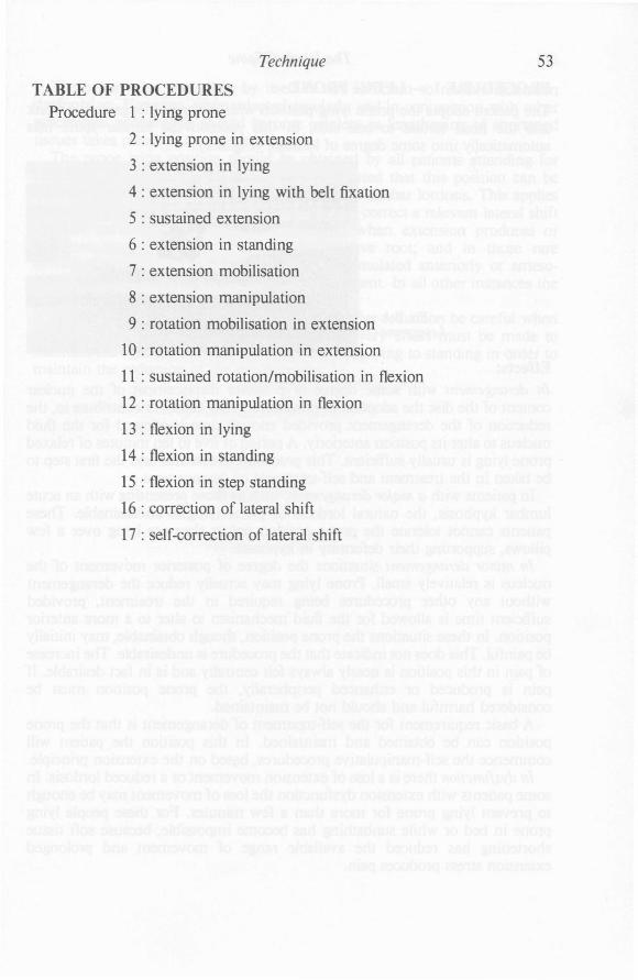
Technique
TABLE OF PROCEDURESProcedure 1: lying prone
2 : lying prone in extension
3 : extension in lying
4: extension in lying with belt fixation
5 : sustained extension
6 : extension in standing
7 : extension mobilisation
8 : extension manipulation
9 : rotation mobilisation in extension
10: rotation manipulation in extension
11 : sustained rotation/mobilisation in flexion
12 : rotation manipulation in flexion
13 : flexion in lying
14 : flexion in standing
15 : flexion in step standing
16 : correction of lateral shift
17 : self-correction of lateral shift
53
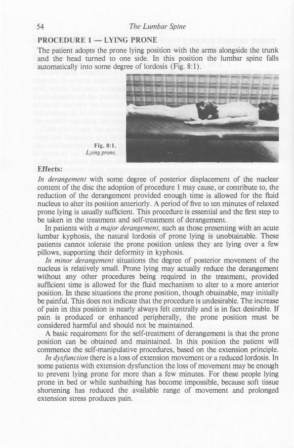
54 The Lumbar Spine
PROCEDURE 1 - LYING PRONEThe patient adopts the prone lying position with the arms alongside the trunkand the head turned to one side. In this position the lumbar spine fallsautomatically into some degree of lordosis (Fig. 8:1).
Fig. 8:1.Lying prone.
Effects:In derangement with some degree of posterior displacement of the nuclearcontent of the disc the adoption of procedure 1 may cause, or contribute to, thereduction of the derangement provided enough time is allowed for the fluidnucleus to alter its position anteriorly. A period of five to ten minutes of relaxedprone lying is usually sufficient. This procedure is essential and the first step tobe taken in the treatment and self-treatment of derangement.
In patients with a major derangement, such as those presenting with an acutelumbar kyphosis, the natural lordosis of prone lying is unobtainable. Thesepatients cannot tolerate the prone position unless they are lying over a fewpillows, supporting their deformity in kyphosis.
In minor derangement situations the degree of posterior movement of thenucleus is relatively small. Prone lying may actually reduce the derangementwithout any other procedures being required in the treatment, providedsufficient time is allowed for the fluid mechanism to alter to a more anteriorposition. In these situations the prone position, though obtainable, may initiallybe painful. This does not indicate that the procedure is undesirable. The increaseof pain in this position is nearly always felt centrally and is in fact desirable. Ifpain is produced or enhanced peripherally, the prone position must beconsidered harmful and should not be maintained.
A basic requirement for the self-treatment of derangement is that the proneposition can be obtained and maintained. In this position the patient willcommence the self-manipulative procedures, based on the extension principle.
In dYsfunction there is a loss of extension movement or a reduced lordosis. Insome patients with extension dysfunction the loss of movement may be enoughto prevent lying prone for more than a few minutes. For these people lyingprone in bed or while sunbathing has become impossible, because soft tissueshortening has reduced the available range of movement and prolongedextension stress produces pain.
Technique 55
The prone lying procedure by itself is not sufficient to resolve extensiondysfunction. However, when adopted regularly and in conjunction with otherprocedures, prone lying should become painless as lengthening of shortenedtissues takes place.
The prone lying position should be obtained by all patients attending fortreatment of low back pain. It has been suggested that this position can beharmful because it increases and accentuates the lumbar lordosis. This appliesonly in a few situations: when we have failed to correct a relevant lateral shiftprior to assuming the prone lying position; when extension produces orincreases the compression on the sciatic nerve root; and in those rarederangements where nuclear material has accumulated anteriorly or anterolaterally, and prone lying increases the derangement. In all other instances theprone lying position is highly beneficial.
Patients with posterior derangement should after reduction be careful whenarising from the prone position to standing. Every effort must be made tomaintain the restored lordosis while moving from lying to standing in order tomaintain the reduction of the derangement.

56 The Lumbar Spine
PROCEDURE 2 - LYING PRONE IN EXTENSIONThe patient, already lying prone, places the elbows under the shoulders andraises the top half of his body so that he comes to lean on elbows and forearmswhile pelvis and thighs remain on the couch. In this position the lumbar lordosisis automatically increased. Emphasis must be placed on allowing the low back tosag and the lordosis to increase (Fig. 8:2) .
Fig. 8:2.Lying prone in extension.
Effects:Procedure 2 is a progression of procedure 1 and merely enhances its effects byincreasing extension.
Again, in derangement some time must be allowed to affect the contents ofthe disc and, if possible patients should remain in this position for five to tenminutes. In more acute patients sustained extension may not be well tolerateddue to pain, and initially we must rely on the use of intermittent extension.

Technique 57
PROCEDURE 3 - EXTENSION IN LYINGThe patient, already lying prone, places the hands (palms down) near theshoulders as for the traditional press-up exercise. He now presses the top half ofhis body up by straightening the arms, while the bottom half, from the pelvisdown is allowed to sag with gravity. The top half of the body is then loweredand the exercise is repeated about ten times. The first two or three movementsshould be carried out with some caution, but once these are found to be safe theremaining extension stresses may become successively stronger until the lastmovement is made to the maximum possible extension range. If the first seriesof exercises appears beneficial, then a second series may be indicated. Morevigour can be applied and a better effect will be obtained if the last two or threeextension stresses are sustained for a few seconds.
It is essential to obtain the maximum elevation by the tenth excursion andonce obtained the lumbar spine should be permitted to relax into the mostextreme 'sagged' position (Fig. 8:3).
Fig. 8:3.Extension in lying.
Effects:This procedure is a further progression of the previous two. Instead of asustained extension stress on the contents and surrounding structures of thelumbar segments, there is now an intermittent extension stress, having apumping as well as a stretching effect.
This procedure is the most important and effective in the treatment ofderangement as well as extension dysfunction. The very maximum degree of

58 The Lumbar Spine
extension possible without external assistance, is obtained with this exercise.An increase of central low back pain at maximum elevation can be expected andshould not cause any concern as it will gradually wear off. It is usually describedas a strain pain and differs from the pain which has caused initial consultation.
In addition to the effects on the disc and periarticular structures there are twoother physiologically related phenomena that could possibly result from theperformance of this exercise.
The self sealing phenomenon
Evidence gathered by Markolf and Morris46 suggests that a self sealingmechanism exists within the disc and appears shortly after injury. The initialinjury weakens the annulus but appropriate stress applied subsequently resultsin restoration of near normal strength, suggesting that the disc has a remarkablerecovery ability and that certain stresses may enhance rapid recovery. Whiteand Panjabi44 conclude that the self sealing phenomena is mechanical in natureand is not dependent on the viscosity or softness of the disc, for the study wasperformed on degenerative as well as normal discs.
My question arising from this information is ... Does the performance ofrepeated passive extension in lying cause a reversal of the posterior migration ofthe nucleus into the developing radial fissure? Does the movement then initiatethe self sealing phenomena?
Cartilaginous repair
Following trauma articular surfaces are normally rested or immobilised topermit healing. It is well known that scar tissue is laid down under thesecircumstances and damaged articular cartilage is replaced with fibrous collagen.Recent investigations by Salter48 suggest that if passive continuous motion isapplied to joints containing .traumatised intra articular cartilage, the damagedcartilage is replaced by true cartilaginous cells instead of scar tissue, and further,these joints do not develop arthritic changes subsequently. The evidence has yetto be confirmed in human studies. We can now pose the question ... Does theregular performance of passive extension following lumbar disc damageenhance the quality or improve the nature of the healing tissues of the posteriorannulus?

Technique 59
PROCEDURE 4 - EXTENSION IN LYING WITH BELT FIXATIONThe patient's position and the exercise are the same as in the third procedure,but now a fixating belt is placed at or just below the segments to be extended.The safety belt is the first simple external aid, used to enhance maximumextension. It does so by preventing the pelvis and lumbar spine lifting from thecouch. Other methods of restraint may be used effectively, for example the bodyweight of a young son or daughter when exercising at home (Fig. 8:4).
Fig. 8:4.Extension in lying with belt
fixation.
Effects:This procedure creates a greater and more localised passive extension stressthan the previous ones. It is particularly suitable for stretching in the case ofextension dysfunction, and is more often required in dysfunction than inderangement.
In dysfunction some pain will be experienced in the small of the back whileexercising, because contracted tissues are being stretched. In derangement therules pertaining to the centralisation phenomenon must be observed, and theprocedure stopped if peripheral pain is produced or increased.
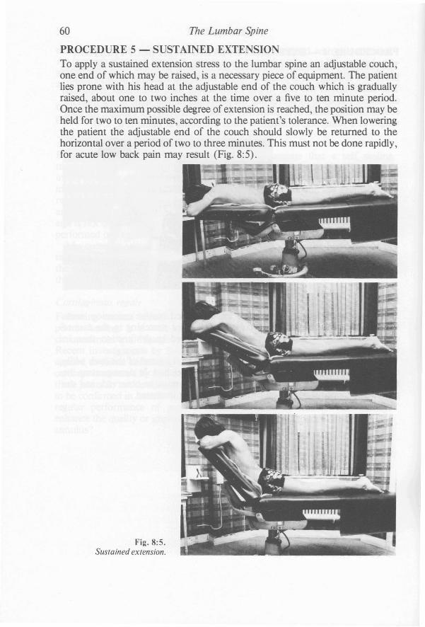
60 The Lumbar Spine
PROCEDURE 5 - SUSTAINED EXTENSIONTo apply a sustained extension stress to the lumbar spine an adjustable couch,one end of which may be raised, is a necessary piece of equipment. The patientlies prone with his head at the adjustable end of the couch which is graduallyraised, about one to two inches at the time over a five to ten minute period.Once the maximum possible degree of extension is reached, the position may beheld for two to ten minutes, according to the patient's tolerance. When loweringthe patient the adjustable end of the couch should slowly be returned to thehorizontal over a period of two to three minutes. This must not be done rapidly,for acute low back pain may result (Fig. 8:5).
Fig. 8:5.Sustained extension.
Technique 61
Effects:The procedure is predominantly used in the treatment of derangement. Theeffect is similar to that of the third procedure, but a time factor is added with thegraduated increase and the sustained nature of the extension. In certaincircumstances a sustained extension stress is preferable to a repeated extensionstress.
The centralisation phenomenon must be watched closely. Any suggestionthat the pain is moving or increasing peripherally must lead to the immediatebut slow lowering of the couch. It is interesting to note that an increase incentral low back pain as the couch is lowered nearly always indicates a goodresponse to the treatment, whereas when there is no increase in central painpatients tend to have little or no improvement following this procedure.

62 The Lumbar Spine
PROCEDURE 6 - EXTENSION IN STANDINGThe patient stands with the feet well apart and places the hands (fingerspointing backwards) in the small of the back across the belt line. He leansbackwards as far as possible, using the hands as a fulcrum, and then returns toneutral standing. The exercise is repeated about ten times. As with extension inlying it is necessary to move to the very maximum to obtain the desired result(Fig. 8:6).
Fig. 8:6.Extension in standing.
Effects:Extension in standing produces similar effects on derangement and dysfunctionas extension in lying, but it is less effective in the earlier treatment stages of bothsyndromes. Whenever extension in lying is prevented by circumstances, anextension stress can be given by extension in standing.
In derangement, extension in standing is designed to reduce accumulation ofnuclear material in the posterior compartment of the intervertebral joint,provided this accumulation is not gross. In the latter case extension in lying willhave to be performed first. The procedure is very important in the prevention ofthe onset of low back pain during or after prolonged sitting or activitiesinvolving prolonged stooping, and is very effective when performed before painis actually felt.

Technique 63
PROCEDURE 7 - EXTE SION MOBILISATIOThe patient lies prone as for procedure 1. The therapist stands to one side of thepatient, crosses the arms and places the heels of the hands on the transverseprocesses of the appropriate lumbar segment. A gentle pressure is appliedsymmetrically and immediately released, but the. hands must not lose contact.This is repeated rhythmically to the same segment about ten times. Eachpressure is a little stronger than the previous one, depending on the patient'stolerance and the behaviour of the pain. The procedure should be applied to theadjacent segments, one at a time, until all the areas affected have been mobilised(Fig. 8:7).
Fig.8:7a.
Fig.8:7b.
Fig.8:7c.
Fig. 8:7a and b.Positioning ofhands prior toextension mobilisation.
Fig.8:7c.Extension mobilisation.
64 The Lumbar Spine
Effects:In this procedure the external force applied by the therapist enhances the effectson derangement and dysfunction as described for the previous extensionprocedures.
In general, symmetrical pressures are used on patients with central andbilateral symptoms. Therapist-technique must be added when the patient isunable to reduce derangement or resolve dysfunction by the self-treatmentprocedures. That situation appears in derangement when instead ofprogressively lessening pain extension in lying (procedure 3) causes the samepain with each repetition. Under those circumstances extension mobilisation isindicated.

Technique 65
PROCEDURE 8 - EXTENSION MANIPULATIONThere are many techniques devised for manipulation of the lumbar spine inextension. It is not important which technique is used, provided the technique isperformed on the properly selected patient and applied in the correct direction.The technique that I recommend is similar to the first two manipulationsdescribed by Cyriax31 for the reduction of a lumbar disc lesion.
The patient lies prone as for procedure 1. The therapist stands to one side ofthe patient and, having selected the affected segment, places the hands on eitherside of the spine as for the technique of extension mobilisation (procedure 7),which is always applied as a premanipulative testing procedure. If followingtesting the manipulation is indicated, the therapist leans over the patient withthe arms at right angles to the spine and forces slowly downwards until thespine feels taut. Then a high velocity thrust of very short amplitude is appliedand immediately released (Fig. 8:8) .
Fig. 8:8.Extension manipulation.
Effects:The effects of the external force and the reasons for its use are the same as forprocedure 7. When the desired result is not obtained with the mobilisingtechniques, manipulation is indicated under certain circumstances.
The extension thrust is used by many manipulators, and there is difference ofopinion regarding the structures that may be influenced by this technique.Cyriax31 states that it reduces derangement of an annular fragment of the disc.Others propose reduction of facet locking, tearing of adhesions and reduction ofnerve root entrapment. Whatever the true mechanism may be, properly selectedpatients often experience a click or a dull thud. In most instances the click isfollowed by a change, usually an improvement, in the patient's signs andsymptoms.

66 The Lumbar Spine
PROCEDURE 9 - ROTATION MOBILISATION IN EXTENSIONThe position of patient and therapist is the same as for procedure 7. Bymodifying the technique of extension mobilisation so that the pressure isapplied first to the transverse process on the one side and then on the other sideof the appropriate segment a rocking effect is obtained. Each time the vertebra isrotated away from the side to which the pressure is applied - for example,pressure on the right transverse process of the fourth lumbar vertebra causes leftrotation of the same vertebra. The technique should be repeated about ten timeson the involved segment and, if indicated, adjacent segments should be treatedas well (Fig. 8:9).
Fig. 8:9.Rotation mobilisation in
extension.
Effects:Also here the external force applied by the therapist enhances the effects onderangement and dysfunction as described for the previous extensionprocedures. The reasons for adding therapist-technique are the same as forprocedure 7.
In general, unilateral techniques are likely to effect unilateral or asymmetricalsymptoms sooner and more efficiently than bilateral or central techniques. Butonce centralisation of symptoms has taken place, treatment may be continuedwith central or bilateral techniques. Thus, in derangement rotation mobilisationin extension may have to be performed first to bring about centralisation ofnuclear material in the disc. This is followed by symmetrical extensionmobilisation to restore the nucleus to its more anterior position.
Occasionally a click is felt during mobilisation. A click often indicates areduction of derangement, and we should immediately assess if this is the case.If the patient has improved significantly as a result of this technique, any furthertreatment may disturb the reduction and the treatment session should beterminated at this point.
During the procedure the patient may describe an enhancement of the painby pressure on one side with a corresponding reduction of the pain by pressureon the other side. This is valuable information on which further treatment will

Technique 67
be based. We must keep in mind that we are affecting the pain by increasing ordecreasing mechanical deformation. In dysfunction an increase in mechanicaldeformation with certain limits is desirable and pain should be produced orincreased with the application of the technique. In derangement an increase inmechanical deformation is extremely undesirable, and we should aim for adecrease instead with centralisation, reduction or abolition of the pain.Therefore, precise identification of the syndrome to be treated is essential todetermine whether rotation mobilisation should be performed towards thepainful or the painfree side.
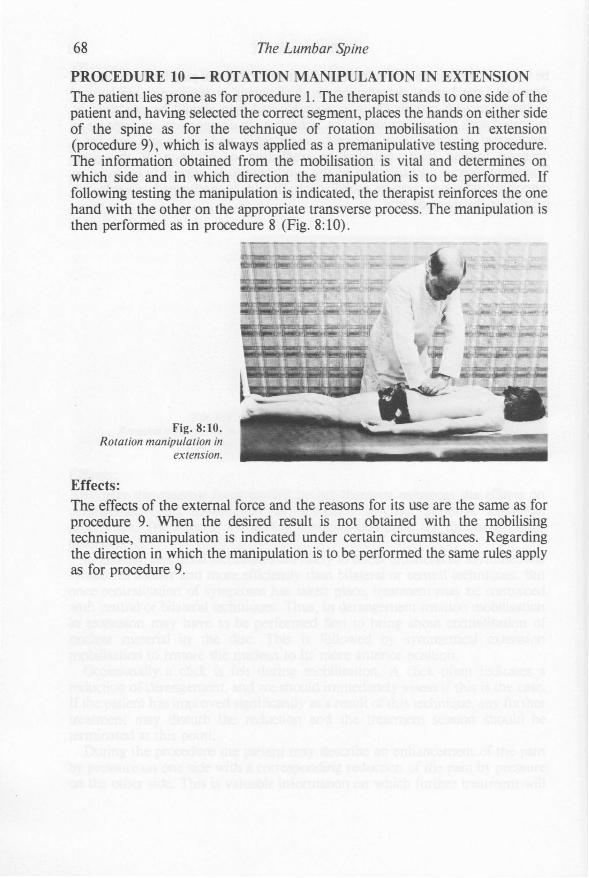
68 The Lumbar Spine
PROCEDURE 10 - ROTATION MANIPULATION IN EXTENSIONThe patient lies prone as for procedure 1. The therapist stands to one side of thepatient and, having selected the correct segment, places the hands on either sideof the spine as for the technique of rotation mobilisation in extension(procedure 9), which is always applied as a premanipulative testing procedure.The information obtained from the mobilisation is vital and determines onwhich side and in which direction the manipulation is to be performed. Iffollowing testing the manipulation is indicated, the therapist reinforces the onehand with the other on the appropriate transverse process. The manipulation isthen performed as in procedure 8 (Fig. 8: 10).
Fig. 8:10.Rotation manipulation in
extension.
Effects:The effects of the external force and the reasons for its use are the same as forprocedure 9. When the desired result is not obtained with the mobilisingtechnique, manipulation is indicated under certain circumstances. Regardingthe direction in which the manipulation is to be performed the same rules applyas for procedure 9.

Technique 69
PROCEDURE 11 -SUSTAINED ROTATIO IMOBILISATION IN FLEXIOThe patient lies supine on the couch, and the therapist stands on the side towhich the legs are to be drawn, facing the head end of the couch. The patient'sfar shoulder is held firmly on the couch by the therapist's near hand, providingfixation and stabilisation. With the other hand the therapist flexes the patient'ships and knees to a rightangle and carries them towards himself, causing thelumbar spine to rotate. With the patient's ankles resting on the therapist's thighthe knees are allowed to sink as far as possible and the legs are permitted to restin that extreme position. The lumbar spine is now hanging on its ligaments in aposition which combines side bending and rotation. By pushing the kneesfurther towards the floor the therapist applies overpressure to take up theremaining slack in the lumbar spine. Depending on the purpose for which theprocedure is used, the position of extreme rotation is maintained for a shorter orlonger period (Fig. 8: 11) .
Fig. 8:11.Sustained
rotation/mobilisation inflexion.
Effects:The procedure is mainly used in derangement. Sustained rotation for aboutthirty to forty seconds provides the time factor required to allow alteration of theposition of the fluid nucleus within the disc. In those situations where time isimportant in the reduction this procedure may effect relief that will not beobtained by the much quicker performed rotation thrust (procedure 12). Dtlfingthe period that rotation is sustained the patient should be watched closely andasked constantly about the behaviour of pain. Any sign of peripheralisation ofsymptoms indicates that more than enough time has been spent in this position.
The procedure may also be used as a mobilising technique in dysfunction, oras premanipulative testing in dysfunction as well as in derangement. In thesecases the rotation is less sustained or performed in a rhythmical mobilisingmanner.
If a small therapist cannot reach across the patient to stabilise his shoulder, aseat belt fastened firmly across the patient's upper chest provides adequatefixation. Alternatively, a second person may be used to hold the patient down.

70 The Lumbar Spine
PROCEDURE 12 - ROTATION MANIPULATION IN FLEXIONThe sequence of procedure 11 must be followed completely to perform therequired pre-manipulative testing. If the manipulation is indicated a suddenthrust of high velocity and small amplitude is performed, moving the spine intoextreme side bending and rotation (Fig. 8:12).
Fig. 8:12.Rotation manipulation in
flexion.
Technique 71
Effects:There are many techniques devised for rotation manipulation of the lumbarspine. When rotation of the lumbar spine is achieved by using the legs of thepatient as a lever or fulcrum of movement, confusion arises as to the direction inwhich the lumbar spine rotates. This is judged by the movement of the uppervertebrae in relation to the lower - for example, if the patient is lying supineand the legs are taken to the right, then the lumbar spine rotates to the left.
It has become widely accepted that rotation manipulation of the spine shouldbe performed by rotation away from the painful side. This has applied toderangement as well as dysfunction, because hitherto no differentiation hasbeen made between these syndromes. I wish to emphasise that, while thispractise is correct for the majority of derangements, it is useless at best whenapplied to dysfunction. Because there are more low back pain patients withdysfunction than with derangement, it is essential to determine precisely whichsyndrome is present.
Premanipulative testing with the lumbar spine held in full rotation stretchprior to the administration of the manipulative thrust will indicate if we havechosen the correct direction in which the manipulation should be performed. Inderangement this will always be in the direction that causes a decrease,centralisation or abolition of unilateral pain. Reduction of symptoms may beachieved with the lumbar spine rotated either towards or away from the painfulside. The deciding factor for the direction of the manipulation must be thereduction of mechanical deformation, irrespective of the fact whether this isachieved with movement towards or away from the painful side. In dysfunctionthe patient should experience an enhancement ofpain, but the pain must neverperipheralise. The patient with dysfunction may have to be manipulated in bothdirections, which is rarely the case in derangement.
A rotation manipulation is often described as having a gapping effect on thefacet joints. Although this idea is widely held, it is almost impossible to detecton x-rays, taken during manipulation, that movement occurs at the facet joints.However, significant movement can be,observed between the vertebral bodies.It is my contention that a rotation manipulation influences the nucleus andannulus of the disc more than the facet joints. Due to the torsion and sidebending provided with the procedure, the annular wall must become tightenedand under increased tension. This could possibly influence a distorted nucleus,at least as long as the annular wall is intact.
Manipulation consistently applied with the painful side uppermost in thebelief that gapping of the facets is required to relieve the patient's symptoms, isno longer tenable. We must be guided by the increase and decrease ofmechanical deformation instead of conjecture.
The term 'rotation manipulation' is perhaps incorrect, as there is a muchgreater side bending than rotation component when spinal rotation isperformed.
Lightly built therapists who feel that they have inadequate weight to performthe rotation thrust procedure in flexion, can achieve equally satisfactory resultsby using the sustained rotation procedure.

72 The Lumbar Spine
PROCEDURE 13 - FLEXION IN LYINGThe patient lies supine with the knees and hips flexed to about forty-five degreesand the feet flat on the couch. He bends the knees up towards the chest, firmlyclasps the hands about them and applies overpressure to achieve maximumstress. The knees are then released and the feet placed back on the couch. Thesequence is repeated about ten times. The first two or three flexion stresses areapplied cautiously, but when the procedure is found to be safe the remainingpressures may become successively stronger, the last two or three being appliedto the maximum possible (Fig. 8:13).
Fig. 8:13.Flexion in lying.
Effects:Flexion in lying causes a stretching of the posterior wall of the annulus, theposterior longitudinal ligament, the capsules of the facet joints, and other softtissues. As the movement takes place from below upwards the lower lumbarand lumbo-sacral joints are placed on full stretch at the beginning of the exerciseas soon as movement is initiated. Thus, the procedure is very important inflexion dysfunction when shortening of posterior soft tissues has occurred.
The procedure should always be performed following stabilisation of areduced posterior derangement. This ensures that no flexion loss remains afterthe patient has become symptom free. By keeping the patient in extension al1davoiding flexion as healing takes place, we permit scar formation with the jointsin a shortened position. This shortened position will be held by the scar as itcontracts, the patient remaining painfree but unable to flex. Any attempts to
Technique 73
perform flexion beyond the limits imposed by the contracting scar, will producepain. Therefore, further flexion will be avoided and adaptive shorteninggradually worsens.
Flexion in lying performed regularly following reduction of posteriorderangement allows the formation ofan extensible scar in the midst ofan elasticstructure. Should we permit an inextensible scar to remain in the midst of anelastic structure, - the disc in this case - then sooner or later the patient willinadvertently move beyond the limitations of the scar, which results in furthertearing of soft tissues and apparent recurrence of the derangement condition.This basic complication of healing exists throughout the muscular as well as thearticular systems.
Flexion in lying also causes a posterior movement of the fluid nucleus andwill be utilised in anterior derangement situations (Derangement seven) toreverse the excessive anterior position of the nucleus.
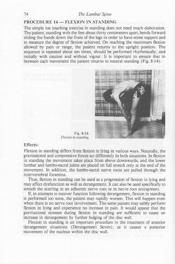
74 The Lumbar Spine
PROCEDURE 14 - FLEXION IN STANDINGThe simple toe touching exercise in standing does not need much elaboration.The patient, standing with the feet about thirty centimeters apart, bends forwardsliding the hands down the front of the legs in order to have some support andto measure the degree of flexion achieved. On reaching the maximurn flexionallowed by pain or range, the patient returns to the upright position. Thesequence is repeated about ten times, should be performed rhythmically, andinitially with caution and without vigour. It is important to ensure that inbetween each movement the patient returns to neutral standing (Fig. 8:14).
Fig. 8:14.Flexion in standing.
Effects:Flexion in standing differs from flexion in lying in various ways. Naturally, thegravitational and compressive forces act differently in both situations. In flexionin standing the movement takes place from above downwards, and the lowerlumbar and lurnbo-sacral joints are placed on full stretch only at the end of themovement. In addition, the lumbo-sacral nerve roots are pulled through theintervertebral foramina.
Thus, flexion in standing can be used as a progression of flexion in lying andmay affect dysfunction as well as derangement. It can also be used specifically tostretch the scarring in an adherent nerve root or in nerve root entrapment.
If, in attempts to recover function following derangement, flexion in standingis performed too soon, the patient may rapidly worsen. This will happen evenwhen there is no nerve root involvement. The same patient may safely performflexion in lying and experience no increase in pain. It would appear that thegravitational stresses during flexion in standing are sufficient to cause anincrease in derangement by further bulging of the disc wall.
Flexion in standing is an important procedure in the treatment of anteriorderangement situations (Derangement Seven), as it causes a posteriormovement of the nucleus within the disc wall.

Technique 75
PROCEDURE 15 - FLEXION IN STEP STANDINGIn this procedure the patient stands on one leg while the other leg rests with thefoot on a stool so that hip and knee are about ninety degrees flexed. Keeping theweight bearing leg straight the patient draws himself into a flexed position,firmly approximating the shoulder and the already raised knee (both being onthe same side). If possible the shoulder should be moved even lower than theknee. The patient may apply further pressure by pulling on the ankle of theraised foot. The pressure is then released and the patient returns to the uprightposition. The sequence is repeated about six to ten times. It is important that thepatient returns to neutral standing and restores the lordosis in between eachmovement (Fig. 8:15).
Fig. 8:15. Flexion in step standing.
Effects:This procedure causes an asymmetrical flexion stress on the affected segments.It is applied when there is a deviation in flexion, which may occur indysfunction as well as derangement. Both in dysfunction and derangement theleg to be raised is that opposite to the side to which the deviation in flexion istaking place - for example, in deviation in flexion to the left the right leg has tobe raised.
In dysfunction asymmetrically shortened structures are stretched by flexionin step standing, provided it is performed often enough with the application ofsufficient stress.
In derangement the procedure will influence the off-center nucleus so that itmoves to a more central position, thus allowing the normal pathway of flexionto be regained. Where deviation in flexion is due to derangement some patientswill experience a reversal of the deviation if the procedure is performed toooften. Thus the exercise must be repeated only five to six times before checkingif flexion in standing has been reduced to normal.

76 The Lumbar Spine
PROCEDURE 16 - CORRECTION OF LATERAL SHIFTThis procedure has two parts: first the deformity in scoliosis is corrected; then, ifpresent, the deformity in kyphosis is reduced and full extension is restored.
The patient, standing with the feet about thirty centimeters apart, is asked toclearly define the areas where pain is being felt at present. The therapist standson the side to which the patient is deviating and places the patient's near elbowat a right angle by his side. The elbow will be used to increase the lateralpressure against the patient's rib cage.
The therapist's arms encircle the patient's trunk, clasping the hands about therim of the pelvis. Now the therapist presses his shoulder against the patient'selbow, pushing the patient's rib cage, thoracic and upper lumbar spine awaywhile at the same time drawing the patient's pelvis towards himself. In thismanner the deformity in scoliosis is reduced and, if possible slightlyovercorrected.
Initially, there will be significant resistance to the procedure, which mayactually cause an increase in pain. It is quite safe to continue with correction aslong as centralisation of pain takes place, and therefore the patient must bequestioned continually about the behaviour of his pain. Relaxation of the patientduring the procedure is very important and we should always try to get thepatient to 'let it all go'. The first pressure in the series should be a gentle gradualsqueeze which is held momentarily and then released. After this an accurateassessment of the patient's reactions must be made. Experience has taught methat too much pressure or too fast a correction in the initial stages may result infainting and collapse of the patient. If well tolerated the pressure is applied alittle further each time. As correction progresses over ten to fifteen rhythmicallyapplied pressures, the patient usually describes that the pain moves from aunilateral to a central position, and by the time over correction is achieved therewill be a significant reduction in intensity of the pain or the pain may havemoved slightly to the opposite side. If after a few rhythmical pressures noprogress is made in the correction, it may be necessary to apply a longer andmore sustained pressure.
Sometimes reduction may be felt clearly by the therapist and the patient'strunk is felt to move slowly but surely from its previously held position. Inlightly-built or tall and slender patients shift correction may occur quite easily,and only a few minutes of ten to fifteen pressures are required to reduce thederangement. On the other hand, some acute lateral shifts are extremelydifficult to reduce and one may have to perform five or six series of correctivepressures.
Assuming that correction of the deformity in scoliosis has been achieved, wemust now proceed with restoring the lumbar lordosis. This is preferablycommenced in the standing position. The patient no longer exhibits a lumbarscoliosis but may still have a kyphosis. The therapist, holding the patient as forcorrection of the scoliosis, must maintain slight over correction while movingthe low back of the patient into the beginning of extension. A few movementswill indicate the ease with which the lordosis will be restored. If the extension

Technique 77
range improves rapidly it is usually better to recover as much extension aspossible in the standing position. If extension does not increase rapidly, then itis better to change to extension in lying. This procedure should produce a steadyand continuing reduction of central pain, and it should automatically follow forall patients with a postero-lateral derangement once the scoliosis has beencorrected and the symptoms have centralised (Fig. 8: 16).
Fig. 8:16.Correction of lateral shift.
Effects:These will be discussed following the next procedure.

78 The Lumbar Spine
PROCEDURE 17 - SELF-CORRECTION OF LATERAL SHIFTHaving corrected the lateral shift and the blockage to extension, it is nowessential to teach the patient to perform self-correction by side gliding instanding followed by extension in standing. This must be done on the very firstday, so that the patient is equipped with a means of reducing the derangementhimself at first sign of regression. Failure to teach self-correction will lead torecurrence within hours, ruining the initial reduction, and the patient will returnthe next day with the same deformity as on his first visit.
I have discarded the technique of self-correction as described previously12 andinstead I now teach patients to respond to pressures applied laterally againstshoulder and pelvis. Initially, therapist' assistance is required. Patient andtherapist stand facing each other. The therapist places one hand on the patient'sshoulder on the side to which he deviates, and the other hand on the patient'sopposite iliac crest. The therapist applies pressure by squeezing the patientbetween his hands, ensuring that the patient's shoulders remain parallel to theground. When over correction has been achieved by the therapist, the mobilitymust be maintained by the patient who is therefore taught to actively respond tothe pressures applied by the therapist. After some practise, remembering to keepthe shoulders parallel to the ground, the heels on the ground and the kneesstraight, the patient can correct his own deformity. It is important to make thepatient stand for a minute or two in the extreme over corrected position.Immediately following correction of the lateral deformity full extension must berestored. In the corrected standing position the patient must perform about tenrepetitions of extension in standing.
Fig. 8: 17. Self-correction of lateral shift.

Technique 79
It must be emphasised that as long as the lordosis is retained there is littlechance of recurrence of the derangement. If the patient is unable to maintain thereduction, he must perform self-correction during the day at regular intervals.The patient is advised to perform a series of extension in lying exercises aftereach session of self-correction (Fig. 8: 17).
Effects and points to note:It must be obvious that the last two procedures mainly influence the disc inderangement situations. In all instances time must be allowed for the reductionof the derangement to take place. Failure to correct the common lateral shift isusually the fault of the therapist, who has not taken enough time to allow achange in the contents of the disc to occur. The lateral shift correction includingthe restoration of extension must be an unhurried process and may take up toforty-five minutes in difficult patients. Constant repetition of the correctiveprocedures is necessary.
During the correction period there must be a continual reference to thepatient's symptoms. When there is any suggestion of the production orenhancement of limb pain, treatment must be applied with great caution and achange should be made to the angle of flexion or extension in which the lateralshift is being corrected.
Maintenance of the lordosis following the reduction of the derangement mustbe emphasised from the first day, and we must ensure that the patient hasadequate knowledge to retain his lordosis while sitting. The most commoncause of regression or recurrence of symptoms within a few hours of reductionis a poor sitting posture. For example, after a successful reduction a patient maydrive home for twenty to thirty minutes and on leaving the car full derangementhas recurred. This occurs commonly and must be anticipated. In order to copewith this problem the patient should be provided with a lumbar support - forexample, a rolled towel or something similar - to accentuate and support thelordosis in sitting.
In acute cases patients should go home (and not back to work) after reductionhas been achieved. On arrival home they should move directly to a mirror andcheck if derangement has recurred. If so, they should perform the selfcorrection procedures before it becomes too difficult to reduce the lateral shiftwithout outside help. Then they must lie prone for a few minutes on a bed or onthe floor prior to performing a series of extension in lying exercises. This patternshould be repeated each hour or whenever possible throughout the day, andbetween exercise sessions the patient must be lying and not sitting. On retiringfor bed the patient must lie supine in the over corrected position, with a lumbarsupport in the small of the back to maintain the lordosis, for about thirtyminutes before going to sleep. The next morning there is usually a significantreduction in the deformity and pain after the correction procedure has beenperformed once or twice, although when first awaking the pain may be quitenoticeable on movement.

80 The Lumbar Spine
Most patients with an acute lateral shift should be pain free in about seventytwo hours. A few difficult patients may take longer and many will take less timeto reduce. Failure usually occurs because self-correction has not been taughtadequately on the first visit.
&iatic scoliosis with the complication of nerve root compression is moredifficult to reduce than the uncomplicated lumbar scoliosis because the damageto the annulus is of such magnitude that outer fibres are distended. Reversal atthis stage in the development of the derangement is always difficult. Treatmentmay also be rather difficult when patients have a lateral shift towards the painfulside instead of the more common shift away from the pain. This may be becausean escape of nuclear material has allowed the vertebral margins to approximate.Whatever the reason for this phenomenon, patients in this group are slow toprogress and a few may completely fail to respond to treatment. Recovery canbe so slow that one is never certain whether it is due to the treatment or just tothe passage of time. Treatment procedures are the same as for patients who havea lateral shift away from the painful side, and the deformity must be correctedwith due regard of the centralisation phenomenon. As in all cases, once the painhas resolved we must ensure that the function is restored, the posture iscorrected and prophylaxis is taught.
There is a significantly higher incidence of sciatica in patients with a lateralshift towards the painful side and the incidence of failure to reductive measuresis also higher in this group.
CHAPTER 9
The Postural SyndromeDEFINITIONI would define the postural syndrome as mechanical deformation of posturalorigin causing pain of a strictly intermittent nature, which appears when the softtissues surrounding the lumbar segments are placed under prolonged stress.This occurs when a person performs activities which keep the lumbar spine in arelatively static position (as in vacuuming, gardening) or when they maintainend positions for any length of time (as in prolonged sitting).
HistoryPatients with postural pain are usually aged thirty or under. Frequently theyhave a sedentary occupation and in general they lack physical fitness. Inaddition to low back pain they often describe pains in the mid-thoracic andcervical areas. They state that the pain is produced by positions and not bymovement, is intermittent and may sometimes disappear for two to three daysat a time. It is often found that, when patients are more active at weekends playing tennis and dancing - they have relatively little or no trouble. Thereason is that, although activity places more stress on the lumbar spine thandoes the adoption of static postures, with movement the stresses are continuallychanging and pain does not occur. The stresses arising from static postures,although less than those occuring during activity, are sustained and will, ifmaintained, eventually cause pain.
ExaminationOn examination no deformity is evident, no loss of movement will be detectedand the test movements prove to be painfree. X-rays are normal and laboratorytests are negative. The patient's sitting and often the standing posture will bepoor, and usually this is the only objective finding.
Clinical exampleLet us look at the clinical example of a typical patient with the posturalsyndrome (Fig. 9:1). The patient has a bad posture indeed, and the pain cannotbe reproduced by the test movements. To reproduce the appropriate posturalstress, the patient must assume and maintain the position that is stated to causepain - that is, the sitting posture. Only after the passage of sufficient time will
81
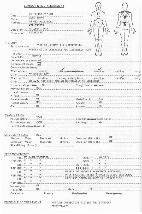
HISTORYPAIN AT LUMBAR 3 4 5 CENTRALLYSymptoms now ..
.........................~9.RQ!?~ .. 9.7..~.'r1 ~~M.~~X @.fP ..G.e.~fr.M.~.l.X T. 56at onset .
Present for ~ ~~.s. ..~:~;::r:~~ ;;a::~~u Ig ..CQl'l~/lntermittent. J ../When worse - bending, sitting GI'-~siRg frQ~ standing, walking, lying.Other J\.T. ..F;@.. .9.F. I>t.":l y /When better - bending, sitting or rising from, standing, walking, lying.Other ~~ ..~.!.~.~ ~~ ..~~ ~~.T.~~ ..~.~~~.c::~~.L.~X ..~~ ~~~.~~ .Disturbed sleep - ~ No Cough/sneeze -loW -vePrevious history ~.~.~ ..
and treatment .X-Rays ~~ .General health ~~ Meds/Steroids ~)~ ..Recent surgery ~~~ Accident ~g ..Gait .9.~ Bladder .9J .
29 FEBRUARY 1981Date : ..Name. ~~X ..~!:!.I::rff ..Address ~~ ..~.~ ...M.~~.~ ..~().~ ..
WELLINGTON..............................................................................•.................................
Date of birth ..3.1 !\P.~~.L. .. ~.~.~1 .Occupation ~.~~~.~Tli.Iff .
LUMBAR SPINE ASSESSMENT
EXAMINATIONPosture sitting ~~?~ .Posture standing r.99.~ ..Lateral shift l-R-~~I:+ornil
Lordosis ~/accentuated ..Leg length 9.1: ..
MOVEMENT LOSS NILFlexion: Major. Moderate. Minimal.Extension: Major. Moderate. Minimal.Side Gliding: (R) or (Ll .
Deviation (R) or (L) y~ ..Deviation (R) or (L) 9.x: ..
TEST MOVEMENTSFIS Jf.9 P.J\.PL.r.~9.I>~9.~P......................................... SGIS (R) ~9 f~I~ .
REP. FIS ~~ " '.'.................................................... SGIS (L) '.' '.~ ..EIS :~ '.' ~ REP. SGIS (R) '~ '.' ..
REP. EIS ~~ '.' '.' REP. SGIS (L) ~ ~~ .
REP. ~: ~ :::;;::::::::~~::::::::::::::::::::::::::::::::::::::: ::::::::::::::~~~j~~~~~~::~~:~~~~!~H.~~:~:s:~~~~:j:~~~H.~ ~~::::::::::::::ElL ~~ '.' '.' ~.t:-.~~ ~~~.~.~~.J? .. ().N. ..~~!u.~.~ ..~.g~~.?.~!.~'?~.~ .
"" "REP. EI L ..Neurological '?~ ..Hip joints '?~ ~.~.r..~ <?~ ..Conclusion: Posture ~~f1_ ~e~
PRINCIPLE OF TREATMENT POSTURE CORRECTION SITTING AND STANDINGPROPHYLAXIS

The Postural Syndrome 83
the symptoms appear in this position, and up to half an hour may be requiredbefore pain is felt. Once pain has been produced by adoption of a certainposture, it will be abolished by correction of that posture. Now our suspicionsare confirmed and a diagnosis can be made. In short, the patient with thepostural syndrome has no clinical or laboratory findings indicating a particularpathology and all functions appear perfectly normal.
Thousands of people are seeking treatment for pain resulting from badpostures; they consult doctors who often are unsuited to deal with the problemand, taking the easiest way out, prescribe pain relieving drugs instead ofrecommending postural correction; disillusioned with drug therapy patientsattend a chiropractor, osteopath, physiotherapist or some fringe manipulatorwho, mainly out of ignorance, proceeds to manipulate joints in which there is nopathology and certainly nothing 'out of place'.
I must emphasise that in many patients presenting with postural pain nopathology needs to exist. All patients with low back pain have an increase inpain when postural stresses are added. In derangement and dysfunction there isa pathological cause for the pain, and postural stresses may enhance thepathological state. But in the postural syndrome no pathology is present, and theonly treatment that is required is postural correction and re-education andinstruction in prophylaxis.
Postures involvedEvery patient with pain of postural origin has a different description for thecircumstances leading to the production of pain. Sitting (Fig. 9:2), by no means
Fig. 9:2. Sitting postures.
Fig. 9: 1. (opposite) Clinical example of a typical patient with the postural syndrome.

84 The Lumbar Spine
the only postural situation causing and prolonging low back pain, is the mostfrequent cause of postural pain. Some patients will name the sitting positionpurely and simply as causative, and they complain that pain is produced as soonas they sp~nd more than a certain amount of time, say ten minutes, in any sortof chair or car seat. Others will describe sitting at the typewriter as the only timethat pain is felt. Bus, taxi, and car drivers all complain of being worse whileseated for long periods in their vehicles; both pilots and passengers complainabout the seating in airplanes.
Working in prolonged standing positions also may cause postural pain, butthe opportunity to move and change position is greater in standing than insitting and the avenues for relief are more numerous. Consequently, there areless complaints of pain arising from the standing position than from sitting.People who work in cramped positions, be it standing or sitting, are also likely tocomplain of low back pain. The incidence of low back pain is very high in peoplewho work in continuously stooped positions (Fig. 9:3).
Fig. 9:3. Standing postures.
The lying position (Fig. 9:4) may be an additional source of stress enhancinglow back pain, and if pain predominantly occurs while lying it requires thoroughinvestigation.
Fig. 9:4.Lying posture.

The Postural Syndrome 85
TREATMENT OF THE POSTURAL SYNDROMEEvery patient must be examined and analysed individually, and educated for hisown particular postural stress. Education is probably the most important part ofthe treatment for low back pain of postural origin. The patient must have a clearand unambiguous explanation of the mechanism that produces his pain. Hemust realise that, when he assumes the positions of stress causing pain, he is infact pulling the ligaments apart; and all that is required to stop his postural pain,is to stop stressing the ligaments for about ten days. I also explain to the patientthat once he commences the correction regime, he will and should develop somenew pains which are commonly felt higher in the back. This is merely theconsequence of adjustment to a new postural habit.
The more often pain is triggered, the more readily it will occur. And the lessoften pain is triggered, the more difficult it is to be produced. Thus, poor sittingpositions maintained regularly will cause pain after the passage of less and lesstime. Conversely, good sitting postures will enable the patient to remain painfree for longer and longer periods, and when slouching next occurs it will takemuch more time for the pain to arise. After two weeks of correct sitting patientswill be able to slouch for short periods without having pain. However, no oneshould be permitted to slouch for extended periods. For example, a patient whousually gets low back pain after ten minutes of slouched sitting, may after acouple of weeks of sitting correctly revert to the slouched position and only.·experience pain after twenty minutes in that position. This painfree slouchedsitting period can be progressed up to a limit, so that at the end of ten weeks ofcorrect sitting a patient may well be able to slouch for an hour or two. A possibleexplanation for this phenomenon is forthcoming from Professor P. D. Wall. 35
Correction of the sitting postureAll patients who have low back pain produced or enhanced by prolonged sitting,should receive an adequate explanation regarding the cause of pain and the needfor maintenance of the correct sitting posture.
We must explain that when a person sits his spine will sooner or later take upa relaxed posture. Unless a special lumbar support is given or a conscious effortis made to maintain the lordosis, the lumbar spine will move into a fully flexedposition placing various ligamentous structures on full stretch. The nucleus ofthe intervertebral discs is forced posteriorly, the intradiscal pressure rises, andthe stresses on the posterior wall of the annulus are increased. At this stagethere are many reasons for the spine to feel uncomfortable. If this position ismaintained for a long period, the spine will become painful as well and in sOqlecases derangement may occur. Few patients fail to comprehend ourexplanations, provided these are couched in terms understandable to thelayman.
To convince the patient that our suspicions about his sitting posture arecorrect it is necessary to prove this to him. Pain of postural origin arising bysitting incorrectly will be abolished by sitting correctly. During the firsttreatment session we must reproduce the pain by positioning the patient and

86 The Lumbar Spine
allowing enough time for postural stresses to build up. Once pain is felt thepatient is easily convinced that it is posture-related, when on adopting thecorrect sitting posture the pain ceases. If we cannot reproduce the symptomsduring the first treatment, we must instruct the patient to assess the relationshipbetween posture and pain himself by correction of the sitting posture the nexttime pain is felt.
To achieve correction of the sitting posture the following is necessary:(1) firstly the patient must be able to obtain the correct sitting posture;(2) then he must know how to maintain it when sitting for prolonged periods.
To obtain the correct sitting posture
The patient must have a good understanding of the correct sitting posture, andhis control of the muscles and joints involved in obtaining it must be restored.Therefore, it is necessary that he be acquainted with the extreme of the goodand bad sitting positions before he is instructed regarding the correct sittingposture.
In order to achieve this we have to introduce the 'slouch-overcorrect'procedure. The patient must sit slouched on a backless chair or stool, allow thelumbar spine to rest on the ligaments in the fully flexed position, and permithead and chin to protrude. Then he must smoothly move into the erect sittingposture with the lordosis at its maximum and the head held directly over thespine with the chin pulled in. This sequence should be repeated in a flowingrhythmical manner, so that the patient moves from ~':c extreme of the good tothe extreme of the bad position (Fig. 9:5).
Fig.9:5a.Slouched sitting.
Fig.9:5b.Overcorrected sitting.
Fig.9:5c.Correct sitting.
Fiiures 5a and b together form the 'slouch-overcorrect' procedure, and Figure 5c shows the tenpercent release from the overcorrected position.
The Postural Syndrome 87
After some practise at this most patients are able to find the extreme of thegood sitting position. They should become so good at it, that at the snap of thefingers they can assume the overcorrected sitting posture and hold it for a fewminutes. Once this can be achieved patients are advised to follow this procedurewhenever pain is felt and to maintain the extreme of the good sitting positionfor a few minutes. Pain induced by poor sitting is nearly always quicklyabolished by this method. On discovering the relationship between sittingpostures and pain by this simple exercise few patients fail to carry out ouradvice. Postural correction and exercises related to pain are easily understoodand performed by most people.
Once the patient has a good understanding of the good and bad postures hecan assume while sitting, he must be taught which position is the correct sittingposture. The extreme of the good position is of course closest to the desirable,but it is impossible to hold this position for any length of time because variousstructures are on full stretch and will become painful with time. Therefore, thepatient is instructed to move into the extreme of lordosis and then release thelast ten percent of the movement. After this release from fully strained erectsitting the position can easily be maintained if necessary. This is the positionthat must be adopted habitually in the future. It must be emphasised that in thecorrect sitting posture the lumbar spine always has a certain amount but not themaximum of lordosis. If postural pain arises in the sitting position, it is causedby an insufficient or lost lordosis and postural correction will abolish the pain.
Thus, in order to learn how to assume the correct sitting posture with alumbar lordosis patients must be instructed to carry out the 'slouch-overcorrect'procedure three times daily, fifteen to twenty times at each session. At the endof each session they must release the last ten percent of the extreme good sittingposition. They have now found the correct sitting posture. This routine must bekept up for three to four days at least, longer if necessary, until the correctposture becomes automatic.
To maintain the correct sitting posture
When sitting for prolonged periods it is essential that a certain amount oflordosis be maintained at all times. From the very first day the patient must beshown how this can be achieved. The lumbar spine may be held in lordosis intwo ways:
(a) actively by consciolliS l:ontrol of the lordosis, when sitting on a seatwithout backrest.
(b) passively by the use of a lumbar support, when sitting on a seat with abackrest. The purpose of the lumbar supportive roll is to hold the lumbarspine in a good but not extreme lordosis in the sitting position whilerelaxing, working and driving the car. Without the support the lordosiswill be lost as soon as a person leans back in a chair or concentrates onanything other than maintaining the lordosis.

88 The Lumbar Spine
The lumbar roll as sitting support:
A roll inserted in the small of the back provides adequate support for the lumbarspine in sitting, provided the apex of the support maintains the lordosis justshort of its maximum. When placed at or just above the belt line, affectingapproximately the area of the third and fourth lumbar vertebrae, it produces theoptimum lordosis, provided one sits with the sacrum against the back of thechair (Fig. 9:6). A cushion is not a suitable lumbar support because, whenplaced behind the low back, it merely pushes the whole spine a few centimetersaway from the back of the chair without in any way influencing the angle ofextension or degree of lordosis in the lumbar spine.
Fig.9:6a.Use of lumbar roll in office chair.
Fig.9:6b.Use of lumbar roll in easy chair. This roll canalso be used for support in a car seat.
Various rolls can be made for the different situations in which they may berequired - for example, lounge chair, office chair and car seat. If a lounge chairor car seat is designed in such a way that the roll is absorbed by the upholstery, itmay be necessary to place one or more cushions in the chair first and then addthe lumbar roll.
Patients frequently complain about the effort they must expend to maintainthe correct sitting posture, more so when this is done actively than with the helpof a lumbar roll. In fact, many patients will describe a strain pain or say that thenew position is a painful one. It is important that these pains are recognised asnew postural stresses which should normally occur. If after a day or two ofcorrect sitting a patient has not complained of 'new pains', it is likely that he hasnot maintained the corrected position often and long enough. Adjustment to a

The Postural Syndrome 89
new posture results in shortlived transitional aching of a different quality andlocation than the pain which initially forced consultation. It should not lastlonger than five to six days.
It is reported22 that most people in North America sit with the lumbar lordosisaccentuated. On several visits to the United States and Canada I have had thepleasure of treating some hundreds of patients, and I was relieved to notice thatmost of these, as well as most of the doctors and physiotherapists attending mycourses, sat with the lumber spine held in flexion after about half an hour ofsitting. I was relieved because I had been wondering whether patients in NorthAmerica just might be different from those in Australasia and Europe, in whichcase the treatments I have developed would be most unsuitable in the NorthAmerican continent. I mention this to draw attention to a fundamental error inthe clinical observation of the basic mechanics of the sitting posture, which hasled to the development of an inflexible concept of treatment for low back painthroughout North America based on the assumption that the lordosis isundesirable, extension harmful and flexion beneficial.
In that continent, the treatment of low back pain has been influenced byauthors who argue that damage in the posterior compartment of the disc is theresult of compressive forces exerted by the lordotic posture which they believeis a predominant feature of western cultures. The treatment recommendedinvolves a combination of flexion exercises and postures all carefully calculatedto reduce the lumbar lordosis. Supporters of the flexion philosophy argue thatAfrican and Asian cultures do not have as high an incidence of back pain as isfound in western society and this is largely because of the flexed spinal posturesadopted by these cultures. Other authorities, Arrnstrong,14 and more recentlyHickey and Hukins,43 state that annular failure under compression is unlikely tobe a significant cause of low back pain. White and Panjabi44 were not aware ofany investigation in which cross cultural and racial comparisons had ever beensatisfactorily studied and related to low back pain. They also state that Williamsflexion exercises are based on the assumption that achieving and maintaining aflexed lumbar spine is preferable, but "this has not been proved and is contraryto evidence of studies in vivo of intradiscal pressure and electromyographicstudies".44
Another widespread misconception, held by many doctors and therapists,suggests that postural correction can be achieved by strengthening the musclesof the spine. Strengthening of muscles has no effect on posture. Nostrengthening exercises will educate muscles to maintain the correct posture.Actively maintaining the correct posture is the only way to achieve posturalcorrection. This has the added bonus that the muscles required to maintain thisposition are automatically strengthened merely by performing the task forwhich they were originally designed.
In postural retraining the problem does not lie in the inability to assume thecorrect posture, but in a loss of awareness of the correct posture, if indeed suchan awareness existed. To restore this it is necessary to retrain postural concepts,which is essentially a matter of the will. Indeed, willpower motivated by pain
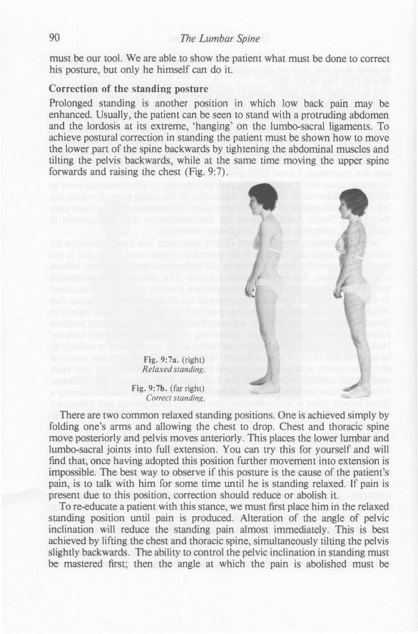
90 The Lumbar Spine
must be our tool. We are able to show the patient what must be done to correcthis posture, but only he himself can do it.
Correction of the standing postureProlonged standing is another position in which low back pain may beenhanced. Usually, the patient can be seen to stand with a protruding abdomenand the lordosis at its extreme, 'hanging' on the lumbo-sacral ligaments. Toachieve postural correction in standing the patient must be shown how to movethe lower part of the spine backwards by tightening the abdominal muscles andtilting the pelvis backwards, while at the same time moving the upper spineforwards and raising the chest (Fig. 9:7).
Fig. 9:7a. (right)Relaxed standing.
Fig. 9:7b. (far right)Correct standinl{.
There are two common relaxed standing positions. One is achieved simply byfolding one's arms and allowing the chest to drop. Chest and thoracic spinemove posteriorly and pelvis moves anteriorly. This places the lower lumbar andlumbo-sacral joints into full extension. You can try this for yourself and willfind that, once having adopted this position further movement into extension isimpossible. The best way to observe if this posture is the cause of the patient'spain, is to talk with him for some time until he is standing relaxed. If pain ispresent due to this position, correction should reduce or abolish it.
To re-educate a patient with this stance, we must first place him in the relaxedstanding position until pain is produced. Alteration of the angle of pelvicinclination will reduce the standing pain almost immediately. This is bestachieved by lifting the chest and thoracic spine, simultaneously tilting the pelvisslightly backwards. The ability to control the pelvic inclination in standing mustbe mastered first; then the angle at which the pain is abolished must be

The Postural Syndrome 91
established and maintained. If pain in standing can not be reproduced on thefirst examination the patient must be instructed to evaluate the relationshipbetween posture and pain himself by postural correction the next time pain isfelt.
The second relaxed standing position is obtained by taking all the bodyweight on one leg, while the knee of the other leg is allowed to bend causing thepelvis to droop at the same side. The lumbar spine moves into a full sidegliding/rotation position. Again, you can try this for yourself and will find thathaving adopted this position no further movement into side gliding or extensionis possible. If this position produces pain it is easily corrected and avoidance ofthe standing habit should suffice.
Correction of the lying posturePain in the lying position is common. It causes considerable distress when itinterferes with sleep over a long enough period, and it requires attention whenthe patient regularly wakes up with pain in the morning, the pain abating as theday progresses. There are two factors to be investigated:
(a) The lying posture itself. This is different for each person and must bedealt with individually. The lying posture during sleep is difficult toinfluence.
(b) The surface on which one is lying. For the majority of people themattress itself should not be too hard, whereas the base on which themattress rests must be firm and unyielding. This allows adequate supportfor the contours of the body without placing stresses on the spine.Usually, the surface on which one is lying is easily corrected or modified.
When dealing with pain produced by lying in bed, I have threerecommendations to make which may be worth considering:
Due to the natural contours of the body - that is, wider at shoulders andpelvis than at the waist - and due to the lordotic curvature of the lumbar spine,the lumbar area may be placed under stress in the prone, supine or side lyingposition. This is particularly so when a hard mattress is supported by a firm andunyielding base. If this is thought to be the cause of the problem, the patientshould use a lumbar supportive roll. When lying prone the roll will preventextreme extension in the lumbar spine. When lying supine with the legsoutstretched the roll will fill the gap between the lumbar spine and the mattressand prevent sagging of the spine into flexion. When lying on the side it will fillthe gap between pelvis and ribs and prevent sagging of the spine into sidebending. This type of lumbar support in bed usually works quickly or not at all;and should be tried for about three nights.
The lumbar roll as lying support
A beach towel folded end to end and then rolled cross-wise usually fits aroundthe average middle. If this is too big a bath towel folded length-wise can be usedinstead. Each patient will have to experiment to find the correct size of lumbar

92 The Lumbar Spine
roll required in his particular case. He should wrap the towel around the belt lineand attach the two ends to each other. If left loose the roll will not remain inplace and may, when positioned anywhere else than at the waist, furtherincrease the stresses placed on the spine (Fig. 9: 8).
Fig. 9:8.Use of lumbar roll in lying. The
roll is not fastened in the picturein order to show the shape of the
spine.
When the base of the mattress is not firm enough or the mattress itself is toosoft, stresses may also be placed on the lumbar spine. Because of the costsinvolved in replacing a mattress or its base, I always recommend that themattress be placed on the floor if it is felt that a firm flat base is required. If thereis no improvement after sleeping three to four nights on a flat surface, it isunlikely that this is the answer to the patient's problems.
There is a small number of people who require a sagging mattress. Usuallythese people fall under the flexion principle category. The sagging mattress caneasily be created by placing pillows at both ends of the bed in between themattress and its base to form a dish shape.
ConclusionWe should now have equipped the patient with sufficient information enablinghim to sensibly control mechanical stresses and deal with symptoms himself.The essence of treatment of the postural syndrome is that, if it is possible forpatients to stop their pain, it is also possible for them to prevent the onset of thepain. I feel that it is negligent of the medical and physiotherapy professions tocontinue giving relief for episodic pain without familiarising patients with themanner in which their pain arises and providing them with the means toprevent the onset of such pain. It is my experience that patients with posturalpain, when properly instructed and advised, treat themselves ably andadequately. They can well control their postural stresses, and only needassistance when excessive and sometimes unexpected external forces have beenplaced on their joints - for example, following lifting heavy weights, steppingunexpectedly from the pavement, being hit by a motor vehicle, a sudden bout ofcoughing or sneezing while sitting or bent forwards.
When treatment is completed successfully we must explain to the patientthat, although the present pain has been relieved, recurrence of similarsymptoms is possible whenever he forgets postural care for extended periods.The consequences of postural neglect should be discussed when appropriate.

The Postural Syndrome 93
Consequences of postural neglectThe effects of postural habits on the shape of man are obvious when we observepeople around us. If head and chin are allowed to protrude long and oftenenough, the ability to glide the head dorsally will be lost which results in apermanently protruding head and a dowager's hump. As age advances this oncereversible situation will become irreversible. People with this type of posturehave a flattened lumbar spine as well, and by the age of seventy the ability tostand erect is lost so that they walk with a slight stoop. Movement that was onceeasily obtained is lost forever. But this postural stoop is not the -inevitableconsequence of ageing. Loss of function can be prevented if movements in thedesired direction are performed adequately and often enough.
Initially, poor postural habits will only produce pain without loss of function.If as a result of continuous slouched sitting, flexion is regularly performed butextension never, the anterior structures of the joints involved will shorten andthe posterior structures will lengthen. In this way flexion remains readilyobtainable, but extension becomes more and more difficult and will therefore beavoided. Thus, the consequences of postural neglect are adaptive shorteningleading to dysfunction.
Adaptive shortening implies loss of function and movement. In addition tothe production of pain whenever the shortened structures are placed on stress,this loss of movement and function must inevitably lead to impairment ofnutrition in an avascular structure like the disc. This will become one of thecontributing factors of disc degeneration.
We should point out to people engaged in sedentary occupations that adaptiveshortening and dysfunction due to poor posture can be prevented by regularpostural correction and adequate performance of the appropriate exercisesbefore adaptive shortening is allowed to develop.
TYPICAL TREATMENT PROGRESSION THE POSTURAL SYNDROMEThe days referred to in the treatment progression are related to treatmentsessions which do not necessarily take place on consecutive days. This alsoapplies for the treatment progressions of the dysfunction and derangementsyndromes.
Day one- Assessment and conclusion/diagnosis.- Postural discussion ensuring adequate explanation of the nature of the
problem. The patient must understand the cause of pain. I usually give thesimple example of pain arising from the passively bent forefinger.
- We must satisfy ourselves and the patient that the pain can be inducedand abolished by positioning. If it is not possible to induce pain during thefirst treatment session, the patient must be instructed mow to abolish painby postural correction when next it appears.

94 The Lumbar Spine
- Commence with postural correction exercises and give postural advice; donot try to teach too much the first visit.
- Discuss the importance of maintenance of the lordosis while sittingprolonged, and demonstrate the use of lumbar supports in sitting andlying.
Day two
- Confirm diagnosis.- Check results. If the patient was unsuccessful in controlling the postural
pain on his own, it is possible that we have not taught correction wellenough. It also may be that the patient has not corrected his postureadequately or maintained the corrected posture long enough. Whenconfronted with such a suggestion in an accusing manner, patients oftenfeel offended and deny having slouched. We must be tactful whendiscussing these points.
- If possible have the patient produce and abolish the pain; otherwiseenquire as to his ability to abolish the pain during the preceeding twentyfour hours by correcting the posture whenever pain appeared.
- Check the exercises. It is surprising how often patients alter the exerciseswithout realising it.
- Repeat the postural advice in full.- Inform the patient that 'new pains' are to be expected as a result of
adjustment to different postural habits.
Day three
- Treatment as for day two.- Once the patient is adequately controlling his postural stresses, treatment
may be altered from a daily basis to every second or third day.- Once the pain occurs only occasionally and can be well controlled, the
patient may stop the 'slouch-overcorrect' exercise.- Reassure regarding the onset of 'new' postural pains.
Day four and five
- Check exercises and progress.- Deal with any other postural pain that may have become apparent.- Deal with other situations which may have previously been overlooked.
Further treatments
- A few check-ups at greater intervals may be necessary to ensure thepatient has full control of his postural pain.
- We must ensure that the patient has adequately stressed the joints and isengaged in all normal activities.
- Discuss the consequences of postural neglect.- Before discharge prophylaxis must be discussed in detail.
CHAPTER 10
The Dysfunction SyndromeThe word 'dysfunction' chosen by Mennell36 to describe the loss of movementcommonly known as 'joint play' or 'accessory movement' seems infinitelypreferable to the terms 'osteopathic lesion' and 'chiropractic subluxation',neither of which means anything and both of which mean everything.'Dysfunction' or 'not functioning correctly' at least acknowledges thatsomething is wrong without going through the sham procedure of pretendingthat only those who belong to the club really understand the terminology. Foryears osteopaths and chiropractors have claimed that only the people, properlytrained in their particular calling, have the necessary knowledge to understandtheir terminology. There may be some truth in that.
Although I believe that the term 'dysfunction' as used by Mennell36 does notstrictly cover the loss of movement caused by adaptive shortening, I havechosen to use this term instead of repeatedly referring to 'adaptive shortening'.
Essentially, the mechanism of pain production in dysfunction is the same asin normal tissues - that is, when overstretching of soft tissues causes sufficientmechanical deformation of the free nerve endings in these tissues, pain willarise. In dysfunction soft tissues in or around the segment involved areshortened or contain contracted scar tissue. When normal movement isattempted these structures are placed on full stretch somewhat prematurely.While normally movement in the joint would take place over a certain distancebefore being stopped by ligamentous tension, it is now brought to a halt afteronly part of that distance is completed. Attempts to move further towards endrange will result in overstretching and produce pain. The pain is felt at the endof the existing range and ceases immediately after end range stretch is released.Repeated uncontrolled stretching of contracted soft tissues will lead to furthermicro-traumata and pain. The patient then avoids the movement which ispainful, and adaptive shortening of the scar reduces the existing range ofmovement even more.
DEFINITIONDeveloped as a result of poor postural habit, spondylosis, trauma orderangement, the dysfunction syndrome is the condition in which adaptiveshortening and resultant loss of mobility causes pain prematurely - that is,before achievement of full normal end range movement. Essentially, thecondition arises because movement is performed inadequately at a time thatcontraction of soft tissues is taking place.
95

96 The Lumbar Spine
HistoryThere are two possibilities regarding the cause of dysfunction. Whendysfunction develops following trauma or derangement, the patient will beaware of the onset. He will describe the symptoms from the date of trauma orderangement, but the pain produced by trauma or derangement will no longerbe present and the symptoms are now related to the resultant loss of mobilityand function. When dysfunction is the result of poor posture or spondylosis thepatient will be unaware of the onset. He will be unable to relate the cause of thepain to a particular incident and usually describes a gradual slow onset of paincommencing for no apparent reason.
Patients in the dysfunction category are likely to be over thirty years of age.However, younger patients may welI present, who have had previous low backpain or trauma which resulted in loss of function that has not been detected ortreated.
The pain is felt at the end range of certaincmovements, or before end range isachieved, and this may interfere with the performance of simple tasks. Forexample, loss of function in the neck is often first noticed when the motoristturns the head while reversing the car; in loss of function in the low back it isoften difficult to put stockings on the feet or to get into the trousers.
Initially patients with dysfunction are stiff first thing in the morning,loosening as the day progresses. But as time passes flexion and extensionbecome reduced and the morn~l1g stiffness does not pass. In extensiondysfunction lying prone for any length of time - for example, on the beach cannot be tolerated. Due to inadequate extension in the lumbar spine theligamentous structures are placed on full stretch prematurely while lying proneand pain is produced.
Often the patient with dysfunction states that he feels better when he is activeand moving about than when at rest. The reasons for this are obvious: duringregular and not excessive activity end range of movement is seldom requiredand, if so, only momentarily; on the other hand, during resting end positions arereadily assumed and as soon as they are maintained they may prove painful.
In dysfunction the pain is intermittent, occurring only when periarticularstructures are placed on full stretch. This happens much sooner in a patient withdysfunction than in a normal person, hence the much more frequentprovocation of pain in dysfunction. The greater the loss of function, the moreoften will the pain occur.
Pain from dysfunction sometimes develops in an episodic manner andappears to resemble derangement. This episodic pain is triggered by excessiveuse, for example a vigorous afternoon in the garden. Overstretching ofcontracted soft tissues causes minor traumata and produces or increases pain. Ifthe patient rests for a few days the pain subsides, but further scarring andhealing contractures will increasingly limit the available range of movement.This becomes a vicious circle which will only be broken by treatmentprocedures as described for dysfunction.
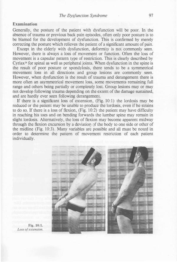
The Dysfunction Syndrome 97
ExaminationGenerally, the posture of the patient with dysfunction will be poor. In theabsence of trauma or previous back pain episodes, often only poor posture is tobe blamed for the development of dysfunction. This is confirmed by merelycorrecting the posture which relieves the patient of a significant amount of pain.
Except in the elderly with dysfunction, deformity is not commonly seen.However, there is always a loss of movement or function. Often the loss ofmovement is a capsular pattern type of restriction. This is clearly described byCyriax24 for spinal as well as peripheral joints. When dysfunction in the spine isthe result of poor posture or spondylosis, there tends to be a symmetricalmovement loss in all directions and group lesions are commonly seen.However, when dysfunction is the result of trauma and derangement there ismore often an asymmetrical movement loss, some movements remaining fullrange and others being partially or completely lost. Group lesions mayor maynot develop following trauma depending on the extent of the damage sustained,and are hardly ever seen following derangement.
If there is a significant loss of extension, (Fig. 10: 1) the lordosis may bereduced or the patient may be unable to produce the lordosis, even if he strainsto do so. If there is a loss of flexion, (Fig. 10:2) the patient may have difficultyin reaching his toes and on bending forwards the lumbar spine may remain inslight lordosis. Alternatively, the loss of flexion may become apparent midwaythrough the flexion excursion by a deviation ;1f the body to one side or other ofthe midline (Fig. 10:3). Many variables are possible and all must be noted inorder to determine the pattern of movement restriction of each patientindividually.
Fig. 10:1.Loss ojextension.

98 The Lumbar Spine
Fig. 10:3.Loss offiexion with deviation.
The test movementsIt will not be difficult to reproduce the symptoms with the test movements. Dueto the reduced end range of some movements pain is elicited readily as soon asstretching of these movements is performed, and each time the stress is releasedthe pain subsides quickly.
Following the test movements the patient should be allowed to move aboutand perhaps have a short walk. The object of this is to determine the effect ofthe test movements on the general pain pattern. A patient with dysfunction may

The Dysfunction Syndrome 99
be slightly more aware of his pain after the examination, but he will neverremain significantly worse, provided tissue damage due to overstretching ofshortened structures has not taken place. Following the test movements themovement pattern will not have altered - that is, if we were to repeat the wholesequence the same movements would produce the same pain as in the firstsession.
Clinical example
Let us look at the example of a typical patient with dysfunction (Fig. 10:4). Inparticular we must assess the effects of the test movements on the pain. In thispatient pain is produced at the point of full stretch in flexion and extension,which are both restricted in range of movement. Repetition of the testmovements does not make the symptoms better or worse, and on release of thestress the pain subsides leaving the patient no worse than before testing. Rapidchanges ofsymptoms do not occur in dYsfunction. It takes weeks for soft tissuesto become contracted and adaptively shortened and, likewise, it will take a longtime for them to lengthen again.
Treatment of the dysfunction syndromePatients who fall in the dysfunction category will still require posturalinstruction. When planning treatment we must include from the first day all theprocedures laid down for the patient with postural pain. The patient withdysfunction can learn quickly to control those symptoms which are caused orenhanced by bad posture.
The symptoms of dysfunction are more related to movement and becomeevident in the difficulty or inability of the patient to accomplish end range ofmovement, most noticeable in the extremes of flexion and extension. Thesesymptoms will remain present until the length of the shortened tissues isincreased and the range of movement is improved. This will be achieved inabout four to six weeks, provided the treatment procedures are used in a preciseand clearly defined manner. The very nature of adaptive shortening of softtissues adjacent to articular structures prohibits the rapid recovery offunction ina few days.
Stretching must be performed in such a way that it allows elongation ofligamentous structures and scar tissue without causing micro-traumata. Painproduced by stretching should stop shortly after the stress is released. Whenpain persists long after stretching has occurred overstretching has taken place.
To achieve lengthening of soft tissues it is not sufficient to perform stretchingonce a week, neither is it sufficient to do this once a day. Too manyphysiotherapy clinics make their patients return daily for ten minutes ofmobilisation or, worse still, one minute of manipulation with the expresspurpose of restoring mobility. This concept of treatment may well be acceptable,if the patient is given adequate exercises to perform regularly in betweentreatment sessions. However, often enough this is not done; or else, when the
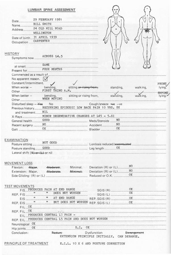
HISTORYACROSS 14.5
Symptoms now .
~~.~.~~~~ .. ::::::::::::::::::::s.~~:::::::::::::::::::::::::::::::::::::::::::::::::::::::::::::::Present for ~?~.R.,.J:ll?~~ .~:~;:anr~~~ ~:a~~~u I@ ..
Constant/lntermittent.'/ / PRONE/When worse - bending, sitting ef-1'i~1'e1'l't; standing, walking, lying.
~~hee: b~~~~~·~········ f.~:~~·i·~::·I.N.~ t::.~.M~~·~~i~~·;~··~;~i~~·f~·~·~·.. ···············~~~~~~:···· .. ········ ..;;~~~:"''''''''''''~r;~~Other ~~.~.. ~~\:'.~.~~ .
Disturbed sleep - ~ No Cough/sneeze -lo\Ie -vePrevious history R.F;<;:.~~.Pf~ F;.J:'.~~9.D.~<;: ~.~..~g.~ I'~.W ..1.~ ~.S.~ ~.~ ..
and treatment ~.U ..X-Rays ~m~.9.~ I;?F;9.~~.~n~ g.I:l.@.~~~ ~~ ~.~ ::- ~.~.~X ..General health <?~P Meds/Steroids ~~ .
Recent surgery N.C? Accident ~~ .Gait <?; Bladder ~.~ .
Date ~? :.~.B.~~~.~:...~.?..~.1 ..
Name. ~.~.L.~ S.1'1.I.T.~ ..Address ~~ ~~ .. !:f.~.~~ ~~.~ .
WELLINGTON................................................................................................................Date of birth ~~ ~I'.R.~~ ~.9..3.? .Occupation ~~.R.~~.~.~ .
LUMBAR SPINE ASSESSMENT
EXAMINATION
Posture sitting ~~.~..~.?? .Posture standing gQ9.I;? .Lateral shift (~)-9fo+b+ornil
Lordosis reduced/aeeeRll:lalel!l .
Leg length 9.1; ..
MOVEMENT LOSSFlexion: ~_. ~.a-le. Minimal.
Extension: Major. ~~ Ml~1.
Side Gliding: (R) or (L) .
Deviation (R) or (L) ~? ..Deviation (R) or (L) N.C? .Reduced or O.K <?; ..
TEST MOVEMENTS
FIS I'.R.~p.y.c;~.? ..I'~~~ ..t::~ ~~ I0.~~.~................ SsGG IISS (( RL
)) g~ .REP. F IS '.' '.~ ??~~ ~! ~.~~.~ ..
EIS '.' ~~ ~~ ~~.~ REP. SGIS (R) C?.r ..REP. EIS :: ~.':r..!?<?~.~ ~~':r ..~<?~.~.~~ REP. SGIS (L) Q.I .
FIL 9.~ ..REP. FIL 9.~ (.,., ..
ElL I'.R.qp..~c;~.?..~~~.~~.~ .. ~.~ ~~.~~ :7 ..REP. ElL I'.R.qp..~c;.~~..~~~!~.~ ..~.? ..~~.!~ t::~ ..~.~~ N.~! ..~~~~.~ ..Neurological ..9.I ..Hip joints C?~ ~.~.~.~ ~~ .Conclusion: ~st~ Dysfunction ge~
EXTENSION PRINCIPLE INITIALLY. CAN DERANGE.
PRINCIPLE OF TREATMENT E.I.L. 10 X 6 AND POSTURE CORRECTION
The Dysfunction Syndrome 101
patient is instructed in a self-treatment programme he is carefully warned not todo the exercises if they cause pain, thus defeating the purpose of the stretching.
If a patient receives manipulation in an attempt to lengthen contractedstructures, minor trauma must follow and the dysfunction cycle will beperpetuated. If the patient receives only mobilisation procedures and does notperform exercises, the stretching that occurred during the ten minutes oftreatment will be entirely lost by the contraction that is allowed to take placeover the next twenty-three hours and fifty minutes, or whatever time periodseparates the treatment sessions. Even if exercises are given, no benefit willresult unless the patient is instructed to move to the extreme range where some,but not great, discomfort or pain should be experienced. If no strain pain isproduced during the performance of exercises for the recovery of lost movement,the contracted soft tissues are not being stretched enough to enhance elongationof the shortened structures. Furthermore, if the stretching procedures are notperformed often enough, no benefit will result either. If the rest periods betweenthe stretching procedures are too long, the length of time when no stretchingtakes place negates the effect of stretching.
We can have a significant influence on the remodelling of tissue expeciallyduring the process ofrepair.45
In all dysfunction situations exercises for the restoration of movement andfunction must be performed about ten times per day from first thing in themorning to last thing at night. On each of the ten occasions a minimum of tenmovements will be performed. In other words, the patient will perform onehundred stretches per day in groups of ten. The following instructions must begiven to the patient:
- if the exercises do not produce some minor pain, the movement has notbeen performed far enough into the end range;
- the type of discomfort aimed at is not unlike the pain felt when bendingthe finger backwards beyond the normal position;
- the pain should have subsided within ten to twenty minutes aftercompletion of the exercises;
- when pain produced by the stretching procedures lasts continuouly and isstill evident the next day, overstretching - that is, too much stretching- has taken place; in this case the number of exercises in each sequenceor the frequency of the sequences must be reduced.
It is accepted that due to circumstances many patients will be unable tostrictly follow the recommended frequency of exercising. Where it is notpossible to perform stretching as often as instructed, recovery of full function islikely to take a little longer.
During the course of one treatment session we should not use more than onenew procedure; nor should that procedure, if it is a manipulative thrUSttElChnique, be performed more than once. Following the application of a newprocedure or a manipulation we must wait, if necessary twenty-four hours, toassess the response of the patient.
Fig. 10:4. (opposite) Clinical example of a typical patient with the dysfunction syndrome.

102 The Lumbar Spine
TREATMENT OF EXTENSION DYSFUNCTION
By far the most common form of dysfunction is that involving loss ofextension. Having already explained and taught the postural requirements, wemust now instruct the patient in the methods required to regain lost extension(Fig. 10:5). We must explain to him the reasons for the need to recover theextension movement. The patient must realise that without an adequate rangeof extension it is not possible to sit with a lordosis, even when a lumbar supportis used. For some patients it is imperative that the range of extension beimproved, otherwise they will be unable to sit correctly. It is my experience that,following adequate explanation, patients will co-operate with the treatment andwork hard at their recovery. They will perform exercises that cause discomfortor even pain, as long as they understand the reasons for doing so.
Fig. 10:5.Recovery ofloss of
extension, using theprocedure ofextension in
lying.
Exercises
In order to systematically stretch the lumbar spine in extension, I have adopteda system in which the patient is able to use gravity and his own body weight toapply enough force for adequate passive stretching of the joints of the lumbarspine. This procedure, a modified press-up exercise, is extension in lying(Proc. 8:3)*. If with this exercise the desired result is not obtained quicklyenough or if progress ceases, extension in lying with belt fixation (Proc. 8:4)must be commenced.
If due to circumstances it is absolutely impossible to perform extensionexercises in lying, extension in standing (Proc. 8:6) must be performedinstead. But it must be emphasised that a far better extension stretch is obtainedwith extension exercises in lying (Proc. 8:3 and 8:4).
• Procedures will be referred to as follows:(Proc. 8:3) means Chapter 8; Procedure 3.

The Dysfunction Syndrome 103
The patient should be instructed to perform the exercise ten times on eachoccasion, and to repeat the series ten times a day at intervals of approximatelytwo hours. It is most important to ensure that stretching occurs very regularlyand the patient does not let more than two to three hours pass by without doingso.
The exercise routine should result in an increase of localised central back painwhich subsides within ten to twenty minutes. The patient should also developsome new pains higher up in the spine and across the shoulders. These arenormally the resul t of performing new exercises and holding a new posture. It isnecessary to explain that the combination of the new posture and exercises willresult in discomfort felt in other places; that this new aching is unavoidable andindeed necessary, but will pass after a week or so. Patients who do not complainof these transitional pains are probably not exercising adequately.
Irrespective of the category in which they may fall, all patients should bewarned of the significance of producing peripheral pain. If exercises are found toproduce peripheral pain, the patient should stop and wait until the nexttreatment when further advice should be sought.
The loss of function in patients in this group is usually resolved graduallyover a period of about four to six weeks. After this period the patient mayreduce the number of times the exercises are performed to four sessions per day,maintaining the number of ten repetitions at each session. I instruct myextension dysfunction patients that they should continue the programme andperform ten exercises twice daily for the rest of their lives.
Often it is desirable to keep some record of progress and the therapist maychoose to take photographs to evaluate the improvement in the lumbarextension curve. The improvement is most evident in the first week, andtherefore the first photographs should be taken on the first day prior to thecommencement of the self-treatment programme.
Special techniques
As soon as progress slows down or ceases it is time to add mobilisationtechniques (Proc. 8:7 and 8:9). If after three to four mobilisation treatments nochange is evident, the patient should be manipulated (Proc. 8:8 and 8:10).Special techniques of mobilisation and manipulation are indicated, when thepatient is unable to fully restore lumbar extension himself. These proceduresmay ensure full recovery of extension provided the extension exercises in lying(Proc. 8:3 and 8:4) are continued as well.
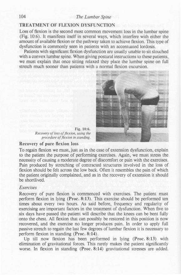
104 The Lumbar Spine
TREATMENT OF FLEXION DYSFUNCTIONLoss of flexion is the second most common movement loss in the lumbar spine(Fig. 10:6). It manifests itself in several ways, which interfere with either theamount of available flexion or the pathway taken to achieve flexion. This type ofdysfunction is commonly seen in patients with an accentuated lordosis.
Patients with significant flexion dysfunction are usually unable to sit slouchedwith a convex lumbar spine. When giving postural instructions to these patients,we must explain that once sitting relaxed they place the lumbar spine on fullstretch much sooner than patients with a normal flexion excursion.
Fig. 10:6.Recovery oj loss ojflexion, using the
procedure ojflexion in standing.
Recovery of pure flexion loss
To regain flexion we must, just as in the case of extension dysfunction, explainto the patient the purpose of performing exercises. Again, we must stress thenecessity of causing a moderate degree of discomfort or pain with the exercises.Pain produced by stretching of contracted structures involved in the loss offlexion should be felt across the low back. Often it resembles the pain of whichthe patient originally complained, and as in the recovery of extension it shouldbe shortlived.
Exercises
Recovery of pure flexion is commenced with exercises. The patient mustperform flexion in lying (Proc. 8:13). This exercise should be performed tentimes about every two hours. As said before, frequency and regularity ofexercising are important factors in the treatment of dysfunction. When five tosix days have passed the patient will describe that the knees can be bent fullyonto the chest. All flexion that can possibly be restored in this position is nowrecovered, and the exercise no longer produces pain. In order to apply fullpassive stretch to regain the last few degrees of lumbar flexion it is necessary toperform flexion in standing (Proc. 8:14).
Up till now flexion has been performed in lying (Proc. 8:13) withelimination of gravitational forces. This rarely makes the patient significantlyworse. In flexion in standing (Proc. 8:14) gravitational stresses are added.

The Dysfunction Syndrome 105
When applied to dysfunction resulting from recent derangement, flexion instanding (Proc.8:14) may sometimes exacerbate the condition. Therefore,when commencing flexion in standing (Proc. 8:14) the patient should reducethe number of exercises performed at each session as well as the frequency ofthe sessions per day - for example, five to six repetitions of the exercise shouldbe done five to six times per day. This way there is little risk of exacerbation,and after four to five days the patient may progress to a full programme.Eventually the patient will recover his flexion and will almost reach the groundbefore any strain pain is felt. No discomfort should be experienced on returningto the erect position.Special techniques
If full flexion cannot be restored by the patient's own efforts, the application ofspecial procedures may be indicated, such as rotation mobilisation andmanipulation in flexion (Proc. 8:11 and 8:12).
Occasionally, a patient with the derangement syndrome will mistakenly beplaced in the dysfunction category. If this occurs the flexion procedures mayimmediately and significantly worsen the symptoms, and in this way the truenature of the condition will be revealed. Consequently, the treatmentprogramme must be altered appropriately.N.B. Remember that the recovery of function in flexion following recent
derangement, is hazardous. Extension in lying (Proc. 8:3) should alwaysfollow flexion movements in order that any posterior disturbance iscorrected immediately.
Treatment of flexion with deviation
There are two common types of deviation in flexion resulting from dysfunction.The first one occurs in the patient who is unable to reach full flexion via thenormal sagittal pathway, no matter how hard he tries. Due to adaptivelyshortened structures within the intervertebral segment he is forced to deviate toone side during flexion.
Treatment for this type of flexion loss may follow the course recommendedfor recovery of pure flexion loss. Thus, treatment commences with flexion inlying (Proc. 8:13). Provided the initial twenty-four hour period of flexion inlying (Proc. 8:13) has not caused a lasting increase or peripheralisation of pain,the use of flexion in step standing (Proc. 8:15) is indicated at an early stage oftreatment to correct the deviation in flexion. This is later followed by flexion instanding (Proc. 8:14) to ensure recovery of full flexion movement. Whencommencing flexion in step standing (Proc. 8:15) the same precautions shouldbe taken as discussed previously (that is, when commencing flexion in standing(Proc. 8:14) in treatment recommended for recovery of pure flexion).
If the initial twenty-four hour period of flexion in lying (Proc. 8: 13) causes alasting increase or peripheralisation of symptoms, our diagnosis is incorrect andthe most likely reason for the deviation is internal derangement of a lumbardisc. Consequently, we must alter our treatment approach.
The second type of deviation in flexion resulting from dysfunction is causedby an adherent nerve root, and its treatment will be discussed in conjunctionwith the treatment of sciatica.

106 The Lumbar Spine
TREATMENT OF SIDE GLIDING DYSFUNCTION CORRECTION OF SECONDARY LATERAL SHIFT
Having observed thousands of lumbar spines it has become clear to me thatasymmetry is the 'norm' and symmetry is almost atypical. Therefore, whenexamining dysfunction patients it is important to realise that many exhibit aminor scoliosis or lateral shift, the direction of which is sometimes extremelydifficult to determine. With careful observation it can be seen that the top half ofthe patient's body is not correctly related to the bottom half, and the patient hasshifted laterally about the lumbar area (Fig. 10:7). The anomalies include anumber of lateral shifts now dysfunctional in character. These lateral shifts arereferred to as secondary whereas those caused by derangement are primary.
Fig. 10:7.Recovery ofloss ofside gliding,
teaching the procedure ofselfcorrection ofsecondary lateral shift.
As discussed previously, we must determine whether the lateral shift isrelevant to the present symptoms or is merely a congenital or developmentalanomaly. If side gliding produces pain there is likely to be adaptive shorteningwithin or about the disc and recovery of the side gliding movement must beattempted. As it is not easy to apply overPressure in the side gliding exercise, itmay be difficult to recover this movement.
The patient must be fully instructed in self-correction of lateral shift(Proc. 8:17). He should perform the procedure ten times per day., at eachsession moving ten times into the overcorrected position. The last movementshould be held firmly for about thirty to forty seconds. The patient should alsobe encouraged to stand in the overcorrected position whenever an opportunityarises during the day. If by the end of the first week pain produced by lateralshift correction is much less, the procedure will most likely have the desired
The Dysfunction Syndrome 107
result and must be continued for about three to four weeks in an attempt torestore full function. But if no change is evident after one week, there is littlehope of improving this aspect of dysfunction.
TYPICAL TREATMENT PROGRESSION THE DYSFUNCTION SY DROMEDay one
- Assessment and conclusion/diagnosis.- Explanation of the cause of dysfunction and the treatment approach.- Postural correction and instructions, especially regarding sitting;
demonstrate the use of a lumbar support.- Commence with exercises to recover function - that is, extension in
lying, flexion in lying, or side gliding in standing, whatever procedure isindicated.
- Emphasise the need to experience some discomfort during the exercises,and the importance of frequent exercising during the day.
- If flexion in lying is recommended, we must warn to stop exercising if thesymptoms quickly worsen. We may have overlooked derangement, orcommenced the procedure too early following recent derangement.
- Always follow flexion exercises with some extension.
Day two- Confirm diagnosis.- Check postural correction.- Completely repeat'postural correction and instructions.- Check exercises. If improving nothing should be changed.- If not improving, ensure that exercises are performed far enough into end
range, maintained long enough during the last three repetitions, andperformed often enough during the day.
- Warn for 'new pains'.
Day three- Check posture and exercises.- If no improvement, commence mobilisation procedures. Several
mobilisation treatments may be required.- Patient must continue the self-treatment exercises as directed.

108 The Lumbar Spine
Day four and five- Check exercises and progress.- If in treatment for flexion dysfunction no further progress is possible with
flexion in lying, change to flexion in standing, possibly flexion in stepstanding.
- Take necessary precautions when starting flexion in standing.- Ensure that patient has 'new pains'.
Further treatments- I prefer to see patients in this category three or four days in succession. If
progress is adequate and the patient understands the self-treatmentprogramme, treatment may change to alternate days and later to twice perweek if required.
- It usually takes ten to twelve treatments, spread over four to six weeks, tosuccessfully treat dysfunction.
- If towards the middle of the treatment period the patient ceases toimprove and especially if the remaining pain is unilateral, then a rotationmanipulation may be required. This may have to be repeated two or threetimes and should be combined with mobilising and exercising proceduresalready being applied.
- Before discharge prophylaxis must be discussed in detail.

CHAPTER 11
The Derangement SyndromeOf all mechanical low back problems that are encountered in general medicalpractise, mechanical derangement of the intervertebral disc is potentially themost disabling. It is my belief that in the lumbar spine, if in no other area,disturbance of the intervertebral disc m~hanism is responsible for theproduction of symptoms in as many as ninety-five percent of our patients.Twenty-five years of clinical observation and treatment of lumbar conditionshave convinced me that certain phenomena and the various movements whichaffect them, can occur only because of the hydrostatic properties invested in theintervertebral disc.
For thirty years Cyriax24 has attributed lumbar pain to internal derangementof the intervertebral disc mechanism. He has outlined the cause of lumbago, andproposed that pain of a slow onset is likely to be produced by a nuclearprotrusion while that of a sudden onset is caused by a displaced annularfragment. Although at present we are unable to prove either of thesehypotheses, I support his general view that the disc is the cause of much lumbarpain; but I cannot believe that slow and sudden onsets of pain can be so clearlyseparated and ascribed to different structures within the same disc.
Various authorities describe phenomena - observed clinically, at surgery,and in cadaveric experiments - that indicate aberrant behaviour of structureswithin the disc complex. Mathews37 reports that a small bulge of a lumbar discmay cause pain in the back and asymmetry of the involved intervertebralarticulation, leading to scoliosis and asymmetrical limitation of movement of thelumbar spine. Nachemson38 describes bulging of the annulus under loadingconditions, and increased bulging with movements of the spinal column.Vernon-Roberts23 states that during surgical explorations of disc prolapses someprotrusions were seen to alternately protrude into the spinal canal and retreatinto the disc substance, depending on the type of movement the lumbar spinewas subjected to. Moreover, according to Mathews37 myelograrns were seen tobe positive only when taken with patients in the painful position, a furtherindication of the relationship between disc bulge and increased pain onmovements. Thus, it is evident that minor disc bulging may cause deformity andlimitation of movement, and that certain movements of the spinal columnincrease the bulge while others may reduce it.
The ability of the nucleus to move anteriorly in extension, posteriorly inflexion and laterally in lateral flexion 13,21 depends very much on the integrity of
109
110 The Lumbar Spine
the annular wall, for it has been found that the position of the nuclear fluids canbe affected by movement only as long as the hydrostatic mechanism is intact.Park27 has shown that in frank nuclear protrusions the opaque medium, injectedin the disc, remains contained in a localised area irrespective of the position ofthe spine. Once nuclear material has escaped through the annular wall, thehydrostatic mechanism is no longer intact and internal derangement of the disccannot be reduced significantly by movements of the spinal column. On theother hand, movements of the spinal column can be utilised to reverse internalderangement of the disc as long as the integrity of the disc wall is maintained.
In conclusion, we have now ample evidence that movements of the spinalcolumn effect the position of the nucleus within the disc under certaincircumstances; and there is enough proof to incriminate the disc as the cause ofthe deformities so often encountered in patients with low back pain, particularlyin the acute stages. It cannot be disputed that certain movements and positionslead to the development of these deformities; and, likewise, that othermovements and positions can be utilised to reduce these deformities.
Many doctors are still under the impression that the pain of 'lumbago'necessarily has an inflammatory component. If this were true, reduction ofderangement would never provide immediate and lasting relief from pain asoften occurs.
It must be well understood that pain resulting from inflammation has achemical origin, and will only be present as long as there is enough chemicalirritation of the free nerve endings. Inflammatory pain cannot possibly on onemovement appear in the buttock, and on another movement disappear from thebuttock and appear in the midline of the back. Pain that changes site withmovement must result from mechanical deformation - that is, beforemovement took place different structures or different parts of the samestructure were subjected to mechanical deformation than during or aftermovement. The actual cause of the mechanical deformation - the structureproducing it - has changed its position or shape during the movement.
DEFINITIONI would define derangement as the situation in which the normal restingposition of the articular surfaces of two adjacent vertebrae is disturbed as a resultof a change in the position of the fluid nucleus between these surfaces. Thealteration in the position of the nucleus may also disturb annular material. Thischange within the joint will affect the ability of the joint surfaces to move intheir normal relative pathways and departures from these pathways arefrequently seen. In some patients movements may only be reduced but in othersthey will be lost entirely. Usually, the movement involved is either flexion orextension and often a loss of side gliding can be seen in conjunction.
In addition to causing losses of movement in the lumbar segments,derangement of the disc can cause the deformities of kyphosis and scoliosis.

The Derangement Syndrome III
Acute lumbar kyphosisThe stresses most likely to cause an acute lumbar kyphosis are sustained flexionstresses such as those applied during gardening and in sitting. Prolongedmaintenance of flexed positions and frequent repetition of movements inflexion, especially when extension is never fully recovered, may both lead toexcessive accumulation of the fluid nucleus in the posterior compartmentbetween the vertebral bodies. Once this accumulation is great enough it maybecome a blockage and prevent the erect position from being obtained. Anyattempt by the patient to straighten up quickly leads to significant pain, as thecompressive forces create bulging of the posterior longitudinal ligament andannulus which are now under extreme tangential stress. The patient mustreturn to the flexed position in order to reduce the intensity of the pain. We nowhave an acute lumbar kyphosis. The patient is not held in this position bymuscle spasm.24
In acute lumbar kyphosis the incidence of sciatica is relatively low. Thepostero-central disc bulging which causes the kyphosis, must move laterallybefore it can reach the nerve root. Once it has moved laterally the patient willdevelop a scoliosis. Consequently, most patients with sciatica have some degreeof lateral shift or scoliosis.
Acute lumbar scoliosisIt appears reasonable to suggest that the posterior longitudinal ligament mayprevent postero-central disturbances in the disc wall. However, sooner or laterasymmetrical stresses will force the fluid nucleus laterally where the outerannulus will distend at its weakest point. There is greater elasticity anddecreased resistance to distention at the posterior surface of the disc, especiallywhere the larger radius curve meets the smaller one. 39 Here the annulus isnarrowest and less firmly attached to the bone, and is not reinforced by theposterior longitudinal ligament. Therefore, bulging of the annulus is more likelyto occur at this point than anywhere else.
To recapitulate, a patient with lumbago may obtain the deformity of kyphosisby a posterior movement of the nucleus. Should this symmetrical posterioraccumulation become asymmetrical and move to a more postero-lateral positionwithin the intervertebral compartment, the patient is likely to develop sciaticaand will acquire the deformity of scoliosis. Thus, a patient with lumbar kyphosisand low back pain merely has to move to one side or other to run the risk ofchanging his deformity to a scoliosis. For example, a side bending or torsionmovement to the left will cause the nucleus to move to the right; with sufficientaccumulation of nuclear material on the right side the patient will havedifficuiting in obtaining the erect posture and will develop a lateral shift to theleft.
I have recorded the progression from acute kyphosis to acute scoliosis manytimes clinically. During the development of my treatment methods I discoveredthat it is possible to induce a scoliosis in a patient presenting with an acutekyphosis and then to reduce it, provided the appropriate procedure is applied intime.

112 The Lumbar Spine
HistoryPatients with low back pain caused by derangement are commonly betweentwenty and fifty-five years of age. From that time on the incidence of thederangement syndrome reduces gradually as degenerative processes developand progress. Men appear to be affected by derangement more often thanwomen. 10. 12
Low back pain is notorious for its recurrent nature. It is suggested that ninetypercent of people with low back pain will have recurrent problems.40 I estimatethat sixty-two of every hundred patients will become recurrent. The majority ofpatients with recurrent problems occur in the derangement syndrome.
Derangement of the lumbar intervertebral disc may arise from a single severestrain,9 a less severe strain applied more frequently, or a sustained flexion strain.The latter is most common and occurs in people who have a sedentaryoccupation or work in stooped positions. Sustained flexion positions force thenucleus to the extreme within its cavity and, as a result of time, posterioraccumulation of nuclear material beyond the normal limit may prevent theattainment of movements and positions opposite to the sustained flexedposition. The length ot time that a particular position has been held oftendetermines the degree of derangement.
When movements are constantly varied in direction the fluid nucleus neveroccupies a fixed position for any length of time and continuously alters itssilhouette. Provided movements do not cause excessive strain and are notrepeated too regularly, accumulation of nuclear material will not occur.Likewise, the simple action of flexion - that is, bending forwards once withoutplacing great strains on the lumbar spine and without having been in sustainedflexion prior to bending - is unlikely to cause an episode of low back pain.Many patients blame the simple forward bending movement for the onset oftheir pain, but on close questioning there has usually been a position or anactivity involving prolonged flexion preceding the onset of pain. Let me furtherillustrate this.
Clinical examples:
A lady states that her low back pain came on as she merely stooped to pick up abutton. However, further questioning reveals that she had been in sustainedflexion - sitting in front of her sewing machine - for some time immediatelyprior to bending down.
Similarly, the man who removed his golf clubs from the rear compartment ofhis car did not develop pain because he suddenly flexed. He had been drivinghis car and sat in flexion for some time before he got out of the car and bentover to pick up his golf clubs. It is unlikely that he straightened up fully afterleaving the car, and the additional flexion to lift his golf clubs from the carsurely raised the intradiscal pressure in a joint that was already underconsiderable stress. These are typical examples of derangement with a rapidonset.
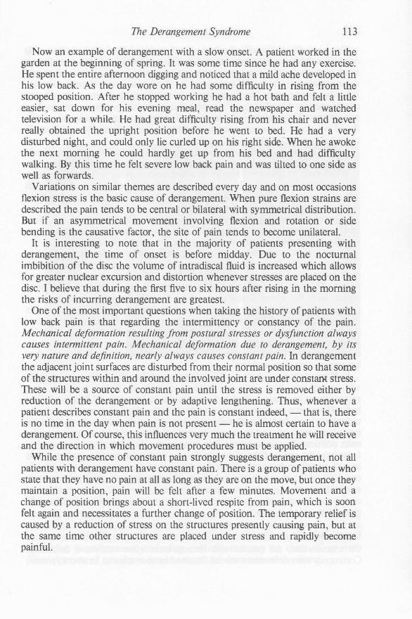
The Derangement Syndrome 113
Now an example of derangement with a slow onset. A patient worked in thegarden at the beginning of spring. It was some time since he had any exercise.He spent the entire afternoon digging and noticed that a mild ache developed inhis low back. As the day wore on he had some difficulty in rising from thestooped position. After he stopped working he had a hot bath and felt a littleeasier, sat down for his evening meal, read the newspaper and watchedtelevision for a while. He had great difficulty rising from his chair and neverreally obtained the upright position before he went to bed. He had a verydisturbed night, and could only lie curled up on hIs right side. When he awokethe next morning he could hardly get up from his bed and had difficultywalking. By this time he felt severe low back pain and was tilted to one side aswell as forwards.
Variations on similar themes are described every day and on most occasionsflexion stress is the basic cause of derangement. When pure flexion strains aredescribed the pain tends to be central or bilateral with symmetrical distribution.But if an asymmetrical movement involving flexion and rotation or sidebending is the causative factor, the site of pain tends to become unilateral.
It is interesting to note that in the majority of patients presenting withderangement, the time of onset is before midday. Due to the nocturnalimbibition of the disc the volume of intradiscal fluid is increased which allowsfor greater nuclear excursion and distortion whenever stresses are placed on thedisc. I believe that during the first five to six hours after rising in the morningthe risks of incurring derangement are greatest.
One of the most important questions when taking the history of patients withlow back pain is that regarding the intermittency or constancy of the pain.Mechanical deformation resulting from postural stresses or dysfunction alwayscauses intermittent pain. Mechanical deformation due to derangement, by itsvery nature and definition, nearly always causes constant pain. In derangementthe adjacent joint surfaces are disturbed from their normal position so that someof the structures within and around the involved joint are under constant stress.These will be a source of constant pain until the stress is removed either byreduction of the derangement or by adaptive lengthening. Thus, whenever apatient describes constant pain and the pain is constant indeed, - that is, thereis no time in the day when pain is not present - he is almost certain to have aderangement. Of course, this influences very much the treatment he will receiveand the direction in which movement procedures must be applied.
While the presence of constant pain strongly suggests derangement, not allpatients with derangement have constant pain. There is a group of patients whostate that they have no pain at all as long as they are on the move, but once theymaintain a position, pain will be felt after a few minutes. Movement and achange of position brings about a short-lived respite from pain, which is soonfelt again and necessitates a further change of position. The temporary relief iscaused by a reduction of stress on the structures presently causing pain, but atthe same time other structures are placed under stress and rapidly becomepainful.

114 The Lumbar Spine
There are also patients in whom, during the course of the day and dependingon their activities, minor derangements are alternatively produced and reducedspontaneously. In these patients the intermittent pain is caused by intermittentderangements which are self-reducing.
In the derangement syndrome the pain will be made worse under certaincircumstances. Usually these circumstances involve positions rather thanmovements. The majority of patients will experience an increase of pain inflexed positions, and when a scoliosis or lateral shift is present extension willcause an increase in pain as well as flexion.
Sitting is the most common aggravating factor for all low back painsyndromes. Rising from sitting increases pain in derangement syn,drome only.
When patients describe a history in which both walking and sitting enhancepain, they are likely to exhibit a lateral shift. lO In the presence of a lateral shiftany attempt to extend the lumbar spine will cause or increase pain, and walkingproduces enough extension to enhance pain.
In general, if the derangement is small patients feel better when on the moveand worse when at rest. However, if the derangement is large and causessignificant deformity patients usually feel better when lying down.
Patients with derangement often have difficulty in finding a comfortablesleeping position. They cannot lie supine with the legs out straight because theavailable range of extension is inadequate. Prone lying is prevented for the samereason. The only position in which some comfort can be derived is by lyingsupine with the knees flexed or on the side with the knees bent up, bothpositions placing the lumbar spine in flexion. Following either of tllese, patientshave considerable difficulty arising in the morning. In sub-acute patientsextension remains blocked for up to an hour and is only gradually regained if atall. In acute patients the pain is so severe that they prefer to return to bed, wherethey resume the flexed position which relieves their pain for the present timebut perpetuates their problem.
In North America, where the flexion therapy philosophy is strongest, patientswith acute low back pain are advised to rest in flexed positions, andextraordinary measures are taken to ensure the maintenance of flexion while inbed. However, on questioning patients with derangement they almostuniversally complain that, after sleeping in such a position, they have difficultyon rising from lying and their pain is markedly increased when coming to theerect position. Pain experienced first thing in the morning when getting up isfrequently the result of the overnight posture of the patient. If the spine is heldin such a position during the night that the nucleus accumulates posteriorly, it isextremely difficult and painful to reduce this derangement rapidly. Sometimesseveral hours must pass before reduction can be achieved. The same mechanismcauses the increase of pain felt when the patient with posterior derangementrises from sitting to standing.
ExaminationOn examination, the patient with derangement often exhibits a deformity.Commonly seen deformities are the flattened lumbar spine or lumbar kyphosis,
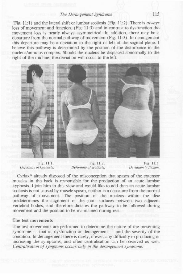
The Derangement Syndrome 115
(Fig. 11: 1) and the lateral shift or lumbar scoliosis (Fig. 11:2). There is alwaysloss of movement and function, (Fig. 11:3) and in contrast to dysfunction themovement loss is nearly always asymmetrical. In addition, there may be adeparture from the normal pathway of movement (Fig. 11:3). In derangementthis departure may be a deviation to the right or left of the sagittal plane. Ibelieve this pathway is determined by the position of the disturbance in thenucleus/annulus complex. Should the nucleus be displaced abnormally to theright of the midline, the deviation will occur to the left.
Fig. 11:1.Deformity ofkyphosis.
Fig. 11:2.Deformity ofscoliosis.
Fig. 11:3.Deviation in flexion.
Cyriax24 already disposed of the misconception that spasm of the extensormuscles in the back is responsible for the production of an acute lumbarkyphosis. I join him in this view and would like to add than an acute lumbarscoliosis is not caused by muscle spasm, neither is a departure from the normalpathway of movement. The position of the nucleus within the discpredetermines the alignment of the joint surfaces between two adjacentvertebral bodies, and therefore dictates the pathway to be followed duringmovement and the position to be maintained during rest.
The test movementsThe test movements are performed to determine the nature of the presentingsyndrome - that is, dysfunction or derangement - and the severity of thecondition. In derangement there is rarely, if ever, any difficulty in producing orincreasing the symptoms, and often centralisation can be observed as well.Centralisation of symptoms occurs only in the derangement syndrome.
116 The Lumbar Spine
The test movements are designed to reveal the presence of derangement.They alternately increase and decrease the disturbance by changing the positionof the nucleus within the intervertebral disc and altering the stresses on thesurrounding annulus and posterior longitudinal ligament. When the historysuggests that a potentially disabling situation is present, it is not desirable todevelop the flexion testing procedures to the bitter end. In these cases flexion instanding is often too painful to be repeated, and each successive movement mayincrease or peripheralise the pain. Sometimes repeated flexion in lying has thesame effect. In derangement, the increase or peripheralisation of pain with eachrepeated movement indicates a rapidly increasing derangement. When thisoccurs we should not insist on completing the recommended number of testmovements.
When the test movements affect pain it is important to state clearly whetherpain is produced or increased. The former indicates that there was no pain beforethe test movements were performed and the test movements actually producedpain. The latter means that pain, present before the performance of testmovements, has increased as a result of them. It is imperative that we establishthe existing pain state before commencing the test movements in order to beable to assess the effects of these test movements on pain.
If a patient with derangement describes changes in the pain pattern followingthe test movements, there should also be observable changes in range ofmovement and deformity. In other words, a patient who describes a significantincrease in pain should exhibit an increase in the mechanical blockage ofmovement and deformity - that is, a reduction of movement range and anincrease of deformity; and the patient who describes a reduction in pain mustsimultaneously exhibit an increase in range of movement and a reduction ofdeformity.
In derangement the repetition of the test movements can have a rapid effecton the condition and the patient may improve or worsen in a matter of minutesdepending on the direction in which the movements are performed. When thetest movements are repeated in the direction which increases the accumulationof nuclear material, pain will increase with each successive movement and afterthe treatment the patient may remain significantly worse as a result of increasedderangement. The opposite applies when the test movements are repeated in thedirection which reduces the derangement. In this case the patient improves witheach successive movement and remains improved subsequently. In general,rapid and lasting changes in the condition as a result of the test movements arean indication of derangement.
If the annular wall is breached, we cannot influence the position of thenucleus with normal movements of the spine. This situation appears clinicallyin the patient with sciatica who has constant pain and cannot find relief by eitherpositioning or movement. Most of the test movements can be seen to enhancehis leg pain and no test movement will reduce it. Thus, the performance of thetest movements clarifies the severity of the derangement situation.
Fig. 11:4. (opposite) Clinical example of a typical patient with the derangement syndrome.
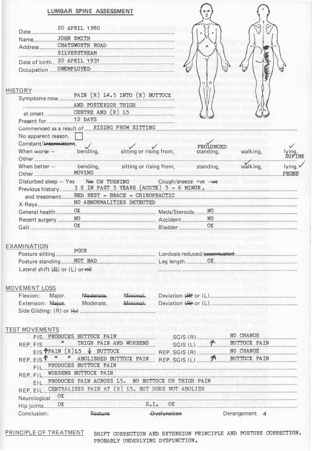
HISTORYPAIN (R) IA.5 INTO (R) BUTIDCI:
Symptoms now ......................................~ffi? ..r9.~~.!U9K.r~tG.Ii ..at onset 9.~~':r:~~ ~m?.<.~) ~?. .
Present for ~.~ J?~X~ .Commenced as a result of Rl~Jff.9...f.R9.!':! ~U.'r:r:t~9 ..No apparent reason. DConstant/W'l.8'Jtlli~eAt. -/ -./../ PRO'GNGED -./When worse - bending, sitting or rising from, standing, walking, lying.
SUPINEOther V /When better - bending, sitting or rising from, standing, walking, lying.Other w.?:vJ~9. .P.R.ONEDisturbed sleep - Yes Ne ON TURNING Cough/sneeze +ve --oW
Previous history ) ..?!: ..I~ ..!.'A~~ ~ ..~.~~ ...c~9!.~.L ..~ :7 ? ~.P~~.~.! ..and treatment ~.~~ ~~.'L::- ~~.9.~ :7..9.~~.~g!.'~q.T.~q ..
X-Rays ~~ ~~~~~~.~.~~~.~ !?~~!.~ ..General health g~ Meds/Steroids ~Q ..Recent surgery )~Q Accident ~Q ..Gait ~~ Bladder <?~ .
20 APRIL 1980Date ..Name. ~9.Wf ~~.r~ ..Address g~!.~.~~.~ ~!\l? .
SILVERSTREAM................................................................................................................Date of birth ~.l?.A!.'.~~~ )..~)) .Occupation .v~ffl:'.~Q~!? .
LUMBAR SPINE ASSESSMENT
EXAMINATIONPosture sitting :.??~................................................. Lordosis reduced/aeeeAttjatee .Posture standing J'fQ.T...MJ:? Leg length 9.~ ..Lateral shift (.8.) or (L) orofltl ..
MOVEMENT LOSSFlexion: Major. MQgllra~ll. ~
Extension: ~. Moderate. ~~
Side Gliding: (R) or ~ .
Deviation ~or (L) ..Deviation l-Aoror (L) .
TEST MOVEMENTSFIS r..:RQP.JJ.c;;r;.~ ..);lJJ.rnC;;~ p.!H.tL......................... SGIS (R) ~~ 9.~~~.~ ..
REP. FIS ~ ~~~.~ ..!.'~~~ ~!.iP. ..~~~.~~~..... SGIS (L) t.' ~.~.~r. ~~.~~ ..EIS f.r.A~~...c~J.~~ * ~y.':l:'I9.9.~ REP. SGIS (R) ~9 9.~~~.¢.... ..
REP. EISt. ~ ~ ~.?gp~~.~~ ~·!I~q.~..rA~~ REP. SGIS (L) ~ ~~.T.~.9.~ ~~.~.~ ..FIL ~.~~~?~~.~ ..~?!!?~~ ~~.~~ ..
REP. FI L ~.?~.~.~~ ~~.~~ ~~.~~ ..ElL .. X~~!?.'!.~~.~ ..!:~~~ ~~.~~~.~ ~~~ ~~ ~!:'.~.~~ ~~ '!:~.~.~~ ~~.~.~ .
REP. EI L 9.~~!:"}~.~~ r:.,:O:p~ ..~.~ .. .<.~.t ..~~.~ ~.~ ..p.9.~~ ~~!. ..~.~<?~.~.~~ ..Neurological ~.~ .Hip joints 9.~ ~.~.~.! l?~ ..Conclusion: ~.. ~_tlefl Derangement 4
PRINCIPLE OF TREATMENT SHIFT CORRECTION AND EXTENSION PRINCIPLE AND POSTURE CORRECTION.
PROBABLY UNDERLYING DYSFUNCTION.

118 The Lumbar Spine
Clinical examples:
Let us now look at the example of a typical patient with derangement(Fig. 11:4). He states that he has constant pain, but is better on the move thanin any of the positions. Walking and lying prone also improve the symptoms.The test movements clearly show increase and peripheralisation of pain on theflexion movements, and centralisation of symptoms occurs during extension.This patient will benefit from the extension principle, and hopefully his constantpain will become intermittent with the regular use of extension procedures.
A few more examples will demonstrate the importance of centralisation ofsymptoms during test movements and treatment of the patient withderangement.
A patient complains of pain extending evenly across the low back to about tencentimeters on either side of the midline. This pain has been present for somemonths and is usually worse when working in flexed positions or sitting for awhile. The test movements reveal that on repeated flexion the pain spreadsfurther across the low back, and on repeated extension the pain increases butmoves towards the midline. Further extension movements cause a reduction inintensity of that pain. Extension clearly is the movement that reduces thederangement and should be used in the treatment. If extension is continued andperformed regularly over the next twenty-four hours, the pain should becomeless and less and should be under control within that time period.
A young woman complains of pain across the low back and aching into bothbuttocks and thighs. She states that the pain increases in the back duringprolonged standing - extension - and moves into buttocks and thighs duringwalking - increased extension. - However, on sitting down - flexion - thepain leaves the legs and is felt only in the back, and after ten minutes of sitting- sustained flexion - it has subsided completely. Again the centralisationphenomenon shows us that in this case extension movements are increasing thederangement and therefore should be avoided. On the other hand, flexion isreducing the derangement and is the correct principle of treatment for thispatient.
Straight-leg-raisingPatients with sciatica, especially those who also exhibit a deformity of scoliosisand have constant pain, will have a limitation of straight-leg-raising on the sideof the sciatica. Furthermore, straight-leg-raising will be limited in patients whoare in the acute stage of a severe derangement condition. In these patients allmovements aggravate the symptoms, and often relief is obtained only whenlying in bed and keeping as still as possible.
Let us examine the relevance of straight-leg-raising, performed as anobjective testing procedure. When a limitation of straight-leg-raising is found,there is always a restriction of flexion in standing as well. It is virtuallyimpossible to have a limitation of straight-leg-raising and be able to flex pelvisand lumbar spine in a normal fashion. Some patients appear to have full flexionin standing, but on close observation they can be seen to deviate in flexion and

The Derangement Syndrome 119
to move in an arc to one side or other rather than in the sagittal plane. Inpatients with sciatica and root entrapment, deviation occurs always to the side ofreferred pain as this is the side where nerve root impingement takes place. Thepatients are unable to control the deviation in flexion themselves.
There is nothing that can be elicited by straight-leg-raising tests, that is notevident in careful observation of flexion in standing. When there is increasedtension of the nerve root the straight-leg-raising limitation appears to occurmuch sooner than does the restriction of flexion in standing. This is because thestraight-leg-raising test is performed in supine lying, and in this positiondeviation of the spine is difficult. There only appears to be a better range offlexion in standing, because deviation of the spine allows more flexion towardsthe side of root tension. If the deviation is prevented from occurring by holdingthe patient in the sagittal plane, an immediate reduction in the flexion rangebecomes apparent. This reduction in the range of flexion will now coincide withthe degree of straight-leg-raising.
Another test may help to understand the mechanism involved in restrictionof straight-leg-raising. When performing straight-leg-raising, do not stop themovement immediately when sciatica is produced or increased. Instead,continue to raise the leg firmly but slowly, and at the same time observe thepelvis. At first it can be seen to rotate to the opposite side, then it tilts backwardswhile the lumbar spine flattens and commences to flex. This is merely aninverted version of the deviation of flexion in standing.
Straight-leg-raising is an extremely unreliable test and as an objective toolonly of use in determining the progress of patients with entrapment.
TREATMENT OF THE DERANGEMENT SYNDROMEOf all patients with low back pain those having derangement of theintervertebral disc are the most interesting and rewarding to treat. As indysfunction, it is essential in derangement that from the very first treatmentcorrection of the sitting posture be achieved, but in the early and acute stages ofderangement emphasis is placed on the maintenance of lordosis rather than theobtaining of the correct posture. Failure in this respect means failure of whatotherwise might be a successful reduction of the derangement. So often it occursthat a patient describes a significant relief from pain and is visibly improvedimmediately following treatment, but later that same day after sitting for sometime he is unable to straighten up on rising from sitting and the symptoms havereturned just as they were before treatment. Usually the patient clearlyunderstands the dangers of bending and stooping and carefully avoids thesemovements. But the hidden dangers of sustained flexion incurred in the sittingposition is rarely recognised by patient or therapist.
Two of every three patients with low back pain have symptoms commencingfor no apparent reason. to Where there is no recognisable precipitating strain inthe production of mechanical back pain, we must assume that the symptomscommenced as a result of the patient's normal daily pursuits. In other words, inthe course of every day living the patient has performed a series of movements
120 The Lumbar Spine
or adopted certain positions which have led to mechanical derangement withinthe lumbar spine. I believe that it is possible to equip the patient with thenecessary information and instruct him in the methods required to reverse themechanical disturbances he unwittingly created and to prevent further episodesof low back pain. This can be achieved if instructions and explanations are givenin an adequate but simple manner.
If the patient adopted a position or performed a movement that damaged thedisc mechanism, utilisation of the patient's movements can reverse thatderangement if we understand the mechanism involved.
Where time is a crucial factor in the production of derangement, it must beutilised to its advantage in the reduction of the same. For example, if pain isstated to arise commonly after half an hour of sitting and is caused byderangement, it is unlikely to appear clinically after only two minutes of flexion;and if it takes thirty minutes to produce pain clinically it is unlikely to disappearin two minutes. Throughout the treatment of derangement ample time must beallowed for the distorted nucleus to alter its silhouette and for reversal of theflow of displaced nuclear gel within the disc. In the reduction of derangement,time is obtained by sustaining positions or repeating movements.
During the course of one treatment session we should not use more than onenew procedure; nor should that procedure, if it is a manipulative thrusttechnique, be performed more than once. Following the application of a newprocedure or a manipulation we must wait, if necessary twenty-four hours, toassess the response of the patient.
There are several derangements which commonly occur in the lumbar spine.I realise that my classification of the derangements may oversimplify the trueposition, but for adequate explanation simplification is necessary. It must beappreciated that many variations of the derangements are possible and not allpatients will neatly fit into the system.
Table of DerangementsDerangement One:
Central or symmetrical pain across L4/5.Rarely buttock or thigh pain.No deformity.
Derangement Two:Central or symmetrical pain across L4/5.With or without buttock and/or thigh pain.With deformity of lumbar kyphosis.
Derangement Three:Unilateral or asymmetrical pain across L4/5.With or without buttock and/or thigh pain.No deformity.

The Derangement Syndrome
Derangement Four:
Unilateral or asymmetrical pain across L4/5.With or without buttock and/or thigh pain.With deformity of lumbar scoliosis.
Derangement Five:
Unilateral or asymmetrical pain across L4/5.With or without buttock and/or thigh pain.With leg pain extending below the knee.No deformity.
Derangement Six:
Unilateral or asymmetrical pain across L4/5.With or without buttock and/or thigh pain.With leg pain extending below the knee.With deformity of sciatic scoliosis.
121
Derangement Seven:Symmetrical or asymmetrical pain across L4/5.With or without buttock and/or thigh pain.With deformity of accentuated lumbar lordosis.
I believe that the postero-central and postero-Iateral derangements(Derangements One to Six) are all progressions of the same disturbance withinthe intervertebral disc: commencing with Derangement One, which is theembryonic stage of posterior disc disturbance exhibiting central pain, eachsuccessive derangement shows peripheralisation of pain or development ofdeformity. The principle aim of treatment is to centralise pain and reducedeformity in order to reverse all derangements to Derangement One. Patientswith Derangement One are able to treat themselves.
Under Derangement Seven fall the less common anterior and antero-lateraldisc disturbances. The treatment follows a different course than for the posteriorderangements, but also here the principle treatment aim is centralisation of painand reduction of deformity.
In general, the treatment of derangement has four stages:(1) reduction of derangement.(2) maintenance of reduction.(3) recovery of function.(4) prevention of recurrence.If possible, the first two stages will be achieved during the initial treatment
session. Correction of sitting posture and instruction in a simple means of selfreduction in case of recurrence usually follow. Recovery of function will only becommenced once reduction of derangement has proven to be stable and thepatient has been painfree for a few days. Before discharging the patient a fullprophylactic programme is given. Self treatment is essential in prophylaxis.
Prophylaxis is impossible without self understanding.

CHAPTER 12
The Derangements and TheirTreatment
DERANGEMENT ONE:Central or symmetrical pain across L4/5.Rarely buttock or thigh painNo deformity
In Derangement One the disturbance within the disc is at a comparativelyembryonic stage. Due to minor posterior migration of the nucleus and itsinvasion of a small radial fissure in the inner annulus, there is a minimaldisturbance of disc material. This causes mechanical deformation of structuresposteriorly within and about the disc, resulting in central or symmetrical lowback pain. The accumulation of disc material also leads to a minor blockage inthe affected joint preventing full extension, but the blockage is not enough toforce the deformity of kyphosis upon the joint.
In patients with Derangement One the history, symptoms and signs areusually typical of the syndrome, and the test movements confirm the diagnosisof derangement. Because the disturbance within the joint is relatively small itresponds well to the patients' own movements, and the majority of patients areable to reduce the derangement themselves by applying self-mobilisingprocedures. It is vitally important that all patients realise and experience theextent to which their own efforts of self-treatment contribute to the reduction ofDerangement One. Therefore, it is undesirable to use therapist-technique in thefirst twenty-four hours of treatment.
TreatmentReduction of derangement
The patient with Derangement One will require the application of the extensionprinciple. He should start lying prone (Proc.8:1)* for about five minutes,followed by lying prone in extension (Proc. 8:2) for another five minutes. Hemust then lie relaxed for a short while before commencing extension in lying(Proc. 8:3). This exercise, a modified press-up, should be done in groups of ten,
• Procedures will be referred to as follows:(Proc. 8:1) means (Chapter 8, Procedure 1).
122

The Derangements and their Treatment 123
and during the treatment session the groups are to be repeated five or six timeswith a rest period of about two minutes in between. During this time the patientshould be questioned repeatedly to ensure that reduction of the derangement istaking place. At the completion of thirty or' forty press-ups the range ofextension should have improved significantly and the pain, if previously feltacross the low back, should be localised more centrally. If before exercising thepain was already felt centrally, it should now be reduced in intensity. Whenreduction is almost complete patients often state that the original pain is gone buta strain pain or stiffness is felt instead. This can be achieved even during thefirst treatment session. No other procedure should be used if extension in lying(Proc. 8:3) culminates in centralisation or decrease in intensity of thepresenting pain. If this occurs we can assume that there is a reduction in themagnitude of the derangement, and steps should now be taken to ensure thatthe reduction is maintained.
Maintenance of reduction
In order to maintain reduction of posterior derangement it is essential that thelordosis is maintained at all times. The patient should be instructed that at notime should he allow the lumbar spine to become flattened or convex. We mustpoint out that if he is painfree in the prone lying position with the lumbar spinein extension, there is no reason why pain should arise in other positions such assitting and standing or during movements and walking, provided that the samedegree of extension is retained. If, with the lumbar spine in lordosis, the angle atwhich the affected joint is held prevents mechanical deformation, no pain willoccur providing that angle is maintained.
Successful reduction of derangement will be shortlived if the patientsubsequently sits without a lordosis. Sitting is usually the most troublesomeposition for patients with Derangement One. Bad sitting positions may causeand will definitely prolong symptoms of derangement. However, bymaintaining the joints in the position of near maximum lordosis while sitting,the patient ensures that the fluid nucleus is held anteriorly. Of all the sittingpositions the intradiscal pressure is at its lowest when sitting with a lordosis.Mechanical deformation will be minimal and the pain level will be greatlyreduced in this position.
At this stage the patient should be shown how to maintain the lordosis insitting. The slouch-overcorrect procedure should not be introduced as yet as itallows the patient to lose his lordosis. This procedure can be added oncereduction of the derangement proves stable and the patient is ready for flexionprocedures. In the early stages of treatment of Derangment One the patient isinstructed to maintain the lordosis with his own muscular effort or with the useof a lumbar support. All patients with this type of derangement are advised to sitwith a lumbar support, especially when driving the car or sitting in an easychair.
Besides maintaining the lordosis the patient must be instructed to repeat theexercises every waking hour for the next twenty-four hours. Hourly repetition of

124 The Lumbar Spine
the passively performed extension in lying (Proc. 8:3) ensures that nosignificant accumulation of nuclear material recurs in the posterior compartmentof the joint. One group of ten repetitions of extension in lying (Proc. 8:3) issufficient and it requires less than one minute to complete this. If circumstancesprevent the performance of extension in lying (Proc. 8:3) it must be replacedby extension in standing (Proc. 8:6).
If following successful reduction, pain returns or increases to its former levelat some time later in the day, it is likely that the patient has permitted someflexion to occur and has consequently lost the lordosis. He must take immediatesteps to recover it, and usually one group of ten repetitions of extension in lying(Proc. 8:3) is sufficient but sometimes more may be required. Again, ifcircumstances prevent the performance of extension in lying (Proc. 8:3),extension in standing (Proc. 8:6) must be substituted.
Following reduction of posterior derangement patients should be very carefulwhen rising from the lying and sitting position to standing. Normally, the spineis momentarily flexed when standing up and this must now be prevented.Patients must be shown how to lordose when rising from lying and sitting,because without help they will not easily master this. Again, we must emphasisethat even a momentary loss of lordosis may cause recurrence of thederangement.
If patients are uncomfortable while lying in bed, it may be advisable to sleepwith a lumbar supportive roll around the waist. This helps to maintain thelordosis when lying supine, and prevents the spine from sagging when lying onthe side. On awaking the next morning the patient may feel stiff and painful, butafter about an hour he will loosen up and find himself much improvedcompared with the previous day.
The second treatment should be given twenty-four hours after the firstsession. Only after this time may we confirm that we have made the correctdiagnosis and have chosen the correct principle of treatment. If on returning thepatient reports improvement, there is usually a definite change in the symptoms- that is, the pain has centralised or decreased in intensity; or its frequency isreduced; or both. Examination of the range of extension reveals improvementand the patient is better indeed. Our diagnosis is confirmed and we shouldcontinue with extension principle procedures. It is a basic rule of treatment,applicable to all syndromes, that technique presently resulting in improvementshould not be added to, modified or replaced in any way until all improvementceases. Thus, the same procedures can be continued safely over a number ofdays, provided the symptoms continue to improve. When the patient no longerhas constant pain he may discontinue lying prone and lying prone in extension(Proc. 8:1 and 8:2). His intermittent pain is now most likely caused by loss ofthe lordosis, especially in slouched sitting, and we must emphasise theimportance of the correct sitting posture once more. At this stage theintroduction of the slouch overcorrect procedure is desirable in order that thepatient may fully appreciate that slouched sitting will produce low back painwhereas correct sitting abolishes it. As further improvement becomes apparent,

The Derangements and their Treatment 125
extension in lying (Proc. 8:3) may be reduced to two or three times per daypreferably morning, noon and evening, and may be replaced during the day byextension in standing (Proc. 8:6) whenever it is felt necessary.
We must warn the patient that after having started the treatment programmehe is likely to experience 'new pains'. These may be felt higher in the back,between the shoulder blades, and possibly in the arms. They are different fromthe original pain for which treatment was sought, and are the result ofadjustment by the body to new positions and movements. New pains should beexpected and will wear off in a few days to a week, provided the exercises arecontinued.
All patients should be instructed that, if they have severe pain which worsensor peripheralises at the time of exercising, they should stop the exercises and askfurther advice. We must make sure it is well understood that, to be guilty ofaggravation of symptoms, the exercises must actually increase the pain at thetime ofperformance and not two hours afterwards. Pain felt immediately afterexercising can be a result of the exercise. Pain appearing two hours afterwards iscommonly felt because of the position occupied at that time - for example,sitting slouched while watching television.
I have outlined this far the routine that should apply in most DerangementOne patients and which should result in the rapid and uneventful resolution ofthe symptoms. However symptoms and signs do not always respond as wewould wish. Where reduction of posterior derangement proves difficult itbecomes necessary to apply progressively increasing stresses in order to achievereduction, and there is a certain order in which these progressions should bemade.
Let us assume that the patient already described as an example does notcontinue to improve, that after the first twenty-four hours improvement ceases.Either the self applied reduction stresses are not adequate to fully reduce thederangement or our diagnosis may be incorrect or the patient may haveinadvertently or unwittingly caused recurrence of the derangement. Reexamination is necessary to exclude incorrect diagnosis and re-instructionnecessary to ensure the patient understands the self treatment proceduresespecially those regarding posture, and reduction. Having excluded otherpossible causes for non-improvement we must conclude that therapist techniqueshould be applied before self treatment procedures will again become effective.
Under normal circumstances repeated extension will result in a progressivereduction of pain and or centralisation will occur, but in this instance such aresult is not forthcoming. Pain is not continuing to reduce and is still present atthe maximum point of extension in lying (Proc. 8:3). Whenever difficultyarises in obtaining reduction of Derangement One the following progressionsshould be applied.
The first progression should be the application of extension mobilisationcombined at intervals with rotation mobilisation in extension (Proc. 8:7 and8:9) so that eight to ten of each is applied in succession to each segmentindicated. As well as the affected segment, the segment above and below should
126 The Lumbar Spine
be mobilised. This should immediately be followed by extension in lying(Proc. 8:3) which now will be increasing in range with concomrnitantreduction of or centralisation of pain. The effects of mobilisation will beapparent following twenty-four hours and if improvement has resulted theprogression should be repeated.
The second progression should be applied only if the first progression hasfailed to reduce derangement. Again apply the technique of extensionmobilisation (Proc.8:7). After ten pressures to each of the appropriatesegments maintain a continuous pressure at the level affected and ask thepatient to perform about ten extension in lying movements (Proc. 8:3) againstthe pressure of your hands. Ensure your pressure is moderate but sufficient toprevent the pelvis moving from the treatment table. This increased stresssurprisingly, (in derangement) permits freer painless movement rather thanmore difficult painful movement. This progression will frequently but notalways remove the last obstruction to full extension. It may be necessary toapply these sequences on two or three successive days. Should reduction still beelusive a further progression may be required.
The third progression is the extension thrust manipulation (Proc.8:8) andshould only be applied if the preceding progressions have failed. Prior tomanipUlation, extension mobilisation (Proc. 8:7) and rotation mobilisation inextension (Proc. 8:9) should be applied in order to relax the patient and toprovide the therapist with the necessary pre-manipulative information.Remember that repeating the mobilisation should be centralising or reducingpain as the derangement reduces. Providing this is occuring the manipulationcan be administered. Should peripheralisation or increasing pain appear then themanipulation is inappropriate. This information can only be acquired by pretesting. If the manipulation is successful it is usually evident immediately, andfurther treatment at this session is inadvisable. The patient should proceed withthe self treatment programme as instructed on the first visit.
Following each progression the patient should continue through the followingtwenty-four hours with all advice and exercises initially prescribed. It should beemphasised again here that the progressions should only be initiated when allimprovement utilising the patients own movements and abilities have beenexhausted. Do not touch the patient unless you are certain that reduction cannotbe achieved by any other means.
It can safely be assumed that reduction of Derangement One has beenaccomplished when full maximum extension in lying is painless even thoughthe patient may describe and be aware of a 'strain pain' in this position. It is myexperience that patients are well able to differentiate the 'strain pain' from thepain that caused consultation. It is necessary always to ask them to make thedifferentiation, for when asked, 'in maximum extension do you have pain?', theanswer in invariably 'yes' indicating to the unwary that the derangement is notreduced. However if you further ask 'is it pain, or strain' the answer is usually'strain'. A very subjective line of questioning I know, but very important for Ihave seen therapists abandon the extension principle of treatment merely

The Derangements and their Treatment 127
because the patient described a pain at the extreme of extension, that occurscommonly as end range strain in any normal joint.
Reduction of the derangement can be assumed when extension is painless.The patient may still describe a pain that appears when he sits and also a painthat appears when he flexes the lumbar spine past a certain point. Thisdescription can also be misleading and creates the impression that thederangement is still present. Posterior derangement cannot exist whenextension is full and free and painless. The pain the patient is now describingappears in sitting and in flexion because the collagen laid down in the healingstructures is now under stress as the posterior vertebral margins are separated bythe action of slouching or flexing. These are not the pains of derangement,although derangement could perhaps develop if the positions are maintained fortoo long. These pains are the pains that arise from dysfunction and are now thesole cause of the remaining symptoms. If left untreated they can remain foryears and cause end range pain whenever the patient moves to the extreme offlexion.
It appears that the first collagen fibres appear at about the fifth day after tissuedamage occurs and these are the first foundations of scar tissue. Withoutmovement this collagen will be laid down in a haphazard and disorderly fashionand as this disorderly structured tissue contracts with time an inelastic scarforms within the elastic annulus.
It is important that we recognise the signs that indicate when it is appropriateto commence the procedures that when applied enhance the quality of thedeveloping collagen. By applying the appropriate stress, we can influence thedirection in which the newly formed collagen fibres will lie so enhancing thestrength and quality of the new tissue. At the same time we ensure that the scarthat forms is an extensible scar and will not interfere with the adjacent stillhealthy annular structures. Function must always be restored followingposterior derangement.
Recovery offull function
When commencing with flexion procedures initially only flexion in lying(Proc. 8:13) should be considered. Because of its additional gravitational stressflexion in standing (Proc. 8:14) should not be applied yet. To ensure thatflexion in lying (Proc.8:13) may be started safely reassessment of the testmovement of flexion in lying is essential. The first time flexion in lying isperformed it may cause pain as already some adaptive shortening has takenplace in the posterior joint structures due to the maintenance oflordosis. Flexionbecoming more painful on repetition indicates that it is too early to performstretching in flexion; if continued, derangement is likely to recur or shortenedstructures may be damaged as a result of premature stretching. But flexionbecoming less painful on repetition indicates that adaptively shortened structuresare gradually being lengthened without further damage; flexion procedures maynow be started.
In order to recover flexion the patient must commence with flexion in lying(Proc. 8:13). Because of the risks involved when performing flexion exercises

128 The Lumbar Spine
following recent derangement, he should initially be careful and the followingprecautions must be taken:
When starting flexion in lying (Proc. 8:13) the patient should reduce thenumber of exercises performed at each session, as well as the frequency of thesessions per day - for example, five or six repetitions of the exercise shouldbe done five or six times per day. Once the condition proves stable, he maygradually work towards a full programme of ten repetitions performed eachhour or hour and a half.Flexion in lying (Proc. 8:13) must always be followed by extension in lying(Proc. 8:3) to ensure that after exercising, the fluid nucleus is restored to theoptimal position within the disc, thus removing the risk of recurrence. If dueto circumstances the performance of extension in lying (Proc. 8:3) isimpossible, extension in standing (Proc. 8:6) must be performed instead.Flexion in lying (Proc. 8:13) should not be done during the first few hoursof the day. In this time period the risk of incurring derangement is increaseddue to an increase in the volume of nuclear fluid as a result of reabsorptionand imbibition properties of the disc. This has previously been discussed(Chapter 11).When no further flexion can be gained with flexion in lying (Proc. 8:13) the
patient should progress to flexion in standing (Proc. 8:14). The sameprecautions must be taken as for flexion in lying (Proc. 8:13). Thus, flexion instanding (Proc. 8:14) should be started on a reduced scale, should always befollowed by extension in lying (Proc. 8:3) or extension in standing (Proc. 8:6)as a second choice, and should never be performed first thing in the morning.Once full flexion is recovered flexion in standing (Proc. 8:14) can bediscontinued.
Recovery of flexion is considered to be complete when on performance offlexion in lying or standing full range of movement is achieved without pain,though a strain may be felt.
Prevention of Recurrence
Once recovery of function is achieved, the patient is advised to continue for atleast six weeks, possibly longer, with extension in lying (Proc. 8:3) twice perday - early in the morning and late at night - flexion in lying (Proc. 8:13)once per day - late at night; extension in standing (Proc. 8:6) whenevernecessary during the day; and the slouch-correct exercise whenever becomingnegligent regarding the correct sitting posture.
Very few patients require to reduce or discontinue activities followingderangement of the lumbar spine. We must explain to the patient that he mayresume all the activities he is used to and enjoys doing - for example, sportsactivities, gardening, concreting, activities involving lifting - provided hefollows the advice and instructions given to prevent recurrence of derangement.Prophylactic measures are outlined in detail in Chapter 14.
I believe that failure to prevent recurrence is often the result of our failure tore!;tore full function following derangement or trauma; our failure to ensure the

The Derangements and their Treatment 129
patient has adequate knowledge and full understanding of the prophylacticmea')ures; and, not less often, the patient's failure to adhere to the prophylacticmeasures and to apply self-treatment p'rocedures when these are called for.
TYPICAL TREATMENT PROGRESSION - DERANGEMENT ONE
Day one:- Assessment and conclusion/diagnosis.- Explanation of cause of derangement and treatment approach.- Reduction of derangement: commence with lying prone, lying prone in
extension, extension in lying.- Instruct to maintain lordosis at all times, must sit with lordosis and insert
lumbar support. May benefit from supportive roll in bed.- Repeat extension procedures each hour to maintain reduction and
prevent recurrence.- If extension in lying is not possible, it must be replaced by extension in
standing.- On recurrence of symptoms watch maintenance of lordosis even more,
and immediately perform extension in lying.- Demonstrate the use of lumbar supports in sitting and lying.
Day two:- Confirm diagnosis.- Check sitting posture and exercises.- If improving then change nothing other than reducing extension in lying
to once every two hours; replace extension in lying with extension instanding when necessary.
- If no improvement at all then check vhat exercises are performed farenough into extension, often enough during the day, and that the lordosisis well kept.
- Add extension mobilisation, possibly rotation mobilisation in extension.- Warn for 'new pains'.
Day three:- Check sitting posture and exercises.- If improving, continue with procedures as directed.- Once constant pain has changed to intermittent pain, stop lying prone and
lying prone in extension; start the slouch-over correct exercise.- If no improvement, concentrate on mobilisation techniques and add
extension manipulation.
130 The Lumbar Spine
Day four:- Check exercises and progress.- If progress is satisfactory, reduce treatment to three times per week.- Continue with same programme until painfree for three days at least.- If progress is unsatisfactory, repeat mobilisation and manipulation
techniques.
Day five and seven:- Check exercises and progress.- Once painfree for three days reduce extension in lying to three times per
day and replace it by extension in standing whenever necessary duringthe day.
- Commencing flexion in lying; take all necessary precautions.- Flexion in lying must be followed by extension in lying.
Further treatments:- I prefer to see patients with derangement every day until the reduction is
stable and patients are in control. This may take up to five days. Then thetreatment may be reduced to alternate days.
- Once reduction of derangement proves stable and the patient has beenpainfree for at least three days, flexion exercises may be started to recoverfunction.
- All flexion exercises must be followed by extension in lying; if this is notpossible extension in standing must be performed.
- When no further flexion can be gained with flexion in lying, the patientmust start flexion in standing.
- When function is recovered flexion in standing may cease.- The patient is advised to continue with the exercises for another six weeks
to prevent recurrence: he will do extension in lying in the morning;flexion in lying followed by extension in lying in the evening; extensionin standing whenever necessary during the day; and possibly the slouchovercorrect exercise, whenever becoming negligent regarding sitting.
- Before discharge prophylaxis and self-treatment must be discussed indetail. We must emphasise that self-treatment is infinitely preferable todependence on therapy.
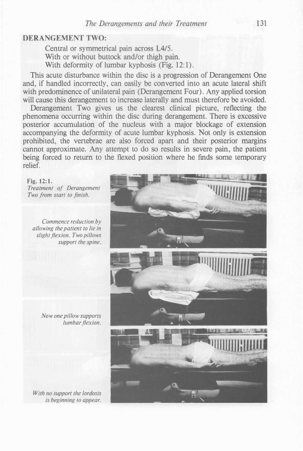
The Derangements and their Treatment 131
DERANGEMENT TWO:Central or symmetrical pain across L4/5.With or without buttock and/or thigh pain.With deformity of lumbar kyphosis (Fig. 12:1).
This acute disturbance within the disc is a progression of Derangement Oneand, if handled incorrectly, can easily be converted into an acute lateral shiftwith predominence of unilateral pain (Derangement Four). Any applied torsionwill cause this derangement to increase laterally and must therefore be avoided.
Derangement Two gives us the clearest clinical picture, reflecting thephenomena occurring within the disc during derangement. There is excessiveposterior accumulation of the nucleus with a major blockage of extensionaccompanying the deformity of acute lumbar kyphosis. Not only is extensionprohibited, the vertebrae are also forced apart arid their posterior marginscannot approximate. Any attempt to do so results in severe pain, the patientbeing forced to return to the flexed position where he finds some temporaryrelief.
Fig. 12:1.Treatment of DerangementTwo from start to finish.
Commence reduction byallowing the patient to lie in
slight flexion. Two pillowssupport the spine.
Now one pillow supportslumbar flexion.
With no support the lordosisis beginning to appear.
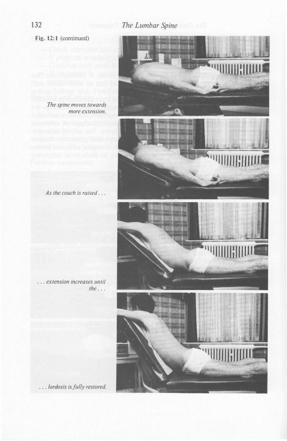
132
Fig. 12: 1 (continued)
The spine moves towardsmore extension.
As the couch is raised . ..
. . . extension increases untilthe . ..
. . . lordosis is fully restored.
The Lumbar Spine

The Derangements and their Treatment 133
Fig. 12: 1. (continued)
After resting fiatfor aminute or two, the patient
should . ..
. . .then apply the extension
... movements himself . ..
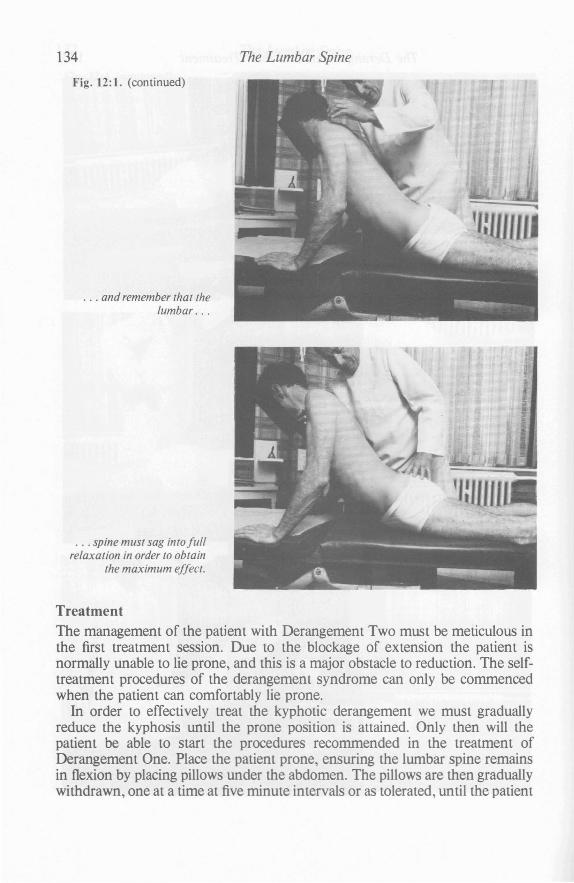
134
Fig. 12:1. (continued)
... and remember that thelumbar . ..
. . . spine must sag into fullrelaxation in order to obtain
the maximum effect.
The Lumbar Spine
TreatmentThe management of the patient with Derangement Two must be meticulous inthe first treatment session. Due to the blockage of extension the patient isnormally unable to lie prone, and this is a major obstacle to reduction. The selftreatment procedures of the derangement syndrome can only be commencedwhen the patient can comfortably lie prone.
In order to effectively treat the kyphotic derangement we must graduallyreduce the kyphosis until the prone position is attained. Only then will thepatient be able to start the procedures recommended in the treatment ofDerangement One. Place the patient prone, ensuring the lumbar spine remainsin flexion by placing pillows under the abdomen. The pillows are then graduallywithdrawn, one at a time at five minute intervals or as tolerated, until the patient
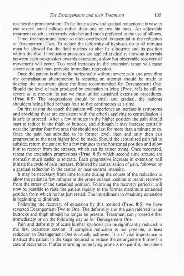
The Derangements and their Treatment 135
reaches the prone position. To facilitate a slow and gradual reduction it is wise touse several small pillows rather than one or two big ones. An adjustabletreatment couch is extremely valuable and much preferred to the use of pillows.
Time, the important factor so often overlooked, is essential in the reductionof Derangement Two. To reduce the deformity of kyphosis up to 45 minutesmust be allowed for the fluid nucleus to alter its silhouette and its positionwithin the disc. If reduction pressures are applied gradually, allowing intervalsbetween each progression towards extension, a slow but observable recovery ofmovement will occur. Too rapid increases in the extension range will causesevere pain and may provoke immediate regression.
Once the patient is able to lie horizontally without severe pain and providingthe centralisation phenomenon is occuring an attempt should be made todevelop the treatment along the lines recommended for Derangement One.Should the level of pain produced by extension in lying (Proc. 8:3) be still sosevere as to prevent its use we must utilise sustained extension procedures.(Proc. 8:5) The progressions should be small and gradual, the patientsshoulders being lifted perhaps four to five centimetres at a time.
On first raising the couch the patient will experience an increase in symptomsand providing these are consistent with the criteria applying to centralisation itis safe to proceed. After a few minutes in the higher position the pain shouldstart to reduce in the thigh or buttock, and although it may increase centrallynear the lumbar four five area this should not last for more than a minute or so.Once the pain has subsided to its former level, then and only then canprogression to the next higher level be made. Should the centralised pain fail tosubside, return the patient for a few minutes to the horizontal position and allowhim to recover from the stresses, which can be rather trying. Once recovered,repeat the extension progressions (Proc. 8:5) which second time around arenormally much easier to tolerate. Each progressive increase in extension willinitiate the cycle of pain increase, followed by centralisation of pain, followed bya gradual reduction in the central or near central intensity.
It may be necessary from time to time during the course of the reduction toallow the patient a few minutes in the prone relaxed position to permit recoveryfrom the stress of the sustained position. Following the recovery period it willnow be possible to raise the patient rapidly to the former maximum extendedposition from which he has just rested. The impediment to obtaining extensionis beginning to diminish.
Following the recovery of extension by this method (Proc.8:5) we havereversed Derangement Two to One. The deformity and the pain referred to thebuttocks and thigh should no longer be present. Treatment can proceed eitherimmediately or on the following day as for Derangement One.
Pain and deformity of .acute lumbar kyphosis can be significantly reduced inthe first treatment session. If complete reduction is not possible, at leastreduction to Derangement One is usually achieved. It is of vital importance toinstruct the patient in the steps required to reduce the derangement himself incase of recurrence. If after returning home lying prone is too painful, the patient
136 The Lumbar Spine
must repeat the sequence of lying over pillows and then gradually loweringhimself until the prone position is obtained. This can be done on the floor overcushions or in bed over pillows. If reduction of derangement is maintained, thepatient will usually be able to commence the extension principle procedures asfor Derangement One the next day.
It may take up to twenty-four hours, occassionally longer, to reduceDerangement Two to Derangement One. Once this is accomplished treatmentshould progress on a daily basis exactly as described for Derangement One, andthe recovery time will be the same as for patients with Derangement One.
The Derangements and their Treatment 137
DERANGEME T THREE:Unilateral or asymmetrical pain across L4/5.With or without buttock and/or thigh pain.No deformity.
In Derangement Three the disturbance within the disc is located morepostero-Iaterally rather than the postero-central position of Derangement One.This may be a progression of Derangement One, but it can also be the primarysite of derangement.
TreatmentInitially the treatment should be the same as for Derangement One. The firsttwenty-four hours of treatment will determine whether the procedures forreduction of Derangement One can successfully be applied to DerangementThree. Very often the application of the extension principle for DerangementThree reduces the disturbance to Derangement One within twenty-four hours.
If on the second visit the patient is improving and the unilateral pain isreducing or centralising, or has moved evenly across the back, self-reduction isprogressing and the extension principle as laid out for Derangement One mustbe continued.
On the second visit, should the patient show no improvement, reexamination and or re-instruction may be necessary; or, as was the case in thereduction of Derangement One the pressures may be inadequate. Should theapplied reductive pressures prove inadequate it will again be necessary toprogressively increase these and additionally alter the direction in which theymay have to be applied.
The first progression in Derangement Three should be precisely the same asthat applied as the first progression in the treatment of Derangement One. Thisprogression should be given on the second day.
The second progression should be applied if the patient returns on the thirdday and is unimproved. With the patient in the prone position a lateral shift isformed by the therapist laterally gliding the patients pelvis away from thepainful side. (This in effect creates a lateral shift towards the painful side.) Withthe pelvis in this off centre position the patient should be asked to perform asequence of extension in lying (Proc.8:3). There is often an initialcentralisation of pain as the extensions are commenced. Should the painsubsequently move laterally or peripherally it may be necessary for the therapistto maintain the off centre position passively as the extensions are performed.Once the repetition of extension results in reduction of pain, or pain feltconsistently in the mid line, the therapist can release the passive maintenance ofthe shifted pelvis. Providing the pains are now reduced or remaining centrally,treatment can progress as for the reduction of Derangement One. It is importantto teach the patient how to place the pelvis laterally on his own, for he may haveto make this lateral correction at home, in order for extension in lying to beeffective.

138 The Lumbar Spine
(An early indication that this progression may be necessary is often obtainedduring the course of examination of the test movements. Where lateral glidingin standing increases or decreases the patients symptoms or where thesemovements cause the initiation of centralisation it can be assumed thatprogression two in Derangement Three is likely to succeed.)
The patient should apply the second progression during the performance ofthe self treatment procedure of extension in lying (Proc.8:3) whenever hedoes these during the next twenty-four hours.
The third, fourth and fifth progressions should be applied if on the fourth daythere is still no improvement.
The third progression sustained rotation, (Proc. 8:11) should be applied andits effects recorded. The patients legs should be moved towards the painful sideas the first rotation is applied. (My records indicate that pressure applied in thisdirection is most consistently followed by rapid improvement. Thecentralisation phenomenon is the ultimate guide however.) The position shouldbe held for up to two minutes depending on the patients tolerance andcentralisation.
The fourth progression should be rotation manipulation in extension(Proc.8:10).
The fifth progression should be rotation manipulation in flexion (Proc. 8:12).Centralisation or reduction of pain must always be our guide regarding the
side to which the technique should be performed or pressure applied. It is myexperience that manipulation has a rapid effect and if there is no change in thesymptoms after two manipulations in one direction, performed on successivedays, the technique should be applied in the opposite direction providedcentralisation and reduction of pain occur.
Once full centralisation is achieved Derangement Three is reversed toDerangement One. The treatment should now be continued and progressedexactly as for Derangement One.
The Derangements and their Treatment 139
DERANGEMENT FOUR:Unilateral or asymmetrical pain across L4/5.With or without buttock and/or thigh pain.With deformity of lumbar scoliosis (Fig. 12:2).
In 1956 I was fortunate enough to observe an acute lumbar scoliosis in apatient friend, who had more than the average faith in my ability as well as anintense fear of remaining deformed for life. The lateral shift with which hepresented was more generous than usually seen, and the poor fellow was ingreat trouble indeed. At that time I did not know of any treatment methodwhich would restore him rapidly to normal, and there did not appear to be anysuch reference in the literature either. My friend's desire to become painfree, hisintense fear of remaining deformed for life coupled with my own curiosityregarding the mechanism causing the scoliosis led me to an abortive attempt torectify the problem and correct the lateral shift. Needless to say, my friend wasno better as a result but he was no worse either. He had to retire to his bed forten days, and four weeks later he was painfree.
I tell this story for a specific reason. The fact that my friend left me neitherbetter nor worse was of little concern to me at the time. However, the fact thathis right sided pain had become left sided and the tilt of his trunk to the left hadchanged to the right, caused great consternation. I wondered what possiblepathology could behave in this manner. In the only book available at that timeto assist in the treatment of spinal disorders, Cyriax24 provided the clue that theintervertebral disc is the most likely structure to be responsible for the lateralshift phenomenon.
From then on, bearing the disc in mind, I closely observed every lateral shiftthat presented and attempted to reduce the derangement. For two years I wasunsuccessful, regularly bringing patients to the upright position only tohopelessly watch them fall back into scoliosis. It was not until the significance offacet apposition in extension occurred to me, that I applied the extensionprinciple following lateral shift correction to ensure maintenance of the erectposture.
In the past ten years I have had ample opportunity to treat patients in manyparts of the world. On such occasions I am always provided with lateral shiftpatients who failed to respond to other treatment methods. Apart from thepatients whose lateral shift had been present for too long, most respondedrapidly and well to my correction techniques. In general, no more than four daysare required to reduce the deformity of scoliosis. The procedure of lateral shiftcorrection has now been adopted by doctors and therapists in many parts of theworld.
Derangement FourMany theories have been put forward to explain the symptoms and peculiarbehaviour of the derangement presented. These theories involve either disc,facet joint, ligaments and muscles, or combinations of any of these structures.
140 The Lumbar Spine
Because in its most extreme form the lateral shift co-exists with a prolapsedintervertebral disc exhibiting an accomplished protrusion, we can assume thatthe disc is heavily involved in the production of this deformity. It is my opinionthat Derangement Four is a lateral progression of the posterior disc disturbanceof Derangement Two, and a simple progression of Derangement Three.
There is evidence that the incidence of deformity of scoliosis is higher in menthan in women. My own statistics show that of all the patients presenting with alateral shift in a certain time period seventy percent were male and thirtypercent female. 12 The results of a similar study reports a proportion of ninetypercent male to only ten percent female patientsY
Although the literature reports an even distribution of back pain among thesexes, I would dispute this, in patients with the derangement syndrome.
Does the rather more accentuated lordosis of the female have anything to dowith a lower incidence of lumbar scoliosis?
The majority of patients are seen to develop a lateral shift away from thepainful side. My statistics show that ninety percent behave in this fashion,whereas the remaining ten percent deviate towards the painful side. 12 In thelatter group reduction is much more difficult to achieve and recovery may takeup to three times longer than in patients of the first group.
A small number of patients exhibit an alternating scoliosis. I believe thatwhen a minor disc bulge exists close to the midline very little movement canunder certain circumstances cause the bulging to move to the opposite side,again occupying a postero-lateral position. As the disc bulge changes positionthe lateral shift deformity resulting from the bulge will also change sides. Thepatient under examination can sometimes create the" alternating deformity atwill, once he has observed its nature and the mechanism effecting it.
The incidence of a relevant lateral shift in patients with low back pain is very"high. My statistics show that fifty-two percent of patients with low back painhave a relevant lateral shift lO that is, alteration of the lateral shift causes a changein intensity or site of pain or both.
The significance of derangement with deformity of scoliosis is much greaterthan is generally recognised. In the presence of a lateral shift either flexion orextension in the lumbar spine may be impaired, and sometimes both areaffected. Lateral flexion to one side is similarly reduced. The loss of movementis always visible, sometimes markedly so. Furthermore, patients with even aminor lumbar scoliosis who are extended by exercises, mobilisation ormanipulation, will become significantly worse with enhancement andperipheralisation of pain. The application of an extension thrust manipulation toa patient with a barely discernable lateral shift may precipitate a disc prolapse.Many patients with an undetected minor lateral shift have entered aclinic fortreatment of their back pain only to walk out with sciatica. Again, I mustemphasise that it is necessary to test all patients with low back pain for evidenceof a relevant lateral shift.
I believe that, to a large extent, the disrepute surrounding the application ofextension exercises has developed because of failure to detect minor lumbar
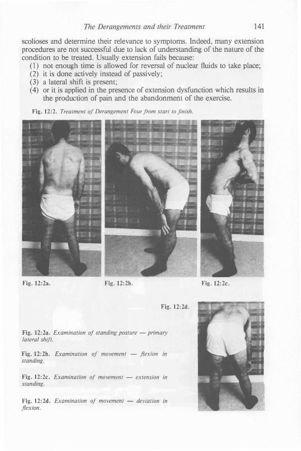
The Derangements and their Treatment 141
scolioses and determine their relevance to symptoms. Indeed, many extensionprocedures are not successful due to lack of understanding of the nature of thecondition to be treated. Usually extension fails because:
(1) not enough time is allowed for reversal of nuclear fluids to take place;(2) it is done actively instead of passively;(3) a lateral shift is present;(4) or it is applied in the presence of extension dysfunction which results in
the production of pain and the abandonment of the exercise.
Fig. 12:2. Treatment of Derangement Four from start to finish.
Fig. 12:2a. Fig. 12:2b. Fig. 12:2c.
Fig. 12:2d.
Fig. 12:2a. Examination of standing posture - primarylateral shift.
Fig. 12:2b. Examination of movement - flexion instanding.
Fig. 12:2c. Examination of movement - extension instanding.
Fig. 12:2d. Examination of movement - deviation inflexion.
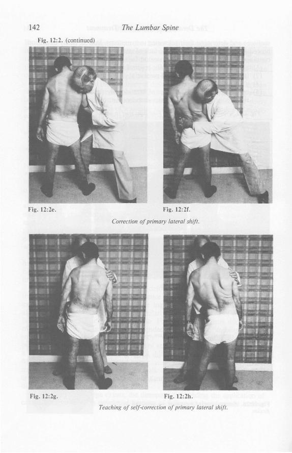
142
Fig. 12:2. (continued)
The Lumbar Spine
Fig. 12:2e.
Fig. 12:2g.
Fig. 12:2f.
Correction ojprimary lateral shift.
Fig. 12:2h.
Teaching oj self-eorrection oj primary lateral shift.
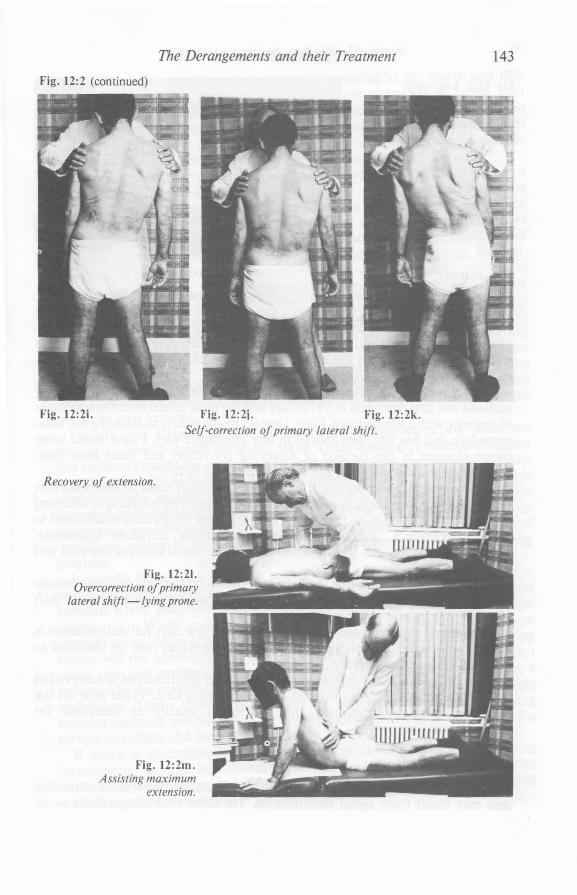
The Derangements and their Treatment
Fig. 12:2 (continued)
143
Fig. 12:2i. Fig. 12:2j. Fig. 12:2k.Self-correction of primary lateral shift.
Recovery of extension.
Fig. 12:21.Overcorrection ofprimary
lateral shift -lying prone.
Fig. 12:2m.Assisting maximum
extension.
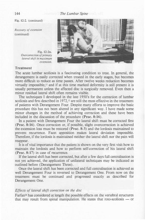
144
Fig. 12:2. (continued)
Recovery of extension
(continued)
Fig. 12:20.Overcorrection ofprimarylateral shift in maximum
extension.
The Lumbar Spine
TreatmentThe acute lumbar scoliosis is a fascinating condition to treat. In general, thederangement is easily corrected when treated in the early stages, but becomesmore difficult to reduce as time passes. After twelve weeks reduction becomesvirtually impossible,12 and if at this time marked deformity is still present it isusually permanent unless the affected disc is surgically removed. Even then aminor residual lateral shift often remains visible.
The techniques I developed in the late 1950's for the correction of lumbarscoliosis and firSt described in 1972,12 are still the most effective in the treatmentof patients with Derangement Four. Despite many efforts to improve the basicprocedure this has not been altered in any significant way. I have made someminor changes in the method of achieving correction and these have beenincluded in the discussion of the procedure (Proc. 8:16).
In a patient with Derangement Four the lateral shift must be corrected first(Proc. 8:16). Once correction or, if possible, slight overcorrection is achievedthe extension loss must be restored (Proc. 8:3) and the lordosis maintained toprevent recurrence. Facet apposition makes lateral deviation impossible.Therefore, if the lordosis is maintained neither the lateral shift nor the pain willreappear.
It is of vital importance that the patient is shown on the very first visit how tomaintain the lordosis and how to perform self-correction of his lateral shift(Proc. 8:17) in case of recurrence.
If the lateral shift has been corrected, but after a few days full centralisation isnot yet achieved, the application of unilateral techniques may be indicated asoutlined before (Derangement Three).
Once the lateral shift has been corrected and full centralisation is achieved aswell Derangement Four is reversed to Derangement One. From now on thetreatment must be continued and progressed exactly as described forDerangement One.
Effects of lateral shift correction on the discFarfan26 has considered at length the possible effects on the vertebral structuresthat may result from spinal manipulation. He states that roto-scoliosis - or

The Derangements and their Treatment 145
lateral shift - is caused by rotation usually occurring at the fourth and fifthlumbar vertebrae. He believes that the McKenzie technique of lateral shiftcorrection is effective because firstly it opens the intervertebral space laterally inassociation with a slight flexion movement which is then followed by anextension movement in the lumbar spine. This produces reduction of the discdisturbance in a manner similar to squeezing a melon seed.
In my experience correction of many hundreds of lateral shifts has only on afew occasions caused a 'pop'-like effect. When perceivable the correctionappears to be a slow flowing process rather than the effects one would associatewith squeezing the melon seed. Whatever the true mechanics eventually proveto be, correction of lumbar scoliosis usually leads to a rapid reduction ofdeformity and symptoms with a corresponding restoration of function. Straightleg-raising, if reduced before correction, is frequently dramatically restored tonormal. I believe that the side bending deformity occurring with lumbarscoliosis is responsible for the reduction in straight-leg-raising on the painfulside when the lateral shift is away from the painful side. In this case theelongation caused by the side bending deformity towards the painfree side willreduce the available root length range in straight-leg-raising. Thus, it is notnecessarily a bulge in the annulus which limits straight-leg-raising. If thenucleus is disturbed enough to lead to the deformity oflumbar scoliosis withoutcausing the outer annulus to bulge, the deformity by itself will be responsible forthe nerve root tension.
Farfan26 has stated that the procedure of straight-leg-raising does notnecessarily have to stretch the sciatic nerve. There is enough evidence to suggestthat reduced straight-leg-raising can be caused by a tight nerve root due to therotation deformity as in sciatic or, as he prefers to call it, roto-scoliosis. Thiswould partially explain why straight-leg-raising can quickly and simply beincreased and reduced merely by laterally altering the patient's position insupine prior to performing the test. Under these circumstances the objectivity ofstraight-leg-raising is suspect, and the test is therefore irrelevant in judgingprogress.
It should be pointed out that testing of straight-leg-raising can be verymisleading and is only reliable as an objective guide when the patient hasconstant sciatic pain. Constant sciatic pain is caused by constant mechanicaldeformation. When a disc protrusion is of sufficient magnitude to causeconstant compression of the nerve root, alteration of the lateral shift while lyingsupine will not allow a better range of straight-leg-raising. If there is constantsciatic pain and straight-leg-raising improves as a result of some manoeuvre, areduction in the magnitude of the protrusion must have occurred following thismanoeuvre. Under these circumstances the objectivity of straight-leg-raisingremains constant and therefore the test is useful in assessing the value of acertain procedure and judging progress.
If there is intermittent sciatic pain, the pressure on the nerve root isintermittent. In this case a minor annular bulge may be influenced bymovements and the patient's symptoms can be created or abolished at willwithout significant alteration in his condition.
146 The Lumbar Spine
DERANGEMENT FIVE:Unilateral or asymmetrical pain across L4/5.With or without buttock and/or thigh pain.With leg pain extending below the knee.No deformity.
Derangement Five is a straight progression of Derangement Three. InDerangement Five the magnitude and location of the disc disturbance are suchthat impingement of the nerve root and dural sleeve occurs, but is not sufficientto force deformity. If the history is of recent onset, the patient must be treatedwith great care. It is essential to determine whether the sciatica is constant orintermittent, because the treatment will be different accordingly. If the sciaticahas been present for several weeks or months, as sometimes occurs, constant legpain may have altered to intermittent pain. Generally, if the patient presentswith a deformity of scoliosis, sciatica is likely to be constant, and if there is nodeformity, sciatica is usually intermittent. Thus, Derangement Five patients aremore likely to have intermittent sciatica though exceptions occur, expecially inacute stages of derangement.
Intermittent sciaticaUsually, the patient with Derangement Five presents without a deformity in thelumbar spine and the sciatica will be intermittent - that is, there are times inthe day when the sciatica is not present. Neurological deficit is infrequentlyenc'ountered in these patients as even short periods without root compressionallow physiological recovery to some extent. Straight-leg-raising mayor may notbe limited.
If the root irritation is indeed intermittent, the patient can be treated withsome caution using mechanical therapy. In the treatment we must make use ofthose positions and movements which are found to reduce or abolish thesciatica. It must be emphasised that any position or movement which producesor enhances sciatica should not be further developed. When neurological deficitis present the chances of success are greatly reduced.
Intermittent sciatica may be caused by a small disc bulge which is, dependingon the patient's activities, alternately protruding into the canal and retreatinginto the disc substance,u but is not large enough to force deformity upon thejoint.
Intermittent sciatica may also be caused by an adherent nerve root orentrapment which is placed under increased tension when certain movementsare performed and positions are adopted. For example, a patient with sciaticamay have recovered recently from an acute disc prolapse and his symptoms arenow becoming intermittent; we must determine whether his sciatica is caused
.by an increase of disc bulging or by an increased nerve root tension due toentrapment, adherence or scarring; and when adherence of the nerve root isthought to be the cause, we must determine whether it is safe at this stage tostretch the nerve root without causing further disc prolapse.
The Derangements and their Treatment 147
To differentiate between sciatica caused by compression from a disc bulge, orthat due to increased root tension from adherence, careful assessment of theflexion test movements is essential. Flexion in standing will enhance the sciaticain both situations - that is, in derangement and in root adherence. The answerto the problem will be found in flexion in lying, which will not enhance sciaticain apatient with an adherent root, but will always enhance it when the disc is in aweakened state. Thus, production or enhancement of sciatica which remainsworse as a result of performing flexion in lying indicates that the increase inmechanical deformation is caused by a bulging disc. With such a response to thetest movements it is dangerous for the patient to perform flexion exercises instanding as well as in lying, as this may lead to recurrence or increase ofderangement.
TreatmentShould testing of flexion in lying indicate that sciatica is caused by a discderangement and the disc wall is able to distend during this movement, it islikely that testing of extension in lying will have the opposite effect and reducethe derangement. If this is the case the patient must be placed under theextension principle for a period of twenty-four hours to confirm the diagnosis. Ifafter this time there is a positive response to extension, the extension principlemay be continued and the patient must be treated as for Derangement Three. Ifthe response to extension appears to be negative, or if progress appears to beslow, the use of unilateral mobilisation and manipulation procedures is indicatedas outlined before (Derangement Three). It will be understood that, once fullcentralisation is obtained treatment should be continued and progressed exactlyas described for Derangement One.
Should the test movements indicate that sciatica is caused by entrapment root adherence or scarring, the treatment described for this under DerangementSix should be applied. Remember however that the treatment must beconsidered to be that required for dysfunction. The derangement stage has nowpassed and the damage repaired.

148 The Lumbar Spine
DERANGEMENT SIX:Unilateral or asymmetrical pain across L4/5.With or without buttock and/or thigh pain.With leg pain extending below the knee.With deformity of sciatic scoliosis. (Fig. 12:3)
Derangement Six clearly is a progression of Derangement Four and Five. Thepatient commonly exhibits a sciatic scoliosis as well as a reduced lordosis. Thesciatica is more likely to be constant and there is little chance of reducing thesymptoms by positioning or movement. Often the patient states that movementbrings relief of pain; however, movement or a change of position gives a shortlived respite only. Neurological deficit commonly occurs.
If in a patient with Derangement Six and constant sciatica most testmovements are found to enhance the leg pain and no movement reduces it, aprovisional diagnosis of accomplished disc protrusion can be made provided thepresence of neurological deficit and a positive myelogram support this finding.Remember, that at this stage the condition is irreversible using mechanicaltherapy. Subsequent surgery usually confirms our diagnosis.
Fig. 12:3.Patient with Derangement Six. This patient shows a
similar deformity as the patient with DerangementFour - a primary lateral shift.
Constant sciatica
If there is no position in which the patient is painfree, we must be careful whensending the patient home on bed rest. It is certainly not desirable that the patientassumes positions which increase the symptoms. Before placing a patient on bedrest we must examine the various positions and their effects on sciatica and lowback pain, and advise the patient accordingly. If there is a position whichproduces significant reduction in leg pain, the patient should be instructed toadopt and maintain that position for long periods at a time.
The Derangements and their Treatment 149
If no test movements or positions can be found to reduce the pain referredbelow the knee then, I believe, there are no techniques available which will havea beneficial effect on the derangement. Intermittent traction in the order oftwenty minutes a day on an out-patient basis has been described as useful, but ifprogress is made it is difficult to attribute this to the treatment rather than to thepassage of time. Continuous traction in bed is a different matter and oftenproduces good enough results to make the procedure an attractive alternative tosurgery.
Treatment
If test movements indicate that reduction of the derangement is possible,treatment should be commenced following the lines of that for DerangementFour and Five. During the treatment great care should be taken to checkregularly if there is a change in peripheral symptoms. Increased tingling ornumbness felt in the foot must never be ignored, and if these are presenttreatment should be modified immediately.
If there is improvement but full centralisation has not yet been achieved, theapplication of mobilising and manipulation procedures may be indicated.Techniques outlined previously (Derangement Three) may be utilised, providedpre-manipulative testing is performed to ensure centralisation of symptoms istaking place. If peripheral pain is enhanced in the pre-manipulative testingposition, the manipulation should not be performed. Whatever procedure youhave in mind, never manipulate a patient without pre-manipulative testing.
Should lateral shift correction produce reduction of the derangement, thenprogress usually is uncomplicated and treatment should continue as outlined forDerangement Four. The recovery of full flexion or the treatment of any residualflexion dysfunction must be delayed much longer after the resolution of a severesciatica than is usually the case with pure lumbar pain. I consider eight to tenweeks from the time of onset of peripheral symptoms to be the minimum timebefore implementing the flexion dysfunction procedures to the maximum.
Stretching of adherent nerve root
Once the acute episode has subsided and the patient is able to attend to hisoccupation, nerve root adherence as a result of scarring may cause a persistentsciatic reference which, in some cases, may last for years. This disability will notproduce neurological deficit if the initial episode has not already done so. It isdesirable to reduce the nerve root entrapment or adherence, and in manypatients this may be done successfully by using the procedures for flexiondysfunction - that is, the shortened structures should be stretched frequentlyand firmly every day until the adherence is resolved. To achieve this flexion inlying (Proc. 8:13) should be regularly performed for one week. This is followedby flexion in standing (Proc. 8:14) which must be performed often enough toelongate and stretch scarred and adherent tissue without causing further bulgingof the disc wall (Fig. 12:4).

150
Fig. 12:4.Treatment ofadherent
sciatic nerve root, usingflexion in standing.
The Lumbar Spine
When treatment of dysfunction due to an adherent nerve root is commencedit should not be attempted during the first four to five hours of the day. Duringthis time period the disc is likely to be under increased pressure as a result of itsnocturnal imbibition and reabsorbtion of fluid. I recommend that in the initialstages of treatment the patient performs ten repetitions of flexion in standing(Proc. 8:14) from midday on, every three hours until retirement. When thederangement appears stable and the integrity of the disc wall is established, thepatient may commence the exercise a little earlier in the day and repeat it everytwo hours. However, it is inadvisable to perform flexion in standing(Proc. 8:14) on awaking.
Because flexion in standing (Proc. 8:14) places stress on the disc wall andforces the disc posteriorly, there are always risks involved in stretching anadherent nerve root, especially in the initial stages. Therefore, flexion instanding (Proc. 8:14) must always and immediately be followed by extensionin lying (Proc.8:3), or if this is not possible by extension in standing(Proc.8:6).
As the nerve root stretching becomes effective, the patient may perceive thesame amount of pain whereas his flexion range has increased. Gradually hisfingertips will move closer to his ankles, and where a certain amount of flexionproduced significant pain before it may have become painfree now. It willbecome easier to drive the car and to walk uphill.
It is my belief that intermittent stress applied to joints in this mannerstrengthens ligamentous structures rather than damages them, while scar tissuewill stretch and yield to such stress.45 However, if a sustained stress is applieddamage may occur to both ligamentous and scar tissue.
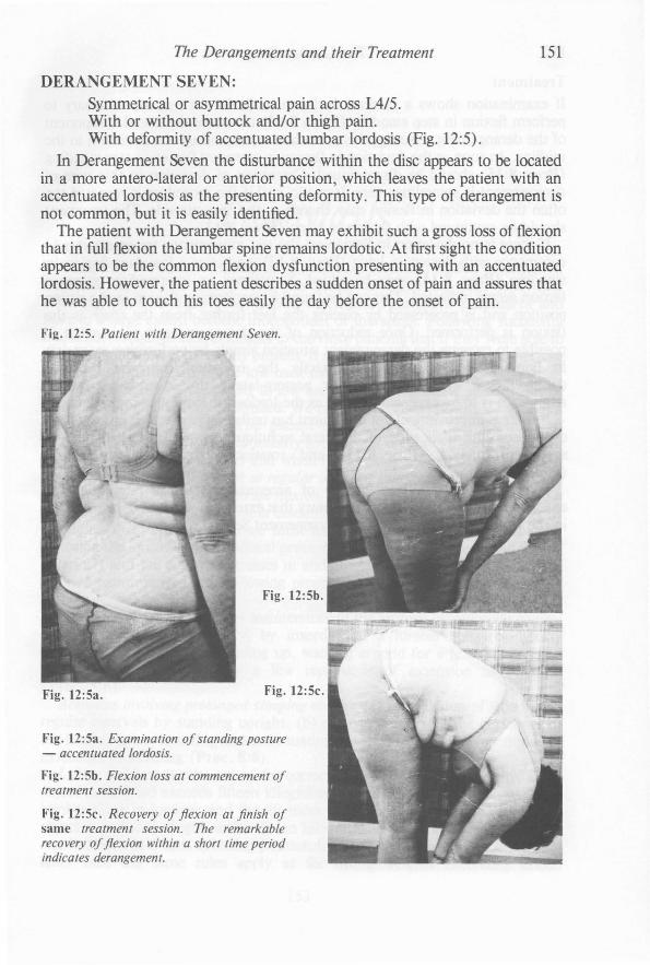
The Derangements and their Treatment 151
DERANGEMENT SEVEN:Symmetrical or asymmetrical pain across L4/5.With or without buttock and/or thigh pain.With deformity of accentuated lumbar lordosis (Fig. 12:5).
In Derangement Seven the disturbance within the disc appears to be locatedin a more antero-Iateral or anterior position, which leaves the patient with anaccentuated lordosis as the presenting deformity. This type of derangement isnot common, but it is easily identified.
The patient with Derangement Seven may exhibit such a gross loss of flexionthat in full flexion the lumbar spine remains lordotic. At first sight the conditionappears to be the common flexion dysfunction presenting with an accentuatedlordosis. However, the patient describes a sudden onset of pain and assures thathe was able to touch his toes easily the day before the onset of pain.
Fig. 12:5. Patient with Derangement Seven.
Fig. 12:5c. Recovery of flexion at finish ofsame treatment session. The remarkablerecovery offlexion within a short time periodindicates derangement.
Fig. 12:5a. Examination of standing posture- accentuated lordosis.
Fig. 12:5b. Flexion loss at commencement oftreatment session.
Fig. 12:5c.
Fig. 12:5b.
Fig. 12:5a.
152 The Lumbar Spine
Treatment
If examination shows a marked deviation in flexion, it may be necessary toperform flexion in step standing (Proc. 8:15) to reduce the lateral componentof the derangement. Usually, this procedure rapidly brings the deviation to themidline. As soon as the deviation in flexion is corrected flexion in step standing(Proc. 8:15) should be discontinued. Correction of deviation in this type ofderangement is sometimes very dramatic, and if the procedure is repeated toooften the deviation in flexion may change to the opposite side. The treatmentshould be continued with flexion in lying (Proc. 8:13).
If there is no deviation in flexion on the test movements, treatment shouldcommence with flexion in lying (Proc. 8.13), and after a few days flexion instanding (Proc. 8:14) may be added. Sometimes an intermediate stage offlexion seems indicated. This can be found by performing flexion in the sittingposition and is progressed by placing the feet further from the chair as theflexion is performed. Once reduction of the derangement is achieved it ispossible to recreate the derangement situation simply by performing extensionin lying (Proc.8:3). This is exactly the opposite from the previousderangements with a posterior or postero-Iateral disc disturbance whererecurrence is to be expected as soon as the lordosis is lost following reduction.
If there is improvement but the patient has unilateral pain which has not fullycentralised, the application of unilateral techniques may be indicated such assustained rotation (Proc. 8:11) and rotation manipulation in flexion(Proc.8:12).
Because patients with deformity of accentuated lordosis rarely developextension dysfunction, it is not necessary that extension procedures be includedin the treatment of patients with Derangement Seven.

CHAPTER 13
ProphylaxisThe majority of patients responding to basic extension and flexion principles oftreatment have been educated in the means of achieving pain relief andrestoring function. They have carried out the self-treatment procedures andhave to a large extent become independent of therapists. Following successfultreatment it requires little emphasis to convince patients that if they were able toreduce and abolish pain already present, it should also be possible to prevent theonset of any significant future low back pain.
Of all the factors predisposing to low back pain only postural stresses can beeasily influenced and fully controlled. We must develop this potential ingredientof prophylaxis to the full. The patient must understand that the risks ofincurring low back pain are particularly great when the lumbar spine is held insustained flexed positions; and that when the lordosis is reduced or eliminatedfor prolonged periods, he must at regular intervals and before the onset ofpainmake a conscious effort to interrupt flexion, restore the lordosis and accentuate itmomentarily to the maximum. It is essential that the patient knows the reasonsfor doing this, and therefore we must explain to him in lay terms that onrestoring the lordosis the intradiscal pressure decreases, the nuclear fluid movesanteriorly and the posterior stresses in and around the disc are reduced.
Briefly summarised, the following prophylactic measures should always betaken:
Prolonged sitting requires (a) maintenance of the lordosis by muscular controlof the posture or, preferably, by insertion of a lumbar roll, (b) hourlyinterruption of sitting by standing up, walking around for a few minutes andaccentuating the lordosis by a few repetitions of extension in standing(Proc.8:6).
Activities involving prolonged stooping require (a) interruption of stooping atregular intervals by standing upright, (b) regularly reversing the curvature ofthe lumbar spine, restoring and accentuating the lordosis by a few repetitions ofextension in standing (Proc. 8:6).
Lifting requires (a) the use of the correct lifting technique. Generally, if theobject to be lifted exceeds fifteen kilograms, the strain must be taken with thelumbar spine in lordosis and the lift must be performed using the legs. If theobject to be lifted weighs under fifteen kilograms less care is necessary, unlessone has been in a bent or sitting position for some time prior to the lift. In thelatter case the same rules apply as for lifting weights exceeding fifteen
153

154 The Lumbar Spine
kilograms, (b) accentuation of the lordosis before and after lifting by a fewrepetitions of extension in standing (Proc. 8:6).
If inadvertently pain has developed during sitting, stooping or lifting, thepatient should immediately commence extension in lying (Proc.8:3).Extension in standing (Proc. 8:6), very effective in preventing the onset ofpain, is less effective when used to reduce present pain. Extension in lying(Proc. 83) is the technique of first aid for back pain.
Recurrence: At the first sign of recurrence the patient should immediatelycommence the procedures which previously led to recovery. Although 9J1episode of low back pain can commence suddenly and without warning, manypatients are aware of a minor degree of discomfort before the onset of severepain. If this type of warning is given, the patient has an excellent chance toprevent the development of symptoms, provided the appropriate procedure isapplied immediately.
It is not possible for patients to remember all verbal instructions and advicegiven during the first treatment. To avoid tedious repetition and to ensure thenecessary information is conveyed to the patient, a list of instructions is suppliedon the first visit. This list firstly deals with information for patients in the acutestage of low back pain, and secondly provides information required oncerecovery has taken place. These instructions form an important part of selftreatment, because when followed properly they will help in reduction ofpresent symptoms and prevention of their recurrence.
WELLINGTON PHYSIOTHERAPY CLINIC
General Instructions - When in AcuteLow Back Pain
You must retain the lordosis at all times (lordosis is the hollowin the lower back). Bending forwards as in touching the toeswill only stretch and weaken the supporting structures of the backand lead to further injury. Losing the lordosis when sitting will alsocause further strain.
SITTING• When in acute pain you should sit as little as possible, and then
only for short periods only.• At all times you must sit with a lordosis. Therefore you must
place a supportive roll in the small of the back, especially whensitting in a car or lounge chair.

Prophylaxis 155
• If you have the choice you must sit on a firm, high chair with astraight back such as a kitchen chair. You should avoid sittingon a low, soft couch with a deep seat; this will force you to sitwith hips lower than knees, and you will round the back andlose the lordosis.
• The legs must never be kept straight out in front as in sitting inbed, in the bath or on the floor; in this position you are forced tolose the lordosis.
• When rising from sitting you must retain the lordosis; move tothe front of the seat, stand up by straightening the legs, andavoid bending forwards at the waist.
• Poor sitting postures are certain to keep you in pain or makeyou worse.
DRIVING A CAR• When in acute pain you should drive the car as little as pos
sible. It is better to be a passenger than to drive yourself.
• When driving, your seat must be close enough to the steeringwheel to allow you to maintain the lordosis. If in this positionyour hips are lower than your knees you may be able to raiseyourself by sitting on a pillow.
BENDING FORWARDS• When in acute pain you should avoid activities which require
bending forwards or stooping, as you will be forced to lose thelordosis.
• You may be able to retain the lordosis by kneeling - forexample, when making the bed, vacuuming, cleaning the floor,or weeding the garden.
LIFTING• When in acute pain you should avoid lifting altogether.• If this is not possible you should at least not lift objects that are
awkward or heavier than about thirty pounds.• You must always use the correct lifting technique: during lifting
the back must remain upright and never stoop or bend forwards; stand close to the load, have a firm footing and widestance; bend the knees and keep the back straight; have asecure grip on the load; lift by straightening the knees; take asteady lift and do not jerk; shift your feet to turn and do not twistyour back.

156 The Lumbar Spine
LYING• A good firm support is usually desirable when lying. If your bed
is sagging, slats or plywood supports between mattress andbase will firm it. You can also place the mattress on the floor, asimple but temporary solution.
• You may be more comfortable at night when you use a supportive roll. A rolled up towel, wound around your waist and tieddown in front, is usually satisfactory.
• When rising from lying you must retain the lordosis; turn on oneside, draw both knees up and drop the feet over the edge of thebed; sit up by pushing yourself up with the hands and avoidbending forwards at the waist.
COUGHING AND SNEEZING• When in acute pain you must try to stand up, bend backwards
and increase the lordosis while you cough and sneeze.
REMEMBER:• At all times you must retain the lordosis; if you slouch you will
have discomfort and pain.• Good posture is the key to spinal comfort.
General Instructions WhenRecovered From Acute Low Back Pain
You have recovered from the acute episode because of yourability to master the exercises which relieved your pain. Theseexercises must be repeated whenever situations arise whichhave previously caused pain. You must perform the correctivemovements before the onset of pain. This is essential.
If you carry out the following instruction, you can resume yournormal activities without the fear of recurrence.
SITTING• When sitting for prolonged periods the maintenance of the
lordosis is essential. It does not matter if you maintain this withyour own muscles or with the help of a supportive roll, placed inthe small of your back.
Prophylaxis 157
• In addition to sitting correctly with a lumbar support, you shouldinterrupt prolonged sitting at regular intervals. On extended carjourneys you should get out of the car every hour or two, standupright, bend backwards five or six times, and walk around fora few minutes.
BENDING FORWARDS• When engaged in activities which require prolonged forward
bending or stooping - for example, gardening, vacuuming,concreting - you must stand upright, restore the lordosis andbend backwards five or six times before pain commences.
• Frequent interruption of prolonged bending by reversing thecurve in the low back should enable you to continue with mostactivities you enjoy, even with some you do not enjoy.
LIFTING• If the load to be lifted weighs over thirty pounds, the strain must
be taken with the low back in lordosis and you must lift bystraightening your legs.
• If the object weighs under thirty pounds less care is required,unless you have been in a bent or sitting position for some timeprior to lifting. In the latter case you must lift as if the weightexceeds thirty pounds.
• In addition to correct lifting technique, you must stand uprightand bend backwards five or six times after lifting.
RECURRENCE• At the first signs of recurrence of low back pain you should
immediately start the exercises which previously led to recovery, and follow the instructions given for when in acute pain.
• If this episode of low back pain seems to be different than onprevious occasions, and if your pain persists despite followingthe instructions, you should contact a manipulative therapist.
REMEMBER:• If you lose the lordosis for any length of time, you are risking
recurrence of low back pain.
Robin McKenzie16 The Terrace
Wellington

158 The Lumbar Spine
It seems that at this stage in the development of spinal therapy theresponsibility for successful management and prophylaxis in low back pain lieswith the manipulative therapist. As education is the only way of preventing lowback pain induced and increased by poor posture, I would hope that in thefuture, prophylactic postural advice be provided at school level. This shouldreally be the province of the physical education instructor, but unfortunately atthe present time this profession does not yet have the necessary knowledgeregarding prophylaxis for any spinal pain.
CHAPTER 14
Contra-IndicationsAlthough it has been accepted throughout that all patients have receivedadequate medical screening, occasionally patients with serious pathology ormechanical disorders unsuited to mechanical treatment are encountered duringroutine examination. If in the examination no position or movement can be foundwhich reduces the presenting pain, the patient is unsuited for mechanicaltherapy, at least at this time.
The existence of serious pathology should be considered when the historystates that there has been no apparent reason for the onset of symptoms; thatthe symptoms have been present for many weeks or months, and have duringthat time increased in intensity; and that they are constant; and the patient feelsthat he is gradually getting worse. On examination the pain remains exactly thesame, irrespective of positions assumed or movements performed. Usually thereis little loss of function if any, and postural deformity is not often seen. Inaddition to the examination finding, the patient often looks unwell and maycomplain of feeling unwell. Mechanical procedures described in this bookshould never be applied to patients presenting with signs and symptoms of thisnature.
Saddle anaesthesia and bladder weakness are indications of compression ofthe fourth sacral nerve root. These symptoms occur in major disc herniationand, when present, form a definite contra-indication to manipulativeprocedures.
Patients who exhibit signs of extreme pain - that is, who become transfixedon movement, and freeze and immobilise the spine when palpation is attempted- are considered unsuitable for mechanical therapy, at least at this stage.
Developmental or acquired anomalies of bone structures which may lead toweakness or instability of mechanical articulations are hazards to manipulativeprocedures. Architectural faults should be excluded from mechanical therapy..Exceptions are minor grades of spondylolisthesis where symptoms are mild andintermittent. The presence of spondylolisthesis at a certain segment is notnecessarily an indication that symptoms are forthcoming from that segment. Itis seen very often indeed that pain is the result of disc derangement at adifferent level.
A simple clinical test may determine whether spondylolisthesis is responsiblefor the presenting pain, as it will often reduce or abolish pain in the presence ofthat condition. Place one hand across the sacrum of the standing patient and the
159

160 The Lumbar Spine
other firmly against the abdomen. By further compressing the abdominalcontent while at the same time increasing pressure on the sacrum, pain instanding arising from spondylolisthesis is markedly reduced or abolished. Onthe other hand, pain arising from derangement of any of the lumbar discs willusually be enhanced by this procedure, and postural or dysfunctional painremains unaffected. Thus, if pain is increased with this test the patient should betreated as for the derangement syndrome, but if pain is reduced the presence ofspondylolisthesis must be investigated and the necessary precautions must betaken in the treatmen.t.
Bed restPatients with mechanical low back pain are often advised to rest as much aspossible, preferably in bed. In many instances this is inappropriate if not badadvice. The nutrition of the intervertebral discs depends entirely on osmosis,and movements of the spine are essential for the flow of fluids containingnutriments. To rest the joints of patients with mechanical low back pain oftenonly adds to the problems of restoration of function and full rehabilitation. Theonly patient who should be placed on bed rest is the very acute patient withsevere constant low back pain with or without sciatica whose symptoms areconsiderably worse during weight bearing, and in whom no movement or positioncan be found to reduce or centralise the pain.
In order to prevent the development of dysfunction within or about theinvolved intervertebral joint following derangement, regular assessment of thepatient on bed rest is required and treatment must be instituted as early aspossible. This is particularly important in the patient who responds very well tobed rest, because without movement this patient will become painfree but maydevelop a significant loss of function.
SupportsIf, following reduction of derangement, all efforts at prevention of immediaterecurrence are unsuccessful, it may be desirable to supply the patient with acorset for short term support and stability. Long term use of a corset isundesirable as it merely hastens the development of dysfunction.
SurgeryThe question of surgical intervention inevitably arises in the minds of doctors,therapists and patients alike when progress is slow or non-existent. The decisionas to whether one should or should not operate must always concern theorthopaedic surgeon.
It is my experience that, in New Zealand, surgery will be performed onlywhen conservative treatment methods have failed. This does not always includeadequate mechanical therapy, but usually it has allowed adequate passage oftime. Fortunately, few patients in New Zealand receive surgery for low backpain only, and the procedures of laminectomy and discectomy are reserved forpatients suffering significant nerve root compression.

Contra-Indications 161
The difficulties surrounding the diagnostic criteria for surgical interventionare enormous. One study showed a large number of positive myelograms inlumbar spines of patients who were asymptomatic and had been admitted andX-Rayed for conditions other than low back pain.5 Thus the value of themyelogram, the good old standby relied upon by many doctors, has becomesomewhat suspect.
The results of surgery can also be seen to vary significantly.5 Randomlyselected patients with low back pain, referred pain to one leg, and positivemyelograms coupled with clinical signs, were divided into two groups. Thepatients in the one group received surgery, those in the other group not. It wasfound that at the end of one year ninety percent of the non-operated patientswere better; that the operated patients were only better in the first two monthsfollowing surgery; and that at the end of one year the results in the group thathad received surgery were the same as in the group that had not been operatedupon.
The final result of a disc lesion is not endangered by a three months' waitingperiod before surgery is undertaken. However, a longer wait may be prejudicialas it may leave the patient with residual disability.5
Fielding5states that Nachemson, Rothman and Hirsch have all found thatthere is a clear correlation between the early return of symptoms followingsurgery and the formation ofscar tissue. Quoting Fielding:5
"Early return of symptoms usually means the formation of scarring whichcannot be cured by surgery and means that the results will be poor".Rothman39 examined sixty-eight patients who had undergone two or more
unsuccessful spinal surgical interventions. He reports that seventy-five percentof the patients claimed partial or total disability. Half of the failures in multiplesurgery are due to scarring or nerve root adherence. Scarring and fibrosis areassociated with a high failure rate and dismal results.
It is appropriate to make clear that 'scarring causes dysfunction, describedearlier in this book. Scarring does not necessarily have to cause problemsprovided it is discovered early enough and dealt with adequately in the mannersuggested in previous chapters. I believe that one procedure is mandatoryfollowing surgery for lumbar disc protrusion: that is, the regular performance ofone full straight-leg-raising movement at two hourly intervals. This shouldreduce significantly the complications of fibrosis and nerve root adherence. Justas recent scars can be stretched and lengthened by dysfunction treatmentprocedures, so should the prevention of inextensible scar formation be feasibleby the even earlier use of the same procedures.

ReferencesI Fisk, 1. W. (1977). "The painful neck and back". (Charles C. Thomas, Springfield, USA)2 Wood, P. H. N. (1976), "Epidemiology of back pain". in: "The Lumbar spine and back pain" ed. M.
Jayson. (Pitman Medical Publishing Co., Tumbridge Wells, Great Britain)3 Anderson, 1. A. D. (1976), "Back pain in industry". in: "The lumbar spine and back pain" ed. M. Jayson.4 Wyke, B. (1976), "Neurological aspects of low back pain". in: "The lumbar spine and back pain" ed. M.
Jayson.5 Fielding, 1. W. (1979), Orthopaedic and Physical Therapy Seminar, University of Cincinatti Medical
Center, Sept. 1979.6. La Rocca, H. and MacNab, I. (1969), "Value of pre-employment radiographic assessment of the lumbar
spine", Canad. Med. Assoc. 1. 101; 383.7 Nachemson, A. (1976), "A critical look at conservative treatment for low back pain". in: "The lumbar
spine and back pain" ed. M. Jayson.8 Dixon, A.St.1. (1976), "Diagnosis of low back pain". in: "The lumbar spine and back pain" ed. M. Jayson.9 Magora, A. (1972), "Investigation of relation between low back pain and occupation", Ind. Med. Surg. 41;
5.10 McKenzie, R. A. (1979), "Prophylaxis in recurrent low back pain", NZ. Med. 1. 89; 22.II Andersson, B. 1. G., et al. (1975), "The sitting posture: An electromyographic and discometric study",
Orth. Cl. N. Am. Vol. 6, o. I.12 McKenzie, R. A. (1972), "Manual correction of sciatic scoliosis", NZ Med. 1. 76; 484.IJ Shah, 1. S., Hampson, W. G. H., and Jayson, M. I. V. (1978), "The distribution of surface strain in the
cadaveric lumbar spine", J. Bone & Jt. Surg. 60B; 246.14 Armstrong, 1. R. (1958), "Lumbar disc lesions" (E. & F. Livingstone Ltd., Edinburgh, London.)15 Andersson G. B. 1., Murphy R. W., Ortengren R., and Nachemson A. (1979), "The influence of backrest
inclination and lumbar support on lumbar lordosis", Spine, Vol. 4, No.1; 52.16 Nachemson A. (1975), "Towards a better understanding of low back pain. A review of the mechanics of
the lumbar disc", Rheum. & Rehab. 14; 129.17 Markolf K. L., and Morris J. M. (1974), "The structural components of the intervertebral disc. A study of
their contributions to the ability of the disc to withstand compressive forces", 1. Bone & Jnt. Surg. 56A;675.
18 Naylor A., and Shentall R. (1976), "Biochemical aspects of intervertebral discs in ageing and disease". in:"The lumbar spine and back pain" ed. M. Jayson.
19 Nachemson A. (1976), "Lumbar intradiscal pressure". in: "The lumbar spine and back pain" ed. M.Jayson.
20 Happey F. (1976), "A biophysical study of the human intervertebral disc" in: "The lumbar spine and backpain" ed. M. Jayson.
21 Farfan H. F. (1973), "Mechanical disorders of the low back". (Lea & Febiger, Philadelphia, USA.)22 Fahrni W. H. (1976)" "Backache: Assessment and treatment". (Musqueam Publishers Ltd., Vancouver,
Canada.)23 Vernon-Roberts B. (1976), "Pathology of degenerative spondylosis" in: "The lumbar spine and back pain"
ed. M. Jayson.24 Cyriax 1. (1969), "Textbook of orthopaedic medicine" Vol. 1, 5th ed. (Bailliere Tindall, London.)25 Charnley J. (1951), "Orthopaedic signs in the diagnosis of disc protrusion", Lancet cclx; 186.26 Farfan H. F. (1979), Orthopaedic and Physical Therapy Seminar, University ofCincinatti Medical Center,
Sept. 1979.21 Park, W. M. (1976), "Radiological investigation of the intervertebral disc" in: "The lumbar spine and back
pain" ed. M. Jayson.28 Yates A. (1976), "Treatment of back pain" in: "The lumbar spine and back pain" ed. M. Jayson.29 Jayson, M. I. V. (1976), Editor's Preface in: "The lumbar spine and back pain" ed. M. Jayson.30 Nachemson A. (1976), Personal communication.31 Cyriax 1. (1971), "Textbook of orthopaedic medicine" Vol. 2, 8th ed. (Bailliere Tindall, London.)
163

164 The Lumbar Spine
J2 Dalseth l. (1974), "Anatomic studies of the cranio-vertebral joints", Man. Med. 6; 130.II Baddeley H. (1976), "Radiology of lumbar spinal stenosis" in: "The lumbar spine and back pain" ed. M.
Jayson.J4 Goldstein M. (Editor) (1975), "The research status of spinal manipulative therapy", US Department of
Health, Education and Welfare.35 Wall P. D. (1977), Proceedings of FIMM Congress, Copenhagen.36 Mennell 1. McM. (1960), "Back pain". (1. & A Churchill Ltd., London.)37 Mathews 1. A. (1976), "Epidurography - A technique for diagnosis and research" in: "The lumbar spine
and back pain" ed. M. Jayson.38 Nachemson A. (1960), "Lumbar intradiscal pressure", Acta Orth. Scand. Suppl. 43.39 Tottle C. R. (1972), "Symposium on the painful back" Royal Coil. Phys., London.40 Fielding J. W. (1979), Quoting Rothman, R. at Orthopaedic and Physical Therapy Seminar, University of
Cincinatti Medical Center, Sept. 1979.41 Kelly P., and Schneider G. (1975), "Manual correction of scoliosis", Proceedings of Australian
Physiotherapy Congress.42 Tucker W. E. (1960), Active Alerted Posture. London. Livingstone.43 Hickey D. S., and Hukins D. W. L. (1980), "Relation Between the Structure of the Annulus Fibrosus and
the Function and Failure of the Intervertebral Disc". Spine Vol. 5. No.2. 106.44 White A A, and Panjabi M. M. (1978), "Clinical Biomechanics of the Spine". 1. B. Lippincott Co.45 Burkart S. (1980)" Proceedings of IFOMT Congress. Christchurch, New Zealand.46 Markolf K. L., and Morris 1. M. (1974), The structural components of the intervertebral disc. 1.B. 1.S. 56A.
675.47 Markolf K. L. (1972), Deformation of the thoraic lumbar intervertebral joint in response to external loads:
a biomechanical study using autopsy material. 1.B. 1.S. 54A. 511.48 Salter R. (1979), Paper presented Canadian Royal College of Physicians and Surgeons Montreal.