
JuMandoctor2017.jumedicine.com/.../7/2018/06/Midterm-Exam.pdfJuMan ... JuMan JuMan

Sarah Jaar
…
9
Anas Abu-Humaidan
Lujain Hamdan
1 | P a g e
Taxonomy
• It’s the ordering of organisms in organized and related groups according to their
characteristics.
• Identification, classification and nomenclature are three separate but interrelated
areas of bacterial taxonomy. Each area is critical to the ultimate goal of accurately
studying the infectious diseases and precisely communicating these to others in the
field.
• Bacterial taxonomy depends on our knowledge about the structure, metabolism and
genetics of bacteria.
❖ Classification is the categorization of organisms into taxonomic groups
using biochemical, physiologic, genetic and morphologic properties.
o Taxonomic ranks are: kingdom, division, class, order, family, genus, species and
subtype.
o What defines a species is the ability to reproduce and give fertile offspring. But there
is no definitive “species" taxon in bacterial classification due to:
➢ Asexual reproduction.
➢ Horizontal gene transfer.
❖ Identification is the practical use of a classification scheme to:
a. isolate and distinguish specific organisms among the mix of complex microbial flora.
b. verify the authenticity or special properties of a culture in a clinical setting.
c. isolate the causative agent of a disease and identify it. This leads to the selection of
specific pharmacologic treatments (e.g. antibiotics).
• This area of microbiology is necessarily dynamic as the tools continue to evolve (e.g. new methods of microscopy, biochemical analysis and computational nucleic acid biology).
❖ Nomenclature refers to the naming of an organism according to scientific
rules, so each name refers to the same organism and is understood by all
scientists, microbiologists, physicians, etc.
• This is arguably the most important component of taxonomy because it allows
medical professionals to communicate with each other. Any professional
associated with an infectious disease should be aware of the evolving taxonomy
of infectious microorganisms.
2 | P a g e
Classification
Bacteria can be classified according to:
1. Macroscopic and microscopic appearance (morphology)
It’s the first step in identification because it’s simple and needs few minutes. To
determine the morphologic characteristics, we stain the bacteria then examine them
under the microscope.
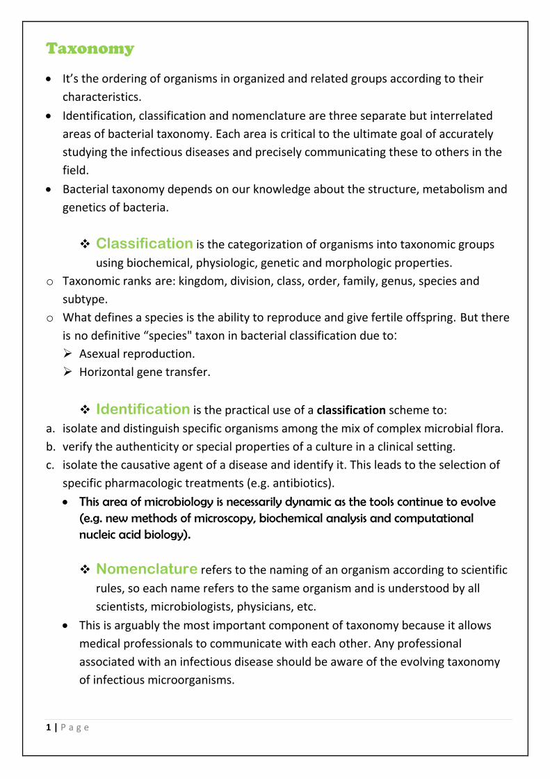
A. The shape:
▪ Cells can have different shapes:
Coccus, Bacillus, Spirillum, Vibrio,
etc.
▪ Many cells can form clusters,
chains or pairs.
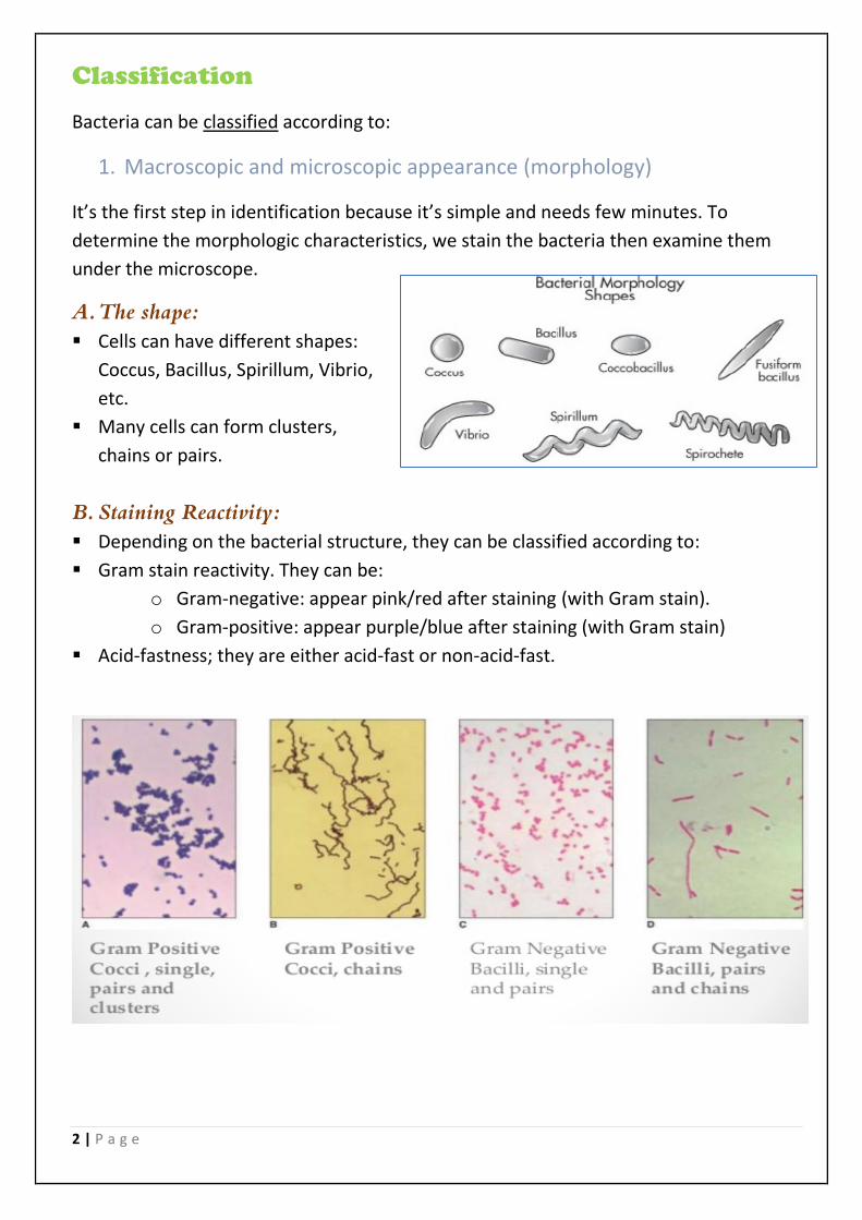
B. Staining Reactivity:
▪ Depending on the bacterial structure, they can be classified according to:
▪ Gram stain reactivity. They can be:
o Gram-negative: appear pink/red after staining (with Gram stain).
o Gram-positive: appear purple/blue after staining (with Gram stain)
▪ Acid-fastness; they are either acid-fast or non-acid-fast.
3 | P a g e
C. Motility: can be detected by the SIM (Sulfide (H2S) Indole motility) medium test.
SIM is generally used to differentiate Enterobacteriaceae members.
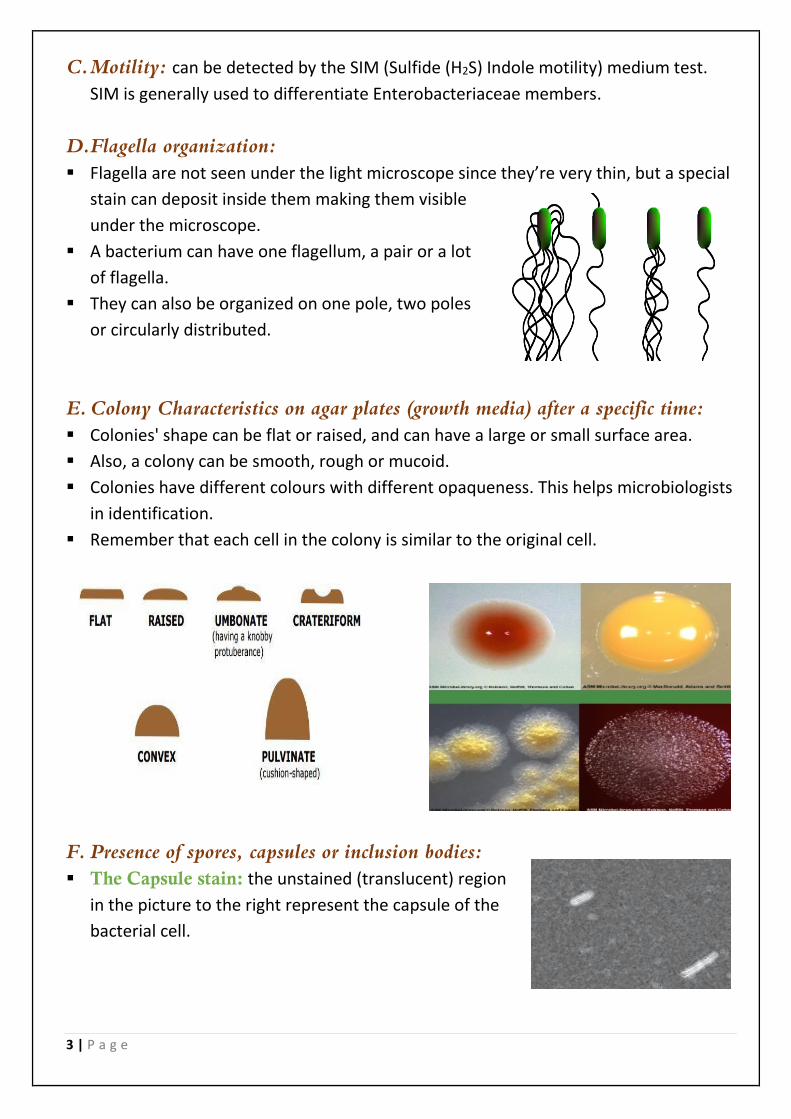
D.Flagella organization:
▪ Flagella are not seen under the light microscope since they’re very thin, but a special
stain can deposit inside them making them visible
under the microscope.
▪ A bacterium can have one flagellum, a pair or a lot
of flagella.
▪ They can also be organized on one pole, two poles
or circularly distributed.
E. Colony Characteristics on agar plates (growth media) after a specific time:
▪ Colonies' shape can be flat or raised, and can have a large or small surface area.
▪ Also, a colony can be smooth, rough or mucoid.
▪ Colonies have different colours with different opaqueness. This helps microbiologists
in identification.
▪ Remember that each cell in the colony is similar to the original cell.
F. Presence of spores, capsules or inclusion bodies:
▪ The Capsule stain: the unstained (translucent) region
in the picture to the right represent the capsule of the
bacterial cell.

4 | P a g e
▪ The Spore stain: Additional info: Spores are most simply observed as intracellular
refractile bodies in unstained cell suspensions or as colourless areas in cells stained
by conventional methods. The spore wall is relatively
impermeable, but dyes can be made to penetrate it by
heating the preparation. This same impermeability then
serves to prevent decolourisation of the spore during a
period of alcohol treatment sufficient to decolorize
vegetative cells. The latter (vegetative cells) can finally be
counterstained. Spores are commonly stained with
malachite green or carbolfuchsin.
2. Growth conditions
A. Oxygenation:
✓ If the bacteria are aerobic: the culture's size increases during oxygenation.
✓ If the bacteria are facultative: the cells can survive and grow with or without oxygen.
✓ If the bacteria are anaerobic: the culture's size decreases during oxygenation due to
the formation of oxygen radicals (toxic byproducts) and the inability to eliminate
them, resulting in cell death.
✓ Actually, these free radicals are formed in aerobic and facultative bacteria as well,
but those bacteria have certain enzymes that can detoxify these radicals like:
catalase, peroxidase and superoxidase dismutase. So H2O2 and other radicals will be
converted to oxygen or water (nontoxic).
5 | P a g e
B. Type of medium used: Because some bacteria need special nutrients.
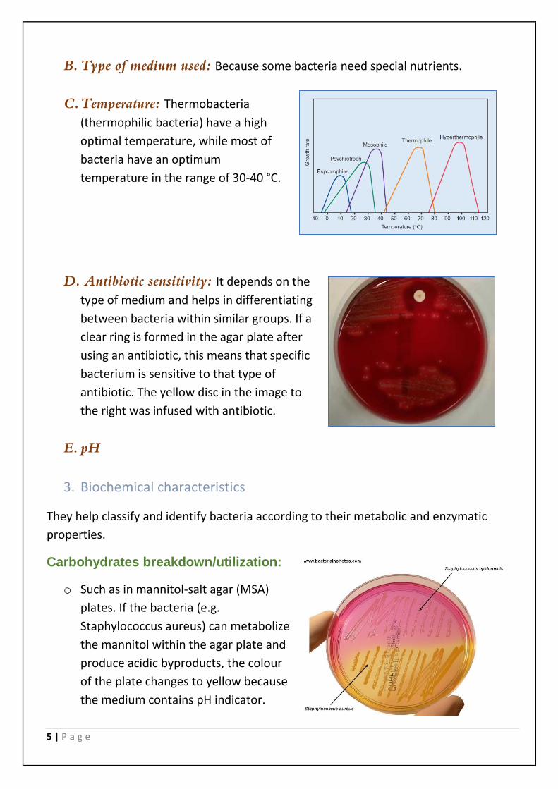
C. Temperature: Thermobacteria
(thermophilic bacteria) have a high
optimal temperature, while most of
bacteria have an optimum
temperature in the range of 30-40 °C.
D. Antibiotic sensitivity: It depends on the
type of medium and helps in differentiating
between bacteria within similar groups. If a
clear ring is formed in the agar plate after
using an antibiotic, this means that specific
bacterium is sensitive to that type of
antibiotic. The yellow disc in the image to
the right was infused with antibiotic.
E. pH
3. Biochemical characteristics
They help classify and identify bacteria according to their metabolic and enzymatic
properties.
Carbohydrates breakdown/utilization:
o Such as in mannitol-salt agar (MSA)
plates. If the bacteria (e.g.
Staphylococcus aureus) can metabolize
the mannitol within the agar plate and
produce acidic byproducts, the colour
of the plate changes to yellow because
the medium contains pH indicator.
6 | P a g e
o The non-pathogenic Staphylococcus epidermidis can grow on MSA plate but can’t
utilize mannitol, so colour remains pink-red.
o The MSA can be both selective or differential. It is selective since it mostly allows
only Staph. bacteria to grow, and is differential since it can differentiate between
Staph. aureus and epidermidis.
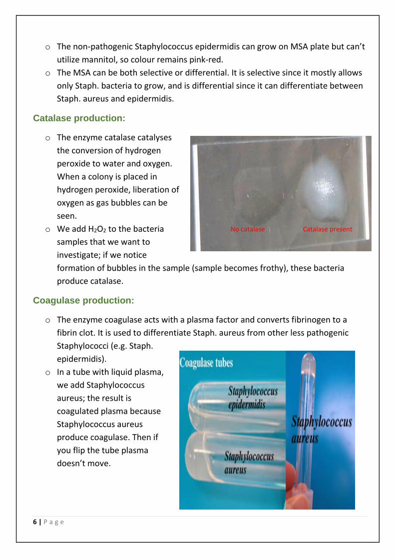
Catalase production:
o The enzyme catalase catalyses
the conversion of hydrogen
peroxide to water and oxygen.
When a colony is placed in
hydrogen peroxide, liberation of
oxygen as gas bubbles can be
seen.
o We add H2O2 to the bacteria
samples that we want to
investigate; if we notice
formation of bubbles in the sample (sample becomes frothy), these bacteria
produce catalase.
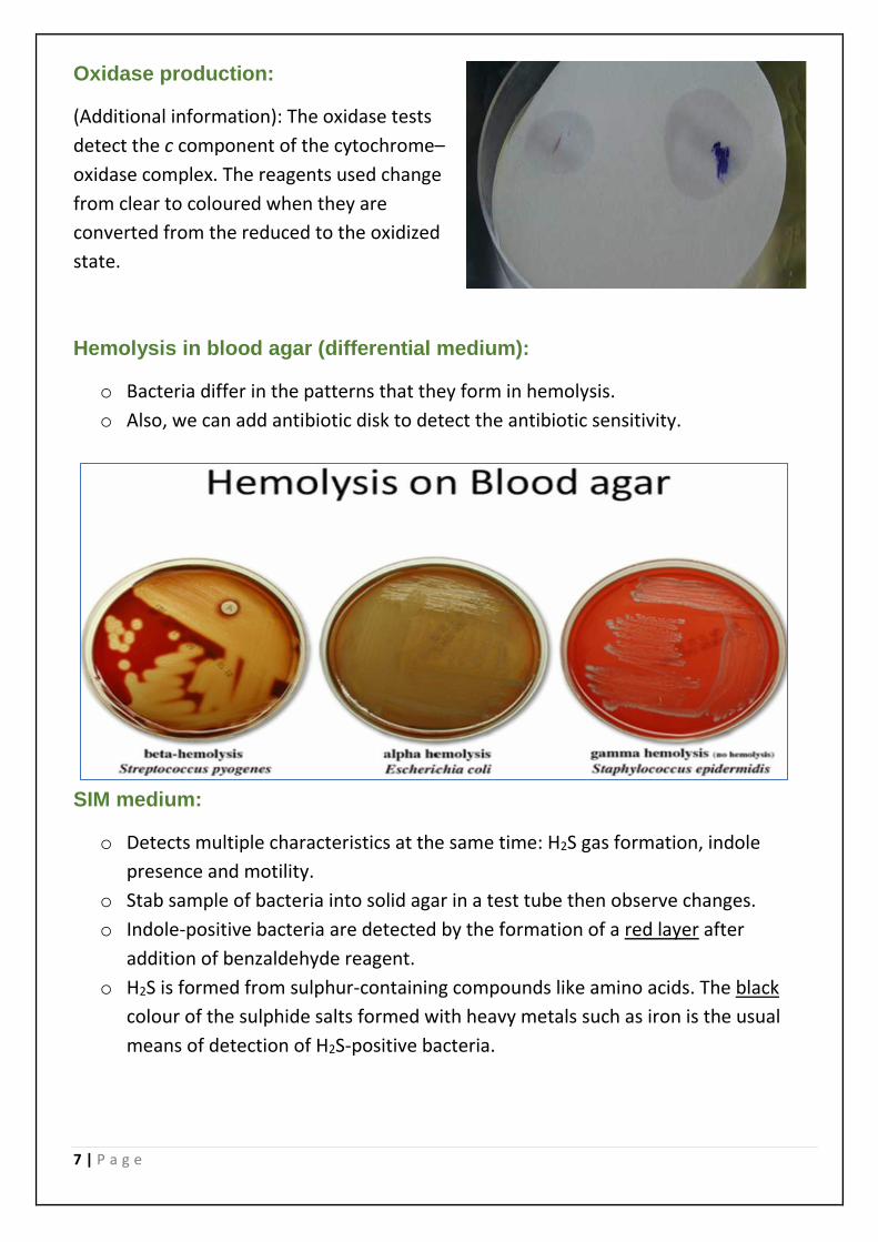
Coagulase production:
o The enzyme coagulase acts with a plasma factor and converts fibrinogen to a
fibrin clot. It is used to differentiate Staph. aureus from other less pathogenic
Staphylococci (e.g. Staph.
epidermidis).
o In a tube with liquid plasma,
we add Staphylococcus
aureus; the result is
coagulated plasma because
Staphylococcus aureus
produce coagulase. Then if
you flip the tube plasma
doesn’t move.
No catalase Catalase present
7 | P a g e
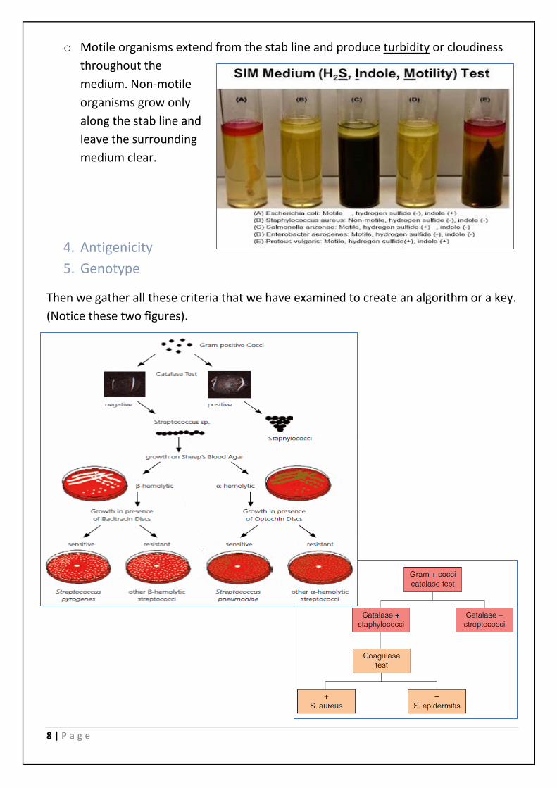
Oxidase production:
(Additional information): The oxidase tests
detect the c component of the cytochrome–
oxidase complex. The reagents used change
from clear to coloured when they are
converted from the reduced to the oxidized
state.
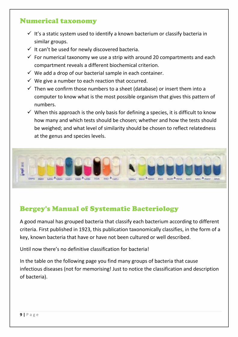
Hemolysis in blood agar (differential medium):
o Bacteria differ in the patterns that they form in hemolysis.
o Also, we can add antibiotic disk to detect the antibiotic sensitivity.
SIM medium:
o Detects multiple characteristics at the same time: H2S gas formation, indole
presence and motility.
o Stab sample of bacteria into solid agar in a test tube then observe changes.
o Indole-positive bacteria are detected by the formation of a red layer after
addition of benzaldehyde reagent.
o H2S is formed from sulphur-containing compounds like amino acids. The black
colour of the sulphide salts formed with heavy metals such as iron is the usual
means of detection of H2S-positive bacteria.
8 | P a g e
o Motile organisms extend from the stab line and produce turbidity or cloudiness
throughout the
medium. Non-motile
organisms grow only
along the stab line and
leave the surrounding
medium clear.
4. Antigenicity
5. Genotype
Then we gather all these criteria that we have examined to create an algorithm or a key.
(Notice these two figures).
9 | P a g e
Numerical taxonomy
✓ It’s a static system used to identify a known bacterium or classify bacteria in
similar groups.
✓ It can’t be used for newly discovered bacteria.
✓ For numerical taxonomy we use a strip with around 20 compartments and each
compartment reveals a different biochemical criterion.
✓ We add a drop of our bacterial sample in each container.
✓ We give a number to each reaction that occurred.
✓ Then we confirm those numbers to a sheet (database) or insert them into a
computer to know what is the most possible organism that gives this pattern of
numbers.
✓ When this approach is the only basis for defining a species, it is difficult to know
how many and which tests should be chosen; whether and how the tests should
be weighed; and what level of similarity should be chosen to reflect relatedness
at the genus and species levels.
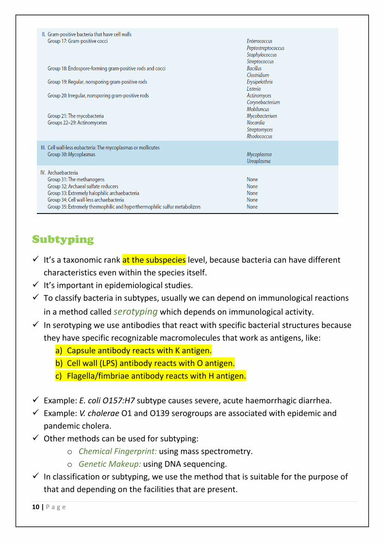
Bergey's Manual of Systematic Bacteriology
A good manual has grouped bacteria that classify each bacterium according to different
criteria. First published in 1923, this publication taxonomically classifies, in the form of a
key, known bacteria that have or have not been cultured or well described.
Until now there’s no definitive classification for bacteria!
In the table on the following page you find many groups of bacteria that cause
infectious diseases (not for memorising! Just to notice the classification and description
of bacteria).
10 | P a g e
Subtyping
✓ It’s a taxonomic rank at the subspecies level, because bacteria can have different
characteristics even within the species itself.
✓ It’s important in epidemiological studies.
✓ To classify bacteria in subtypes, usually we can depend on immunological reactions
in a method called serotyping which depends on immunological activity.
✓ In serotyping we use antibodies that react with specific bacterial structures because
they have specific recognizable macromolecules that work as antigens, like:
a) Capsule antibody reacts with K antigen.
b) Cell wall (LPS) antibody reacts with O antigen.
c) Flagella/fimbriae antibody reacts with H antigen.
✓ Example: E. coli O157:H7 subtype causes severe, acute haemorrhagic diarrhea.
✓ Example: V. cholerae O1 and O139 serogroups are associated with epidemic and
pandemic cholera.
✓ Other methods can be used for subtyping:
o Chemical Fingerprint: using mass spectrometry.
o Genetic Makeup: using DNA sequencing.
✓ In classification or subtyping, we use the method that is suitable for the purpose of
that and depending on the facilities that are present.
11 | P a g e
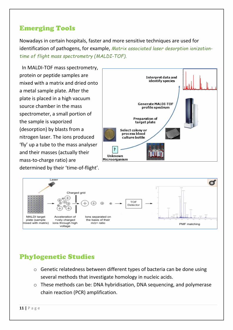
Emerging Tools
Nowadays in certain hospitals, faster and more sensitive techniques are used for
identification of pathogens, for example, Matrix associated laser desorption ionization-
time of flight mass spectrometry (MALDI-TOF).
In MALDI-TOF mass spectrometry,
protein or peptide samples are
mixed with a matrix and dried onto
a metal sample plate. After the
plate is placed in a high vacuum
source chamber in the mass
spectrometer, a small portion of
the sample is vaporized
(desorption) by blasts from a
nitrogen laser. The ions produced
‘fly’ up a tube to the mass analyser
and their masses (actually their
mass-to-charge ratio) are
determined by their ‘time-of-flight’.
Phylogenetic Studies
o Genetic relatedness between different types of bacteria can be done using
several methods that investigate homology in nucleic acids.
o These methods can be: DNA hybridisation, DNA sequencing, and polymerase
chain reaction (PCR) amplification.

12 | P a g e
o Ribosomal RNA: ➢ Ribosomes have an essential role in protein synthesis for all organisms. Genetic
sequence encodings ribosomal RNAs (rRNA s16) and ribosomal proteins (both of
which are required to comprise a functional ribosome) have been highly conserved
throughout evolution and have diverged more slowly than other chromosomal genes
since ribosomes are essential for bacteria survival.
➢ The phylogenetic tree below is based on rRNA data, which shows the three major
domains of biological life as they are currently understood.
➢ From this diagram, two kingdoms- eubacteria (true bacteria) and the archaebacteria-
are distinct from the Eukaryotic branch.
➢ Many organisms have multiple copies (five to seven) of these genes, resulting in
patterns with a sufficient number of bands to provide good discriminatory power;
however, ribotyping is of limited value for some microorganisms, such as
mycobacteria, which have only a single copy of these genes.
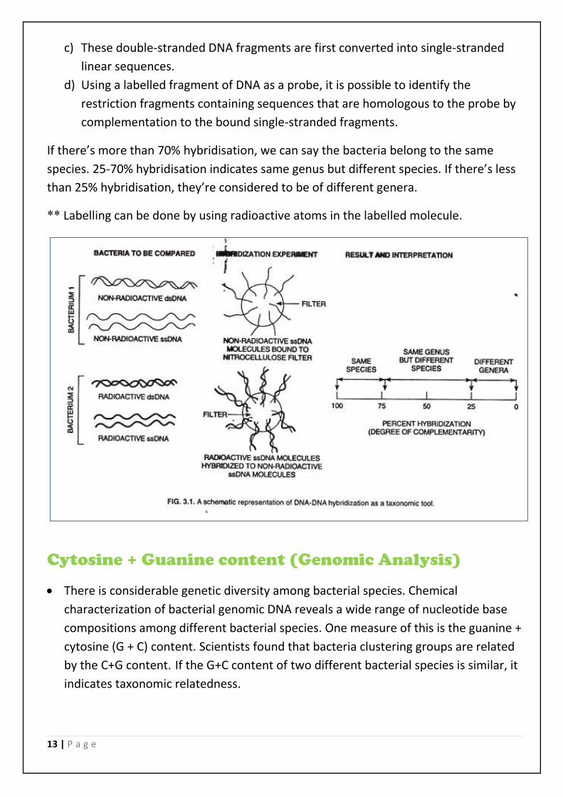
DNA hybridization
It’s done by the technique of Southern blot analysis by these steps:
a) DNA molecule is extracted from the bacteria that we want to compare with each
other.
b) The separated fragments are transferred to a nitrocellulose or nylon filter.
13 | P a g e
c) These double-stranded DNA fragments are first converted into single-stranded
linear sequences.
d) Using a labelled fragment of DNA as a probe, it is possible to identify the
restriction fragments containing sequences that are homologous to the probe by
complementation to the bound single-stranded fragments.
If there’s more than 70% hybridisation, we can say the bacteria belong to the same
species. 25-70% hybridisation indicates same genus but different species. If there’s less
than 25% hybridisation, they’re considered to be of different genera.
** Labelling can be done by using radioactive atoms in the labelled molecule.
Cytosine + Guanine content (Genomic Analysis)
• There is considerable genetic diversity among bacterial species. Chemical
characterization of bacterial genomic DNA reveals a wide range of nucleotide base
compositions among different bacterial species. One measure of this is the guanine +
cytosine (G + C) content. Scientists found that bacteria clustering groups are related
by the C+G content. If the G+C content of two different bacterial species is similar, it
indicates taxonomic relatedness.
14 | P a g e
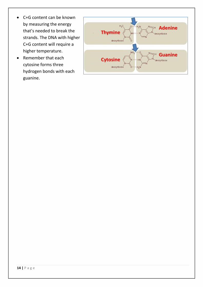
• C+G content can be known
by measuring the energy
that’s needed to break the
strands. The DNA with higher
C+G content will require a
higher temperature.
• Remember that each
cytosine forms three
hydrogen bonds with each
guanine.