Ludwig Brussels 2012 annual scientific report (PDF 5.4 MB)
34
Ludwig Institute for Cancer Research Brussels Branch
-
Upload
phungkhanh -
Category
Documents
-
view
216 -
download
2
Transcript of Ludwig Brussels 2012 annual scientific report (PDF 5.4 MB)

Ludwig Institute for Cancer Research
Brussels Branch

research | 94
Ludwig Institute for Cancer Research
Brussels Branch
Cancer is a major concern in human health. The prospects for bringing cancer under control require linked innovative basic and clinical research. In this view, Daniel K. Ludwig created in 1971 the Ludwig Institute for Cancer Research, an international organization bringing together scientists and clinicians from around the world. Many Ludwig investigators are leaders in many areas of science, involving genetics, bioinformatics, immunology, virology, cell biology and signal transduction.Faithful to the organizing principles laid down by Mr. Ludwig, the Institute conducts its research through ten Branches, located in seven countries. The Branch structure allows the Institute to interact with a number of different research and clinical environments. Each Branch is focused on a research program defined by the Branch Director in relation with the overall objectives of the Institute. The Branches are established in association with University Hospitals, to stimulate close collaborations between research laboratories and the clinic. By organizing and controlling its own clinical trials programs, the Institute has indeed created a continuum that integrates laboratory and clinical research. The biological properties of any given cancer cell constantly change, allowing tumors to spread and become more aggressive. To overcome these obstacles, the Ludwig Institute has developed a broad-based discovery program that seeks to understand the full complexity of cancer. Research is organized according to the four major programmatic themes that define the Institute: genetics, cell biology, cell signalling and immunology. Branch staffs vary in size from 30 to over 90, and internationally the Institute employs some 800 scientists, clinicians and support personnel. The quality of the research is monitored on an ongoing basis by the Institute’s Scientific Committee and by an external peer review process. The Brussels Branch of the Institute was created in 1978. It is composed of 91 members and was headed by Thierry Boon until 2009. The Branch is now headed by Benoît Van den Eynde, the current Branch Director.

research | 95
Administration and General Services
DirectorBenoît Van den Eynde Administration and secretariatDario Florean, Administrator • Aline Bien, Accounting and Administrative Assistant • Julien Doornaert, Administrative Assistant (Part-time) • Julie Klein, Assistant to the Director • Nathalie Krack, Librarian • Carine Lemoine, Scientific and Administrative Assistant • Geneviève Schoonheydt, Administrative Assistant • Pierre-Olivier Seroen, Accounting Assistant •
Pre-clinical Research Resources Group Guy Warnier, Veterinarian and Group Head • Gilles Gaudray, Deputy Group Head • Julien Gossiaux, Technical Assistant • Laurent Hermans, Technical Assistant • Benoît Poux, Technical Assistant •
Technical supportJo Lejeune, Head of Computer Systems • Paul Wauters, Laboratory Manager • Alain Buisseret, Technical Manager • Jacques Van Cleve, Research Assistant in Bioinformatics • Jean-Luc Germis, Technician • André Tonon, Assistant to the Technical Manager • Manuel Orviz, Senior Technical Assistant • Antoinette Van Goetsenhoven, Technical Assistant • Jean-Claude Verhelst, Administrative and Technical Assistant • Evelyne Corbisier, Laboratory Helper • Samira Jebari, Laboratory Helper • Pascale Mahieu-Gaudy, Laboratory Helper • Rafaela Reyes Prieto, Laboratory Helper • Thamma Singharach, Laboratory Helper • Keoprasith Souvannaraj, Laboratory Helper • Ioanis Spanoudis, Laboratory Helper • André Tonon, Assistant
Ludwig Institute - Brussels BranchB1.74.12Avenue Hippocrate 741200 Bruxelles, Belgium
[T] +32 (2) 764 74 59 [F] +32 (2) 762 94 05

research | 96
Tumor immunology and antigen processing
Benoît Van den Eynde
The group follows three main lines of research. The first focuses on the processing of tumor antigens, studying the role of the proteasome and other proteases in the production of tumor antigenic peptides. The second studies mechanisms whereby tumors resist immune rejection. The third develops preclinical melanoma models for cancer immunotherapy. The long-term goal of these projects is to better understand the interaction of tumors with the immune system and devise strategies to improve the efficacy of cancer vaccines.
An antigenic peptide produced by reverse splicing in the proteasome and double asparagine deamidation
A. Dalet, V. Stroobant, N. Vigneron, in collaboration with P. Robbins, K.-I. Hanada and S. Rosenberg, NCI, NIH, Bethesda, USA
Tumor antigens relevant for cancer immunotherapy consist of peptides presented by MHC class I molecules and derived from intracellular tumor proteins. They are recognized by cy-totoxic T lymphocytes (CTL) and result from the degradation of these proteins, which is mainly exerted by the proteasome. We have described a new mode of production of antigenic peptides by the proteasome, which involves the splicing of peptide fragments, either in the normal or the reverse order [1, 2]. We showed that splicing occurs in the proteasome cata-lytic chamber through a reaction of transpeptidation involv-ing an acyl-enzyme intermediate. We have now described four spliced peptides, two of which are spliced in the reverse order. One of these peptides also contains two additional post-translational modifications, resulting in the conversion of asparagines into aspartic acids, through a process a N-glyco-sylation/deglycosylation [3]. It is derived from tyrosinase and recognized by tumor-infiltrating lymphocytes isolated from a melanoma patient. The peptide is made of two noncontigu-ous tyrosinase fragments that are spliced together in the re-verse order. In addition, it contains two aspartate residues that replace the asparagines encoded in the tyrosinase sequence. We confirmed that this peptide is naturally presented at the surface of melanoma cells, and we showed that its processing sequentially requires translation of tyrosinase into the endo-
plasmic reticulum and its retrotranslocation into the cytosol, where deglycosylation of the two asparagines by peptide-N-glycanase turns them into aspartates by deamidation. This process is followed by cleavage and splicing of the appropriate fragments by the standard proteasome and additional trans-port of the resulting peptide into the endoplasmic reticulum through the transporter associated with antigen processing (TAP) (Figure 1).
New proteasome types that are intermediate between the standard proteasome and the immunoproteasome
B. Guillaume, V. Stroobant , A. Busse, E. De Plaen
Using a series of novel antibodies recognizing catalytic subu-nits of the human proteasome in their native conformation, we identified proteasomes that are intermediate between the standard proteasome and the immunoproteasome [4]. They contain only one (ß5i) or two (ß1i and ß5i) of the three induc-ible catalytic subunits of the immunoproteasome. These inter-mediate proteasomes represent 30-54% of the proteasome content of human liver, colon, small intestine and kidney. They are also present in human tumor cells and dendritic cells. We studied the processing of a series of antigenic peptides by these intermediate proteasomes, and identified two tumor antigens that are processed exclusively either by intermediate proteasome ß5i or by intermediate proteasome ß1i-ß5i. Other functional aspects of these intermediate proteasomes are cur-rently evaluated.

research | 97
Antigenic peptide production by insulin-degrading enzyme
N. Parmentier, V. Stroobant
We studied a proteasome-independent peptide derived form tumor protein MAGE-A3, and we identified insulin-degrading enzyme as the protease producing both the C-terminus and the N-terminus of this peptide [5]. This peptide, with sequence EVDPIGHLY, is presented by HLA-A1 and has been widely used in clinical trials of cancer vaccines. Insulin-degrading enzyme (IDE) is a cytosolic metallopeptidase not previously known to play a role in the class I processing pathway. Cytotoxic T lym-phocyte recognition of tumor cells was reduced after metal-lopeptidase inhibition or IDE silencing. Separate inhibition of the metallopeptidase and the proteasome impaired degrada-tion of MAGE-A3 proteins, and simultaneous inhibition of both further stabilized MAGE-A3 proteins. These results suggest that MAGE-A3 proteins are degraded along two parallel path-ways that involve either the proteasome or IDE and produce different sets of antigenic peptides presented by MHC class I molecules.
Inefficient exogenous loading of a tapasin-dependent peptide onto HLA-B*44:02 can be improved by acid treatment or fixation of target cells
N. Vigneron, V. Stroobant , W. Ma, A. Michaux, in collaboration with N. Demotte and P. van der Bruggen, LICR, Brussels, and with RM Leonhardt and P. Cresswell, Yale University, New Haven, CT, USA
We recently identified a new antigenic peptide, which is de-rived from the MAGE-A1-encoded protein and presented to cytotoxic T lymphocytes (CTLs) by HLA-B*44:02 [6]. Although this peptide is encoded by MAGE-A1, processed endogenous-ly and presented by tumor cells, the corresponding synthetic peptide is hardly able to sensitize target cells to CTL recog-nition when pulsed exogenously. We observed that endog-enous processing and presentation of this peptide is strictly dependent on the presence of tapasin, which is believed to help peptide loading by stabilizing a peptide-receptive form of HLA-B*44:02. In line with this, we showed that exogenous loading of the peptide can be dramatically improved by para-formaldehyde fixation of surface molecules or by peptide loading at acidic pH, two strategies presumably generating or stabilizing a peptide-receptive, empty conformation of the HLA. Altogether, our results indicate a potential drawback of
endoplasmic reticulum
N-linkedglycosylations
MHCmolecule ERAD
proteasome
peptide N-glycanase
TAP
cytosol
NN
NN
DD
D D
NH2
COOH
NH2
COOH
NH2
COOH
NH2 COOHIYMDGTADFSF
I.
II.III.
IV.
CTL
V.
tyrosinase
Figure 1. Production of the deamidated reverse-spliced antigenic peptide derived from tyrosinase. Misfolded N-glycosylated tyrosinase proteins are retrotranslocated from the endoplasmic reticulum (ER) into the cytosol (I), where peptide N-glycanase converts two asparagines into aspartates upon removal of the asparagine-associated sugars (II). This is followed by cleavage and reverse splicing of the deamidated protein by the proteasome (III), leading to production of the final antigenic peptide IYMDGTADFSF, which is then transported back into the ER by the TAP transporter (IV) (from reference 3).

research | 98
short peptide-based vaccination strategies and offer possible solutions regarding the use of such problematic epitopes.
A MAGE-C2 antigenic peptide processed by the immunoproteasome is recognized by cytolytic T cells isolated from a melanoma patient after successful immunotherapy
W. Ma, N. Vigneron, J. Chapiro, V. Stroobant, in collaboration with P. Coulie, de Duve Institute
We have pursued our analysis of a melanoma patient who showed almost complete tumor regression following vacci-nation with MAGE-A1 and MAGE-A3 antigens. We previously described high frequencies of tumor-specific CTL precursors in blood samples collected after but also before vaccination. A set of CTL clones were derived that recognized antigens dif-ferent from those of the vaccine. Two of these antigens were peptides encoded by another MAGE gene, MAGE-C2. Here we describe the antigen recognized by another tumor-specific CTL clone [7]. It proved to be a third antigenic peptide encod-ed by gene MAGE-C2, ASSTLYLVF. It is presented by HLA-B57 molecules and proteasome-dependent. Tumor cells exposed to interferon-gamma (IFN-γ) were better recognized by this anti-MAGE-C2 CTL clone. This mainly resulted from a better processing of the peptide by the immunoproteasome as com-pared to the standard proteasome. Mass spectrometric analy-ses showed that the latter destroyed the antigenic peptide by cleaving between two internal hydrophobic residues. Despite its higher "chymotryptic-like" (posthydrophobic) activity, the immunoproteasome did not cleave at this position, in line with the suggestion that hydrophobic residues immediately down-stream from a cleavage site impair cleavage by the immuno-proteasome. We previously reported that one of the other MAGE-C2 peptides recognized by CTL from this patient was also better processed by the immunoproteasome. Together, these results support the notion that the tumor regression of this patient was mediated by an antitumor response shaped by IFN-γ and dominated by CTL directed against peptides that are better produced by the immunoproteasome, such as the MAGE-C2 peptides.
Modulation of tumor antigen expression by inflammatory cytokines
E. De Plaen, O. Kholmanskikh
We observed that treating some melanoma cell lines with the inflammatory cytokine IL-1ß leads to a 4- to 10-fold decrease in the level of Microphtalmia-associated transcription factor (MITF-M) (Kholmanskikh et al, 2010). This effect is NF-kB and JNK-dependent. MITF-M regulates the expression of melano-cyte differentiation genes such as Melan-A, tyrosinase and gp100, which encode antigens recognized on melanoma cells by autologous cytolytic T lymphocytes (CTL). Accordingly,
treating some melanoma cells with IL-1ß reduced by 40-100% their ability to activate such anti-melanoma CTL.
Minimal tolerance to a tumor antigen encoded by a cancer-germline gene
I. Huijbers, C. Uyttenhove, D. Colau, L. Pilotte, C. Powis de Tenbos-sche, in collaboration with AM Schmitt-Verhulst and S. Soudja, CIML, Marseille, France
Cancer-germline genes such as MAGE and NY-ESO1 encode the most relevant antigens for cancer immunotherapy. Some of these genes are expressed at low levels in the thymus, rais-ing the possibility of central immune tolerance that might reduce the immunogenicity of such antigens. We created a mouse line knocked-out for cancer-germline gene P1A, and tested whether P1AKO mice develop stronger immune re-sponses to P1A antigens as compared to wild type mice [8]. We found only slightly stronger P1A-specific immune respons-es in P1AKO mice, even though these responses were suffi-cient to induce tumor rejection in defined experimental set-tings. These results indicate only minimal immune tolerance to antigens encoded by cancer-germline genes and fully confirm their immunogenicity.
Tumoral immune resistance through tryptophan degradation by indoleamine 2,3-dioxygenase
L. Pilotte, P. Larrieu, V. Stroobant, D. Colau, C. Uyttenhove in col-laboration with U. Rohrig, V. Zoete and O. Michielin, LICR, University of Lausanne, Switzerland
An important factor limiting the efficacy of immunotherapy is the development of mechanisms allowing tumors to resist or escape immune rejection. Immune resistance mechanisms often involve modulation of the tumoral microenvironment resulting in local immunosuppression. We described one such mechanism, based on the expression by tumor cells of indoleamine 2,3-dioxygenase (IDO), a tryptophan-degrading enzyme inducing a local tryptophan depletion that severely affects T lymphocyte proliferation [9]. Our data in a preclinical model indicate that the efficacy of therapeutic vaccination of cancer patients could be improved by concomitant adminis-tration of an IDO inhibitor. In collaboration with the group of Olivier Michielin in Lausanne, we used computational struc-ture-based methods to design new compounds able to inhibit IDO with a high potency (Röhrig et al, 2012). This approach yielded highly efficient low-molecular weight inhibitors, the most active being of nanomolar potency both in an enzymatic and in a cellular assay, while showing no cellular toxicity and a high selectivity for IDO1 over tryptophan 2,3-dioxygenase (TDO). These triazole compounds will be further optimized with the goal of developing drug candidates.

research | 99
We have produced a monoclonal antibody against human IDO, which we used to characterize IDO expression in normal and tumoral tissues. Although others reported high expres-sion of IDO in dendritic cells of murine tumor-draining lymph nodes, our results in humans indicate that a subset of mature human dendritic cells express IDO but these cells are present in normal lymph nodes and not enriched in tumor-draining lymph nodes. However, we observed expression of IDO in a high proportion of human tumors, confirming our initial ob-servation.
Reversal of tumoral immune resistance by inhibition of tryptophan 2,3-dioxygenase
L. Pilotte, P. Larrieu, V. Stroobant, D. Colau, E. De Plaen, C. Uyttenhove, in collaboration with E. Dolusic, R. Frédérick, J. Wouters and B. Masereel, NAMEDIC, University of Namur, Belgium
Besides IDO, we recently uncovered the role of another, un-related, tryptophan-degrading enzyme named tryptophan-dioxygenase (TDO) in tumoral immune resistance [10]. TDO is highly expressed in the liver and regulates systemic tryp-tophan levels. We found TDO to be expressed in a high pro-portion of human tumors. We showed that TDO-expressing mouse tumors are no longer rejected by immunized mice. Moreover, we developed a new TDO inhibitor, which, upon systemic treatment, restored the ability of mice to reject tu-mors. These results describe a mechanism of tumoral immune resistance based on TDO expression and establish proof-of-concept for the use of TDO inhibitors in cancer therapy. This concept will be further developed in the frame of a new spin-off company that we recently launched, named iTeos Thera-peutics.
A preclinical melanoma model for cancer immunotherapy
C. Powis de Tenbossche, S. Cane, F. Schramme (in collaboration with C. Uyttenhove, de Duve Institute and A.-M. Schmitt-Verhulst, CIML, Marseille), France
We made transgenic mice developing melanomas with a 70-80% incidence after tamoxifen injection (Huijbers et al, 2006). These tumors express the tumor antigen encoded by cancer-germline gene P1A. Initially highly pigmented and indolent, they later dedifferentiate in unpigmented highly aggressive tumors. Mice bearing aggressive tumors show exacerbated systemic inflammation associated with disruption of second-ary lymphoid organs, accumulation of immature myeloid cells and immunosuppression (Soudja et al, 2010). Current efforts aim at characterizing this immunosuppression and devising effective therapeutic approaches.
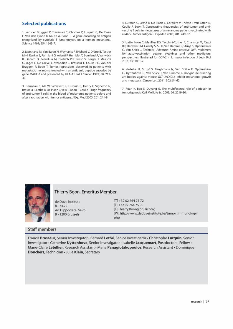
Selected publications
1. Vigneron N, Stroobant V, Chapiro J, Ooms A, Degiovanni G, Morel S, van der Bruggen P, Boon T, Van den Eynde B. An antigenic peptide produced by peptide splicing in the proteasome. Science 2004;304:587-90.
2. Warren EH, Vigneron NJ, Gavin MA, Coulie PG, Stroobant V, Dalet A, Tykodi SS, Xuereb SM, Mito JK, Riddell SR, Van den Eynde BJ. An antigen produced by splicing of noncontiguous peptides in the reverse order. Science 2006;313:1444-7.
3. Dalet A, Robbins PF, Stroobant V, Vigneron N, Li YF, El-Gamil M, Hanada K, Yang JC, Rosenberg SA, Van den Eynde BJ. An antigenic peptide produced by reverse splicing and double asparagine deamidation. Proc Natl Acad Sci USA 2011;108:E323-31.
4. Guillaume B, Chapiro J, Stroobant V, Colau D, Van Holle B, Parvizi G, Bousquet-Dubouch MP, Theate I, Parmentier N, Van den Eynde BJ. Two abundant proteasome subtypes that uniquely process some antigens presented by HLA class I molecules. Proc Natl Acad Sci USA 2010;107:18599-604.
5. Parmentier N, Stroobant V, Colau D, de Diesbach P, Morel S, Chapiro J, van Endert P, Van den Eynde BJ. Production of an antigenic peptide by insulin-degrading enzyme. Nat Immunol 2010;11:449-54.
6. Stroobant V, Demotte N, Luiten RM, Leonhardt RM, Cresswell P, Bonehill A, Michaux A, Ma W, Mulder A, Van den Eynde BJ, van der Bruggen P, Vigneron N. Inefficient exogenous loading of a tapasin-dependent peptide onto HLA-B*44:02 can be improved by acid treatment or fixation of target cells. Eur J Immunol 2012;42:1417-28.
7. Ma W, Vigneron N, Chapiro J, Stroobant V, Germeau C, Boon T, Coulie PG, Van den Eynde BJ. A MAGE-C2 antigenic peptide processed by the immunoproteasome is recognized by cytolytic T cells isolated from a melanoma patient after successful immunotherapy. Int J Cancer 2011;129:2427-34.
8. Huijbers IJ, Soudja SM, Uyttenhove C, Buferne M, Inderberg-Suso EM, Colau D, Pilotte L, Powis de Tenbossche CG, Chomez P, Brasseur F, Schmitt-Verhulst AM, Van den Eynde BJ. Minimal tolerance to a tumor antigen encoded by a cancer-germline gene. J Immunol 2012;188:111-21.
9. Uyttenhove C, Pilotte L, Theate I, Stroobant V, Colau D, Parmentier N, Boon T, Van den Eynde BJ. Evidence for a tumoral immune resistance mechanism based on tryptophan degradation by indoleamine 2,3-dioxygenase. Nat Med 2003;9:1269-74.
10. Pilotte L, Larrieu P, Stroobant V, Colau D, Dolusic E, Frederick R, De Plaen E, Uyttenhove C, Wouters J, Masereel B, Van den Eynde BJ. Reversal of tumoral immune resistance by inhibition of tryptophan 2,3-dioxygenase. Proc Natl Acad Sci USA 2012;109:2497-502.

research | 100
Staff members
Benoît Van den Eynde, Member
Ludwig Institute for Cancer Research B1.74.12 Av. Hippocrate 74-75 B - 1200 Brussels
[T] +32 02 764 75 72[F] +32 02 764 75 90[E] [email protected][W] http://www.deduveinstitute.be/tumor_imunology.php
Etienne De Plaen, Senior Investigator • Vincent Stroobant, Senior Investigator • Stefania Cane, Postdoctoral Fellow • Wenbin Ma, Postdoctoral Fellow • Nathalie Vigneron, Postdoctoral Fellow • Luc Pilotte, Research Associate • Nathalie Arts, Graduate Student • Juliette Lamy, Graduate Student • Alexandre Michaux, Graduate Student • Nicolas Parmentier, Graduate Student • Céline Powis de Tenbossche, Graduate Student • Florence Schramme, Graduate Student • Rui Cheng, Research Assistant • Aline Depasse, Research Assistant • Thérèse Aerts, Technician • Bénédicte Tollet, Technician • Marc Hennequart, Student • Julie Klein, Secretary

research | 101
Regulation of T lymphocyte function in tumors
Pierre van der Bruggen
The identification of tumor-specific antigens recognized by T lymphocytes on human cancer cells has elicited numerous vaccination trials of cancer patients with defined tumor antigens. These treatments have induced T cell responses but have shown a low clinical efficacy in tumor-bearing melanoma patients. We believe that progress depends on unraveling the different blockages for efficient tumor destruction. The analysis of the T cell responses of melanoma patients vaccinated against tumor antigens has led us to consider the possibility that the limiting factor for therapeutic success is not the intensity of the anti-vaccine response but the degree of anergy presented by intratumoral lymphocytes. We aim at a better understanding of dysfunctions of the immune system in tumors and more precisely T lymphocyte dysfunctions.
Previous work in our group: Identification of tumor antigens recognized by T cells
In the 1970s it became clear that T lymphocytes, a subset of the white blood cells, were the major effectors of tumor rejection in mice. In the 1980s, human anti-tumor cytolytic T lympho-cytes (CTL) were isolated in vitro from the blood lymphocytes of cancer patients, mainly those who had melanoma. Most of these CTL were specific, i.e. they did not kill non-tumor cells. This suggested that they target a marker, or antigen, which is expressed exclusively on tumor cells. We started to study the anti-tumor CTL response of a metastatic melanoma patient and contributed to the definition of several distinct tumor antigens recognized by autologous CTL. In the early 1990s, we identi-fied the gene coding for one of these antigens, and defined the antigenic peptide (1). This was the first description of a gene, MAGE-A1, coding for a human tumor antigen recognized by T lymphocytes.Genes such as those of the MAGE family are expressed in many tumors and in male germline cells, but are silent in normal tis-sues. They are therefore referred to as “cancer-germline genes”. They encode tumor specific antigens, which have been used in therapeutic vaccination trials of cancer patients (2). A large set of additional cancer-germline genes have now been identified by different approaches, including purely genetic approaches. As a result, a vast number of sequences are known that can code for tumor-specific shared antigens. The identification of a
larger set of antigenic peptides, which are presented by HLA class I and class II molecules and recognized on tumors by T lymphocytes, could be important for therapeutic vaccination trials of cancer patients and serve as tools for a reliable moni-toring of the immune response of vaccinated patients (3). To that purpose, we have used various approaches that we have loosely named “reverse immunology”, because they use gene sequences as starting point (4). Human tumor antigens recognized by CD4+ or CD8+ T cells are being defined at a regular pace worldwide. Together with col-leagues at the de Duve Institute, we read the new publications and incorporate the newly defined antigens in a database ac-cessible at http://www.cancerimmunity.org/peptidedatabase/Tcellepitopes.htm.
A mechanism causing anergy of CD8 and CD4 T lymphocytes
The identification of specific tumor antigens recognized by T lymphocytes on human cancer cells has elicited numerous clinical trials involving vaccination of tumor-bearing cancer patients with defined tumor antigens. These treatments have shown a low clinical efficacy. Among metastatic melanoma pa-tients, about 5% show a complete or partial clinical response following vaccination, whereas an additional 10% show some evidence of tumor regression without clear clinical benefit. We believe that progress depends on unraveling the different
research | 102
blockages for efficient tumor destruction. The tumors of the patients about to receive the vaccine, already contain T cells directed against tumor antigens. Presumably these T cells are exhausted and this impaired function is main-tained by immunosuppressive factors present in the tumor. The T cell response observed in some vaccinated patients reinforce an hypothesis proposed by Thierry Boon and Pierre Coulie: anti-vaccine CTL are not the effectors that kill the tumor cells but their arrival at the tumor site containing exhausted anti-tumor CTL, generates conditions allowing the reawakening of the ex-hausted CTL and/or activation of new anti-tumor CTL clones, some of them contributing directly to tumor destruction (2, 5). Accordingly, the difference between the responding and the non-responding vaccinated patients is not the intensity of their direct T cell response to the vaccine but the intensity of the im-munosuppression inside the tumor. It is therefore important to know which immunosuppressive mechanisms operate in hu-man tumors.
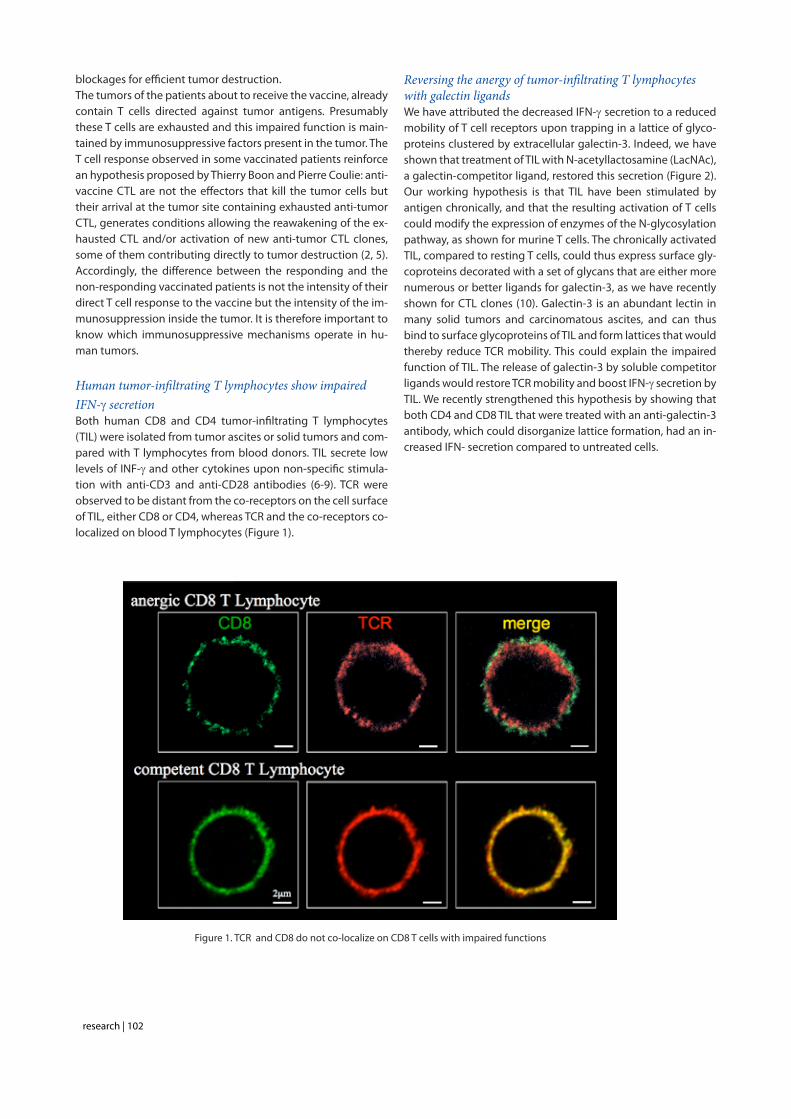
Human tumor-infiltrating T lymphocytes show impaired IFN-g secretionBoth human CD8 and CD4 tumor-infiltrating T lymphocytes (TIL) were isolated from tumor ascites or solid tumors and com-pared with T lymphocytes from blood donors. TIL secrete low levels of INF-g and other cytokines upon non-specific stimula-tion with anti-CD3 and anti-CD28 antibodies (6-9). TCR were observed to be distant from the co-receptors on the cell surface of TIL, either CD8 or CD4, whereas TCR and the co-receptors co-localized on blood T lymphocytes (Figure 1).
Reversing the anergy of tumor-infiltrating T lymphocytes with galectin ligands We have attributed the decreased IFN-g secretion to a reduced mobility of T cell receptors upon trapping in a lattice of glyco-proteins clustered by extracellular galectin-3. Indeed, we have shown that treatment of TIL with N-acetyllactosamine (LacNAc), a galectin-competitor ligand, restored this secretion (Figure 2). Our working hypothesis is that TIL have been stimulated by antigen chronically, and that the resulting activation of T cells could modify the expression of enzymes of the N-glycosylation pathway, as shown for murine T cells. The chronically activated TIL, compared to resting T cells, could thus express surface gly-coproteins decorated with a set of glycans that are either more numerous or better ligands for galectin-3, as we have recently shown for CTL clones (10). Galectin-3 is an abundant lectin in many solid tumors and carcinomatous ascites, and can thus bind to surface glycoproteins of TIL and form lattices that would thereby reduce TCR mobility. This could explain the impaired function of TIL. The release of galectin-3 by soluble competitor ligands would restore TCR mobility and boost IFN-g secretion by TIL. We recently strengthened this hypothesis by showing that both CD4 and CD8 TIL that were treated with an anti-galectin-3 antibody, which could disorganize lattice formation, had an in-creased IFN- secretion compared to untreated cells.
Figure 1. TCR and CD8 do not co-localize on CD8 T cells with impaired functions

research | 103
Towards a clinical trial combining vaccination and galectin-binding polysaccharides Galectin competitor ligands, e.g. disaccharides LacNAc, are rap-idly eliminated in urine, preventing their use in vivo. We recently found that a plant-derived polysaccharide, currently in clinical development, detached galectin-3 from TIL and boosted their IFN- secretion. Importantly, we observed that not only CD8+ TIL but also CD4+ TIL that were treated with this polysaccha-ride secreted more IFN- upon ex vivo re-stimulation. In tumor-bearing mice vaccinated with a tumor antigen, injections of this polysaccharide led to tumor rejection in half of the mice, whereas all control mice died. In non-vaccinated mice, the poly-saccharide had no effect by itself. These results suggest that a combination of galectin-3 ligands and therapeutic vaccination may induce more tumor regressions in cancer patients than vaccination alone. Translation of these results to the clinic was unfortunately impossible because the company producing this polysaccharide got bankrupted. We recently identified an-other plant-derived polysaccharide that binds to galectins and was already used in combination with chemotherapy in phase II clinical trials in colorectal cancer patients. This compound was as effective as LacNAc in boosting the secretion of IFN- by treated TIL. A clinical trial with this new compound, in combina-tion with anti-tumoral vaccination, will start in 2012 in different clinical centers. We are currently trying to understand the very early activation events that are defective in TIL.
Is the spontaneous anti-tumor T cell response of breast carcinoma patients a clinical prognostic factor?
D. Godelaine and V. Ha Thi, in collaboration with Dr J. Carrasco (Grand Hôpital de Charleroi) and Dr J.P. Machiels (Cliniques Universitaires St-Luc)
Several retrospective studies suggest a correlation between the survival of patients with ovarian or colorectal carcinoma and infiltration of their tumors by immune cells. So far, pro-spective data validating these observations do not exist. We set out a prospective study aimed at looking for a correlation between the clinical outcome of patients with non-metastatic breast carcinoma and their spontaneous anti-tumor T cell re-sponse. Considering our experience in quantitative approaches to detect very weak T cell responses in the blood of melanoma patients, D. Godelaine set out to evaluate the frequencies of anti-tumor CD8 T lymphocytes in the blood of non-metastatic breast cancer patients prospectively recruited in several clini-cal centers. Blood samples are collected before and after sur-gery. Frequencies are evaluated by mixed lymphocyte-peptide cultures, carried out with HLA-A2- and A3-restricted HER2/neu and hTERT peptides, followed by detection of specific cells with HLA-peptide tetramers. Tumors removed at surgery are analyzed by immunohistochemistry for infiltration by immune
Tumorsample
CD8+ isolationNon-specific stimulation
with anti-CD3 beads
Detection ofIFN-! secreted
after 18 h
Culture medium+/- LacNAc
for 2 h
IFN
-! s
ecre
ted
by 1
0,0
00
T c
ells
(p
g/m
l)
CD8+ tumor-infiltrating lymphocytes
non-treated
LacNAc-treated
Figure 2. Treatment of tumor-infiltrating lymphocytes with a galectin ligand reverses anergy

research | 104
cells, and fragments are frozen for further genetic analysis of the T cell receptor repertoire. The prospective follow-up of 172 patients will extend over a 5-year-period. So far, 69 patients have been included and 33 have been screened for frequencies of specific CD8 T lymphocytes. Thirty percent of the screened patients have a frequency against the targeted antigens in the range of 3x10⁻⁶ among blood CD8 T cells, whereas the mean value in healthy donors is 3x10⁻⁷. We hope to identify patients with a better prognosis in order to offer them an adapted care avoiding unnecessary heavy treatments.
Selected publications
1. van der Bruggen P, Traversari C, Chomez P, Lurquin C, De Plaen E, Van den Eynde B, Knuth A, Boon T. A gene encoding an antigen recognized by cytolytic T lymphocytes on a human melanoma. Science 1991;254:1643-7
2. Boon T, Coulie PG, Van den Eynde B, van der Bruggen P. Human T cell responses against melanoma. Annu Rev Immunol 2006;24:175-208.
3. Zhang Y, Renkvist N, Sun Z, Schuler-Thurner B, Glaichenhaus N, Schuler G, Boon T, van der Bruggen P, Colau D. A polyclonal anti-vaccine CD4 T cell response detected with HLA-DP4 multimers in a melanoma patient vaccinated with MAGE-3.DP4-peptide-pulsed dendritic cells. Eur J Immunol 2005;35:1066-75.
4. van der Bruggen P, Zhang Y, Chaux P, Stroobant V, Panichelli C, Schultz ES, Chapiro J, Van den Eynde BJ, Brasseur F, Boon T. Tumor-specific shared antigenic peptides recognized by human T cells. Immunol Rev 2002;188:51-64.
5. Carrasco J, Van Pel A, Neyns B, Lethé B, Brasseur F, Renkvist N, van der Bruggen P, van Baren N, Paulus R, Thielemans K, Boon T, Godelaine D. Vaccination of a melanoma patient with mature dendritic cells pulsed with MAGE-3 peptides triggers the activity of nonvaccine anti-tumor cells. J Immunol 2008;180:3585-93.
6. Demotte N, Stroobant V, Courtoy PJ, Van der Smissen P, Colau D, Luescher IF, Hivroz C, Nicaise J, Squifflet JL, Mourad M, Godelaine D, Boon T, van der Bruggen P. Restoring the association of the T cell receptor with CD8 reverses anergy in human tumor-infiltrating lymphocytes. Immunity 2008;28:414-24.
7. François V, Ottaviani S, Renkvist N, Stockis J, Schuler G, Thielemans K, Colau D, Marchand M, Boon T, Lucas S and van der Bruggen P. The CD4+ T-cell response of melanoma patients to a MAGE-A3 peptide vaccine involves potential regulatory T cells. Cancer Res 2009;69:4335-45.
8. Demotte N, Colau D, Ottaviani S, Godelaine D, Van Pel A, Boon T, van der Bruggen P. A reversible functional defect of CD8+ T lymphocytes involving loss of tetramer labeling. Eur J Immunol 2002;32:1688-97.
9. Demotte N, Wieërs G, Van Der Smissen P, Moser M, Schmidt CW, Thielemans K, Squifflet J-L, Weynand B, Carrasco J, Lurquin C, Courtoy PJ, van der Bruggen P. A galectin-3 ligand corrects the impaired function of human CD4 and CD8 tumor-infiltrating lymphocytes and favors tumor rejection in mice. Cancer Res 2010;70:7476-88.
10. Antonopoulos A, Demotte N, Stroobant V, Haslam SM, van der Bruggen P, Dell A. Loss of effector function of human cytolytic T lymphocytes is accompanied by major alterations in N- and O-glycosylation. J Biol Chem 2012;287:11240-51.
Pierre van der Bruggen, Member
Ludwig Institute for Cancer Research B1.74.12 Av. Hippocrate 74-75 B - 1200 Brussels
[T] +32 02 764 74 31[F] +32 02 762 94 05[E] [email protected][W] http://www.deduveinstitute.be/regulation_lympho-cyte.php
Staff members
Danièle Godelaine, Senior Investigator • Nathalie Demotte, Postdoctoral Fellow • Monica Gordon-Alonso, Postdoctoral Fellow (since July 2011) • Claude Wildmann, Research Associate • Anne-Elisabeth Petit, Graduate Student • Grégoire Wieërs, Graduate Student (until September 2011) • Débora Piccolo, Research Assistant • Vinh Ha Thi, Technician • Laurie Vanbiervliet, Technician • Nathalie Krack, Secretary

research | 105
Immunotherapy analysis group
Thierry Boon
The identification in the early 1990’s of human tumor-specific antigens that are recognized by T cells led to widespread attempts at vaccinating cancer patients with these antigens to induce tumor regression [1]. Vaccination of metastatic melanoma patients with MAGE peptides resulted in evidence of tumor regression in about 15% of the patients, with complete and partial clinical responses in only 7% of the patients [2]. Why did most patients fail to respond? A plausible hypothesis was that the anti-MAGE T cell response was too weak. However, none of the numerous attempts to boost the efficacy of the vaccines, for instance with adjuvants or by the use of dendritic cells, resulted in improvement of the clinical efficacy.
Our analysis of a few responding patients led us to a different hypothesis. Several groups reported a long time ago that human tumors contain tumor-infiltrating lymphocytes (TILs). These T lymphocytes could be extracted from the tumors and were capable of destroying tumor cells in vitro after short-term cultivation in the presence of IL-2. However, inside the tumor, they must have become inactive (“anergic”) at one point, since the tumor is progressing. We made the paradoxical observation that, when vaccination causes complete tumor regression, the T lymphocytes directed against the vaccine antigen are present in the tumor in very small numbers, clearly insufficient to cause rejection. But they reactivate the “anergic” tumor infiltrating T lymphocytes that are present in the tumor in large numbers as a result of a past spontaneous immune response of the patient. It is these reactivated TILs which are capable of destroying the bulk of the tumor cells [3, 4]. Our new hypothesis is that what differentiates the non-regressing and the regressing patients is not their direct response to the vaccine but the severity of the anergy of their TILs.
Accordingly, our new strategy to improve anti-tumoral vaccination is to supplement it with a local treatment of the tumor with various cytokines and Toll-like receptor agonists, as well as antibodies directed against inhibitory cytokines such as TGF-ß, to reduce the immunosuppression in the tumor. This should facilitate the action of the anti-vaccine T lymphocytes which provide the “spark” firing the regression response. This approach is proving to be effective in a mouse skin graft model. A small clinical trial involving tumor bearing melanoma patients is under way.

research | 106
Inducing rejection of normally tolerated grafts in the H-Y mouse model
F. Brasseur, I. Jacquemart, B. Lethé, C. Lurquin, C. Uyttenhove, T. Boon
A mouse model of skin grafts was developed that recapitu-lates what happens in cancer patients, where T lymphocytes often infiltrate the tumor without rejecting it. The group tests various approaches to overcome the anergy of such infiltrat-ing T cells. These approaches involve cytokines and Toll-like receptor ligands, as well as antibodies directed against inhibi-tory cytokines such as TGF-β.
Female CBA mice do not reject male skin grafts, even though they are able to mount a cytolytic T cell response against H-Y, a male specific minor histocompatibility antigen. To break this tolerance, repeated local injections of a low dose of IL-12, combined with IFN-α, caused graft rejection in all mice. Like IFN-α, IL-1α, IL-18 and IL-2 were incapable of inducing rejec-tion on their own, but synergized effectively with IL-12. One finding of importance for the clinical application of this pro-cedure is that several weekly cycles of cytokine treatments are necessary for complete rejection of the grafts.
We tested combinations of agents that are approved for clini-cal use. We observed that repeated local injections of a com-bination of low doses of IL-2 (300 ng), GM-CSF (300 ng) and IFNα (105 U) with TLR7 ligand gardiquimod (20 µg) caused 100% rejection. The crucial components appear to be IL-2 and gardiquimod. However, the same doses of IL-2 and IFN-α, without GM-CSF nor gardiquimod, were also effective when combined with both anti-TGF-β and anti-IL-10 antibodies, whereas anti-CTLA-4 antibody needed the additional pres-ence of GM-CSF to be fully effective. Additional molecules in the process of clinical agreement, such as IFNg, anti-PDL1 and anti-CD40 agonist antibodies, are presently under evaluation. Vaccinations with intra-peritoneal injections of male lympho-blasts aimed at enhancing anti-H-Y responses synergize ef-fectively with the local cytokine treatment.
Nicolas van Baren presents elsewhere updated results of a clinical trial involving a local treatment composed of IL-2, GM-CSF, IFN-α and Aldara, a cream containing imiquimod, a TLR7 ligand of the same family as gardiquimod.
Amine-reactive OVA multimers for auto-vaccination against cytokines and other immune mediators
C. Uyttenhove (in collaboration with R. Marillier and J. Van Snick)
Using our amine-reactive OVA multimers, recently described, we have produced a series of monoclonal antibodies inhib-iting murine GCP-2/CXCL6, cytokines GM-CSF, IL-17F, IL-17E/IL-25, IL-27, TGFβ-1 and matrix metalloproteinase-9 [5]. As these monoclonal antibodies were of mouse origin, they can be administered in vivo for long periods without inducing any immune reactions against a foreign protein as observed with rabbit or rat antibodies. The mAb against GCP-2 provided the first demonstration of the essential role played by this chemokine in rapid neutrophil mobilization after Leishmania major infection [5]. In collaboration with the group of Jo Van Damme (Rega Institute, KUL), we showed that neutralization of GCP-2 inhibited growth and metastasis of a melanoma cell line by decreasing angiogenesis [6]. Our mAbs against mouse IL-27, MM27.7B1, and against TGFβ1, 13A1, that potently in-hibited the bioactivity of these cytokines in vitro are currently used to evaluate the contribution of IL-27 or TGFβ1 in anti-tumor activities, skin graft rejection and in a model of GVHD. We also succeded to isolate the first and unique monoclonal antibody directed against the IL-12p35 chain, that contrarily to the existing anti–p40 mAbs, the common chain to IL-12 and IL-23, selectively blocks the IL-12 bioactivity, allowing for the fine dissection of the respective roles of these two cy-tokines in various in vivo models.
Using the same OVA multimers, we immunized mice against periostin/OSF-2, an extracellular matrix protein present in the stroma of many tumors, in mice and humans, that was recently implicated in metastasis development [7]. The first mAbs we isolated are currently being tested (in collaboration with P. Jat and S. Fields, LICR Oxford) to evaluate their ability to block periostin interaction with integrins αvβ3 and αvβ5, one of the mechanisms implicated in cancer cell migration and metastasis establishment.
research | 107
Selected publications
1. van der Bruggen P, Traversari C, Chomez P, Lurquin C, De Plaen E, Van den Eynde B, Knuth A, Boon T. A gene encoding an antigen recognized by cytolytic T lymphocytes on a human melanoma. Science 1991; 254:1643-7.
2. Marchand M, Van Baren N, Weynants P, Brichard V, Dréno B, Tessier M-H, Rankin E, Parmiani G, Arienti F, Humblet Y, Bourland A, Vanwijck R, Liénard D, Beauduin M, Dietrich P-Y, Russo V, Kerger J, Masucci G, Jäger E, De Greve J, Atzpodien J, Brasseur F, Coulie PG, van der Bruggen P, Boon T. Tumor regressions observed in patients with metastatic melanoma treated with an antigenic peptide encoded by gene MAGE-3 and presented by HLA-A1. Int J Cancer 1999; 80: 219-30.
3. Germeau C, Ma W, Schiavetti F, Lurquin C, Henry E, Vigneron N, Brasseur F, Lethé B, De Plaen E, Velu T, Boon T, Coulie P. High frequency of anti-tumor T cells in the blood of melanoma patients before and after vaccination with tumor antigens. J Exp Med 2005; 201: 241-8.
4. Lurquin C, Lethé B, De Plaen E, Corbière V, Théate I, van Baren N, Coulie P, Boon T. Constrasting frequencies of anti-tumor and anti-vaccine T cells in metastases of a melanoma patient vaccinated with a MAGE tumor antigen. J Exp Med 2005; 201: 249-57.
5. Uyttenhove C, Marillier RG, Tacchini-Cottier F, Charmoy M, Caspi RR, Damsker JM, Goriely S, Su D, Van Damme J, Struyf S, Opdenakker G, Van Snick J. Technical Advance: Amine-reactive OVA multimers for auto-vaccination against cytokines and other mediators: perspectives illustrated for GCP-2 in L. major infection. J Leuk Biol 2011; 89: 1001-7.
6. Verbeke H, Struyf S, Berghmans N, Van Coillie E, Opdenakker G, Uyttenhove C, Van Snick J, Van Damme J. Isotypic neutralizing antibodies against mouse GCP-2/CXCL6 inhibit melanoma growth and metastasis. Cancer Lett 2011; 302: 54-62.
7. Ruan K, Bao S, Ouyang G. The multifaceted role of periostin in tumorigenesis. Cell Mol Life Sci 2009; 66: 2219-30.
Thierry Boon, Emeritus Member
de Duve Institute B1.74.72 Av. Hippocrate 74-75 B - 1200 Brussels
[T] +32 02 764 75 72[F] +32 02 764 75 90[E] [email protected][W] http://www.deduveinstitute.be/tumor_immunology.php
Staff members
Francis Brasseur, Senior Investigator • Bernard Lethé, Senior Investigator • Christophe Lurquin, Senior Investigator • Catherine Uyttenhove, Senior Investigator • Isabelle Jacquemart, Postdoctoral Fellow • Marie-Claire Letellier, Research Assistant • Maria Panagiotakopoulos, Research Assistant • Dominique Donckers, Technician • Julie Klein, Secretary

research | 108
Therapeutic vaccination and tumor expression profiling group
Nicolas van Baren
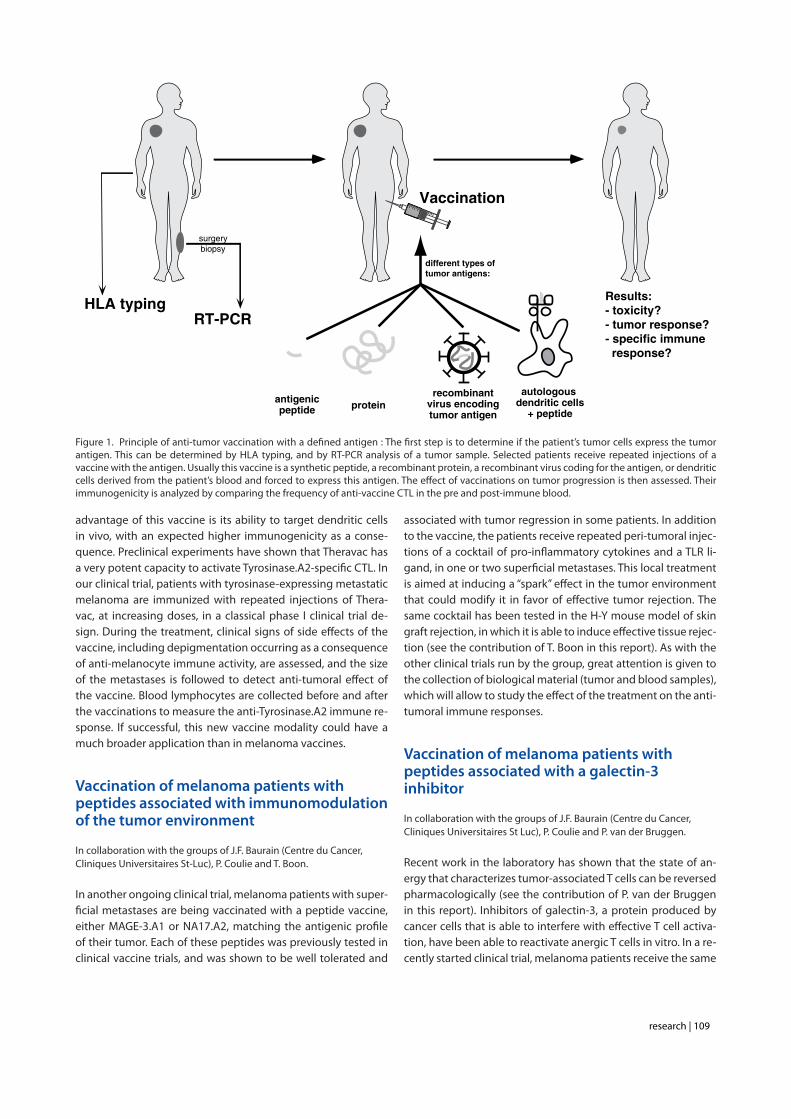
Cancer cells express tumor-specific antigens that can be targeted by cytolytic T lymphocytes (CTL). These antigens are small peptides derived from endogenous proteins presented at the surface of tumor cells by HLA molecules. In vitro, cytolytic T lymphocytes (CTL) lyse selectively tumor cell lines that express their cognate antigen. Our group has developed small scale clinical immunotherapy trials in which patients with advanced cancer, often metastatic melanoma, have been treated repeatedly with a vaccine containing one or several defined tumor antigens that are expressed by their tumor (Fig. 1). Different immunization modalities, such as vaccination with peptides like MAGE-3.A1 and NA17.A2, or with the MAGE-3 recombinant protein, both with or without adjuvant, or with an ALVAC recombinant viral vector, have already been tested. They are all devoid of severe toxicity. A minority of vaccinated melanoma patients (about 10 to 20%) showed regression of metastatic lesions (Fig. 2). This frequency is far beyond the reported incidence of spontaneous regressions of melanoma metastases, estimated at 0.2-0.3%, indicating that these regressions are linked to the vaccinations. However, only 5% of the patients experience a true clinical benefit. Some of the remissions have lasted for several years. There is no evidence that one of the vaccines tested is more effective against the tumors than the others. The most likely explanation for the poor effectiveness of cancer vaccines is the fact that tumors have acquired the ability to resist destruction by anti-tumoral T cells, following repetitive in vivo challenge with spontaneously occurring immune responses. The molecular mechanisms of tumor resistance remain largely unknown, despite the many candidates that have been proposed. Importantly, we have observed that tumor-infiltrating lymphocytes (TIL) purified from melanoma metastases can rapidly recognize and kill autologous tumor cells in vitro, indicating that tumor resistance is a local effect in the tumor environment. We are following two different approaches to try to improve these results: find more immunogenic vaccines, and combine vaccines with treatments that modify the tumor environment in favor of effective tumor rejection.
Vaccination of melanoma patients with Theravac, a new vaccine concept
In collaboration with the groups of J.F. Baurain (Centre du Cancer, Cliniques Universitaires St-Luc), P. Coulie, B. Van den Eynde, and Cl. Leclerc (Institut Pasteur, Paris France)
In an ongoing phase I clinical trial, we are testing the safety, immunogenicity and anti-tumoral effect of a new promising vaccine called Theravac, developed at Institut Pasteur. Theravac is a recombinant chimeric protein vaccine aimed at targeting
dendritic cells (DC) in vivo, and force them to express a Tyrosi-nase.A2 antigen, a peptide derived from the melanocyte and melanoma-specific tyrosinase protein. Theravac is derived from CyaA, a bacterial toxin that binds specifically to CD11b, an ad-hesion molecule expressed by dendritic cells and macrophag-es. Upon binding, a portion of the toxin is internalized and neutralizes its target cell, in order to turn off innate immunity at the infectious site. In the recombinant vaccine protein, the toxin activity has been inactivated by insertional mutagenesis, and coupled to the Tyrosinase.A2 peptide. Thus, the unique
research | 109
advantage of this vaccine is its ability to target dendritic cells in vivo, with an expected higher immunogenicity as a conse-quence. Preclinical experiments have shown that Theravac has a very potent capacity to activate Tyrosinase.A2-specific CTL. In our clinical trial, patients with tyrosinase-expressing metastatic melanoma are immunized with repeated injections of Thera-vac, at increasing doses, in a classical phase I clinical trial de-sign. During the treatment, clinical signs of side effects of the vaccine, including depigmentation occurring as a consequence of anti-melanocyte immune activity, are assessed, and the size of the metastases is followed to detect anti-tumoral effect of the vaccine. Blood lymphocytes are collected before and after the vaccinations to measure the anti-Tyrosinase.A2 immune re-sponse. If successful, this new vaccine modality could have a much broader application than in melanoma vaccines.
Vaccination of melanoma patients with peptides associated with immunomodulation of the tumor environment
In collaboration with the groups of J.F. Baurain (Centre du Cancer, Cliniques Universitaires St-Luc), P. Coulie and T. Boon.
In another ongoing clinical trial, melanoma patients with super-ficial metastases are being vaccinated with a peptide vaccine, either MAGE-3.A1 or NA17.A2, matching the antigenic profile of their tumor. Each of these peptides was previously tested in clinical vaccine trials, and was shown to be well tolerated and
associated with tumor regression in some patients. In addition to the vaccine, the patients receive repeated peri-tumoral injec-tions of a cocktail of pro-inflammatory cytokines and a TLR li-gand, in one or two superficial metastases. This local treatment is aimed at inducing a “spark” effect in the tumor environment that could modify it in favor of effective tumor rejection. The same cocktail has been tested in the H-Y mouse model of skin graft rejection, in which it is able to induce effective tissue rejec-tion (see the contribution of T. Boon in this report). As with the other clinical trials run by the group, great attention is given to the collection of biological material (tumor and blood samples), which will allow to study the effect of the treatment on the anti-tumoral immune responses.
Vaccination of melanoma patients with peptides associated with a galectin-3 inhibitor
In collaboration with the groups of J.F. Baurain (Centre du Cancer, Cliniques Universitaires St Luc), P. Coulie and P. van der Bruggen.
Recent work in the laboratory has shown that the state of an-ergy that characterizes tumor-associated T cells can be reversed pharmacologically (see the contribution of P. van der Bruggen in this report). Inhibitors of galectin-3, a protein produced by cancer cells that is able to interfere with effective T cell activa-tion, have been able to reactivate anergic T cells in vitro. In a re-cently started clinical trial, melanoma patients receive the same
HLA typingRT-PCR
antigenicpeptide
surgerybiopsy
proteinautologous
dendritic cells+ peptide
recombinantvirus encodingtumor antigen
Vaccination
Results:- toxicity?- tumor response?- specific immune response?
different types oftumor antigens:
Figure 1. Principle of anti-tumor vaccination with a defined antigen : The first step is to determine if the patient’s tumor cells express the tumor antigen. This can be determined by HLA typing, and by RT-PCR analysis of a tumor sample. Selected patients receive repeated injections of a vaccine with the antigen. Usually this vaccine is a synthetic peptide, a recombinant protein, a recombinant virus coding for the antigen, or dendritic cells derived from the patient’s blood and forced to express this antigen. The effect of vaccinations on tumor progression is then assessed. Their immunogenicity is analyzed by comparing the frequency of anti-vaccine CTL in the pre and post-immune blood.
research | 110
peptide vaccine as in the previous study, in association with repeated infusions of an experimental drug called Davanat®, a plant-extracted oligosaccharide that binds to and inhibits galectins. Galectin-3 is a protein produced by cancer cells that is able to inhibit T cell activation. The group of Pierre van der Bruggen has shown that the anergy that characterizes tumor-associated T cells can be reversed with galectin inhibitors in-cluding Davanat®. We hope that this combined treatment will favor a synergistic interaction between new anti-tumoral CTL responses induced by the vaccine and the inhibition of tumor resistance by the galectin inhibitor.
Study of the inflammatory environment in melanoma metastases
In collaboration with the group of P. Coulie (Cellular Genetics Unit, de Duve Institute)
Using the microarray technology, we have established the gene expression profile of a series of tumor samples, mainly cutaneous metastases, obtained from melanoma patients. This approach is combined with systematic immunohistological or immunofluorescence analysis of adjacent cryosections, us-ing antibodies directed against tumor cells, T and B cells, mac-rophages, blood vessels, and various molecules involved in in-flammatory reactions (Fig. 3). In addition, adjacent cryosections are analyzed by performing laser capture microdissection of selected areas, e.g. T cell rich areas, followed by RT-qPCR analy-sis of T cell, macrophage, melanoma cell and inflammation as-sociated genes. These complementary approaches help us to characterize the inflammatory events that take place inside the
metastases, and to understand the interaction between the tumor cells and the inflammatory cells at the tumor site. We are currently characterizing an inflammatory signature that is detected in most tumor samples, and that is associated with T cell activation. We also analyze lymphoid structures present in tumors in which B cell responses seem to occur. The informa-tions gathered from these analyses help us to understand the immune pathways that are active or silent in the tumor envi-ronment.
Analysis of melanocyte-derived tumors by non-linear optics techniques.
Our group collaborates with several other European groups in a project aimed at developing innovative imaging micros-copy and endoscopy approaches that might improve cancer diagnosis. These approaches are based on spectroscopical analysis of tissue sections or samples illuminated with one or several laser beams of selected frequencies, using so-called Raman and Coherent Anti-Stokes Raman Spectroscopy (CARS) microscopes. The Raman and CARS effects involve light reflec-tion that depends on the molecular bonds present in the illu-minated sample. The objective is to identify spectral signatures associated with tumor cells, which would allow to detect and quantify these cells in conventional microscope preparations without staining. Eventually, this technique coupled to an en-doscope might allow to detect the presence of cancer cells in vivo. The current project is focused on melanoma and benign naevus samples, and is at an early, proof-of-feasibility stage of development.
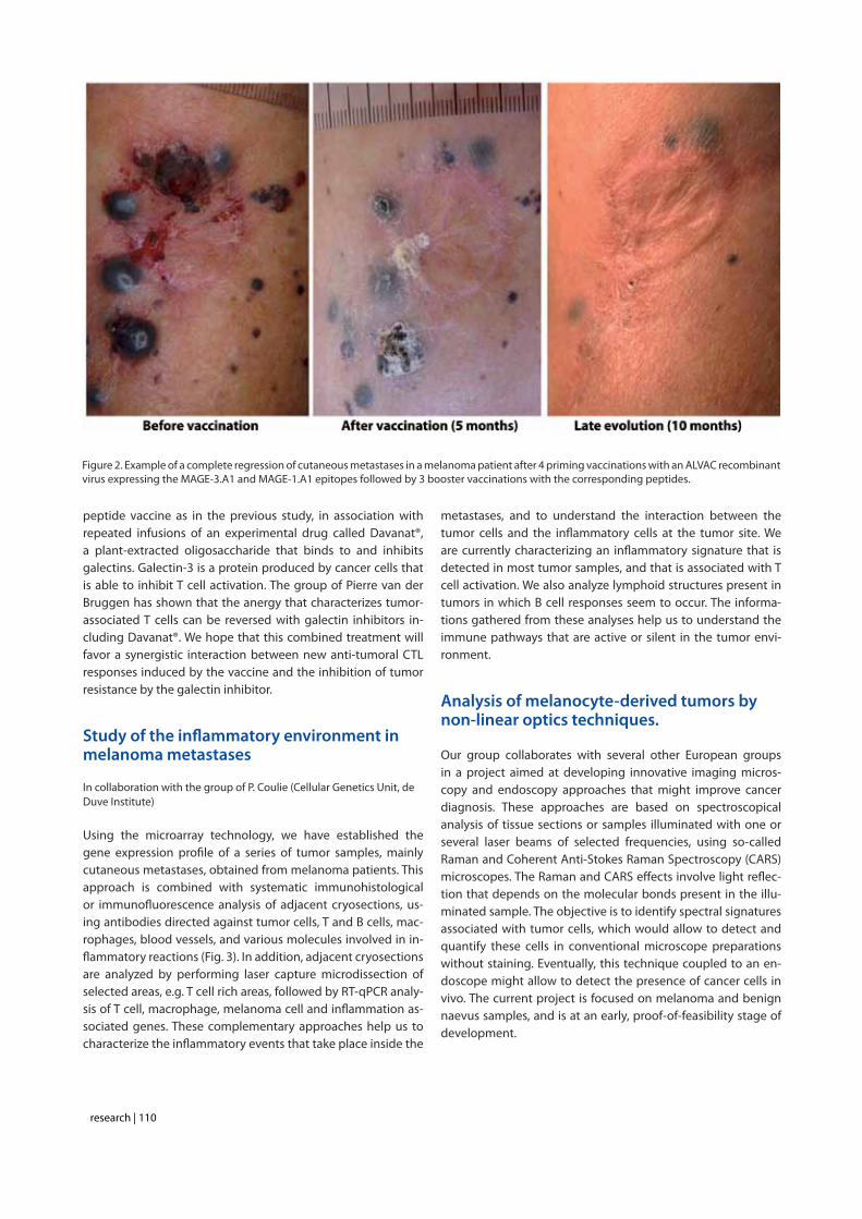
Figure 2. Example of a complete regression of cutaneous metastases in a melanoma patient after 4 priming vaccinations with an ALVAC recombinant virus expressing the MAGE-3.A1 and MAGE-1.A1 epitopes followed by 3 booster vaccinations with the corresponding peptides.

research | 111
Selected publications
1. van Baren N, Brasseur F, Godelaine D, Hames G, Ferrant A, Lehmann F, André M, Ravoet C, Doyen C, Spagnoli GC, Bakkus M, Thielemans K, Boon T. Genes encoding tumor-specific antigens are expressed in human myeloma cells. Blood 1999;94:1156-64.
2. Marchand M, van Baren N, Weynants P, Brichard V, Dréno B, Tessier MH, Rankin E, Parmiani G, Arienti F, Humblet Y, Bourlond A, Vanwijck R, Liénard D, Beauduin M, Dietrich PY, Russo V, Kerger J, Masucci G, Jäger E, De Greve J, Atzpodien J, Brasseur F, Coulie PG, van der Bruggen P, Boon T. Tumor regressions observed in patients with metastatic melanoma treated with an antigenic peptide encoded by gene MAGE-3 and presented by HLA-A1. Int J Cancer 1999;80:219-30.
3. Marchand M, Brichard V, van Baren N, Coulie PG. Biological and clinical developments in melanoma vaccines. Expert Opin Biol Ther 2001;1:497-510.
4. Marchand M, Punt CJ, Aamdal S, Escudier B, Kruit WH, Keilholz U, Hakansson L, van Baren N, Humblet Y, Mulders P, Avril MF, Eggermont AM, Scheibenbogen C, Uiters J, Wanders J, Delire M, Boon T, Stoter G. Immunisation of metastatic cancer patients with MAGE-3 protein combined with adjuvant SBAS-2: a clinical report. Eur J Cancer 2003;39:70-7.
5. Kruit W, van Ojik H, Brichard V, Escudier B, Dorval T, Dréno B, Patel P, van Baren N, Avril M-F, Piperno S, Khammari A, Stas M, Ritter G, Lethé B, Godelaine D, Brasseur F, Zhang Y, van der Bruggen P, Boon T, Eggermont A, Marchand M. Phase I/II study of subcutaneous and intradermal immunization with a recombinant MAGE-3 protein in
patients with detectable non-visceral metastatic melanoma. Int J Cancer 2005;117:596-604.
6. van Baren N, Bonnet MC, Dréno B, Khammari A, Dorval T, Piperno-Neumann S, Liénard D, Speiser D, Marchand M, Brichard VG, Escudier B, Négrier S, Dietrich PY, Maraninchi D, Osanto S, Meyer RG, Ritter G, Moingeon P, Tartaglia J, van der Bruggen P, Coulie PG, Boon T. Tumoral and immunological response following baccination of melanoma patients with an ALVAC virus encoding MAGE antigens recognized by T cells. J Clin Oncol 2005;23:9008-21.
7. Carrasco J, Van Pel A, Neyns B, Lethé B, Brasseur F, Renkvist N, van der Bruggen P, van Baren N, Paulus R, Thielemans K, Boon T, Godelaine D. Vaccination of a melanoma patient with mature dendritic cells pulsed with MAGE-3 peptides triggers the activity of nonvaccine anti-tumor cells. J Immunol 2008;180:3585-93.
8. Corbière V, Chapiro J, Stroobant V, Ma W, Lurquin C, Lethé B, van Baren N, Van den Eynde BJ, Boon T, Coulie PG. Antigen spreading contributes to MAGE vaccination-induced regression of melanoma metastases. Cancer Res. 2011 Feb 15;71:1253-62.
9. Cipponi A, Wieers G, van Baren N, Coulie PG. Tumor-infiltrating lymphocytes: apparently good for melanoma patients. But why? Cancer Immunol Immunother. 2011 60:1153-60. Review.
10. Lucas S, van Baren N, de Smet C, Coulie PG. Demethylation of the FOXP3 gene in human melanoma cells precludes the use of this epigenetic mark for quantification of Tregs in unseparated melanoma samples. Int J Cancer. 2012;130:1960-6.
Figure 3. A melanoma metastatis infiltrated by cytolytic T lymphocytes. A tissue section of the tumor was stained with three antibodies coupled to a different fluorescent dye. Melanoma cells and T cells appear in brown and green, respectively. Some melanoma nuclei express the proliferation marker Ki67 (in red). All cell nuclei are stained with a blue fluorescent dye. This image shows that the tumor continues to grow in the presence of an abundant infiltration by T lymphocytes.
research | 112
Nicolas van Baren, Senior Investigator
Ludwig Institute for Cancer Research B1.74.12 Av. Hippocrate 74-75 B - 1200 Brussels
[T] +32 02 764 75 08[F] +32 02 764 65 65[E] [email protected][W] http://www.deduveinstitute.be/therapeutic_vaccina-tion.php
Staff members
Jérôme Degueldre, Clinical Research Associate • Marjorie Mercier, Research Assistant • Madeleine Swinarska, Research Assistant

research | 113
Cytokines in immunity and inflammation
Jean-Christophe RenauldLaure Dumoutier
The cytokine group studies the biological activities of cytokines in inflammatory and tumoral processes, as well as the molecular mechanisms underlying these activities. Our work focuses on Interleukin-9 (IL-9) and IL-22, two cytokines discovered in our laboratory. IL-9 is produced by a particular T lymphocyte population, called TH9, and plays a role in immune responses against intestinal parasites and asthma. Dysregulation of IL-9 signalling is also implicated in tumoral transformation and this process has been studied in an in vitro tumorigenesis model, leading to the identification of oncogenic mutations of the JAK1 gene. IL-22, originally identified as a gene induced by IL-9 in T lymphocytes, upregulates the production of acute phase reagents and antibacterial proteins in the liver, the lung and intestinal mucosae, and in the skin. IL-22 appears to play a key role in wound healing and skin inflammation processes such as psoriasis. The role of these cytokines in inflammation is currently being investigated using transgenic and gene-targeted mice for these cytokines and their receptors, and by using an original strategy of anti-cytokine vaccination.
Interleukin 9
Interleukin-9 (IL-9) was discovered in our group, through its ability to sustain antigen-independent growth of certain murine T helper clones. Although IL-9 did not turn out to be a T cell growth factor for freshly isolated T cells, it was found particularly potent on T cell lymphomas, as an anti-apoptot-ic agent. To determine the biological activities of this factor, we generated transgenic mice overexpressing this cytokine. Analysis of these animals disclosed two essential properties of IL-9: its activity on mast cells and eosinophils with consecu-tive implications in asthma, and its tumorigenic potential in T lymphocytes.
IL-9-transgenic mice : parasite infections and asthmaAlthough IL-9 overproduction is viable and IL-9 transgenic mice did not show any major abnormality at the first look, they were found to harbor increased numbers of mast cells in the intestinal and respiratory epithelia, and were also charac-terized by a general hypereosinophilia. This phenotypic char-acteristic was found to increase the capacity of these animals to expel nematodes like Trichinella spiralis or Trichuris muris., suggesting that IL-9 administration could protect susceptible
hosts against these parasites. Conversely, blocking IL-9 activity resulted in a failure to expel T. muris parasites and in decreased eosinophilic responses against the parasite (1).The other side of the coin was the discovery that IL-9 over-expression, such as that characterizing the IL-9 transgenic animals, resulted in bronchial hyperresponsiveness upon ex-posure to various allergens. Our observations showed that IL-9 promotes asthma through both IL-13-dependent and IL-13-independent pathways (2), as illustrated in figure 1. The potential aggravating role of IL-9 in asthma was confirmed by genetic analyses performed by others and pointing to both IL-9 and the IL-9 receptor genes as major candidate genes for human asthma. In addition, we found that asthma patients produce increased amounts of IL-9.
IL-9-transgenic mice : T cell lymphomas IL-9 transgenic animals showed normal T cell development and T cell numbers but spontaneously developed thymic lym-phomas at low frequency (5%) when maintained in a conven-tional environment. Two lines of evidence indicate that IL-9 is not a conventional oncogene but rather favors tumor de-velopment in response to exogenous stimuli. First, the tumor incidence was significantly lower when mice were maintained

research | 114
under pathogen-free conditions. Secondly, all IL-9 transgenic mice developed T cell lymphomas when exposed to sublimi-nal doses of a chemical carcinogen or to irradiation, that were innocuous in wild type mice (3). The anti-apoptotic activity of IL-9 provides an attractive explanation for these observations, namely that IL-9 could lead to increased survival of abnormal cells generated by exposure to minimal doses of oncogenic stimuli. The potential implication of IL-9 in oncology was also confirmed in human systems by its constitutive expression in Hodgkin lymphomas.
IL-9 receptor and signal transduction
Analysis of the mode of action of IL-9 at the molecular level was initiated by the cloning of the murine and human IL-9 receptor (IL-9R) cDNAs (4). By further dissecting the signal transduction cascade triggered by IL-9, we showed that, upon IL-9 binding, the IL-9R associates with a co-receptor protein called γc. This induces the phosphorylation of the JAK1 and JAK3 tyrosine kinases, which are associated with IL-9R and γc, respectively. A single tyrosine residue of the IL-9R is then phosphorylated and acts as a docking site for 3 transcription factors of the STAT family, STAT-1, -3 and -5, which become phosphorylated and migrate to the nucleus, where they activate the transcription of a number of genes. This pathway is common to many cy-tokines but is often dispensable for their biological activities. For IL-9, our group demonstrated that activation of the STAT transcription factors is crucial for all the effects of IL-9 studied on various cell lines, including positive and negative regulation of cell proliferation, as well as inhibition of corticoid-induced apoptosis in T cell lymphomas. Further analysis demonstrated
that STAT-1, -3 and -5 play specific, redundant and synergistic roles in the different activities of IL-9 in vitro. The pathways re-sponsible for IL-9-induced proliferation were studied in details, and this process was found to depend mainly on the activation of STAT-5, on the recruitment of the IRS-1 adaptor, and on the activation of the Erk MAP-Kinase pathway.
Role of JAK1 overexpression and mutations in tumor cell transformation
Constitutive activation of the JAK-STAT pathway is frequent in cancer and contributes to oncogenesis. Some of our recent data indicate that JAK overexpression plays a role in such pro-cesses. Using a murine proB cell line that strictly depends on IL-3 for growth in vitro, cytokine-independent and tumorigen-ic clones were derived from a two-step selection process. Cells transfected with a defective IL-9 receptor acquired IL-9 respon-siveness during a first step of selection, and progressed after a second selection step to autonomously growing tumorigenic cells. Microarray analysis pointed to JAK1 overexpression as a key genetic event in this transformation. Overexpression of JAK1 not only increased the sensitivity to IL-9 but most im-portantly allowed a second selection step towards cytokine-independent growth with constitutive STAT activation. This progression was dependent on a functional FERM and kinase JAK1 domain. Similar results were observed after JAK2, JAK3 and TYK2 overexpression. All autonomous cell lines showed an activation of STAT5, ERK1-2 and AKT. Thus, JAK overexpression can be considered as one of the oncogenic events leading to the constitutive activation of the JAK-STAT pathway (5).Recently, we elucidated the mechanism responsible for the second step of this tumoral transformation process, as we found that the majority of the cytokine-independent tumori-genic clones acquired an activating mutation in the kinase or in the pseudokinase domain of JAK1 (figure 2).
Figure 1. Direct and indirect activities of IL-9 in asthma. IL-9 acts directly on mast cells and B lymphocytes to induce an expansion of these cells and IgE production. IL-9 promotes the proliferation of eosinophils indirectly, by upregulating IL-5 production by T cells. Upregulation of IL-13 production by T cells mediates IL-9 activities on lung epithelial cells, including mucus production and secretion of eotaxin, which is required to recruit eosinophils into the lungs (2).
Figure 2. Localization of JAK1 activating mutations in the kinase and pseudokinase domains.

research | 115
In parallel to these observations, in collaboration with Prof. Marco Tartaglia (University of Rome), we identified activating mutations in JAK1 in 20% of T-cell acute lymphoblastic leuke-mia (T-ALL) and in 3% of B-ALL patients, confirming the rele-vance of our in vitro model-derived JAK1 mutations for human malignancies. Further analysis of human ALL samples showed that JAK1-mutated leukemias share a type I IFN transcriptional signature, suggesting that these mutants do not only activate growth-promoting pathways, but also antiviral pathways. Ex-pression of these activating JAK1 mutants in murine hemat-opoietic cell lines recapitulated this signature in the absence of IFN, but also strongly potentiated the in vitro response to IFN. Finally, we also showed in an in vivo leukemia model that cells expressing mutants such as JAK1(A634D) are hypersen-sitive to the anti-proliferative and anti-tumorigenic effect of type I IFN, suggesting that type I IFNs should be considered as a potential therapy for ALL with JAK1 activating mutations (6).
IL-TIF/IL-22 : a new cytokine structurally related to IL-10
Searching for genes specifically regulated by IL-9 in lympho-mas, we identified a new gene that turned out to encode a 179 amino acid long protein, including a potential signal pep-tide, and showing a weak but significant sequence homol-ogy with IL-10. This protein, originally designated IL-TIF for IL-10-related T-cell derived Inducible Factor, was later renamed IL-22. Despite its structural homology with IL-10, IL-22 fails to recapitulate any of IL-10 biological activities. Biological activi-ties of IL- 22 include the induction of acute phase proteins in liver (7) and protection against experimental hepatitis and co-litis (L. Dumoutier, unpublished results). Among the different T cell subset, IL-22 was found to be preferentially produced by TH17 cells, which are associated with several autoimmune and inflammatory processes. We assessed the role of IL-22 in a mouse model where psoriasiform skin inflammation is trig-gered by topical application of the TLR7/8 agonist imiquimod. At the macroscopic level, scaly skin lesions induced by daily applications of imiquimod in wild-type mice were almost to-tally absent in IL-22–deficient mice or in mice treated with a blocking anti–IL-22 Ab. At the microscopic level, IL-22–defi-cient mice showed a dramatic decrease in the development of pustules and neutrophil infiltration and a partial decrease in acanthosis. At the molecular level, the absence or inhibi-tion of IL-22 strongly decreased the expression of chemotac-tic factors such as CCL3 and CXCL3 and of biomarkers such as S100A8, S100A7, and keratin 14, which reflect the antimi-crobial and hyper- proliferative responses of keratinocytes. Contrasting with this proinflammatory effect of IL-22 in skin inflammation, asthma models showed that IL-22 can have a protective anti-inflammatory activity in lungs. This protective effect of IL-22 has been attributed to an inhibition of IL-13 ac-tivity on lung epithelial cells either for CCL17/TARC induction or for IL-25 production. Inhibiting IL-22 in vivo, through anti-body treatment or by gene targeting, increased expression of
these inflammatory mediators, infiltration by eosinophils and broncho-hyperrersponsiveness.
Both in the psoriasis and asthma models have challenged the dogma that IL-22 is mainly produced by TH17 lymphocytes, and gd T cells as well as innate lymphoid cells turned out the be the major producers of this cytokine. We characterized the cells responsible for IL-22 production in response to TLR ago-nists such as LPS or flagelin. We identified a new innate lym-phoid spleen cell population expressing CD25, CCR6 and IL-7R representing 1% of spleen cells from recombination activating gene (Rag2)-deficient mice. This population comprises 60–70% CD4+ cells, which produce IL-22, and are still present in common g chain-deficient mice; the CD4- subset coexpresses IL-22 and IL-17, and is common g chain-dependent. These cells share a transcriptional program with NKp46+ RORgt+ cells found in intestinal mucosae and involved in antibacterial re-sponses. The importance of IL-22 production for the LPS-trig-gered response is highlighted by the fact that IL-22-deficient mice are more resistant to LPS-induced mortality, pointing to the pro-inflammatory activity of this cytokine. Although IL-22 does not share any biological activity with IL-10, these 2 cytokines share a common component of their respective receptor complex, IL-10Rß. Anti-IL-10Rß antibod-ies indeed block the IL-22-induced acute phase response in HepG2 cells (7). All receptor complexes for IL-10-related cy-tokines include a long chain and a short chain, based on the length of the cytoplasmic domain of these transmembrane proteins. IL-10Rß is a typical short chain component, with only 76 amino acids in the cytoplasmic domain, whose main func-tion seems to consist in recruiting the Tyk2 tyrosine kinase. In addition to IL-10R ß, IL-22 signalling requires the expression of a long chain protein, called IL-22R and comprising a 319 amino acid long cytoplasmic domain. This chain associates with JAK1, and is responsible for the activation of cytoplasmic signalling cascades such as the JAK/STAT, ERK, JNK and p38 MAP kinase pathways. An unexpected feature of the IL-22R chain is the fact that the C-terminal domain of this receptor is constitutively as-sociated with STAT3, and that STAT3 activation by this recep-tor does not require the phosphorylation of the receptor, in contrast to the mechanism of STAT activation by most other cytokine receptors (9). Beside this cell membrane IL-22 receptor complex composed of IL-22R and IL-10Rß, we identified a protein of 231 amino acid, showing 33 % amino acid identity with the extracellular domains of IL-22R, respectively, but without any cytoplasmic or transmembrane domain. This soluble receptor has been named IL-22 binding protein (IL-22BP), because it binds IL-22 and blocks its activities in vitro, demonstrating that this pro-tein can act as an IL-22 antagonist.The crystal structure of IL-22, alone and bound to its cellular re-ceptor IL-22R or to its soluble receptor IL-22BP has been char-acterized in collaboration with Prof. Igor Polikarpov (University of Sao Paulo) and is illustrated in figure 3.
In addition to its role in IL-22 binding and signalling, the IL-22R

research | 116
chain also forms a functional heterodimeric receptor complex by associating with IL-20Rß, the second short chain member of the IL-10R-related receptor family. This complex mediates STAT-1 and –3 activation by IL-20 and IL-24, but not by IL-22. In addition, IL-20 and IL-24 can also bind to other complexes consisting of IL-20Rα and IL-20Rß (see ref. 10 for a review of this cytokine family).
Anti-cytokine vaccination
Beside conventional gene targeting strategies, that were used in our lab to generate mice deficient in the IL-9R, in IL-22 or in IL-22R, we developed a new strategy of anti-cytokine vac-cination leading to the production in vaccinated mice of anti-cytokine autoantibody that block the biological activities of endogenous cytokines. Neutralizing auto-antibodies against cytokines such as IL-9, IL-12 and IL-17 have been induced upon vaccination with the autologous cytokines chemically coupled with OVA (IL-9, IL-17) or with the Pan DR T helper epitope PA-DRE (IL-12). This strategy contributed to demonstrate the role of IL-9 in an intestinal helminth infection (1), of IL-12 in athero-sclerosis and of IL-17 in experimental autoimmune encepha-lomyelitis. More recently, we developed a new procedure of anti-cytokine vaccination by taking advantage of tumor cells as a vaccine against peptides presented at their surface in fusion with a human transmembrane protein. These vaccina-tion methods represent simple and convenient approaches to knock down the in vivo activity of soluble regulatory proteins, including cytokines and their receptors, and are currently vali-dated with additional targets in inflammatory models.
Selected publications
1. Richard M, Grencis RK, Humphreys NE, Renauld JC, Van Snick J. Anti-IL-9 vaccination prevents worm expulsion and blood eosinophilia in Trichuris muris-infected mice. Proc Natl Acad Sci U S A 2000; 97:767-72.
2. Steenwinckel V, Louahed J, Orabona C, Huaux F, Warnier G, McKenzie A, Lison D, Levitt R, Renauld JC. IL-13 mediates in vivo IL-9 activities on lung epithelial cells but not on hematopoietic cells. J Immunol 2007; 178:3244-51.
3. Renauld JC, van der Lugt N, Vink A, van Roon M, Godfraind C, Warnier G, Merz H, Feller A, Berns A, Van Snick J. Thymic lymphomas in interleukin 9 transgenic mice. Oncogene 1994; 9:1327-32.
4. Renauld JC, Druez C, Kermouni A, Houssiau F, Uyttenhove C, Van Roost E, Van Snick J. Expression cloning of the murine and human interleukin 9 receptor cDNAs. Proc Natl Acad Sci U S A 1992; 89:5690-4.
5. Knoops L, Hornakova T, Royer Y, Constantinescu SN, Renauld JC. JAK kinases overexpression promotes in vitro cell transformation. Oncogene 2008; 27:1511-9
6. Hornakova T, Chiaretti S, Lemaire ML, Foa R, Ben Abdelali R, Asnafi V, Tartaglia M, Renauld JC, Knoops L. ALL-associated JAK1 mutations confer hypersensitivity to the anti-proliferative effect of Type I interferon. Blood 2010; 115:3287-95.
7. Dumoutier L, Van Roost E, Colau D, Renauld JC. Human interleukin-10-related T cell-derived inducible factor: molecular cloning and functional characterization as an hepatocyte-stimulating factor. Proc Natl Acad Sci U S A 2000; 97:10144-9.
8. Veldhoen M, Hirota K, Westendorf AM, Buer J, Dumoutier L, Renauld JC, Stockinger B. The Aryl hydrocarbon receptor is essential for production of the TH17 cytokine IL-22 and links TH17-mediated autoimmunity to environmental toxins. Nature 2008; 453:106-9.
9. Dumoutier L, de Meester C, Tavernier J, Renauld, JC. A new activation modus of STAT3 : a tyrosine-less region of the IL-22 receptor recruits STAT3 by interacting with its coiled-coil domain. J Biol Chem 2009; 284:26377-84.
10. Renauld JC. Class II cytokine receptors and their ligands: key antiviral and inflammatory modulators. Nature Rev. Immunol. 2003; 3:667-6.
Figure 3. Comparison of IL-22/IL-22BP and IL-22/IL-22R1 binding interfaces. Superposition of IL-22/IL-22BP (cyan/purple blue) and IL-22/IL-22R1 (yellow/red) crystal structures shows their binding interfaces outlined by boxes.

research | 117
Laure Dumoutier, Assistant Member
Ludwig Institute for Cancer Research de Duve Institute MEXP - B1.74.05 Av. Hippocrate 74-75 B - 1200 Brussels
[T] +32 02 764 74 65[F] +32 02 762 94 05[E] [email protected][W] http://www.deduveinstitute.be/cytokines_in_immunity_and_inflammation.php
Staff members
Jacques Van Snick, Member • Reece Marillier, Postdoctoral Fellow • Magali de Heusch, Postdoctoral Fellow • Laurent Knoops, Postdoctoral Fellow • Perrine Cochez, Graduate Student • Muriel Lemaire, Graduate Student • Amandine Saumet, Graduate Student • Lorraine Springuel, Graduate Student • Astrid Van Belle, Graduate Student • Isabelle Bar, Technician • Pamela Cheou, Technician • Emilie Hendrickx, Technician • Monique Stevens, Technician
Jean-Christophe Renauld, Member
Ludwig Institute for Cancer Research de Duve Institute MEXP - B1.74.05 Av. Hippocrate 74-75 B - 1200 Brussels
[T] +32 02 764 74 64[F] +32 02 762 94 05[E] [email protected][W] http://www.deduveinstitute.be/cytokines_in_immunity_and_inflammation.php

research | 118
Signal transduction and molecular hematology group Structure and function of cytokine receptors
Stefan Constantinescu
Cytokines and their receptors govern formation of blood cells and development and function of the immune system. Our broad interest is the understanding of the mechanisms that control assembly in the membrane of cell surface receptors that respond to extracellular cues. We study in detail the structure, function and orientation of several cytokine receptors, such as those for erythropoietin (Epo), thrombopoietin (Tpo), Granulocyte Colony Stimulating Factor (G-CSF), which function as homomeric complexes and of heteromeric receptors such as receptors for several interleukins.
Activation of these receptors is triggered by cytokine-induced changes in receptor dimerization/oligomerization, which is transmitted via juxtamembrane and transmembrane domains to the cytosolic region and ultimately to members of the Janus family of tyrosine kinases (JAKs). We aim to identify: i) the structural basis for transmembrane signaling, especially how transmembrane and juxtamembrane sequences switch-on or -off cytokine receptor signaling; ii) what are the general rules by which hydrophobic membrane-spanning domains interact in the membrane in a sequence-specific manner and how is this dimerization/oligomerization process regulated; and iii) the mechanisms of JAK attachment to receptors, and their subsequent activation, especially the role of pseudokinase domains in JAK kinase domain activation.
The laboratory identified constitutively active oncogenic mutants of JAK2, JAK1 and TYK2 and of cytokine receptors, with some being involved in human blood cancers. Specifically the mechanisms by which JAK2 V617F and TpoR W515 mutants induce, in humans, Myeloproliferative Neoplasms (MPNs) are actively pursued. The mutants we identified induce constitutive STAT activation, and we are interested in determining the gene targets of constitutive STAT5 activation, since we identified several novel target genes induced specifically by constitutively active STAT5, and that are involved in MPN pathogenesis. A close interaction structure has been created with clinicians and clinical biologists at St Luc Hospital for in-depth study of patient-derived cells.
The mechanisms by which the mutant JAK2 V617F induces Polycythemia Vera and other myeloproliferative neoplasms in humans
C. Pecquet, E. Leroy, V. Gryshkova, J.-P. Defour, M. Swinarska, D. Colau
The JAK-STAT pathway mediates signaling by more than 25 cytokine receptors and is constitutively activated in many cancers. Several mutations in genes coding for JAKs have
been identified in the past five years (1). Janus kinases pos-sess two kinase domains, one active and the other, denoted as the pseudokinase domain, predicted to be inactive. Four Janus kinases are coded by the human genome JAK1, JAK2, JAK3 and TYK2. JAK2 is crucial for signaling by EpoR, TpoR, the G-CSFR, the interleukin 3 receptor and several others. JAKs are appended to the cytoplasmic juxtamembrane do-mains of receptors and are switched-on upon ligand binding to the receptors’ extracellular domains.

research | 119
Polycythemia Vera (PV), or the Vaquez-Osler disease, is char-acterized by excessive production of mature red cells and sometimes of platelets and granulocytes. Two other related diseases, Essential Thrombocythemia (ET) and Primary Mye-lofibrosis (PMF) are associated with excessive platelet, granu-locyte production and fibrosis (scaring) of the marrow due to excessive myeloid cell proliferation, enzyme release and col-lagen secretion by marrow fibroblasts.
We have been involved in the discovery of the unique ac-quired somatic JAK2 V617F mutation in collaboration with William Vainchenker at the Institut Gustave Roussy in Paris. JAK2 V617F responsible for >98% of Polycythemia Vera and for >50% of Essential Thrombocythemia (ET) and Primary My-elofibrosis (PMF) cases (2). The mutation in the pseudokinase domain activates the kinase domain and constitutive signal-ing (3) in complexes between JAK2 V617F and cytokine re-ceptors such as EpoR, TpoR and G-CSFR (Figure 1). The homol-
ogous V617F mutations in JAK1 and Tyk2 also enable these kinases to be activated without ligand-binding to cytokine receptors (3). These results suggested that point mutations in JAK proteins might be involved in different forms of can-cers (1), with the already reported example of JAK1 mutations in T-lymphoblastic leukemia.We are interested to understand how a pseudokinase domain mutation can induce kinase domain activation. The aim is to be able to specifically inhibit mutated JAK2 in myeloprolifera-tive neoplasm patients, but not the wild type JAK2, which is crucial for red blood cell and platelet formation. We identified pseudokinase residue F595 as absolutely required for consti-tutive activation by V617F, but not for cytokine-induced ac-tivation of JAK2/JAK2 V617F (4). A region around F617 and F595, involving the middle of helix C of the JAK2 pseudoki-nase domain might be a target for specific JAK2 V617F inhibi-tion (Figure 2). Expression of segments of JAKs is pursued in different cellular systems for structural studies. Furthermore, protein fragment complementation and protein ligation as-
Figure 1. In the absence of cytokine ligands, cytokine receptors (left complex) are preassembled with tyrosine kinases JAK (Janus kinases) in inactive complexes. Cytokine binding to the extracellular domains of receptors (middle complex) induces a conformational change which allows the appended JAKs to cross-phosphorylate and activate each other. In turn, JAKs phosphorylate tyrosine residues (Py) on the cytosolic regions of receptors, which attract SH2- and PTB-containing signaling proteins. These proteins become themselves phosphorylated and either translocate to the nucleus to regulate gene expression (such as STATs, Signal Transducers and Activators of Transcription) or initiate kinase signaling cascades (such as Mitogen Activated Protein-Kinases, MAPK, phosphatydylinositol-3-kinase, PI3K, and Akt). The mutant JAK2 V617F binds to the cytosolic domains of receptors and can trigger signaling in the absence of any cytokine binding to the extracellular receptor domain (right complex). As a consequence, signaling is induced permanently and myeloid progenitors survive, proliferate and differentiate in an uncontrolled manner. (Jean-Michel Heine)

research | 120
says are employed in order to determine the dimerization state and proximity of interaction between mutated JAKs or wild type JAKs themselves and with cytokine receptors.
Involvement of pathologic TpoR signaling in myeloproliferative neoplasms
C. Pecquet, J.-P. Defour, M. Girardot, E. Leroy
Thrombopoietin (Tpo) is a cytokine produced by the liver that is critical for regulation of the formation of platelet cells, the small enucleated blood cells that derive from the cytoplasm of megakaryocytes and that prevent bleeding upon injury. Tpo also regulates the numbers of hematopoietic stem cells and other myeloid cells. TpoR appears to be central to MPNs. First, observations of Jerry Spivak from Johns Hopkins University indicated that
most MPN patients strongly down-modulate TpoR levels in megakaryocytes and platelets. Second, mutations in the TpoR intracellular juxtamembrane motif W515 led to consti-tutive activation of the receptor, and severe in vivo MPN with myelofibrosis. Third, asparagine mutations, which induce di-merization of the transmembrane domain of TpoR also acti-vate TpoR and one such mutation has been shown to be as-sociated with familial ET. Fourth, alterations of TpoR traffic to the cell surface can induce thrombocytosis due to insufficient clearance of Tpo and high sensitivity of early megakaryocytes to high Tpo.We have identified the mechanisms behind the down-modu-lation of TpoR in MPNs, and showed that JAK2 V617F induces ubiquitinylation, inhibition of recycling and degradation of TpoR (5). In addition we discovered that Tpo can induce a strong antiproliferative effect in cells that express high JAK2 levels (5). This effect can be detected in postmitotic mega-
Figure 2. (A) Janus kinase 2 contains several JAK homology domains, JH1, the kinase domain; JH2 the pseudokinase domain; JH3-JH4 the SH2-like domain and JH4-JH7, the FERM (band four point 1, ezrin, radixin, moesin)-like domain. The pseudokinase domain plays a major role in cytokine-dependent activation of the kinase domain, and was implicated in inhibiting the basal activity of the JH1 domain. The V617F mutation is activating the kinase activity of JH1, presumably by preventing the inhibition exerted by JH2 on JH1. The V617F mutation is detected in 98% of PV and approximately 50% of ET and PMF patients. (B) The pseudokinase (JH2) and kinase (JH1) domains of JAK2 are modeled as adopting classical tyrosine kinase structures, interacting with each other and leading to JH1 inhibition. Residue F595 of the helix C of JH2 is required for constitutive activation of JAK2 V617F and of other mutated JAKs proteins, but not for cytokine activation of wild type JAK2. F595 plays a pivotal role in transmitting the conformational change in JH2 to JH1 (red arrow) and eventually in activating the kinase activity of JH1. The region around V617F and the middle of JH2 helix C surrounding F595 could become the target of inhibitors that might specifically decrease constitutive activation of JAK mutants.

research | 121
karyocytes (Plos Biol 2010, 8, e1000476). We showed that selection against the antiproliferative effect of Tpo occurs in JAK2 V617F cells, and is partially responsible for TpoR down-modulation in MPN cells, which then continue to proliferate in the presence of Tpo, unlike normal cells. Biochemically, co-expression of TpoR and JAK2 V617F leads to strong ubiquitinylation of K540 and K544 of TpoR cytosolic domain and proteasome-mediated degradation of TpoR. In the same time, JAK2 itself is ubiquitinylated. Using the Ubi-Scan approach coupled to mass spectrometry we identified a number of proteins that acquire either ubiquitin or ISG15 or other ubiquitin-like molecules in a Tpo-dependent manner and we are establishing the roles of such post-translational modifications in Tpo-signaling and JAK2 V617F-mediated proliferation.
A novel mechanism by which a tryptophan residue regulates TpoR activation
J.-P. Defour, V. Gryshkova, C. Pecquet
When the protein sequences of TpoR and the closely relat-ed EpoR were aligned, we realized that the TpoR contains a
unique amphipathic motif (RWQFP) at the junction between the transmembrane and cytosolic domains. Deletion of this motif (delta5TpoR) results in constitutive activation of the receptor (6), suggesting that these residues maintain the re-ceptor inactive in the absence of TpoR. Mutagenesis of the RWQFP motif showed that W515 (W508 in the murine) is the key residue maintaining human TpoR normally inactive (6). Biophysical experiments performed in collaboration with Dr. Steve Smith at SUNY Stony Brook, and fluorescence comple-mentation studies showed that W515 actually regulates the orientation, tilt and dimerization of the upstream transmem-brane helix, and prevents receptor activation (Defour et al., manuscript in preparation). In vivo, in bone marrow reconsti-tuted mice, the delta5TpoR and TpoR W515A mutant induce massive expansion of platelets, neutrophils and immature erythroid progenitors and myelofibrosis by day 45 (Figure 3). We recently established that the myelofibrosis phenotype induced by TpoR W515 mutants depends on cytosolic Y112 (Y626) of TpoR, and appears to involve excessive STAT3 and MAP-kinase signaling (7). Thus, small molecules targeting phosphorylated Y112 (Y626) might be useful in the treat-ment of myelofibrosis.
Figure 3. Bone marrow adoptive transfer in lethally-irradiated mice with hematopoietic stem cells expressing the constitutively active ∆5TpoR induces severe myeloproliferative disorder, splenomegaly and fibrosis of the spleen. ∆5TpoR is a mutant where the amphipathic RW515QFP motif is deleted, which results in constitutive activation of receptor signaling. (A) Peripheral cell counts recorder 45 days after reconstitution indicates leukocytosis and thrombocytosis induced by ∆5TpoR. (B) Splenomegaly was induced by ∆5TpoR at day 45 post reconstitution. The spleen size in mice reconstituted with bone marrow transduced with wtTpoR was equivalent to that in control healthy mice. Green fluorescence protein (GFP) levels were equal after transduction, but enhanced migration to the spleen and proliferation explain the high GFP levels in ∆5TpoR spleens. (C) Histology of spleen sections of mice reconstituted with the indicated constructs. Silver staining (SL) for reticulin indicates fibrosis of the spleen in the ∆5TpoR mice.

research | 122
Determination of the interface and orientation of the activated cytokine receptors, such as EpoR and TpoR
J.-P. Defour, C. Pecquet, E. Leroy
Epo binding to the erythropoietin receptor (EpoR) results in survival, proliferation and differentiation of erythroid pro-genitors into mature red blood cells. In the absence of Epo, the cell-surface EpoR is dimerized in an inactive conforma-tion, which is stabilized by interactions between the TM se-quences. Epo binding to the extracellular EpoR domain in-duces a conformational change of the receptor, which results in the activation of cytosolic JAK2 proteins. To identify the residues that form the interface between the receptor mono-mers in the activated EpoR dimer we have replaced the EpoR extracellular domain with a coiled-coil dimer of α-helices (8).
Because coiled-coils have a characteristic heptad repeat with hydrophobic residues at positions a (one), d (four), the regis-ter of the coiled-coil α-helices is imposed on the downstream TM α-helix and intracellular domain.
When each of the seven possible dimeric orientations were imposed by the coiled-coil on the fused TM and intracellular domain of the EpoR, only two fusion proteins out of seven stimulated the proliferation of cytokine-dependent cell lines and erythroid differentiation of primary fetal liver cells (8). Since the predicted dimeric interfaces of the two active fu-sion proteins are very close, a unique dimeric EpoR confor-mation appears to be required for activation of signaling. In this active conformation TM residues L241 and L244 and JM residue W258 are predicted to be in the interface.
Figure 4. Different dimeric orientations of the thrombopoietin receptor lead to distinct phenotypes in the bone marrow and in the peripheral blood in bone marrow reconstituted mice. (A) The fusion of the coiled coil protein to differently engineered transmembrane domains of the thrombopoietin receptor (TpoR) imposes distinct dimeric orientations to the receptor. The different effects induced by three such chimeric receptors (cc-TpoR-I, cc-TpoR-III and cc-TpoR-VI) could be observed in the bone marrow and in the peripheral blood of lethally irradiated mice reconstituted with bone marrow cells retrovirally transduced with the indicated cc-TpoR fusion proteins. IC: intracellular, TM: transmembrane. (B) cc-TpoR-I induced a strong megakaryocytic response in the bone marrow with normal cellularity and a correct maturation of the myeloid lineages (May Grunwald stained bone marrow smear X 50, and normal platelet formation. (C) cc-TpoR-III induces granulocytosis, monocytosis and a very weak erythroblastosis in the peripheral blood (May Grunwald stained peripheral blood smear X 50) and weak dysplasia for the megakaryocytic and erythroid lineages in the marrow (not shown). (D) cc-TpoR-VI induced a stronger erythroblastosis and granulocytosis in the peripheral blood than cc-TpoR-III (May Grunwald stained peripheral blood smear X 50). (Jean-Philippe Defour).

research | 123
Similar studies are undertaken for the related TpoR (Figure 4a). Like the EpoR, the TpoR is thought to signal by activation of JAK2, of several STATs (STAT1, 3 and 5) as well as of MAP-kinase, PI-3-kinase and AktB. However, TpoR and EpoR signal quite differently since only TpoR can induce hematopoietic differentiation of embryonic stem cells or stimulate the earli-est stages of hematopoiesis in immature hematopoietic cells. We have shown that TpoR can signal from several distinct dimeric interfaces, and that besides the normal dimeric in-terface that leads to formation of platelets (Figure 4b), other interfaces promote signaling that leads to myeloprolifera-tive and myelodysplastic disorders (Figure 4c and d) (9). One orientation (cc-TpoR-II) corresponds to the inactive receptor state, and another (cc-TpoR-I) induces megakaryocyte dif-ferentiation and physiologic platelet formation (Figure 4b). The dimeric conformations represented by cc-TpoR-III and cc-TpoR-VI induce a combination of myeloproliferation and dysplasia of the erythroid lineage (9). Interestingly, the dimer orientation that induces the highest levels of JAK2 activation, cc-TpoR-IV, also induces strong cell-to-cell adhesion and ex-pansion of early myeloid progenitors, as well as immortali-zation of myeloblasts. Our hypothesis is that in this dimeric orientation the receptor signals to maintain progenitors and possibly hematopoietic stem cells in the niche, and that sig-nals induced by this dimer orientation might be recapitu-lating the quiescence-inducing effects of TpoR in HSCs. We will use this set of seven differently oriented TpoR dimers to dissect the signals induced by TpoR at the different stages of hematopoietic development.
Structure and function of juxtamembrane and transmembrane sequences of transmembrane receptors
R.-I. Albu, A. Dusa, J.-P. Defour, J. Van Hees, C. Mouton,
We have previously shown that the EpoR as well as a fraction of IL2/IL9 receptors exist on the cell surface as a preformed ligand-independent inactive dimers (homomeric and hetero-meric in the case of IL2/IL9 receptor complexes). For the EpoR, transmembrane domain interactions stabilize the inactive di-mer at the surface and the EpoR TM sequence is an example of TM dimer based on purely hydrophobic sequences (Proc. Natl Acad. Sci USA 2001, 98, 4379-84; EMBO J., 1999, 18, 3334-47). We study potential transmembrane interactions in the context of other transmembrane proteins, such as TpoR, G-CSFR. We use cell surface immunofluorescence co-patching of differentially epitope tagged receptors along with protein ligation and protein fragment complementation assays in or-der to determine the ligand-independent state of cell surface complexes. Preformed cytokine receptor oligomers might be important for supporting signaling by mutated JAKs in the absence of ligand. In addition to cytokine receptors, we study the role of transmembrane dimerization in the amyloido-genic processing of Amyloid Precursor Protein (APP) in col-
laboration with the groups of Profs. Jean-Noel Octave in our university and Steven O. Smith (SUNY Stony Brook, NY). We identified three Gly-X-X-X-Gly motifs in the juxtamembrane and transmembrane domain of APP and showed that these consecutive motifs promote transmembrane helix dimeri-zation and amyloidogenic processing of APP (J. Biol. Chem. 2008 283, 7733, Proc. Natl. Acad. Sci. USA 2009, 106, 1421).
Constitutive activation of JAK-STAT signaling pathways and genes targeted by STAT5 in transformed hematopoietic and patient-derived leukemia cells
M. Girardot, J. Van Hees
Cytokine stimulation of cytokine receptors induces tran-sient activation of the JAK-STAT pathway. In contrast, onco-genic forms of receptors or of JAKs (JAK2 V617F) transmit a continuous signal which results in constitutive activation of STAT proteins. In cultured cells this process is studied by expressing oncogenic forms of cytokine receptors or JAKs in cytokine-dependent cells and assaying for their transforma-tion into cells that grow autonomously (1, 8). A similar picture has been noted in patient-derived leukemia cells. The critical question is which genes are specifically regulated by consti-tutively active STAT proteins in leukemic cells. Using chro-matin immunoprecipitation (Chip) and sequencing of native promoters bound by STAT5 we noted that, in transformed cells, STAT5, and mainly STAT5B, can also bind to low affinity N4 (TTCNNNNGAA) DNA sites, not only to the high affinity N3 sites, which are characteristic of ligand-activated STAT5. We are attempting to identify the promoters actually bound by STAT proteins in living cells in physiologic and pathologic sit-uations. We identified one specific target gene of constitutive active STAT5B signaling in megakaryocytes of MPN patients, namely Lipoma Preferred Partner (LPP) (10), a gene found to be translocated in rare leukemias. LPP is the host gene for miR-28, which we found to down-modulate TpoR translation, impair megakaryocyte differentiation (10). miR-28 is patho-logically overexpressed in 30% of MPNs (10). Targets of miR-28, such as E2F6, are critical cell cycle regulators that might influence the phenotype of myeloproliferative disorders. Fur-thermore, miR-28 is specifically associated with megakaryo-cyte proliferation and induces a block in differentiation (10). We are studying the mechanisms of pathologic induction of LPP/miR-28 via constitutively active STAT5.
research | 124
Interaction with St Luc Hospital clinicians and clinical biologists: Identification of the molecular bases of MPNs without known molecular cause
J.-P Defour, J. Van Hees
At present, our laboratory is performing under the auspices of an ARC grant (Action de Recherche Concertée of the Uni-versité catholique de Louvain) with the St Luc Hospital de-partments of Hematology (Prof. Cédric Hermans, Prof. Au-gustin Ferrant, Dr. Laurent Knoops), Clinical Biology (Prof. Dominique Latinne, Dr. Hélène Antoine-Poirel) and groups of de Duve Institute (Prof. Mark Rider, Prof. Jean-Baptiste De-moulin) a large study on the presence and signaling of JAK2, TpoR, and growth factor receptor mutations in patients with myeloproliferative neoplasms. Next generation sequencing will be employed for well-investigated patients, using prima-ry cells that are characterized for functional defects and that do not harbor known mutations in order to unravel novel molecular defects in MPNs and leukemias.
Selected publications
1. Constantinescu SN, Girardot M, Pecquet C. Mining for JAK-STAT mutations in cancer. Trends Biochem Sci. 2008;33:122-31.
2. James C, Ugo V, Le Couédic JP, Staerk J, Delhommeau F, Lacout C, Garçon L, Raslova H, Berger R, Bennaceur-Griscelli A, Villeval JL, Constantinescu SN, Casadevall N, Vainchenker W. A unique clonal JAK2 mutation leading to constitutive signalling causes polycythaemia vera. Nature. 2005;434:1144-8.
3. Staerk J, Kallin A, Demoulin JB, Vainchenker W, Constantinescu SN. JAK1 and Tyk2 activation by the homologous polycythemia vera JAK2 V617F mutation: cross-talk with IGF1 receptor. J Biol Chem. 2005;280:41893-9.
4. Dusa A, Mouton C, Pecquet C, Herman M, Constantinescu SN. JAK2 V617F constitutive activation requires JH2 residue F595: a pseudokinase domain target for specific inhibitors. PLoS One. 2010;5:e11157.
5. Pecquet C, Diaconu CC, Staerk J, Girardot M, Marty C, Royer Y, Defour JP, Dusa A, Besancenot R, Giraudier S, Villeval JL, Knoops L, Courtoy PJ, Vainchenker W, Constantinescu SN. Thrombopoietin receptor down-modulation by JAK2 V617F: restoration of receptor levels by inhibitors of pathologic JAK2 signaling and of proteasomes. Blood. 2012;119:4625-35.
6. Staerk J, Lacout C, Sato T, Smith SO, Vainchenker W, Constantinescu SN. An amphipathic motif at the transmembrane-cytoplasmic junction prevents autonomous activation of the thrombopoietin receptor. Blood. 2006;107:1864-71.
7. Pecquet C, Staerk J, Chaligné R, Goss V, Lee KA, Zhang X, Rush J, Van Hees J, Poirel HA, Scheiff JM, Vainchenker W, Giraudier S, Polakiewicz RD, Constantinescu SN. Induction of myeloproliferative disorder and myelofibrosis by thrombopoietin receptor W515 mutants is mediated by cytosolic tyrosine 112 of the receptor. Blood. 2010;115:1037-48.
8. Seubert N, Royer Y, Staerk J, Kubatzky KF, Moucadel V, Krishnakumar S, Smith SO, Constantinescu SN. Active and inactive orientations of the transmembrane and cytosolic domains of the erythropoietin receptor dimer. Mol Cell. 2003;12:1239-50.
9. Staerk J, Defour JP, Pecquet C, Leroy E, Antoine-Poirel H, Brett I, Itaya M, Smith SO, Vainchenker W, Constantinescu SN. Orientation-specific signalling by thrombopoietin receptor dimers. EMBO J. 2011;30:4398-413.
10. Girardot M, Pecquet C, Boukour S, Knoops L, Ferrant A, Vainchenker W, Giraudier S, Constantinescu SN. miR-28 is a thrombopoietin receptor targeting microRNA detected in a fraction of myeloproliferative neoplasm patient platelets. Blood. 2010;116:437-45.
Links
GroupLudwig Institute for Cancer Research Ltd. NewsLink Sept 2005 of our group (http://www.licr.org/12124501528/news-link/0509/)European Commission Marie Curie Research Training Net-work ReceptEUR (www.recepteur.org)
Research:Whitehead Institute for Biomedical Research, MIT, Lodish Lab (http://www.wi.mit.edu/lodish/)
SUNY Stony Brook, Structural Biology, Smith Labhttp://sos.bio.sunysb.edu lab homepagehttp://csb.sunysb.edu structural biology homepagehttp://csb.sunysb.edu/bsb graduate program in biochemis-try and structural biology
European Hematology Association (EHA)Scientific Program Committee EHA16 (London), EHA17 (Am-sterdam)Hematology (American Society of Hematology Education Book)http://www.asheducationbook.org/
Learning: Biologie moléculaire de la cellule (Molecular Cell Biology H.F. Lodish) French Edition Vainchenker W, Delhommeau F, Constantinescu SN, Bernard OA.http://www.ncbi.nlm.nih.gov/pubmed/21653328"New mu-tations and pathogenesis of myeloproliferative neoplasms.Blood. 2011 Aug 18;118(7):1723-35. Epub 2011 Jun 7. Review.
Bioinformatics:Institute of Bioinformatics Bangalore, India http://www.ibioinformatics.org/

research | 125
Stefan Constantinescu, Member
Ludwig Institute for Cancer Research de Duve Institute SIGN - B1.75.07 Av. Hippocrate 74-75 B - 1200 Brussels
[T] +32 02 764 75 40[F] +32 02 764 75 66[E] [email protected][W] http://www.deduveinstitute.be/signal_transduction_and_molecular_hematology.php
Staff members
Christian Pecquet, Postdoctoral Fellow • Michael Girardot, Postdoctoral Fellow • Vitalina Gryshkova, Postdoctoral Fellow • Roxana-Irina Albu, PhD Student • Emilie Leroy, PhD Student • Jean-Philippe Defour, PhD Student • Céline Mouton, Research Assistant (Attachée de recherche) • Joanne Van Hees, Research Assistant (Attachée de recherche, Lab Manager) • Madeleine Swinarska, Research Assistant • Didier Colau, Senior Investigator • Julien Doornaert, Administrative Assistant

notes | 126
NOTES