“Lucian Blaga” University Sibiu PhD Thesis “Surgically induced ...
36
“Lucian Blaga” University Sibiu PhD Thesis “Surgically induced astigmatism after surgical treatment of cataract” Scientific coordinator: Prof. Univ. Dr. Adriana Stănilă Ph.D. Candidate: Dr. Rodica Pop
Transcript of “Lucian Blaga” University Sibiu PhD Thesis “Surgically induced ...
“Lucian Blaga” University Sibiu
PhD Thesis
“Surgically induced astigmatism after surgical treatment of
cataract”
Scientific coordinator:
Prof. Univ. Dr. Adriana Stănilă
Ph.D. Candidate:
Dr. Rodica Pop
2
I. GENERAL PART
THESIS ABSTRACT
A highly performing visual acuity represents one of the most important human qualities that
are most afraid he could lose and which ensures him a higher quality of life. Cataract is an eye
health problem that primarily affects the elderly population worldwide.
Cataract represents a major eye health problem that affects primarily the elderly population
worldwide. The progress that has been made in the field of physiopathology of corneal wound
healing, in the techniques and instrumentations used, contributed, especially in recent decades, to
a revolution in this area, reducing the risk of postoperative complications to a minimum and
allowing early anatomical and functional rehabilitation after surgery.
I.1. OPERATIVE INDUCED ASTIGMATISM
Operative induced astigmatism is the astigmatism greater than one diopter, which is present at
6-8 weeks postoperatively. This type of astigmatism can be with or against the rule, according to
the more refractive corneal meridian, the vertical or the horizontal. It is a form of regular
astigmatism which is mainly due to abnormalities in the cornea and that is related to the process
of healing and scar reshuffling taking place in the surgical incision.
I.2. PHYSIOPATHOLOGY OF CORNEAL HEALING
In the case of a perforating corneal injury, either by accident or by surgery, the healing process
requires the restoration of two main functions of this tissue: that of external barrier of the eye, by
forming a scar tissue, and that of optical function, by remodeling of this tissue. The quality of the
scar tissue formed is responsible for restoring the optical function of the cornea and the scar
reshuffling occurs well after the injury. The first response that occurs in a corneal aggression of
this type is represented by the corneal epithelial-endothelial remodeling, followed by the repair
of the other tissues.
Primary cicatricial response
It is provided by the epithelial-endothelial barrier restoration and its goal is a swift coaptation
of the wound.
3
Epithelial wound healing
The healing process of an epithelial wound can be divided into three overlapping phases[11]:
• In the first phase there is a loss of normal anchoring structures of the epithelium namely
that of the hemidesmozoms [12], of the desmosomes [13] and that of collagen type VII
[14]. The main phenomenon that occurs is the formation of the pseudopods (lamelipodia
and filopodia) as a result of cellular migration and that occurs up to covering the whole area
of the defect. It also increases the cellular water content, allowing coverage of larger areas.
This phase of cell migration is independent of the cell proliferation phenomenon, but during
the epithelial healing process the two processes complete each other.
• In the second phase occurs the cellular proliferation with the main purpose to repopulate the
epithelial defect area and also with the layering and cell differentiation [20]. This process
also restores cell number and cell mass.
• A very important role in this process have the limbic stem cells, which on one hand can
auto regenerate themselves and on the other hand can give birth to young cells that have the
ability to auto differentiate themselves. These cells contribute to maintain the corneal
epithelial homeostasis [24] and have a longer life than the central corneal epithelial cells
[26].
Due to the regenerative properties of the limbic stem cells, during the epithelial healing
process there is a wave of mitosis which starts at the periphery of the cornea and moves
towards the center continuing until the wound is healed and the epithelium regains its
normal thickness [9].
During an injury, corneal epithelial cell proliferation is intimately linked to the process of
cell differentiation. On the other hand, migration and cell proliferation are dependent on a
metabolic support provided by the glucose present in the aqueous humor and in the
epithelial glycogen stores [31].
• In the third phase, newly formed cells undergo a differentiation process in order to form the
specific structure of the corneal epithelium. If transitory attachments are formed during cell
migration, after the defect is completely covered, occurs the formation of permanent
4
anchorage units. An important role in this process has the extracellular matrix proteins:
fibronectin, fibrinogen / fibrin, laminin and tenascin [32].
Stromal healing
Corneal stromal wound healing is slower than other connective tissues, probably
because it is avascular. This phase of stromal healing includes synthesis, dividing and
forming of new bonds of collagen (cross-linking), phenomena that have as a result the
consolidation and remodelation of the wound [7]. Keratinocytes apoptosis is the earliest
stromal event that occurs immediately after epithelial injury [7] and continues at least for
one week after the occurrence of corneal injury [42].
Proliferation and migration of intact keratinocytes starts at 12-24 hours after injury,
followed by activation of fibroblasts and miofibroblasts, responsible for repopulating the
affected stroma [12].
While the healing of the superficial stromal layer is provided through an epithelial
contribution, its deeper layers are repaired by collagen synthesized by endothelial cells.
Recently, through the studies that were done in this area, the role of the
metalloproteinase in the modulating of the corneal scarring was high lightened. It was
thus demonstrated that MMP-1 (interstitial collagenase), MMP-2 (gelatinases-A), MMP-
3 (stromelysin-1) and MMP-9 (gelatinases B) participate not only in the epithelial
repairing but also in the stromal remodeling [50].
Repairing of the Bowman membrane
This membrane does not recover but it is repaired with defect. [51].
Repairing of the Descemet membrane
Healing and scarring of this is made by fibroblasts, which present an endothelial
morphology, thus forming a neodescemet. It occurs in three weeks after injury and reaches
maximum thickness in about six months [52].
Repairing of the endothelium
In the first 24 hours after the creation of a wound in a cornea, endothelial cells at the
wound edge withdrawal. One day after the injury begins the process of endothelial
5
proliferation, which will form a continuous layer, which by sliding over, will cover the
fibronectin fibrin clot that impregnates the epithelial plug [54].
Secondary cicatricial response
Its aim is to restore the optical function and tissue resistance, which is achieved by remodeling
of the scar tissue, in order to restore partial transparency and mechanical resistance of the corneal
tissue affected. This process takes place in three stages, as following:
Cleaning the scar tissue by activating the fibrinolytic collagenolytic system
Synthesized enzymatic systems by the cells that migrate from the epithelial and stromal
wound, participate in cleaning or extracellular remodeling. Intracellular cleaning system is
provided by polymorphonuclear, macrophages and cheratocite by their phagocytic function.
Reorganization of epithelial architecture
At the edges of the wound occurs a proliferation of the cheratocites, which in turn reject the
epithelial plug, starting from the 2nd day after the occurrence of the injury. Along with the
restoration of epithelial architecture also takes place the nerve regeneration, based on neurites
formation, which subsequently will differentiate into nerve endings.
Base membranes reconstruction
Base membrane remodeling is an integral part of the corneal epithelium healing [24]. This
base membrane has a very important role in the formation of stable cell adhesions [24].
Remodeling of the extracellular cicatricial matrix
An important role in reshaping the cornea scar tissue has the cheratocites. These contain
highly specialized proteins, such as crystalline, which helps maintain the transparency of the
corneal tissue [23].
I.3. CHARACTERISTICS AND IMPLICATIONS OF CORNEAL SCARRING IN
CATARACT SURGERY
Anatomical and functional rehabilitation early after cataract surgery, with the best visual acuity
without optics correction, represent the major desideratum which is expected by both the doctor
and the patient. Much attention is paid to surgical incision and its manner of closure, especially
for the purpose of reducing the corneal scar, with minimal adverse effects.
6
Improvements made in surgical techniques of cataract extraction by phacoemulsification
method, especially in recent decades, have allowed smaller surgical incisions, first with one
suture and afterwards without sutures. For this reason, we thought it appropriate to take into
consideration the peculiarities of the healing of an unsutured wound compared with a sutured
one.
It was demonstrated that sutured wounds and adjacent unsutured ones, from the same cornea,
showed different patterns of healing [64]. Unsutured wounds showed a delayed and abnormal
healing, unlike the sutured ones that showed progression to a normal anatomy of the region [65].
I.4. ETIOLOGY OF THE OPERATIVE INDUCED ASTIGMATISM
Etiological factors that may be associated with operative induced astigmatism after cataract
surgery is: preoperative astigmatism, factors related to the surgical act and factors related to
pesudofak.
Preoperative astigmatism
Preoperative astigmatism of greater value may be associated with increased values of
postoperative astigmatism [71]. Reducing this astigmatism during cataract surgery may improve
postoperative visual outcome. [73] Pre-existing astigmatism can be corrected during cataract
surgery using different methods which include: relaxing peripheral incisions, choice of incision
location, implantation of toric artificial lens [76,77].
Factors related to the incision
• Type of wound – sutured or unsutured
In case of a suture wound operative astigmatism is dependent on the length, depth and
tightness of the suture [78]. Sutures placed close to each other lead to a tissue compression and
cause according to the rule astigmatism. The sutures that are wider and the ones which are tighter
cause an against the rule astigmatism.
7
•
A wide suture allows the cornea to flatten, reducing the curvature in the vertical meridian and
with against the rule astigmatism [80]. If the suture is too tight the cornea stretches vertically
and the curvature in this meridian grows, with according to the rule astigmatism [80].
The tightening degree of the sutures
•
In case of sutured wounds, surgically induced astigmatism differs also on the suture material
used, resorbable or non resorbable. Thus, in the case of the non resorbable sutures, as long as
the wires remain in place, the power and the cylinder axis remain constant. After their removal,
postoperative astigmatism diminishes, due to reducing the tension created in the wound by this
type of sutures. If the case of non resorbable sutures the value and the axis of the cylinder can
also vary and the healing is associated with a local inflammatory reaction.
Suture material
•
The more scleral the incisions are the lower is the operative induced astigmatism. A more
corneal incision determines a more important and against the rule astigmatism and vice versa
[86]. In contrast to these considerations, it was demonstrated that in the case of small incisions,
the effect of anatomical location is less important [84]. For this reason, nowadays, the most
common incisions made with the cataract extraction by phacoemulsification technique are the
ones in clear cornea.
The location of the incision in relation to the limbus
Factors related to the pseudofak
Surgically induced astigmatism can be influenced by the position of the pseudofak in the
posterior chamber, by the shape, size and material of which the artificial implant is made.
8
II. PERSONAL RESEARCH
II.1. INTRODUCTION
Cataract is a major public health problem being responsible for much of the decreases of visual
acuity at the population worldwide. Everywhere in the world the extraction of cataract represents
the largest workload in the surgical departments of ophthalmology.
The improvements that were brought lately in surgical technique focused on the rapid recovery
of the patient, both anatomically as well as functionally. In this regard, the reduction or even the
prevention of postoperative astigmatism was one of the aims pursued by cataract surgeons
worldwide.
Due to the use on a larger scale of foldable crystalline artificial implants, the surgical incision
in clear cornea has lately become the main approach in this type of surgical intervention.
For these reasons and bearing in mind the latest trends in the surgical field, I considered
appropriate that in this clinical study to make a detailed assessment of surgically induced
astigmatism after cataract surgery, taking into consideration the corneal incision and its
characteristics, this actually being the main risk factor in the development of this type of
astigmatism.
II.2. PURPOSE OF THE WORK
This paper aims to assess a group of cases operated of cataract surgery by the
phacoemulsification method with implantation of foldable artificial lens in terms of surgically
induced astigmatism, of the etiologic factors incriminated in its appearance and of the methods
of prevention and treatment.
II.3. MATERIAL AND METHOD
Thesis material
The material of this thesis is represented by a group of 592 patients diagnosed with primitive
cataract primitive which have been operated between 2009 and 2011 at Optilens Clinic in Cluj-
Napoca. The surgical method consisted of cataract extraction by means of phacoemulsification
9
and implantation of foldable artificial lens of posterior chamber. I selected cases of primitive
cataract primitive, present in male and female patients, coming from rural or urban environment,
ages between 40 and 90 years.
Method of work
I conducted a prospective study for each case, which included:
Personal data of the pacient: age, sex, source environment
Clinical preoperative evaluation: lab exams, cardiologic examination with ECG.
Ocular preoperative evaluation: patient history and medical history, visual acuity,
biomicroscopy of anterior pole, pachimetry, direct ophthalmoscopy, intraocular pressure
measurement, measuring the number of endothelial cells, keratometria, ocular biometry,
determination of the power of the artificial implant lens diopter.
Postoperative evaluation: determination of visual acuity with / without optical correction,
refractometry, comments from the patient himself (satisfaction, sensitivity to light).
Criteria for inclusion of the cases in the study: primitive cases of cataract, senile and pre-
senile.
Criteria for exclusion of the cases from the study: ocular ( pre-existing eye disorders or
contemporary to the surgery, other ocular surgery history, eye trauma) and general (diabetes,
autoimmune inflammatory disorders, etc.)
II.4. OPERATING PROTOCOL
Preoperative patient preparation
Preoperative anesthesia: 2% lidocaine solution injected into the inferior fornix, topical
anesthetic with solution Benoxi, administered 2 drops every 1 minute.
Surgery
Cataract lens extraction was performed by the method of phacoemulsification and implantation
of foldable artificial lens of posterior chamber. Surgical incision was made in clear cornea, in a
single plane.
10
Incision was made close to limb, at the level of the vascular arch. The dimensions of the
incisions were 2.75 mm, 2.2 mm and 1.8 mm. The incisions were located supero-temporal,
supero-nasal, superior and temporal.
Intraoperative complications
During surgery are there can be possible intraoperative complications, such as: posterior
capsule rupture with vitreous loss, dislocation of the nucleus or of nuclear fragments in vitreous,
dislocation of the Descemet membrane, etc. None of these complications were present in the
patients in the study group.
Post surgery
Immediately after the surgery local instillations have been carried out with an antibiotic and an
anti-inflammatory steroid, usually in a fixed combination (TOBRADEX, Netildex, Betabioptal),
a short term mydriatic (Tropicamide, Mydrum) and eye dressing for 24 hours.
II.5. STATISTICAL CALCULATION
Statistical analysis was performed using ANOVA.
ANOVA statistical analysis techniques are used when comparing more than two groups of
patients. Anova statistical analysis is also known as analysis of variance. The larger the number
of comparative groups (more than two groups) the more diversify the grades of independent
variables.
II.6 RESULTS
The next stage of our study consisted of conducting a distribution of patients according to
parameters in the study, in order to further establish a detailed analysis of postoperative
functional results. This distribution was carried out as follows:
a. Distribution of cases by environment of origin
b. Distribution of cases by group age
c. Distribution of cases according to clinical type of cataract
11
d. Distribution of cases according to the evolutionary stage of cataract and
preoperative visual acuity
e. Distribution of cases after ocular biometry
f. Distribution of cases by preoperative astigmatism
g. Distribution of cases according to the size of the corneal incision
h. Distribution of cases according to the location of the corneal incision
i. Distribution of cases by postoperative astigmatism
j. Distribution of cases by the pre-operative versus post-operative astigmatism
k. Distribution of cases by astigmatism in six weeks as compared to that of 6 months
postoperatively
l. Global distribution of postoperative astigmatism compared with the preoperative
one
m. Distribution of cases by postoperative visual acuity
n. Distribution of cases by spherical postoperative equivalent
The main concerning parameter was the corneal astigmatism, which was calculated
preoperatively based on the on K1 and K2 values, and postoperatively was determined through
auto-refractometry print. For this purpose, a comparative study was made between postoperative
and preoperative astigmatism, taking into account its distribution by groups of values, the
orientation of the cylinder axis and the clinical form of astigmatism (with or against the rule). I
conducted a comparative study on three groups of patients that were formed according to the size
of the corneal incision. A comparative study was also was performed on groups of patients
divided according to the location of the corneal incision and the value of the preoperative
astigmatism.
Another important parameter in the study was the postoperative visual acuity without and
without optical correction and also the postoperative spherical equivalent. The evolution of the
visual acuity was checked at the two post surgery examinations, at six weeks and at six months
postoperative.
The value of the postoperative spherical resulted from the determination of the ocular
refraction and registered automatically along with the conduction of auto-refractometry. Final
results have been found stable at 6 months postoperative.
12
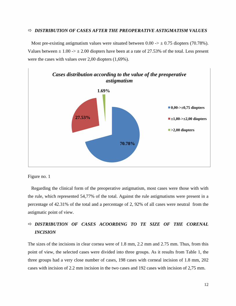
DISTRIBUTION OF CASES AFTER THE PREOPERATIVE ASTIGMATISM VALUES
Most pre-existing astigmatism values were situated between 0.00 -> ± 0.75 diopters (70.78%).
Values between ± 1.00 -> ± 2.00 diopters have been at a rate of 27.53% of the total. Less present
were the cases with values over 2,00 diopters (1,69%).
Figure no. 1
Regarding the clinical form of the preoperative astigmatism, most cases were those with with
the rule, which represented 54,77% of the total. Against the rule astigmatisms were present in a
percentage of 42.31% of the total and a percentage of 2, 92% of all cases were neutral from the
astigmatic point of view.
DISTRIBUTION OF CASES ACOORDING TO TE SIZE OF THE CORENAL
INCISION
The sizes of the incisions in clear cornea were of 1.8 mm, 2.2 mm and 2.75 mm. Thus, from this
point of view, the selected cases were divided into three groups. As it results from Table 1, the
three groups had a very close number of cases, 198 cases with corneal incision of 1.8 mm, 202
cases with incision of 2.2 mm incision in the two cases and 192 cases with incision of 2,75 mm.
70.78%
27.53%
1.69%
Cases distribution according to the value of the preoperative astigmatism
0,00->±0,75 diopters
±1,00->±2,00 diopters
>2,00 diopters
13
Incision 1,8 mm 2,2mm 2,75 mm
No. of cases 198 202 192
Table no. 1
DISTRIBUTION OF CASES ACCORDING TO THE LOCATION OF THE INCISION
Incision location Supero - temporal Supero-nasal Temporal Superior
No. of cases 292 198 57 45
Table no. 2
As noted in the table below. 2, the most corneal incisions were made supero-temporal (292
cases), followed by supero-nasal locations (198 cases). A smaller number of incisions have been
made in the temporal location (57 cases) and in 45 cases the chosen location was the superior
one.
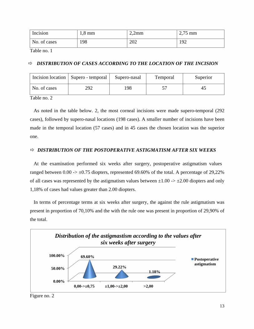
DISTRIBUTION OF THE POSTOPERATIVE ASTIGMATISM AFTER SIX WEEKS
At the examination performed six weeks after surgery, postoperative astigmatism values
ranged between 0.00 -> ±0.75 diopters, represented 69.60% of the total. A percentage of 29,22%
of all cases was represented by the astigmatism values between ±1.00 -> ±2.00 diopters and only
1,18% of cases had values greater than 2.00 diopters.
In terms of percentage terms at six weeks after surgery, the against the rule astigmatism was
present in proportion of 70,10% and the with the rule one was present in proportion of 29,90% of
the total.
Figure no. 2
0.00%
50.00%
100.00%
0,00->±0,75 ±1,00->±2,00 >2,00
69.60%
29.22% 1.18%
Distribution of the astigmastism according to the values after six weeks after surgery
Postoperative astigmatism
14
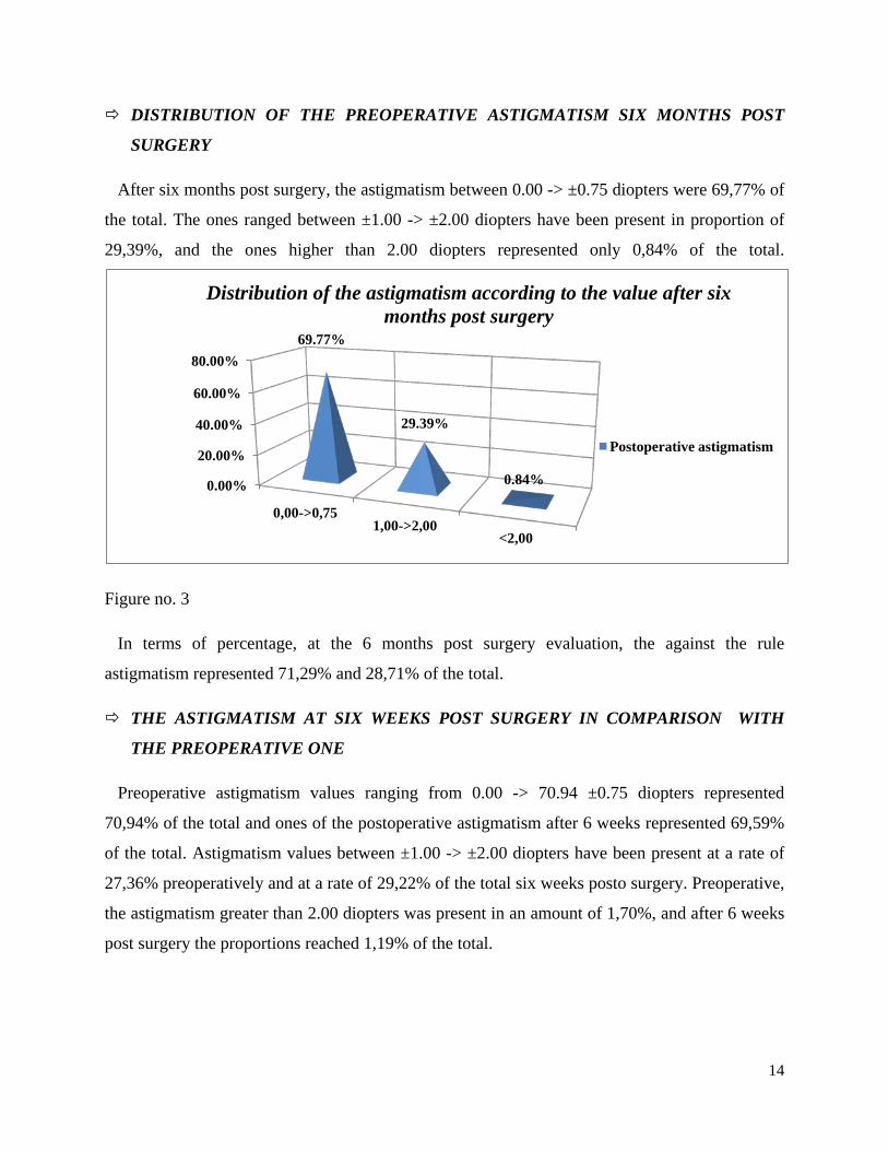
DISTRIBUTION OF THE PREOPERATIVE ASTIGMATISM SIX MONTHS POST
SURGERY
After six months post surgery, the astigmatism between 0.00 -> ±0.75 diopters were 69,77% of
the total. The ones ranged between ±1.00 -> ±2.00 diopters have been present in proportion of
29,39%, and the ones higher than 2.00 diopters represented only 0,84% of the total.
Figure no. 3
In terms of percentage, at the 6 months post surgery evaluation, the against the rule
astigmatism represented 71,29% and 28,71% of the total.
THE ASTIGMATISM AT SIX WEEKS POST SURGERY IN COMPARISON WITH
THE PREOPERATIVE ONE
Preoperative astigmatism values ranging from 0.00 -> 70.94 ±0.75 diopters represented
70,94% of the total and ones of the postoperative astigmatism after 6 weeks represented 69,59%
of the total. Astigmatism values between ±1.00 -> ±2.00 diopters have been present at a rate of
27,36% preoperatively and at a rate of 29,22% of the total six weeks posto surgery. Preoperative,
the astigmatism greater than 2.00 diopters was present in an amount of 1,70%, and after 6 weeks
post surgery the proportions reached 1,19% of the total.
0.00%
20.00%
40.00%
60.00%
80.00%
0,00->0,75 1,00->2,00
<2,00
69.77%
29.39%
0.84%
Distribution of the astigmatism according to the value after six months post surgery
Postoperative astigmatism
15
ASTIGMATISM AT SIX MONTHS POST SURGERY IN COMPARISON WITH THE
PREOPERATIVE ONE
The values ranged between 0.00 -> ±0.75 diopters had a share of 70,94% preoperatively and of
69,76% six months postoperatively (Figure 26). Preoperative, the astigmatism values between
±1.00 -> ±2.00 diopters represented 27,36% of the total and after six months post surgery the
same values represented 29,39% of the total. Astigmatism greater than 2.00 diopters represented
1,70% preoperative and 0,85% six months postoperatively.
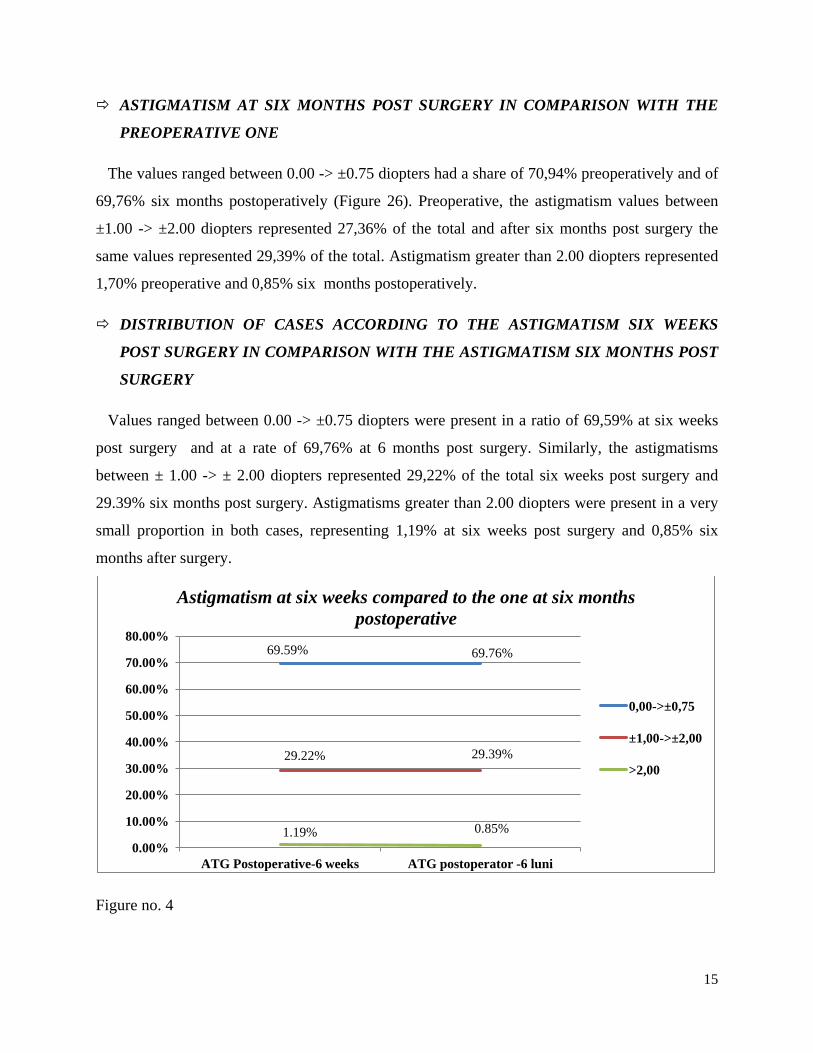
DISTRIBUTION OF CASES ACCORDING TO THE ASTIGMATISM SIX WEEKS
POST SURGERY IN COMPARISON WITH THE ASTIGMATISM SIX MONTHS POST
SURGERY
Values ranged between 0.00 -> ±0.75 diopters were present in a ratio of 69,59% at six weeks
post surgery and at a rate of 69,76% at 6 months post surgery. Similarly, the astigmatisms
between ± 1.00 -> ± 2.00 diopters represented 29,22% of the total six weeks post surgery and
29.39% six months post surgery. Astigmatisms greater than 2.00 diopters were present in a very
small proportion in both cases, representing 1,19% at six weeks post surgery and 0,85% six
months after surgery.
Figure no. 4
69.59% 69.76%
29.22% 29.39%
1.19% 0.85% 0.00%
10.00%
20.00%
30.00%
40.00%
50.00%
60.00%
70.00%
80.00%
ATG Postoperative-6 weeks ATG postoperator -6 luni
Astigmatism at six weeks compared to the one at six months postoperative
0,00->±0,75
±1,00->±2,00
>2,00

16
DISTRIBUTION OF CASES ACCORDING TO THE VISUAL ACUITY AT SIX WEEKS
AFTER SURGERY
The best optical visual acuity without optical correction, six weeks after surgery, was found in
283 cases, representing 52,20% of the total. In the remaining 309 cases the best visual acuity was
obtained with the help of optical correction, these cases representing 47,80% of the total.
DISTRIBUTION OF CASES ACCORDING TO THE VISUAL ACUITY SIX MONTHS
POST SURGERY
At the six months examination postoperative, the best visual acuity with optical correction
was obtained in 296 cases, representing 50% of the total. The other half of the cases the best
visual acuity was obtained with optical correction.
DISTRIBUTION OF CASES ACCORDING TO THE SPHERICAL EQUIVALENT AT
SIX WEEKS POST SURGERY
At six weeks postoperative examination the most spherical equivalent values were between
0.00 -> ±0.75 diopters, representing 63,51% of the total. The values of the spherical equivalent
ranged between ±1.00 -> ±2.00 diopters were present in a ratio of 33,95% of the total. The most
under-represented values of the spherical equivalent were the ones greater than 2,00 diopters,
which were represented in a proportion of 2,54% of the total.
DISTRIBUTION OF CASES ACCORDING TO THE SPHERICAL EQUIVALENT AT
SIX MONTHS POST SURGERY
At the examintion performed at six months postoperative the spherical equivalent values
ranging from 0.00 -> ±0.75 diopters represented 65,71% of the total, those between ±1.00 ->
±2.00 diopters were present in the proportion of 33,44% and those greater than 2,00 diopters
represented 0,85% of the total.
SURGICALLY INDUCED ASTIGMATISM
Applying the ANOVA statistical calculus formula, it was made a detailed analysis of the
preoperative astigmatism in comparison with the postoperative astigmatism, resulting from this
sum, the average and the variation index of the values of the preoperative and postoperative
17
astigmatism, at the two post surgery examinations. For the calculus of the surgically induced
astigmatism (AIC) it was made between the difference between the sum of postoperative
astigmatism values (at six weeks and at six months) and the sum of the preoperative astigmatism
values.
At six weeks post surgery
Surgically induced astigmatism at six weeks post surgery was: 356 – 412,502 = -56,502
At six months post surgery
Surgically induced astigmatism at six months post surgery was of: 378,25- 412,502= - 34,252
ANALYSIS OF THE POSTOPERATIVE ASTIGMATISM IN COMPARISON WITH
THE PREOPERATIVE ON ACCORDING TO THE DIMENSION OF THE CORNEAL
INCISION
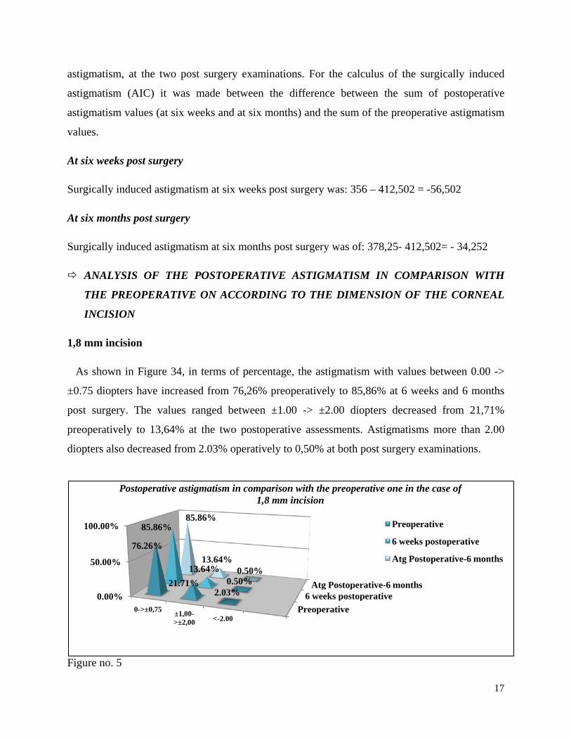
1,8 mm incision
As shown in Figure 34, in terms of percentage, the astigmatism with values between 0.00 ->
±0.75 diopters have increased from 76,26% preoperatively to 85,86% at 6 weeks and 6 months
post surgery. The values ranged between ±1.00 -> ±2.00 diopters decreased from 21,71%
preoperatively to 13,64% at the two postoperative assessments. Astigmatisms more than 2.00
diopters also decreased from 2.03% operatively to 0,50% at both post surgery examinations.
Figure no. 5
Preoperative 6 weeks postoperative
Atg Postoperative-6 months 0.00%
50.00%
100.00%
0->±0,75 ±1,00->±2,00 <-2.00
76.26%
21.71% 2.03%
85.86%
13.64% 0.50%
85.86%
13.64% 0.50%
Postoperative astigmatism in comparison with the preoperative one in the case of 1,8 mm incision
Preoperative
6 weeks postoperative
Atg Postoperative-6 months
18
Surgically induced astigmatism in the 1,8 mm incision was of:
=> At 6 weeks post surgery: 87 - 129= - 42
=> At 6 months post surgery: 97,25 - 129 = -31,75
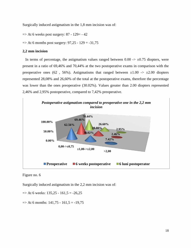
2,2 mm incision
In terms of percentage, the astigmatism values ranged between 0.00 -> ±0.75 diopters, were
present in a ratio of 69,46% and 70,44% at the two postoperative exams in comparison with the
preoperative ones (62 , 56%). Astigmatisms that ranged between ±1.00 -> ±2.00 diopters
represented 28,08% and 26,60% of the total at the postoperative exams, therefore the percentage
was lower than the ones preoperative (30.02%). Values greater than 2.00 diopters represented
2,46% and 2,95% postoperative, compared to 7,42% preoperative.
Figure no. 6
Surgically induced astigmatism in the 2,2 mm incision was of:
=> At 6 weeks: 135,25 - 161,5 = -26,25
=> At 6 months: 141,75 - 161,5 = -19,75
0.00%
50.00%
100.00%
0,00->±0,75 ±1,00->±2,00
>2,00
62.56%
30.02%
7.42%
69.46%
28.08%
2.46%
70.44%
26.60% 2.95%
Postoperative astigmatism compared to preoperative one in the 2,2 mm incision
Preoperative 6 weeks postoperative 6 luni postoperator
19
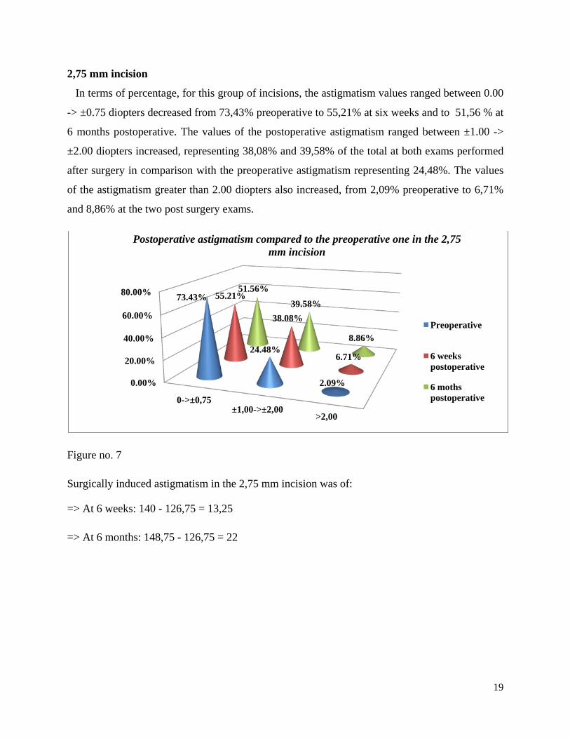
2,75 mm incision
In terms of percentage, for this group of incisions, the astigmatism values ranged between 0.00
-> ±0.75 diopters decreased from 73,43% preoperative to 55,21% at six weeks and to 51,56 % at
6 months postoperative. The values of the postoperative astigmatism ranged between ±1.00 ->
±2.00 diopters increased, representing 38,08% and 39,58% of the total at both exams performed
after surgery in comparison with the preoperative astigmatism representing 24,48%. The values
of the astigmatism greater than 2.00 diopters also increased, from 2,09% preoperative to 6,71%
and 8,86% at the two post surgery exams.
Figure no. 7
Surgically induced astigmatism in the 2,75 mm incision was of:
=> At 6 weeks: 140 - 126,75 = 13,25
=> At 6 months: 148,75 - 126,75 = 22
0.00%
20.00%
40.00%
60.00%
80.00%
0->±0,75 ±1,00->±2,00
>2,00
73.43%
24.48%
2.09%
55.21%
38.08%
6.71%
51.56% 39.58%
8.86%
Postoperative astigmatism compared to the preoperative one in the 2,75 mm incision
Preoperative
6 weeks postoperative
6 moths postoperative
20
ANALYSIS OF THE POSTOPERATIVE ASTIGMATISM COMPARED TO THE
PREOPERATIVE ONE ACCORDING TO THE LOCATION OF THE CORNEAL
INCISION; SURGICALLY INDUCED ASTIGMATISM
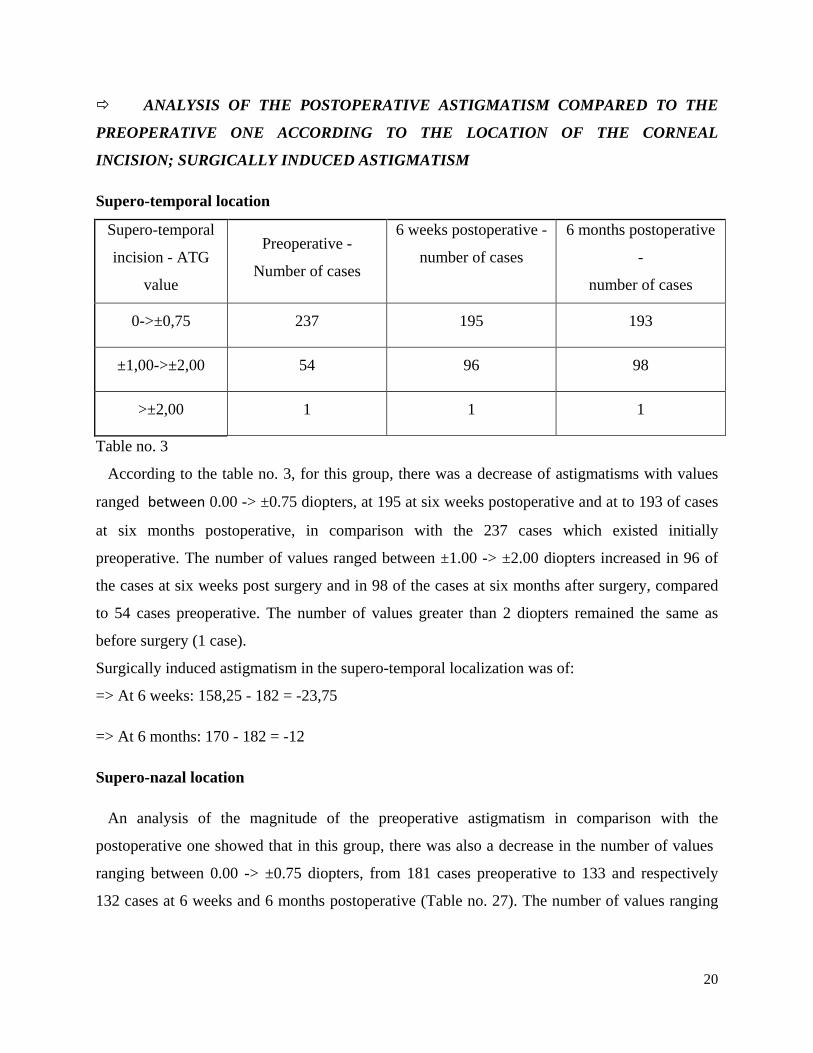
Supero-temporal location
Supero-temporal
incision - ATG
value
Preoperative -
Number of cases
6 weeks postoperative -
number of cases
6 months postoperative
-
number of cases
0->±0,75 237 195 193
±1,00->±2,00 54 96 98
>±2,00 1 1 1
Table no. 3
According to the table no. 3, for this group, there was a decrease of astigmatisms with values
ranged between 0.00 -> ±0.75 diopters, at 195 at six weeks postoperative and at to 193 of cases
at six months postoperative, in comparison with the 237 cases which existed initially
preoperative. The number of values ranged between ±1.00 -> ±2.00 diopters increased in 96 of
the cases at six weeks post surgery and in 98 of the cases at six months after surgery, compared
to 54 cases preoperative. The number of values greater than 2 diopters remained the same as
before surgery (1 case).
Surgically induced astigmatism in the supero-temporal localization was of:
=> At 6 weeks: 158,25 - 182 = -23,75
=> At 6 months: 170 - 182 = -12
Supero-nazal location
An analysis of the magnitude of the preoperative astigmatism in comparison with the
postoperative one showed that in this group, there was also a decrease in the number of values
ranging between 0.00 -> ±0.75 diopters, from 181 cases preoperative to 133 and respectively
132 cases at 6 weeks and 6 months postoperative (Table no. 27). The number of values ranging
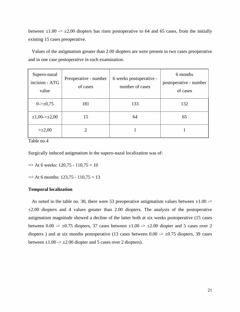
21
between ±1.00 -> ±2.00 diopters has risen postoperative to 64 and 65 cases, from the initially
existing 15 cases preoperative.
Values of the astigmatism greater than 2.00 diopters are were present in two cases preoperative
and in one case postoperative in each examination.
Supero-nazal
incision - ATG
value
Preoperative - number
of cases
6 weeks postoperative -
number of cases
6 months
postoperative - number
of cases
0->±0,75 181 133 132
±1,00->±2,00 15 64 65
>±2,00 2 1 1
Table no.4
Surgically induced astigmatism in the supero-nazal localization was of:
=> At 6 weeks: 120,75 - 110,75 = 10
=> At 6 months: 123,75 - 110,75 = 13
Temporal localization
As noted in the table no. 30, there were 53 preoperative astigmatism values between ±1.00 ->
±2.00 diopters and 4 values greater than 2.00 diopters. The analysis of the postoperative
astigmatism magnitude showed a decline of the latter both at six weeks postoperative (15 cases
between 0.00 -> ±0.75 diopters, 37 cases between ±1.00 -> ±2.00 diopter and 5 cases over 2
diopters ) and at six months postoperative (13 cases between 0.00 -> ±0.75 diopters, 39 cases
between ±1.00 -> ±2.00 diopter and 5 cases over 2 diopters).
22
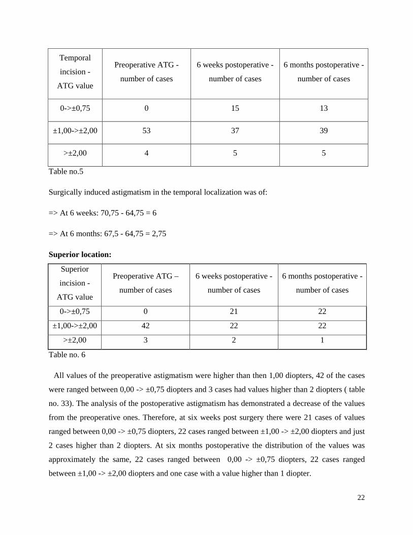
Temporal
incision -
ATG value
Preoperative ATG -
number of cases
6 weeks postoperative -
number of cases
6 months postoperative -
number of cases
0->±0,75 0 15 13
±1,00->±2,00 53 37 39
>±2,00 4 5 5
Table no.5
Surgically induced astigmatism in the temporal localization was of:
=> At 6 weeks: 70,75 - 64,75 = 6
=> At 6 months: 67,5 - 64,75 = 2,75
Superior location:
Superior
incision -
ATG value
Preoperative ATG –
number of cases
6 weeks postoperative -
number of cases
6 months postoperative -
number of cases
0->±0,75 0 21 22
±1,00->±2,00 42 22 22
>±2,00 3 2 1
Table no. 6
All values of the preoperative astigmatism were higher than then 1,00 diopters, 42 of the cases
were ranged between 0,00 -> ±0,75 diopters and 3 cases had values higher than 2 diopters ( table
no. 33). The analysis of the postoperative astigmatism has demonstrated a decrease of the values
from the preoperative ones. Therefore, at six weeks post surgery there were 21 cases of values
ranged between 0,00 -> ±0,75 diopters, 22 cases ranged between ±1,00 -> ±2,00 diopters and just
2 cases higher than 2 diopters. At six months postoperative the distribution of the values was
approximately the same, 22 cases ranged between 0,00 -> ±0,75 diopters, 22 cases ranged
between ±1,00 -> ±2,00 diopters and one case with a value higher than 1 diopter.
23
Surgically induced astigmatism in the temporal localization was of:
=> At 6 weeks postoperative: 34 - 58,5 = -24,5
=> At 6 months postoperative: 40,5 - 58,5 = -18.
ANALYSIS OF POSTOPERATIVE ASTIGMATISM IN COMPARISON WITH THE
PREOPERATIVE ONE ACCORDING TO THE LOCATION AND SIZE OF THE
INCISION; SURGICALLY INDUCED ASTIGMATISM
Supero-temporal location
Expressed in percentages, the number of values ranging between 0.00 ->± 0.75 diopters
represented 85,26% of the total, preoperative, 84,21% and 80,00% at the two postoperative
examinations. The number of values between ±1.00 -> ±2.00 diopters represented 13,68%
preoperative, 15,79% and 20,00% at postoperative examinations . Astigmatisms greater than
2.00 diopters were present only in a very small proportion preoperative (0,76%), missing post
surgery.
Supero-temporal incision of 1,8 mm
The surgically induced astigmatism in the case of the supero-temporal location and incision of
1,8 mm was of:
=> At 6 weeks postoperative: 48,25 - 59,75 = -11,5
=> At 6 months postoperative: 51 - 59,75 = - 8,75
The value curve of the astigmatisms ranging between 0.00 -> ±0.75 diopters has a downward
trajectory from 78,00% preoperative to 59,00% and 58,00% at the two postoperative
examinations. The graphic of the values ranging between ±1.00 -> ±2.00 diopters has a crescent
trajectory from 22,00% preoperative to 41,00% and 42,00% postoperative. Values greater than
2.00 diopters lacked both preoperative and postoperative.
Supero-temporal incision of 2,2 mm
24
The surgically induced astigmatism in the case of the supero-temporal location and incision of
2,2 mm was of:
=> At 6 weeks postoperative: 64,5 - 63,5 = 1
=> At 6 months postoperative: 70,5 - 63,5 = 7
Astigmatism values ranging between 0.00 -> ±0.75 diopters decreased from 82,29%
preoperative to 59,37% at both postoperative examinations. Values ranging between ±1.00 ->
±2.00 diopters increased from 17,71% preoperative to 39,58% at both postoperative
examinations. There were no preoperative values greater than 2.00 diopters, and after the surgery
they were present in an amount of 1.05% at both examinations.
Incizia supero-temporală de 2,75 mm
The surgically induced astigmatism in the case of the supero-temporal location and incision of
2,75 mm was of:
=> At 6 weeks postoperative: 43,75 - 54 = -10,25
=> At 6 months postoperative: 49,5 - 54 = - 4,5
Supero-nazal location
Astigmatism values ranged between 0.00 -> ±0.75 diopters decreased slightly from 94,36%
preoperative to 84,50% and respectively to 80,28% at the two postoperative examinations.
Values ranging between ±1.00 -> ±2.00 diopters have increased at the rate of 5,64% preoperative
to 15,50% and respectively to 19,72% in the two postoperative examinations. There was no case
of astigmatism greater than 2.00 diopters preoperatively or postoperatively. There was no case of
astigmatism greater than 2,00 diopters, preoperative and postoperative.
Supero-nazal incision of 1,8 mm
Surgically induced astigmatism in the supero-nazal location and 1,8 mm incision, was of:
=> At 6 weeks postoperative: 33,75 - 33 = 0,75
=> At 6 months postoperative: 36,5 - 33 = 3,5
25
The number of astigmatisms of small values ranging between 0,00 -> ±0.75 diopters,
decreased from 49 cases before surgery, to 34 and 35 cases at six weeks and six months after
surgery. The number of astigmatisms ranging between ±1.00 -> ±2.00 diopters raised from 9
cases preoperative to 25 and 24 cases ar the two postoperative examinations. The number of
astigmatisms greater than 2.00 diopters has decreased from 2 cases preoperative to one case at
each of the postoperative examinations.
Supero-nazal incision of 2,2 mm
Surgically induced astigmatism in supero-nazal location and incision of 2,2 mm was of:
=> At 6 weeks postoperative: 44,5 - 46 = -1,5
=> At 6 months postoperative: 44,5 – 46 = -1,5
Astigmatisms with values ranging from 0.00 -> ±0.75 diopters decreased from 94,20%
preoperative to 59,42% and 56,52% at the two postoperative examinations. Astigmatism values
ranging between ±1.00 -> ±2.00 diopters increased from 5,80% preoperatively to 40,58% and
43,48% at the two postoperative examinations. Both preoperatively and postoperatively there
was no amount of astigmatism greater than 2.00 diopters.
Supero-nazal incision of 2,2 mm
Surgically induced astigmatism in supero-nazal location and incision of 2,75 mm was of:
=> At 6 weeks postoperative: 41,5 - 34,5 = 7
=> At 6 months postoperative: 44,5 - 34,5 = 10
Temporal location
Astigmatisms ranged between ±1.00 -> ±2.00 diopters decreased from 100% preoperative to
61,53% and respectively 53,84% at the two post surgery examinations. While there was no pre-
operative astigmatism value ranged between 0,00 -> 0.75 diopters, postoperative, these values
were 38,47% at six weeks post surgery and 46,16% at six months post surgery.
Temporal incision of 1,8 mm
26
Surgically induced astigmatism in temporal location and incision of 1,8 mm was of:
=> At 6 weeks postoperative: 8,25 - 15 = - 6,75
=> At 6 months postoperative: 7,5 - 15 = -7,5
If preoperative the astigmatism with values between ±1.00 -> ±2.00 diopters represented
100% of cases, postoperative, in both evaluations, these values were present in a ratio of 66,66%.
Values ranging between 0.00 -> - ±0.75 diopters increased from 0,00% to 23,81% at the teo
postoperative examinations. However, the values greater than 2 diopters have increased from
0.00 preoperative to 9.53% in the postoperative controls.
Temporal incision of 2,2 mm
Surgically induced astigmatism in temporal location and incision of 2,2 mm was of:
=> At 6 weeks postoperative: 27,25 - 22,75 = 4,5
=> At 6 months postoperative: 26,75 - 22,75 = 4
Preoperative astigmatism values ranging between 0.00 -> ±0.75 diopters represented 0.00%,
while at the two postoperative examinations to be present in an amount of 22,72% and
respectively 18, 18% of the total. Values ranging between ±1.00 -> ±2.00 diopters decreased
from 86,36% preoperative to 63,63% and 68,17% at the two postoperative controls. There was
the same proportion of astigmatism with values greater than 2 diopters, both preoperative and
postoperative, at both exmination (13,65%).
Temporal incision of 2,75 mm
Surgically induced astigmatism in temporal location and incision of 2,75 mm was of:
=> At 6 weeks postoperative: 32,25 - 25,75 = 6,5
=> At 6 months postoperative: 32 - 25,75 = 6,25
27
Superior location
Preoperative all of the astigmatism cases in this subgroup had values ranging between ±1.00 ->
±2.00 diopters. At the six weeks postoperative examination, the astigmatisms ranging between
these values represented only 46,66% of the total, the rest were between 0.00 -> ±0.75 diopters.
Superior 1,8 mm incision
Surgically induced astigmatism in superior location and incision of 1,8 mm was of:
=> At 6 weeks postoperative: 3,5 - 17,75 = -14,25
=> At 6 months postoperative: 8 - 17,75 = - 9,75
For this subgroup, astigmatisms ranging between 0.00 -> ±0.75 diopters increased from 0.00%
preoperative to 44,44% and 50,00% at the two postoperative examinations. Astigmatisms
ranging between ±1.00 -> ±2.00 diopters decreased from 88,88% preoperative to 55,56% and
50,00% at postoperative examinations. Values greater than 2.00 diopters decreased from 11,12%
preoperative to 0.00% at postoperative examinations.
Superior de 2,2 mm incision
Surgically induced astigmatism in superior location and incision of 2,2 mm was of:
=> At 6 weeks postoperative: 14,5 - 24,25 = - 9,75
=> At 6 months postoperative: 15 - 24,25 = - 9,25
In terms of percentage, astigmatism values ranged between ±1.00 -> ±2.00 diopters decreased
from 92,85% preoperative to 42,85% and 57,14% at the two postoperative examinations. If there
was no preoperative value between 0.00 -> ±0.75 diopters, postoperative, such values were
found in the proportion of 42,85% and 35,71%. Values greater than 2.00 diopters have been at a
rate of 7,15% before surgery and after six weeks post surgery rose to 14,30%, while at six
months after the surgery, to return to the same number as preoperative (7.15 %).
Superior 2,75 mm incision
28
Surgically induced astigmatism in superior location and incision of 2,75 mm was of:
=> At 6 weeks postoperative: 16 - 18,5 = - 2,5
=> At 6 months postoperative: 15,5 - 18,5 = - 3
II.7. DISCUSSIONS
DISCUSSION OVER DISTRIBUTION OF CASES ACCORDING TO PREOPERATIVE
ASTIGMATISM VALUE
In our study group, low value astigmatism (0.00 -> ±0.75 diopters) was present in proportion
of 70,78% of cases. Preoperative astigmatism values ranging between ±1.00 -> ±2.00 diopters
represented 27,53% of the total. Astigmatism greater than 2.00 diopters was present only in
1,69% of the total. The presence in our study group of the preoperative astigmatism of low value
(≤ ±0.75 diopters) is an aspect which corresponds to the literature data and which indicates that
approximately 15% to 29% of the patients operated of cataract were more than 1.5 diopters,
preexisting astigmatism [72].
DISCUSSION OVER DISTRIBUTION OF CASES ACCORDING TO THE CLINCAL
FORM OF PREOPERATIVE ASTIGMATISM
Regarding the clinical form of preoperative astigmatism there was a slight predominance of
with the rule clinical forms, which represented 54,77% of the total, over those who were against
the rule, which were found in proportion of 42,31% of the total.
DISCUSSIONS OVER THE REPARTITION OF CASES OF POSTOPERATIVE
ASTIGMATISM AT THE SIX WEEKS EXAMINATION IN COMPARISON WITH THE
PREOPERATIVE ONE
The comparative analysis of postoperative astigmatism (at the six weeks examination) with the
preoperative on, there were no significant differences in the distribution of cases of astigmatism
on groups of values, most of them being the low values astigmatisms (0.00 -> ±0.75 diopters)
which postoperative were 69,59% of the total, compared to 70,94% as found preoperatively. In

29
the group of values ranging between ±1.00 -> ±2.00 diopters there was a negligible increase of
their proportion from 27,36% preoperative to 29,22% at the six weeks postoperative
examination.
If preoperative prevailed the according to the rule astigmatisms (57.60%), at 6 weeks
postoperative there was a net change in astigmatism axis orientation, consisting of a clear
majority of those against the rule, which accounted for 70,10% of the total.
DISCUSSIONS OVER THE REPARTITION OF CASES OF POSTOPERATIVE
ASTIGMATISM AT THE SIX MONTHS EXAMINATION IN COMPARISON WITH
THE PREOPERATIVE ONE
At the six months evaluation, it was concluded that in this case there were no significant
changes regarding the magnitude of the values compared to the preoperative one. Thus, there
was only a slight decrease in the percentage of astigmatism with values ranging from 0.00 ->
±0.75 diopters, from 70,94% preoperative to 69,76% postoperative. Values ranging between
±1,00 -> ±2,00 diopters rose slightly from a rate of 27,36% preoperative to 29,39% at six months
postoperative.
At the six months postoperative examination there was a clear predominance of the against the
rule astigmatisms, representing 71,29% of the total.
DISCUSSION OVER THE GLOBAL ANALYSIS OF ASTIGMATISM SIX WEEKS
POST SURGERY IN COMPARISON WITH THE PREOPERATIVE ONE
A global analysis of postoperative astigmatism at the six weeks examination compared with the
preoperative, shows that although the share of low-grade astigmatisms (0.00 -> ±0.75 diopters)
underwent a slight decline from 70,94% preoperative to 69,59% preoperative, the total sum of
the astigmatisms values also showed a decrease from 412,502 preoperative to 356 postoperative.
This decrease in the amount of postoperative astigmatism values at this range can be explained
by the fact that the lowest average of astigmatisms values was recorded postoperative
respectively 0,601 compared to 0,696 preoperative.
30
DISCUSSION OVER GLOBAL ANALYSIS OF ASTIGMATISM SIX MONTHS POST
SURGERY IN COMPARISON WITH THE PREOPERATIVE ONE
An analysis of postoperative astigmatism at six months compared with the preoperative one,
shows the same issues, consisting in the decrease of the sum of values from 412,502 preoperative
to 378,25 after surgery, although in this case there was a slight decrease in low-grade
astigmatisms (between 0.00 -> ±0.75 diopters) from 70,94% preoperative to 69,76%
postoperative. Values ranging between ±1.00 -> ±2.00 diopters have increased slightly from
27,36% preoperative to 29,39% after the operation, while values greater than 2.00 diopters
decreased from 1,70% preoperative to 0,85% after surgery. Decreasing the amount of
postoperative astigmatism values at six months compared with the amount of preoperative
values, might be due, as in the case mentioned above, to the decrease of the average of the
postoperative values (0,638) than the preoperative ones (0,696).
DISCUSSION OVER THE GLOBAL ANALYSIS OF THE SURGICAL INDUCED
ASTIGMATISM
At the six weeks postoperative examination, the overall amount of surgically induced
astigmatism was negative (-56,502) this being the result of the difference between the sum of
astigmatism values at six weeks postoperative (356) and the amount of preoperative values
(412,502).
At the six months postoperative examination there was still a negative value (-34,252), but
slightly higher than the one found at six weeks (-56,502), explained by a slight increase in the
amount of astigmatism values in this assessment compared to the previous (378,25 at six months
compared to 356 at six weeks postoperatively).
The negative value of surgically induced astigmatism in both postoperative examinations can
be translated into a favorable refractive outcome, respectively the lack of this type of
astigmatism at a global level. This aspect was due, as mentioned above, to the stability of the
refractive cornea offered by these small corneal incisions.
31
SURGICALLY INDUCED ASTIGMATISM ACCORDING TO THE DIMENSION OF
THE INCISION
The analysis of surgically induced astigmatism in the three study groups, based on the corneal
incision size, has shown that this type of astigmatism is present only in the 2.75 mm incision
group, missing from the 1.8 mm and 2.2 mm incision groups. There were no major refractive
differences between the 2.2 mm and that of 1.8 mm incisions groups. The magnitude of
surgically induced astigmatism in the above case (incision of 2.75 mm) is small and has not
shown significant changes at the six months examination compared to those found at six weeks
post surgery. In this respect postoperative refractive change was 13.25 at six weeks postoperative
and of 22 at six months postoperative.
SURGICALLY INDUCED ASTIGMATISM ACCORDING TO THE LOCATION OF
THE INCISION
General analysis of surgically induced astigmatism according to the corneal incision location
shows that this type of astigmatism was not revealed in the subgroup of supero-temporal and
superior incisions, but was present in the subgroup of supero-nasal and temporal incisions . From
the data presented above also results that although it was present in the two locations, the
amplitude of surgically induced astigmatism was very low and there were no significant changes
from at the six months examination from the one made at six weeks post surgery.
SURGICALLY INDUCED ASTIGMATISM ACCORDING TO THE LOCATION AND
SIZE OF THE INCISION
Detailed analysis of surgically induced astigmatism depending on the location and size of
peripheral corneal incision showed that the results were varied. Thus, if the case of supero-
temporal location, the corneal incision size was not the main determinant in the development of
surgically induced astigmatism, meaning that this type of astigmatism was absent in the case of
1.8 mm and 2.75 mm incisions, but was present in the case of 2.2 mm incisions, but of very low
amplitude.
32
II.8. PROPHYLAXIS AND TREATMENT OF SURGICALLY INDUCED
ASTIGMATISM AND OF POSTOPERATIVE ASTIGMATISM
- reducing the size of the corneal incision
- making the incision on the more refringent meridian
- making the incisions in clear cornea on opposite meridians
- relaxing limbic incisions technique
- relaxing peripheral corneal incisions, single or in pairs
- correction of intraocular lenses - toric implantation of artificial lens
- postoperative correction of refractive surgery techniques (PRK, LASIK).
II.9. EXAMINATION PROTOCOL AND SURGICAL TREATMENT OF THE PATIENT
IN ORDER TO FIGHT THE SURGICALLY INDUCED ASTIGMATISM
a. Usual preoperative clinical examination:
- determining the distance visual acuity, corrected and uncorrected
- contrast sensibility
- biomicroscopy of the anterior segment
- measuring the number of endothelial cells
- anterior chamber depth measurement
- aplanotonometry
- direct and indirect oftalmoscopy
b. Determination of the patient's preoperative refractive status
- determination of ocular refraction with the help of the Humphrey autorefractometer in the
cases where the evolutionary stage of the cataract is not so advanced and allows this
examination
- keratometry
- corneal topography
c. Ocular biometry and calculation of artificial lens implant (IOL target)
33
d. The choice of surgical incision in terms of size, location in a particular anatomical area
(scleral, corneal Language) and its configuration
e. Choice of type of artificial lens to be implanted in terms of its quality, material, implant design
(spherical, aspherical, design of haptics), optical party size.
f. Preoperative astigmatism treatment
II.10. CONCLUSIONS
* The postoperative astigmatism magnitude map is very slightly different from that of the
preoperative astigmatism, in both cases the most present ones being the ones ranging between
0.00 -> -0.75 diopters
* Regarding the clinical form, postoperative there was a shift of astigmatism axis, meaning that
if preoperative there was a slight predominance of with the rules astigmatism, after surgery there
was a clear predominance of against the rule astigmatisms.
* In the cataract surgery, the practice of small incisions in clear cornea minimizes corneal
damage and postoperative complications that can compromise the functional outcome after
surgery, leading to dissatisfaction of both the surgeon and the patient.
* Such incisions also reduce the time required for visual rehabilitation, restore patient
independence, enabling faster resumption of their normal activity.
34
III. BIBLIOGRAPHY
1. Zeiske J.D, Gipson I.K, Agents that affect corneal wound healing: modulation of structure and
function. In: Principles and Practice of Ophthalmology. 2 ed. Albert DM, Jacobiec FA, Editors.
Philadelphia: WB Saunders Co. 2000: 364 - 372.
2. Klatte DH, Kurpakus MA, Grelling KA, Jones JCR. Immunochemical characterization of
three components of the hemidesmosome and their expression in cultured ephithelias cells. J Cell
Biol. 1989; 109: 3377 - 90.
3. Okada Y, Saika S, Shirai K, Hashizume N, Yamanaka D, Ohnishi Y, Senba E. Disappearance
of desmosomal components in rat corneal ephitelium during wound healing. Ophthalmologica.
2001; 215: 61- 65.
4. Gipson IK, Spurr-Michaud SJ, Tisdale AS. Anchoring fibrils form a complex network in
human and rabbit cornea. Invest Ophthalmol Vis Sci. 1987; 26: 212 - 20.
5. Agrawal V.B, Tsai R.J. Corneal epithelial wound healing. Current ophthalmol. 2003 (51); 1: 5
- 15.
6. Hogan MJ, Alvarado JA, Weddel JE. Histology of the human eye. In: Hogan MJ, Alvarado
JA, Weddel JE, editors. Philadelphia: Saunders Co. 1971, pp 112 - 126.
7. Kinoshita S, Adachi W, Sotozono C, Nishida K, Yokoi N, Quantock AJ, et al. Characteristics
of human ocular surface epithelium. Prog Retina Eye Res. 2001; 20: 639 - 73.
8. Tervo T, van Setten G.B, Beuerman RW, Tervo K, Virtanen I, Tarkkanen A. Appearance of
immunohistochemically detectable cellular fibronectin and tenascin in the experimental rabbit
keratectomy wound. Invest Ophthalmol Vis Sci. 1989; 30:149.
9. Panjwani N, Michalopoulos G, Song J, Zaidi TS, Yogeeswaran G, Baum J. Neutral
glycolipids of migrating and nonmigrating rabbit corneal epithelium in organ and cell culture.
Invest Ophthalmol Vis Sci. 1990; 31: 689 - 695.
10. Eraslan M, Toker E. Mechanisms of corneal wound healing and its modulation following
refractive surgery. Marmara Med. J. 2009; 22: 169-178.
35
11. Fini ME. Keratocyte and fibroblast phenotypes in the repairing cornea. Prog Retin Eye Res.
1999; 18: 529 - 551.
12. Jester JV, Petrol WM, Canavagh HD. Corneal stromal wound healing in refractive surgery:
the role of myofibroblasts. Prog Retin Eye Res. 1999; 18: 311 - 356.
13. Binder PS, Waring GO, Arrowsmith PN, Wang C. Histopathology of traumatic rupture of the
cornea after radiar keratotomy. Arch Ophthalmol. 1988; 106:1584 - 1590.
14. Rigal D. și colab. L´epithelium cornean – Phisiopathologie - La cicatrisation. Ed Masson.
1993; 89 (115): 354 - 357.
15. Sobottka Ventura AC, Walti R, Bohnke M. Corneal thickness and endothelial density before
and after cataract surgery. Br J Ophthalmol. 2001; 85: 18 - 20.
16. Werb Z, Alexander CM, Alder RR. Expression and function of matrix mettaloproteinases in
developement. Matrix Suppl. 1992; 1: 337-343.
17. Hogan MJ, Alvarado JA, Weddel JE. Histology of the human eye. In: Hogan MJ, Alvarado
JA, Weddel JE, editors. Philadelphia: Saunders Co. 1971, pp 112 - 126.
18.Wilson SL, El Haj AJ, Yang Y. Control of scar tissue formation in the cornea: strategies in
clinical and corneal tissue engineering. J. Funct. Biomater. 2012; 3: 642 - 687.
19. Melles G.R.J, Binder P.S, Beekhuis W.H, Wijdh R.H.J, Moore M.N, Anderson J.A,
SundarRaj N. Scar tissue orientation in unsutured and sutured corneal wound healing. BJO.
1995; 79:760 -765.
20. Binder PS, Wickham MG, Zavala EY, Akers PH. Corneal anatomy and wound healing. In:
Trans New Orleans Acad Ophthalmol St Louis; Mosby. 1980: 1- 35.
21.Cho Y.K, Kim M.S. Preoperative modulating factors on astigmatism in sutured cataract
surgery. Korean J Ophthalmol. 2009; 23(4): 240 - 248.
22. Kim I-T, Park H-YL, Kim H-S. Postoperative astigmatic outcomes based on the haptic axis
of intraocular lenses inserted in cataract surgery. Korean J Ophthalmol. 2011 Febr; 25(1): 22-28.
36
23.Carvahlo MJ, Suzuki SH, Freitas Ll, Branco BC, Lima Al. Limbal relaxing incisions to
correct corneal astigmatism during phacoemulsification.. J Refract Surg. 2007; 23(5):4 99 - 504.
24. Amesbury EC, Miller KM. Correction of astigmatism at the time of cataract surgery. Curr.
Opin.Ophthalmol. 2009; 20: 19 - 24.
25. Basti S, Vasavada A, Thomas R, Padhmanabhan P. Extracapsular cataract extraction:
surgical tehniques. India. 1993; 41(4): 195 - 210.
26.Van Rij G, Waring GO. Changes in corneal curvature induced by sutures and incisions. Am J
Ophthalmol. 1984; 98:773 - 83.
27. Biro Z. Bazele facoemulsificării. Ed. Tudomani. Pecs. 2002; 3:39 - 42.
28. Ernest P, Hill W, Potvin R. Minimizing surgically induced astigmatism at time of cataract
surgery using a square posterior limbal incision. Journal of Ophthalmology. 2011;2
(10):1155/2011/233170.
29. Hoffer KJ. Biometry of 7.500 cataractous eyes. Am J Ophthalmol. 1980; 90: 360 - 368.