Lower Leg and Ankle Injuries -...
17
Lower Leg and Ankle Injuries Jon DeBord, PT, MS, ATC, SCS Rehab Summit 2008 Ankle Injuries • Most common injury in sports – 38-45% of all sports- related injuries – 86% are sprains • Mechanism – Forceful inversion with plantarflexion – Eversion – Hyper-plantarflexion Ankle Anatomy • Bones – Tibia – Fibula • Tibio-Fibular Syndesmosis • Lateral Ligaments – Anterior talo-fibular – Calcaneo-fibular – Posterior talo-fibular • Medial Ligament – Deltoid Ligament
Transcript of Lower Leg and Ankle Injuries -...

1
Lower Leg and Ankle Injuries
Jon DeBord, PT, MS, ATC, SCS
Rehab Summit 2008
Ankle Injuries
• Most common injury in sports– 38-45% of all sports-
related injuries– 86% are sprains
• Mechanism– Forceful inversion with
plantarflexion– Eversion– Hyper-plantarflexion
Ankle Anatomy• Bones
– Tibia– Fibula
• Tibio-Fibular Syndesmosis
• Lateral Ligaments– Anterior talo-fibular– Calcaneo-fibular– Posterior talo-fibular
• Medial Ligament– Deltoid Ligament

2
Ankle Sprains
• Inversion Ankle Sprain
• Eversion Ankle Sprain
• Syndesmosis Injury “High Ankle Sprain”
Ankle Sprains
• Grade I– Mild Sprain – Mild tearing of ligament– Mild point tenderness– Little or no swelling – Little or no limitation in motion– Tendency to recurrence– Little or no disability– Quick return to play
Ankle Sprains
• Grade II– Moderate Sprain – Partial tearing of ligament– Moderate point tenderness– Moderate Swelling– Decreased ROM– Local Bruising– Persistent Instability with high recurrence– Return to play average 7-10 days

3
Ankle Sprains
• Grade III– Severe Sprain – Complete rupture of ligament – Severe pain and disability– Severely limited ROM– Possible deformity– Severe swelling– Chronic instability– Sometimes season-ending
Inversion Ankle Sprain• Most common• Mechanism
– Inversion or varus tilt– Forced plantarflexion
• Symptoms– Pain over lateral malleolus– Inability to bear weight
• Signs– Swelling laterally– Bruising laterally or into
foot– Tenderness over lateral
malleolus
Inversion Ankle Sprain• Treatment
– Immediately – R.I.C.E.– Refer to physician– Weight bearing vs. NWB
• Rehabilitation– Decrease swelling/pain– Increase ROM/Strength– Restore proprioception
• Return to play– When able to perform all
agility needed for specific sport
– Bracing/Taping

4
Eversion Ankle Sprain• Mechanism
– Eversion or valgus tilt– Forced external
rotation• Symptoms
– Pain on inside of ankle• Signs
– Tenderness anterior lower leg and medially
– Swelling medially
Eversion Ankle Sprain
• Relatively rare
• Usually involves syndesmosis injury
• Occasionally involves fibula fracture
• Treatment– Protected mobilization– Cast immobilization
• Rehabilitation– Decrease
swelling/pain– Increase
strength/ROM• Return to play
– When able to perform all agility needed for specific sport
Syndesmosis Injury
• High Ankle Sprain• Usually associated
with fracture• Mechanism
– Athlete stops and is pushed back on planted foot
• Signs/Symptoms– Tenderness over distal
tibiofibular ligaments– Tenderness over Deltoid
ligament– Pain with squeeze at mid-
calf
• Treatment – Depends on X-Rays / Laxity Testing– Instability– No instability
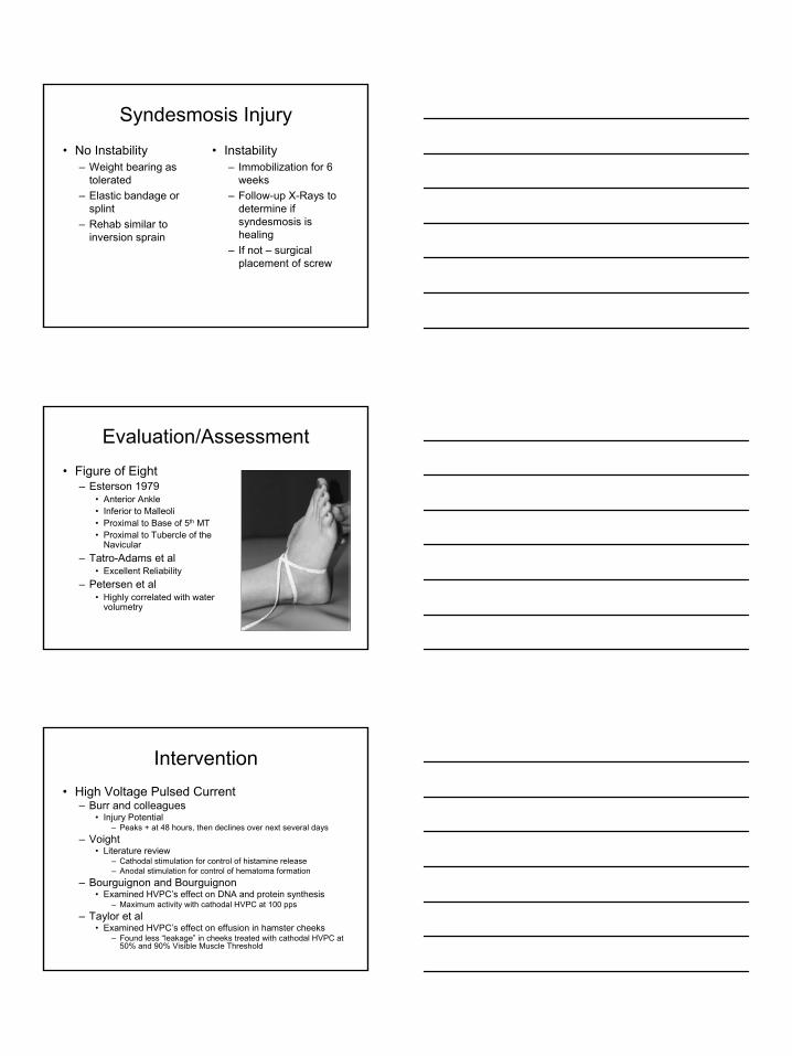
5
Syndesmosis Injury
• No Instability– Weight bearing as
tolerated– Elastic bandage or
splint– Rehab similar to
inversion sprain
• Instability– Immobilization for 6
weeks– Follow-up X-Rays to
determine if syndesmosis is healing
– If not – surgical placement of screw
Evaluation/Assessment• Figure of Eight
– Esterson 1979• Anterior Ankle• Inferior to Malleoli• Proximal to Base of 5th MT• Proximal to Tubercle of the
Navicular– Tatro-Adams et al
• Excellent Reliability– Petersen et al
• Highly correlated with water volumetry
Intervention• High Voltage Pulsed Current
– Burr and colleagues• Injury Potential
– Peaks + at 48 hours, then declines over next several days– Voight
• Literature review– Cathodal stimulation for control of histamine release– Anodal stimulation for control of hematoma formation
– Bourguignon and Bourguignon• Examined HVPC’s effect on DNA and protein synthesis
– Maximum activity with cathodal HVPC at 100 pps– Taylor et al
• Examined HVPC’s effect on effusion in hamster cheeks– Found less “leakage” in cheeks treated with cathodal HVPC at
50% and 90% Visible Muscle Threshold

6
Intervention
• High-Voltage Pulsed Current– 2 Electrodes– Negative polarity– 100pps– Sub-muscular
contraction– 20 minutes
Intervention• Ankle Disk Training
– McGuine and colleagues• Increased postural sway
strongly predictive of ankle sprain
– Wester et al• 12-week training program
– Fewer complaints of re-injury and chronic instability
– Matsusaka and others• Added tactile input• Control Group: normal
balance at 8 weeks• Taped Group: normal
balance at 6 weeks
Intervention
• Ankle Disk Training
– Initiated Day 2 post-injury
– Progressed as tolerated• Bilateral stance• Bilateral support with toes• Unilateral support• Ball height

7
Intervention
• Perturbation Training– Bilateral stance– Bilateral support with
toes– Unilateral stance– Unilateral stance on
compliant surface
Ankle Bracing• AirCast• Positives
– Provides compression and immobilization
– Good for immediately post-injury
• Negatives– Compression not uniform– Bulky – compliance– Does not allow normal
ankle ROM/movement
Ankle Bracing
• ASO Ankle Brace• Positives
– Lace-up/figure-8 straps
– Good compression– Lightweight– Fits in all shoes
• Negatives– Needs to be replaced
annually

8
Lower Leg Anatomy• Bones
– Tibia– Fibula
• Muscles– Tib. Anterior/Posterior– Toe Extensors– Gastrocnemius/Soleus– Toe Flexors
• Tendons• Ligaments/Pseudo-
ligaments
Lower Leg Injuries
• Contusions– Compartment Syndromes
• Achilles Tendon Injuries– Tendonitis– Rupture
• Medial Tibial Stress Syndrome• Fractures
– Stress Fractures– Traumatic Fractures
• Nerve Injuries
Contusions
• Highly exposed to direct trauma
• Most often over the anterior leg (shin)
• Abrasions and Lacerations
• Must rule-out bone injury
• Treat with R.I.C.E.• Complications:
– Compartment Syndrome
– Peroneal Nerve Damage

9
Compartment Syndrome
• Caused by swelling within one of the 4 compartments
• Usually due to trauma• Most commonly
anterior compartment• Increases pressure
on vascular structures and nerves
Compartment Syndrome
• Symptoms– History of trauma– Throbbing/aching pain– Red, distended skin– Increased tissue temp.– Foot Drop– Pain with passive
motion
• Treatment– R.I.C.E.– Immediate referral to
physician• May require surgery to
release pressure
Exertional Compartment Syndrome
• Symptoms similar to Compartment Syndrome, but exercises induced
• Symptoms usually bilateral
• Relief of pain after exercises is stopped
• Treatment– R.I.C.E.– Referral to physician
• Surgery is likely
10
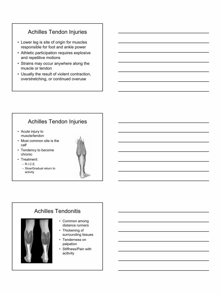
Achilles Tendon Injuries
• Lower leg is site of origin for muscles responsible for foot and ankle power
• Athletic participation requires explosive and repetitive motions
• Strains may occur anywhere along the muscle or tendon
• Usually the result of violent contraction, overstretching, or continued overuse
Achilles Tendon Injuries
• Acute injury to muscle/tendon
• Most common site is the calf
• Tendency to become chronic
• Treatment:– R.I.C.E.– Slow/Gradual return to
activity
Achilles Tendonitis
• Common among distance runners
• Thickening of surrounding tissues
• Tenderness on palpation
• Stiffness/Pain with acitivity

11
Achilles Tendonitis
• Symptoms– Generalized
pain/stiffness around tendon
– Uphill running is worse– Swelling– Decreased ankle ROM– Tendon may feel warm– Crepitus
Achilles Tendonitis
• Treatment– R.I.C.E– Cho-Pat Strap– Proper shoes– Orthotics– Physical Therapy– Stretching– Strengthening
Achilles Tendon Stretching
Gastroc Stretch (knee straight) Soleus Stretch (knee flexed)

12
Achilles Tendon Rupture• Atraumatic injury• Relatively rare• Typically in men age 30-
40• Causes
– Decreased tissue blood supply
– Anatomic limb alignment– Excessive or
uncoordinated muscle contraction
– More likely after period of inactivity
Achilles Tendon Rupture
• Surgical Treatment– Must wear cast/brace for 6-8 weeks– Toe-touch weight bearing– Return to activity 4-6 months after surgery
• Non-Surgical Treatment– Higher risk of re-rupture within first 6 months– Cast/Bracing for 12 weeks or more– Return to activity 6 months or longer
Medial Tibial Stress Syndrome
• “Shin Splints”• Most often occur early
in training program• Inflammation of
tendon and fascia• Disagreement over
exact cause

13
Medial Tibial Stress Syndrome
• Pain anywhere along the leg• Usually limited to muscular
areas anteriorly• Causes
– Muscle inflexibility– Fallen arch– Pronated foot– Ill-fitting footwear– Training techniques– Playing surfaces
Medial Tibial Stress Syndrome
• Treatment– No one best treatment– R.I.C.E.– Correction of possible
causes– Strengthening– Stretching– Taping– Slow return to activity
• Prevention– Gradual progression of
training program– Properly fitted shoes– Identify over-pronation– Strengthening and
flexibility program• Ankle ROM• Ankle strengthening
– Weight bearing– Non-weight bearing
Shin Splints - Taping
• Arch Support– Helps raise arch– Corrects over-
pronation

14
Shin Splints - Taping
• Tibial Taping– Eases symptoms– Provides extra support
to inflamed fascia
Fractures
• Tibia and fibula both susceptible to fracture
• Fibula injured more often– Direct blow to outside of leg– Stress fractures
• Tibia fractures easily identified– Direct blow– Twisting force– Stress fractures
Stress Fractures
• Develop due to abnormal or unusual repetitive stress applied to the bone
• Difficult to differentiate shin splints vs. stress fracture
• More common in people with high arches
• Also more common with over-pronation
• Symptoms– Point tenderness– Swelling– Gradual onset

15
Stress Fractures
34
2420
146
0
10
20
30
40
Perc
ent
Tibia Fibula Metatarsals Femur Pelvis
Bones Involved with Stress Fractures
Stress Fractures
• Causes– Change in shoes– Change in running
surface– Change in distance– Changes in exercise
program– Surgery on other side
• Treatment– Rest– Splinting vs. casting– Non-weight bearing
vs. weight bearing– Gradual return to
activity at 4-6 weeks
Traumatic Fractures
• To ER immediately– Severe local pain– Inability to bear weight– Deformity
• Apply ice and splint extremity in current position
• DO NOT try to straighten deformed leg
16
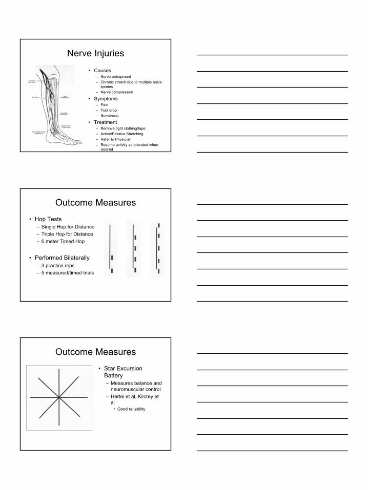
Nerve Injuries
• Causes– Nerve entrapment– Chronic stretch due to multiple ankle
sprains– Nerve compression
• Symptoms– Pain– Foot drop– Numbness
• Treatment– Remove tight clothing/tape– Active/Passive Stretching– Refer to Physician– Resume activity as tolerated when
cleared
Outcome Measures
• Hop Tests– Single Hop for Distance– Triple Hop for Distance– 6 meter Timed Hop
• Performed Bilaterally– 3 practice reps– 5 measured/timed trials
Outcome Measures
• Star Excursion Battery– Measures balance and
neuromuscular control– Hertel et al, Kinzey et
al• Good reliability

17
Outcome Measures
• Star Excursion Battery– Performed bilaterally– 3 practice trials– 5 measured trials