Lower GI Bleeding
21
Lower GI Bleeding Dr. M. Ghanem
-
Upload
kristin-elpida -
Category
Documents
-
view
45 -
download
7
description
Lower GI Bleeding. Dr. M. Ghanem. A less common reason for hospitalization 95% from the colon Etiology usually age related. Presentation. Hematochezia (bright red blood, clots) May present with melena Usually less severe than UGI. Etiology. Diagnosis. Diagnosis. Colonoscopy. - PowerPoint PPT Presentation
Transcript of Lower GI Bleeding
Lower GI Bleeding
Dr. M. Ghanem
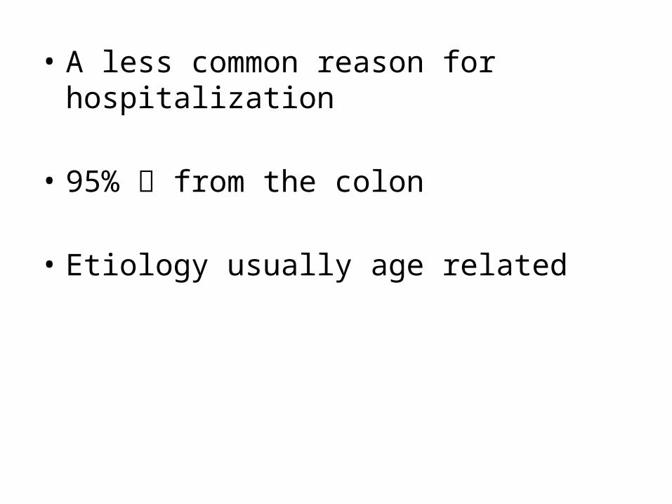
• A less common reason for hospitalization
• 95% from the colon
• Etiology usually age related
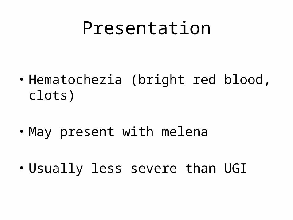
Presentation
• Hematochezia (bright red blood, clots)
• May present with melena
• Usually less severe than UGI
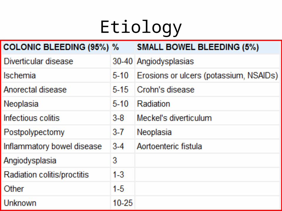
Etiology

Diagnosis
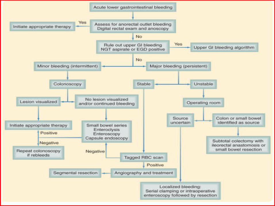
Diagnosis
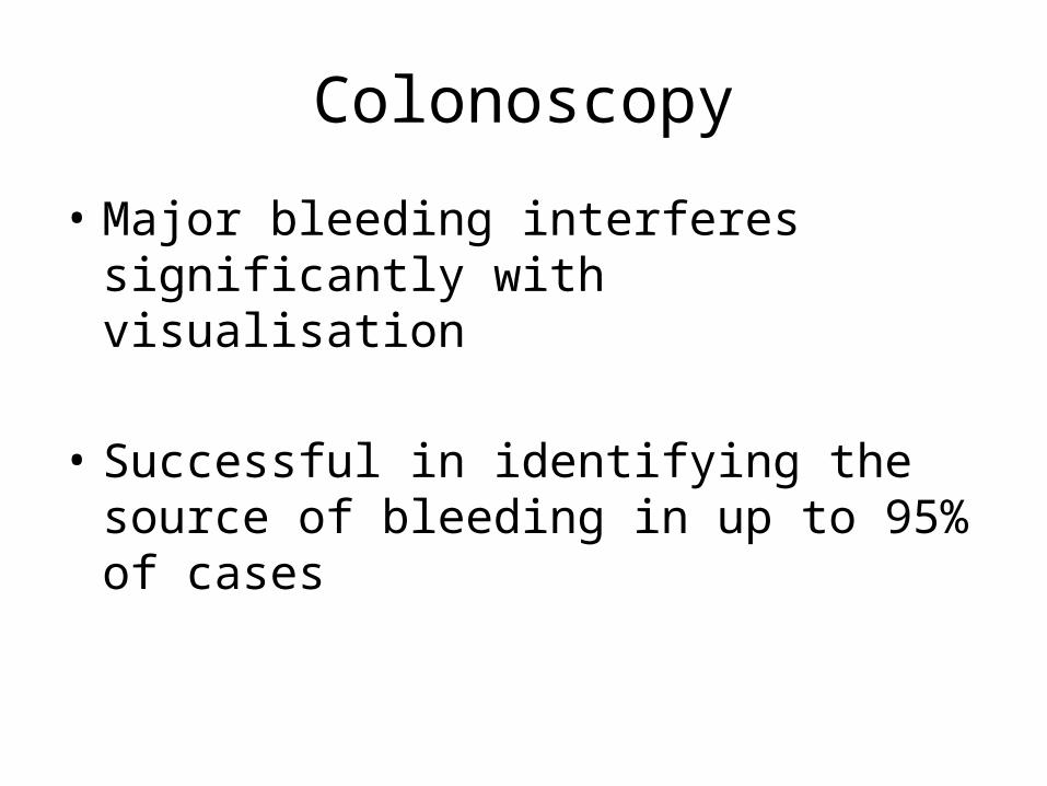
Colonoscopy
• Major bleeding interferes significantly with visualisation
• Successful in identifying the source of bleeding in up to 95% of cases
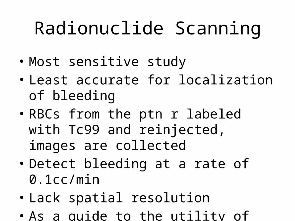
Radionuclide Scanning
• Most sensitive study• Least accurate for localization of bleeding• RBCs from the ptn r labeled with Tc99 and
reinjected, images are collected• Detect bleeding at a rate of 0.1cc/min• Lack spatial resolution• As a guide to the utility of angio
Angiography
• Detect hemorrhage at a rate of 0.5-1 cc/min• Therapeutic advantage (vasopressin inj.,
embolization)• Invasive• Complications include:– Hematoma– Areterial thrombosis– Contrast rxns– ARF
Video Capsule
• A video camera is swallowed
• Identifies the source of bleeding is 90% of cases
• Good in stable ptns
• Doesn’t have a treatment option
Push enteroscopy
• Can reach 70 cms from the ligament of treitz
• Successful in 40% of cases
• Video capsule is usually preferred
Diveticulosis
• The most common cause of LGIB (55%)• >75% stop bleeding spontanously• 10% rebleed within 1 year• 50% rebleed within 10 years• >50% of bleeds from Rt colon
Diveticulosis
• Diagnosis by colonscopy
• Tx by injecting epinephrine, cautery of clip with colonscopy
• Angio carries risk of ischemia
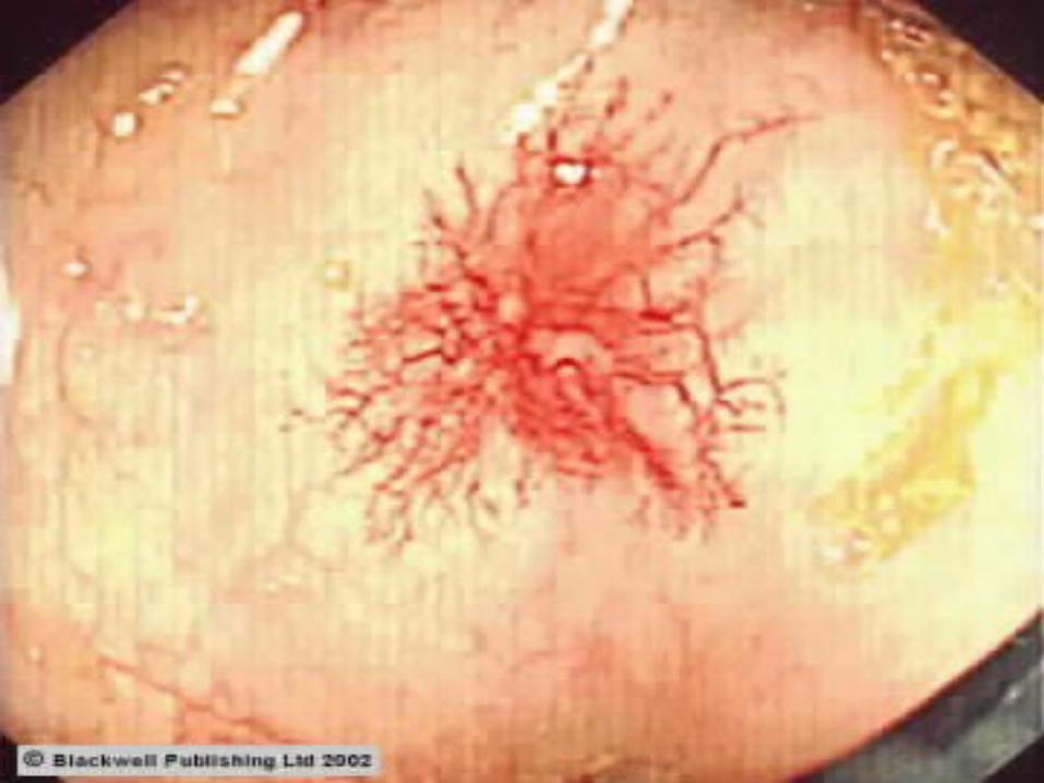
Angiodysplasia
• About 40% of LGIB• Acquired degenerative lesions secondary to
progressive dilation of normal blood vessels within the submucosa of the intestine
• Can occur anywhere in the bowel, cecum most common site
• Diagnosis by colonoscopy, angio • Appear as red stellate lesions with a rim of pale
mucosa
Angiodysplasia
• Tx with sclerotherapy or cautery (colonscopy), embolization by angio
• Rebleeding consider surgery
Neoplasia
• Uncommon cause of significant LGIB
• The first to rule out!!!
• Polyps
• Diagnosis by colonscopy
• Tx depends whether it’s a tumor or polyp, pathology, etc…
Anorectal Disease
• Include internal hemorrhoids, fissures and colorectal neoplasia
• 5-10% of LGIB• Bright-red blood per rectum that is seen in the
toilet bowl and on the toilet paper• Internal hemorrhoids: painless, a lump that
reduces spontanously or by ptn• Fissures: painful
Anorectal Disease
• Malignancy needs to be ruled out before assuming that the bleeding is due to hemorrhoids
• Tx of hemorrhoids: rubber band ligation, sclerotherapy, coagulatiom, surgery
• Tx of fissures: stool softeners, NG, Ca channel blockers
Meckels Diverticulum
• A true diverticulum
• Remnant of the omphalomesentric duct
• Caused by acid production by ectopic musocsa
• Diagnosis by nuclear imaging, angio
• Tx is surgical