Lower Gi Bleed Ng
40
LOWER GI BLEEDNG
-
Upload
bocahbritpop -
Category
Documents
-
view
37 -
download
3
description
sa
Transcript of Lower Gi Bleed Ng
LOWER GI BLEEDNG
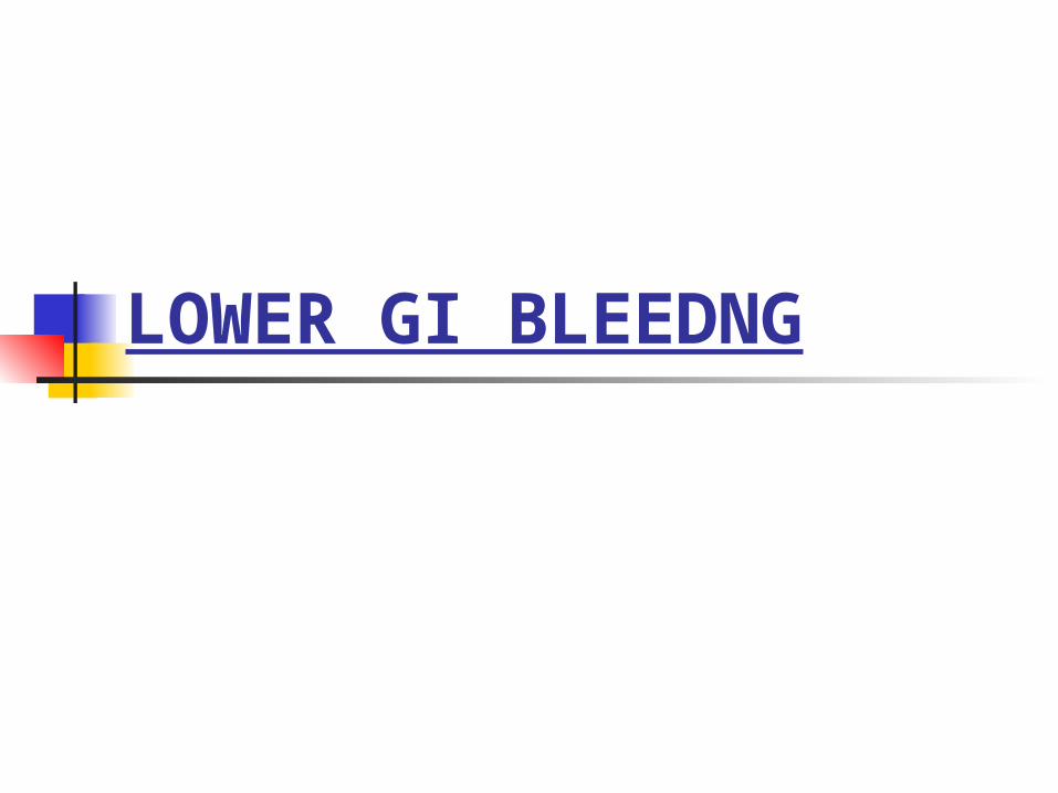
CLINICAL PRESENTATION A 68 yrs old presents to the ER with BRBPR. He has several
episodes, beginning on the evening before presentation. He describes the bleeding as profuse and filling the toilet, he felt
light headed and almost passed out while sitting on the toilet. PMHx: htn Meds: amlodipine P.Ex: B.P. =86/42mm hg, pulse = 134/min,
pt. is orthostatic. Abdominal examination reveals slight
abdominal distention with hyperactive bowel sounds. Rectal exam shows gross blood
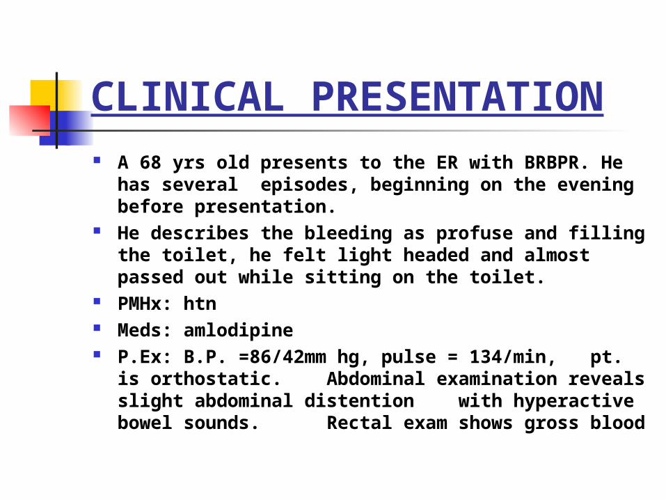
Wbc-10 Hct-28 % Plt-180 Lytes – wnl Coagulation profile wnl LFTs wnl
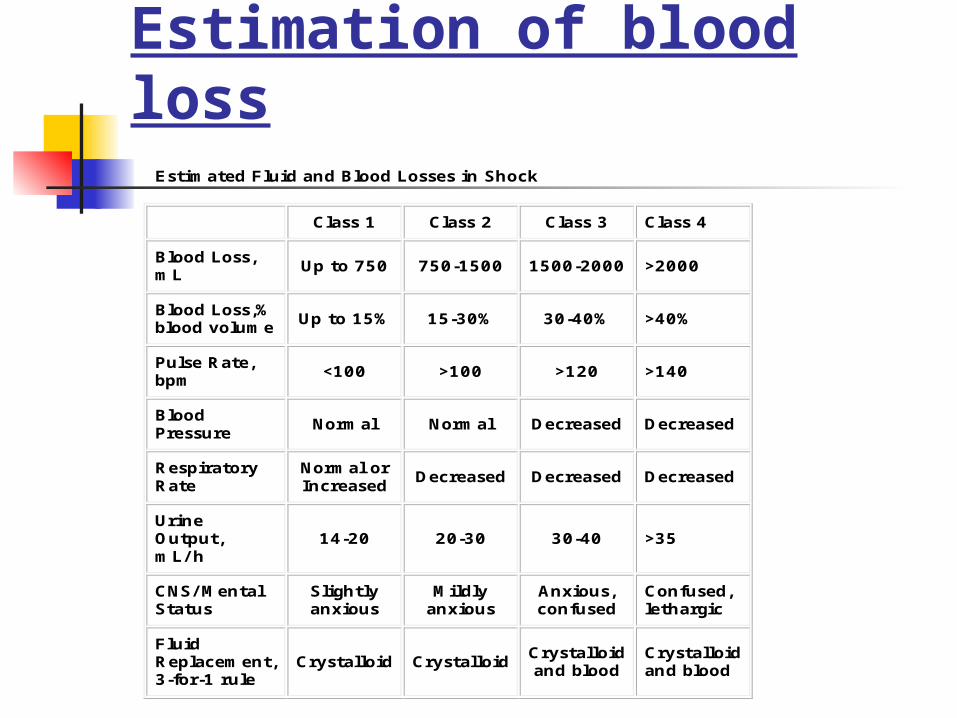
Estimation of blood lossEstimated Fluid and Blood Losses in Shock
Class 1 Class 2 Class 3 Class 4
Blood Loss, mL
Up to 750 750-1500 1500-2000 >2000
Blood Loss,% blood volume Up to 15% 15-30% 30-40% >40%
Pulse Rate, bpm <100 >100 >120 >140
Blood Pressure
Normal Normal Decreased Decreased
Respiratory Rate
Normal or Increased
Decreased Decreased Decreased
Urine Output, mL/ h
14-20 20-30 30-40 >35
CNS/ Mental Status
Slightly anxious
Mildly anxious
Anxious, confused
Confused, lethargic
Fluid Replacement, 3-for-1 rule
Crystalloid Crystalloid Crystalloid and blood
Crystalloid and blood
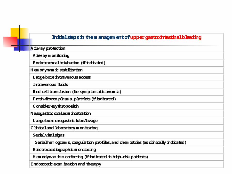
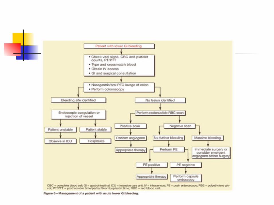
Initial steps in the management of upper gastrointestinal bleeding
Airway protection
Airway monitoring
Endotracheal intubation (if indicated)
Hemodynamic stabilization
Large bore intravenous access
Intravenous fluids
Red cell transfusion (for symptomatic anemia)
Fresh-frozen plasma, platelets (if indicated)
Consider erythropoeitin
Nasogastric oral administration
Large bore orogastric tube/lavage
Clinical and laboratory monitoring
Serial vital signs
Serial hemograms, coagulation profiles, and chemistries (as clinically indicated)
Electrocardiographic monitoring
Hemodynamic monitoring (if indicated in high-risk patients)
Endoscopic examination and therapy
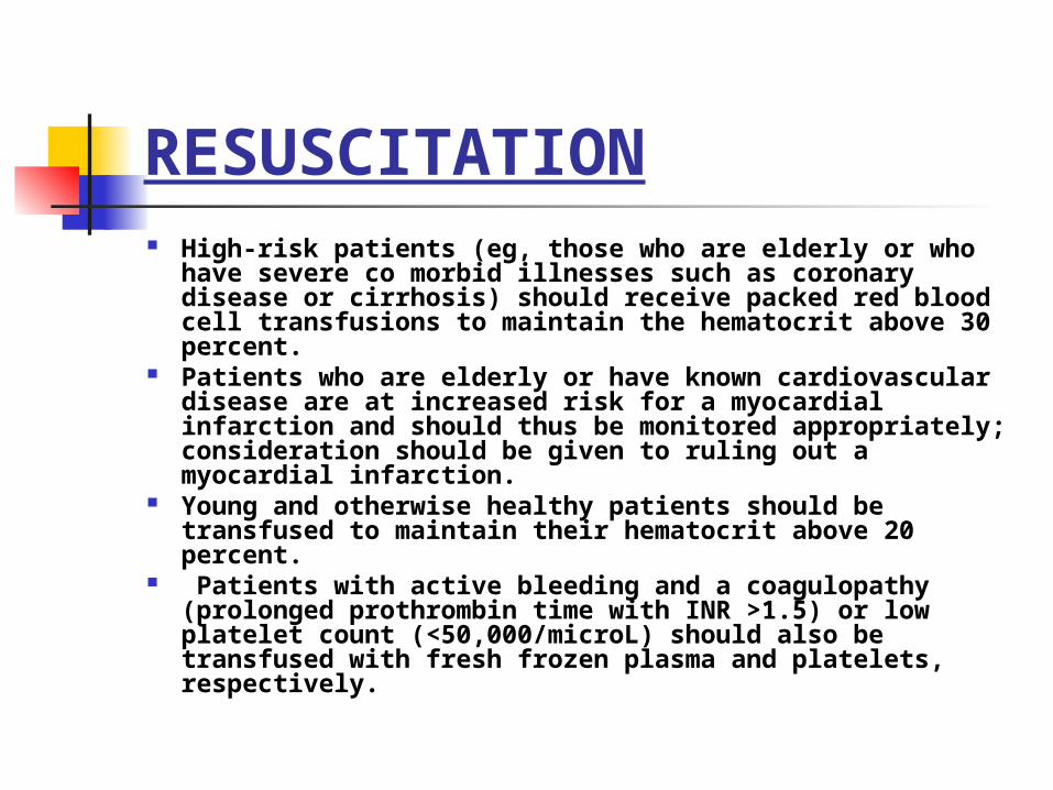
RESUSCITATION High-risk patients (eg, those who are elderly or who have severe
co morbid illnesses such as coronary disease or cirrhosis) should receive packed red blood cell transfusions to maintain the hematocrit above 30 percent.
Patients who are elderly or have known cardiovascular disease are at increased risk for a myocardial infarction and should thus be monitored appropriately; consideration should be given to ruling out a myocardial infarction.
Young and otherwise healthy patients should be transfused to maintain their hematocrit above 20 percent.
Patients with active bleeding and a coagulopathy (prolonged prothrombin time with INR >1.5) or low platelet count (<50,000/microL) should also be transfused with fresh frozen plasma and platelets, respectively.
Lower GI bleeding
Next step
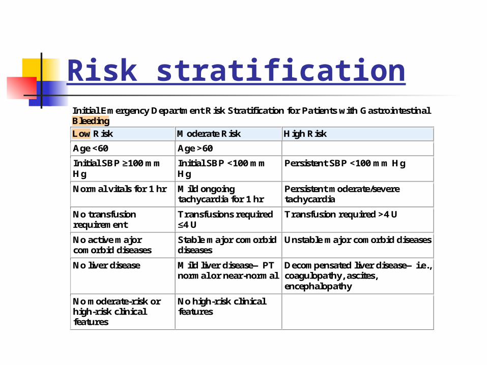
Risk stratificationInitial Emergency Department Risk Stratification for Patients with Gastrointestinal Bleeding
Low Risk Moderate Risk High Risk
Age <60 Age >60
Initial SBP ≥100 mm Hg
Initial SBP <100 mm Hg
Persistent SBP <100 mm Hg
Normal vitals for 1 hr Mild ongoing tachycardia for 1 hr
Persistent moderate/severe tachycardia
No transfusion requirement
Transfusions required ≤4 U
Transfusion required >4 U
No active major comorbid diseases
Stable major comorbid diseases
Unstable major comorbid diseases
No liver disease Mild liver disease—PT normal or near-normal
Decompensated liver disease—i.e., coagulopathy, ascites, encephalopathy
No moderate-risk or high-risk clinical features
No high-risk clinical features
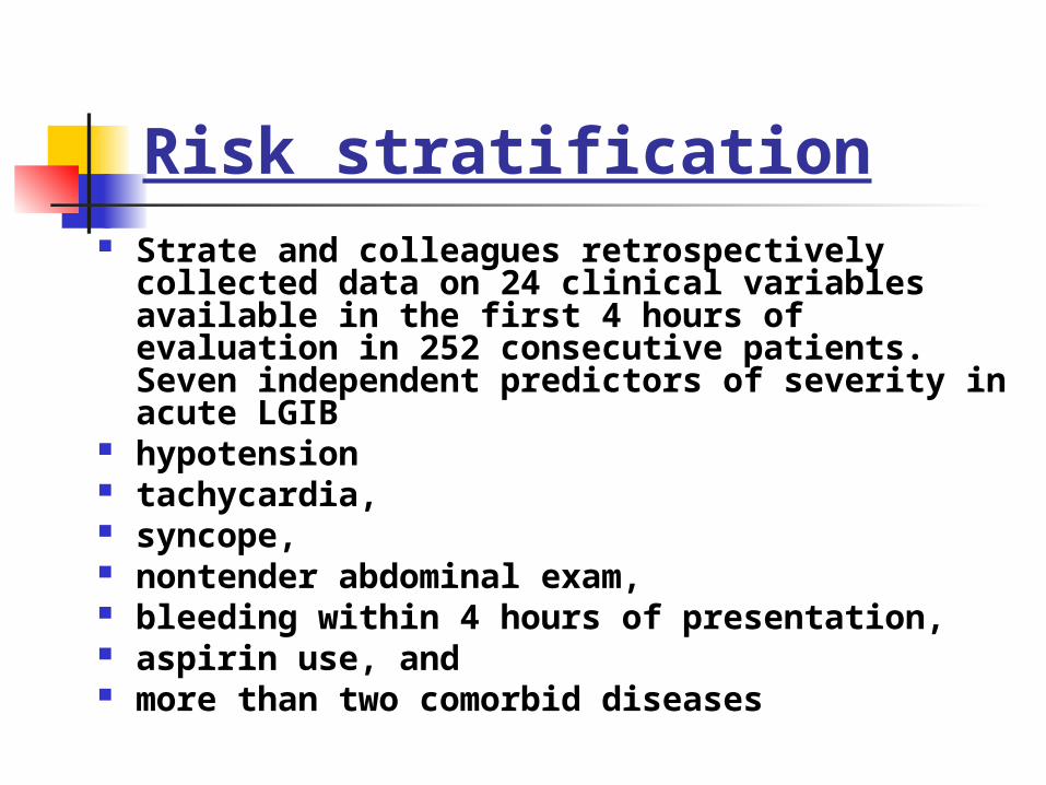
Risk stratification Strate and colleagues retrospectively collected data on 24
clinical variables available in the first 4 hours of evaluation in 252 consecutive patients. Seven independent predictors of severity in acute LGIB
hypotension tachycardia, syncope, nontender abdominal exam, bleeding within 4 hours of presentation, aspirin use, and more than two comorbid diseases
Risk stratification
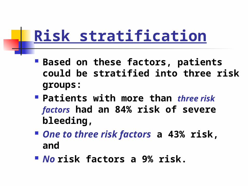
Based on these factors, patients could be stratified into three risk groups:
Patients with more than three risk factors had an 84% risk of severe bleeding,
One to three risk factors a 43% risk, and No risk factors a 9% risk.
Risk stratification
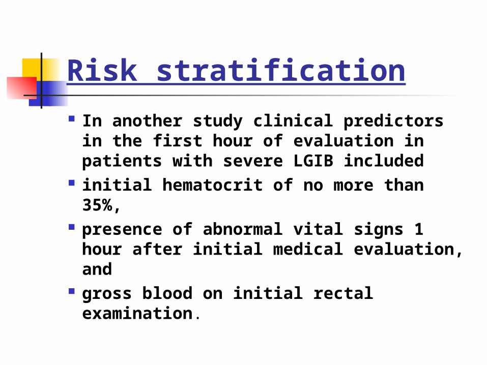
In another study clinical predictors in the first hour of evaluation in patients with severe LGIB included
initial hematocrit of no more than 35%, presence of abnormal vital signs 1 hour
after initial medical evaluation, and gross blood on initial rectal examination.
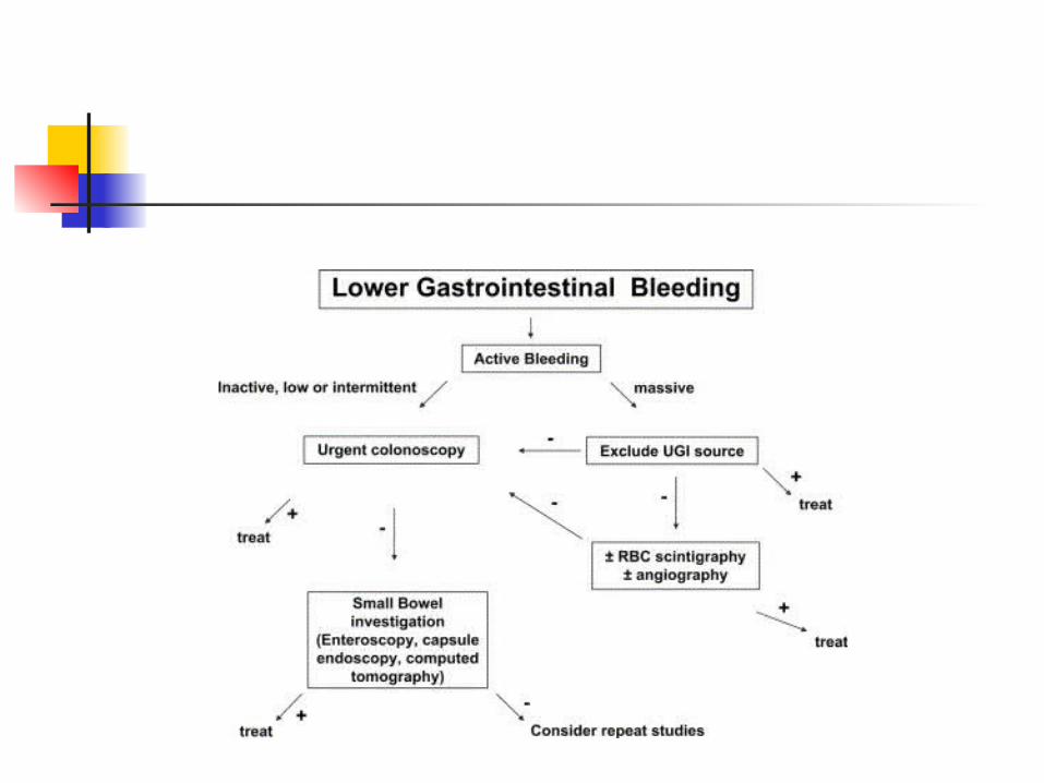
Next step
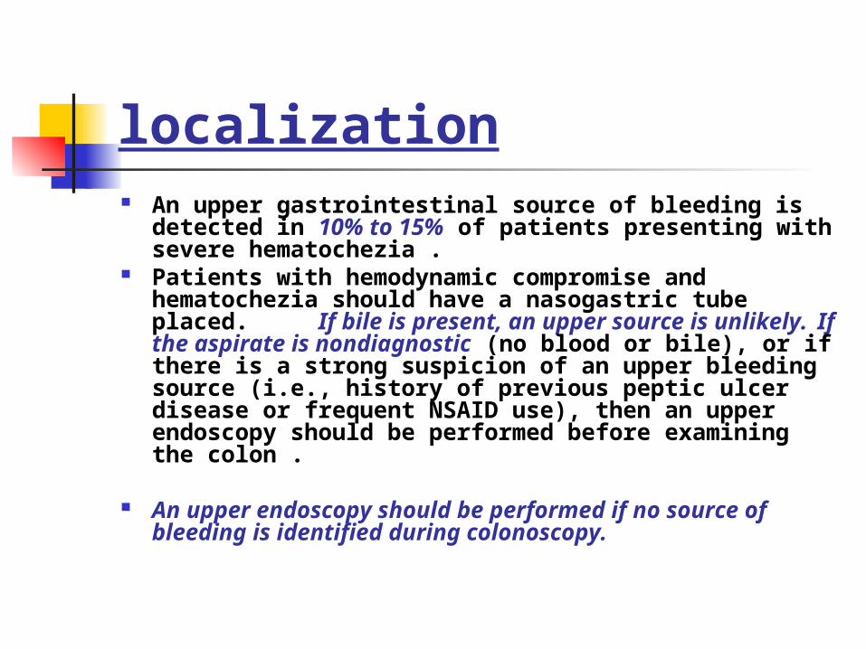
localization An upper gastrointestinal source of bleeding is detected in 10% to
15% of patients presenting with severe hematochezia . Patients with hemodynamic compromise and hematochezia should
have a nasogastric tube placed. If bile is present, an upper source is unlikely.
If the aspirate is nondiagnostic (no blood or bile), or if there is a strong suspicion of an upper bleeding source (i.e., history of previous peptic ulcer disease or frequent NSAID use), then an upper endoscopy should be performed before examining the colon .
An upper endoscopy should be performed if no source of bleeding is identified during colonoscopy.
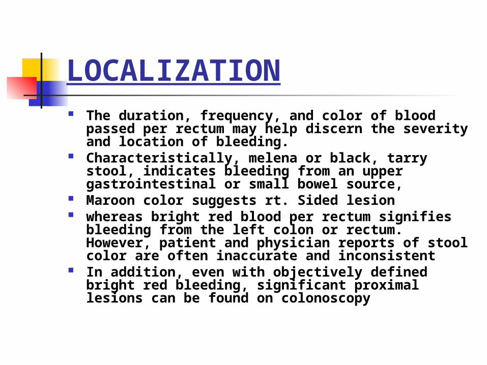
LOCALIZATION The duration, frequency, and color of blood passed per rectum
may help discern the severity and location of bleeding. Characteristically, melena or black, tarry stool, indicates bleeding
from an upper gastrointestinal or small bowel source, Maroon color suggests rt. Sided lesion whereas bright red blood per rectum signifies bleeding from the
left colon or rectum. However, patient and physician reports of stool color are often inaccurate and inconsistent
In addition, even with objectively defined bright red bleeding, significant proximal lesions can be found on colonoscopy
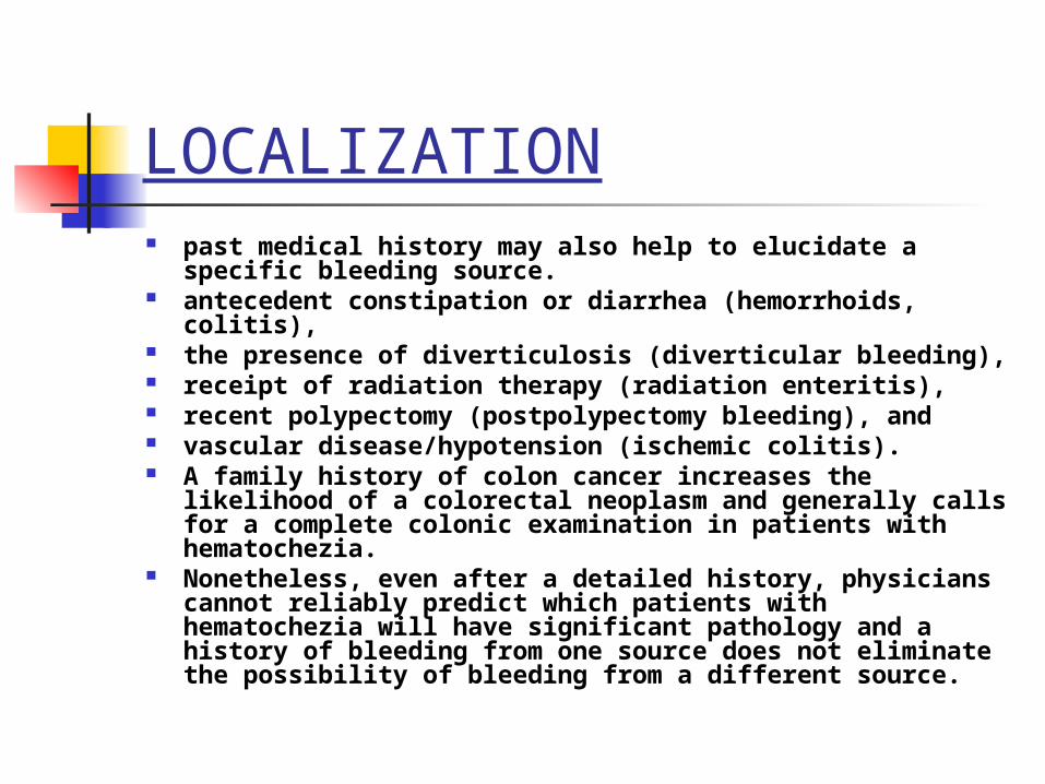
LOCALIZATION past medical history may also help to elucidate a specific bleeding source. antecedent constipation or diarrhea (hemorrhoids, colitis), the presence of diverticulosis (diverticular bleeding), receipt of radiation therapy (radiation enteritis), recent polypectomy (postpolypectomy bleeding), and vascular disease/hypotension (ischemic colitis). A family history of colon cancer increases the likelihood of a colorectal
neoplasm and generally calls for a complete colonic examination in patients with hematochezia.
Nonetheless, even after a detailed history, physicians cannot reliably predict which patients with hematochezia will have significant pathology and a history of bleeding from one source does not eliminate the possibility of bleeding from a different source.
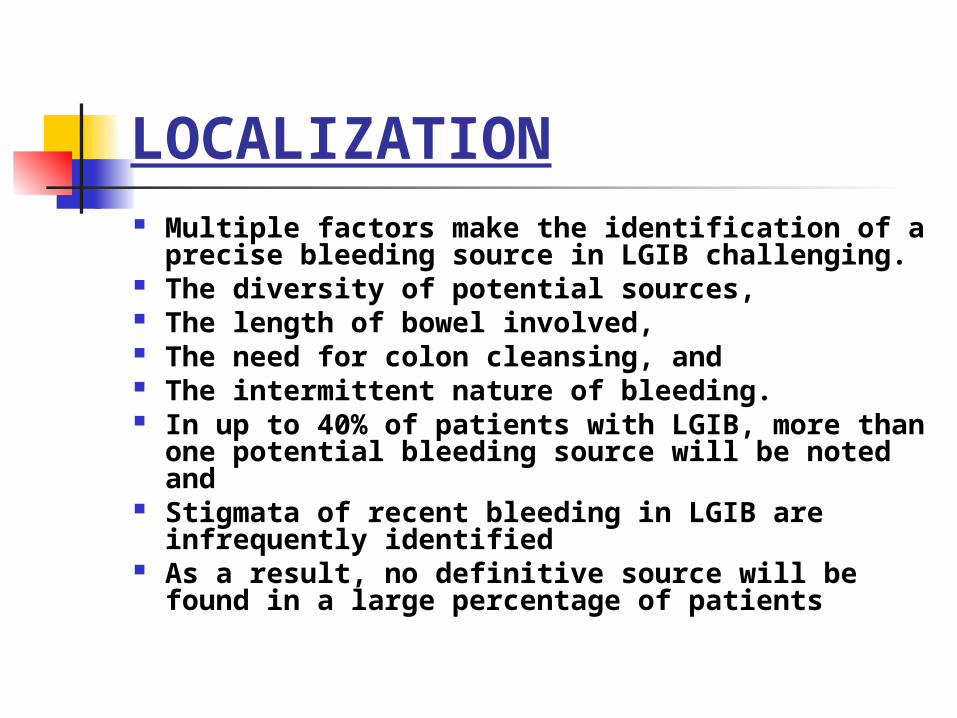
LOCALIZATION Multiple factors make the identification of a precise
bleeding source in LGIB challenging. The diversity of potential sources, The length of bowel involved, The need for colon cleansing, and The intermittent nature of bleeding. In up to 40% of patients with LGIB, more than one
potential bleeding source will be noted and Stigmata of recent bleeding in LGIB are infrequently
identified As a result, no definitive source will be found in a large
percentage of patients
Clinical scenarios
Pt. continued to bleed with hypotension and tachycardia. Patient requires 2 units of PRBCs
Pt. stopped bleeding. Vitals normalizes
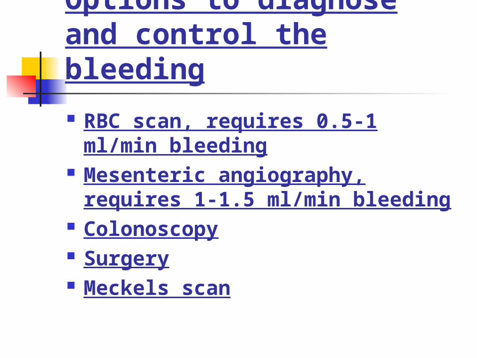
Options to diagnose and control the bleeding
RBC scan, requires 0.5-1 ml/min bleeding
Mesenteric angiography, requires 1-1.5 ml/min bleeding
Colonoscopy Surgery Meckels scan
Scenario one- Pt. continues to bleed
and is unstable.

Rbc scan vs colonoscopy
COLONOSCOPY
Colonoscopy is undoubtedly the best test for confirming the source of LGIB and for excluding ominous diagnoses, such as malignancy.
The diagnostic yield of colonoscopy ranges from 45% to 95% Perform after golytely prep(w/in 12-24h) Identifies lesion in 75 % or more Can provide endoscopic therapy Early colonoscopy associated with reduced stay Complications 0.5-1 % most patients undergoing radiographic evaluation for LGIB
regardless of findings and interventions will subsequently require
a colonoscopy to establish the cause of bleeding.
URGENT COLONOSCOPY Jensen et al
Reduced rate of rebleeding and emergency surgery from diverticular bleed when compared to historical controls
Green et al100 randomized to urgent (w/in 8hrs)
colonoscopy to standard careDefinitive source more common in urgent
group No difference in multiple clinical outcomes
Issue is still not resolved
CLINICAL SCENARIO
Patient continues to bleed RBC scan is positive on the left side?
How much true this information is?? What to do next? surgery, ?angio with
embolization?
RADIONUCLIDE SCAN radionuclide scanning has variable accuracy, cannot confirm the
source of bleeding, and may delay other diagnostic and therapeutic procedures.
Correct localization rate is 41-100% Accuracy appears to be best when the scan becomes positive within a
short period of time In one study, 42% of patients underwent an incorrect surgical
procedure based on scintigraphy results. In addition, several studies have found that regardless of accuracy, scintigraphy did not affect surgical management
Predictors of positive response-hemodynamic instability= 62% vs. 21%
->2units transfused within 24 hrs= 64% vs. 32%
CLINICAL SCENARIO
Patient underwent angiogram with embolization
Vitals improved What are the chances that pt. will
rebleed? Colonoscopy?
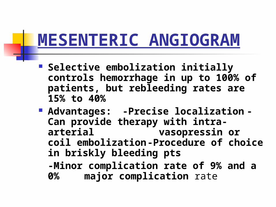
MESENTERIC ANGIOGRAM Selective embolization initially controls
hemorrhage in up to 100% of patients, but rebleeding rates are 15% to 40%
Advantages:-Precise localization-Can provide therapy with intra-arterial vasopressin or coil embolization-Procedure of choice in briskly bleeding pts-Minor complication rate of 9% and a 0% major complication rate
Disadvantages:-Invasive
-Less sensitive in detecting venous bleeding
-Can cause ischemia, contrast reactions, arterial injury
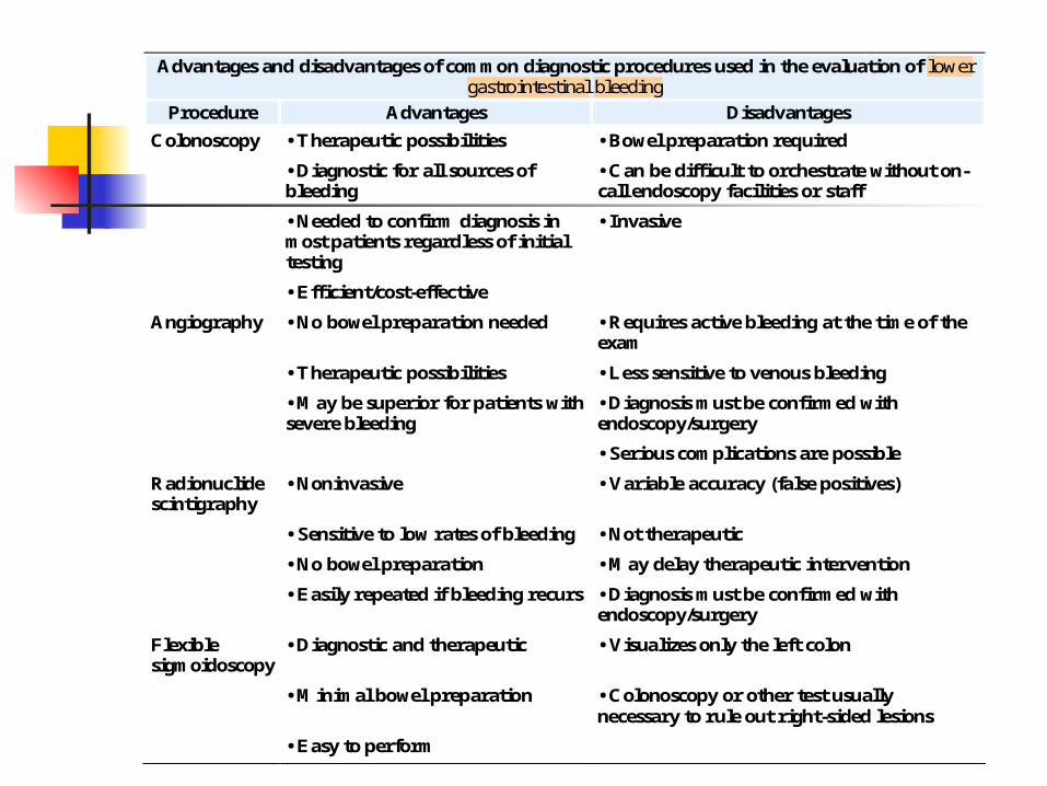
Advantages and disadvantages of common diagnostic procedures used in the evaluation of lower gastrointestinal bleeding
Procedure Advantages Disadvantages
Colonoscopy • Therapeutic possibilities • Bowel preparation required
• Diagnostic for all sources of bleeding
• Can be difficult to orchestrate without on-call endoscopy facilities or staff
• Needed to confirm diagnosis in most patients regardless of initial testing
• Invasive
• Efficient/cost-effective
Angiography • No bowel preparation needed • Requires active bleeding at the time of the exam
• Therapeutic possibilities • Less sensitive to venous bleeding
• May be superior for patients with severe bleeding
• Diagnosis must be confirmed with endoscopy/surgery
• Serious complications are possible
Radionuclide scintigraphy
• Noninvasive • Variable accuracy (false positives)
• Sensitive to low rates of bleeding • Not therapeutic
• No bowel preparation • May delay therapeutic intervention
• Easily repeated if bleeding recurs • Diagnosis must be confirmed with endoscopy/surgery
Flexible sigmoidoscopy
• Diagnostic and therapeutic • Visualizes only the left colon
• Minimal bowel preparation • Colonoscopy or other test usually necessary to rule out right-sided lesions
• Easy to perform
Pt. under went colonoscopy for definitive diagnosis.
In how many patients there will be more than one potential diagnosis?
In how many patients there will be no diagnosis found?
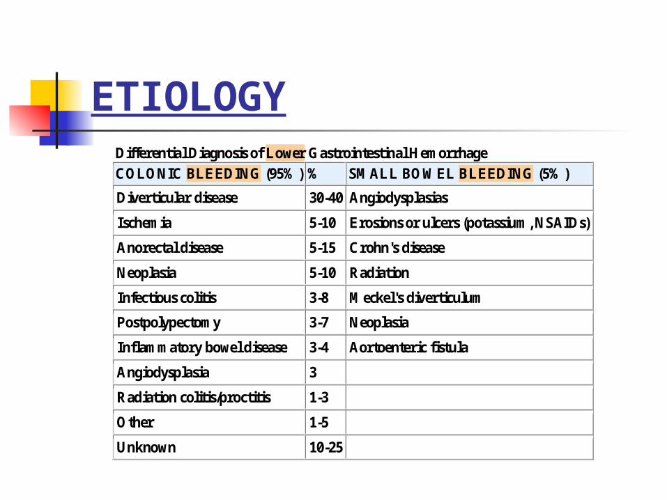
ETIOLOGYDifferential Diagnosis of Lower Gastrointestinal Hemorrhage
COLONIC BLEEDING (95%) % SMALL BOWEL BLEEDING (5%)
Diverticular disease 30-40 Angiodysplasias
Ischemia 5-10 Erosions or ulcers (potassium, NSAIDs)
Anorectal disease 5-15 Crohn's disease
Neoplasia 5-10 Radiation
Infectious colitis 3-8 Meckel's diverticulum
Postpolypectomy 3-7 Neoplasia
Inflammatory bowel disease 3-4 Aortoenteric fistula
Angiodysplasia 3
Radiation colitis/proctitis 1-3
Other 1-5
Unknown 10-25
DIAGNOSTIC DIFFICULTIES
When compared with EGD for upper GI bleeding, the diagnostic modalities for lower GI bleeding are not as sensitive or specific in making an accurate diagnosis.
Diagnostic evaluation is further complicated by the observation that, in up to 40% of patients with lower GI bleeding, more than one potential source of hemorrhage is identified.
If more than one source is identified, it is critical to confirm the responsible lesion before initiating aggressive therapy.
This approach may occasionally require a period of observation with several episodes of bleeding before a definitive diagnosis can be made.
In fact, in up to 25% of patients with lower GI hemorrhage, the bleeding source is never accurately identified.
CLINICAL SCENARIO
COLONOSCOPY SHOWEDold and BRB in mid colon
tics seen throughout Dx= probably diverticular beed Pt was d/c home
CLINICAL SCENARIO
2 wks later readmitted with rebleed and syncope
Hct 32--- 24 Urgent tagged RBC scan – neg Deep mid AC diverticulum with clot that
could not be removed What is the next step
SURGERY Surgery usually is employed for hemorrhage in two settings:
massive or recurrent bleeding. It is required in 15% to 25% of patients who have diverticular
bleeding and is recommended for patients with a high transfusion requirement (generally more than four units within a 24-hour period or greater than 10 units total)
Recurrent bleeding from diverticula occurs in 20% to 40% of patients and generally is considered an indication for surgery
In patients with serious comorbid medical conditions and without exsanguinating hemorrhage, this decision should be made carefully.
Great effort should be made to accurately localize the site of bleeding preoperatively so that segmental rather than subtotal colectomy can be performed Operative mortality is 10% even with accurate localization and up to 57% with blind subtotal colectomy.