Lower Extremity Ultrasound-Guided Regional Anesthesia · · 2014-05-08Lower Extremity...
50
Lower Extremity Ultrasound-Guided Regional Anesthesia Stephanie Duffy, CRNA Regional Anesthesia Faculty Acute Pain Service NMCSD
-
Upload
truonghanh -
Category
Documents
-
view
228 -
download
3
Transcript of Lower Extremity Ultrasound-Guided Regional Anesthesia · · 2014-05-08Lower Extremity...

Lower Extremity Ultrasound-Guided
Regional Anesthesia
Stephanie Duffy, CRNA Regional Anesthesia Faculty Acute Pain Service NMCSD


Objectives • Review anatomy of
lumbosacral plexus • Lumbar plexus blocks
• Psoas • Femoral
• Saphenous • Lateral Femoral Cutaneous
• Sacral plexus blocks • Classic Landmarks • Parasacral • Subgluteal • Popliteal
• Rectus sheath block • Transversus Abdominal Plane
(TAP)

Lumbar Plexus Anatomy
• Iliohypogastric • Ilioinguinal
• Genitofemoral
• Lateral Femoral Cutaneous
• Obturator
• Femoral
Interested In Getting Lunch On Friday?

Lumbar Plexus Anatomy • Anterior rami of T12
to L4 • Gives rise to
• Ilioinguinal • Iliohypogastric • Genitofemoral • LFC • Obturator • Femoral

Lumbar Plexus Anatomy
• Femoral Nerve • Motor: hip flexors, quadriceps, leg extension
• Sensory: anterior thigh and knee
• Terminates as the saphenous nerve
• LFC: lateral thigh sensation • Obturator: adductors and medial thigh
sensation


Femoral Nerve
• Enveloped by Fascia Iliacus • Femoral Artery (5), Femoral Vein (6) • After Inguinal Ligament (4)
• Anterior Division • Sartorius and Cutaneous Branches (3)
• Posterior Division (1) • Quadriceps • Saphenous
LATE
RA
L ME
DIA
L


Lumbar Plexus Block • AKA Psoas Compartment
Block
• Good for complete lumbar plexus coverage • Obturator
• LFC
• Femoral • Useful for hip procedures
• May be unilateral alternative to neuraxial block

Lumbar Plexus Block • Prone or lateral • Landmarks
• Iliac crest • Spine (midline) • PSIS • 2/3 dist PSIS line
• Large curvilinear probe at intersection
• Visualize transverse processes; note depth
• Stimulate quadriceps twitch (or psoas on US)

Lumbar Plexus Block
• Transverse process is a good and safe gauge of depth

Complications
v Highly vascular psoas m.
v Hematoma
v LA toxicity
v Beware multiple needle passes!

Complications
• Psoas Hematoma • Psoas: 3rd most
common site of iatrogenic bleed with coumadin
• Avoid multiple needle passes
• Anticoagulation is contraindication
NEUROMUSCULAR DISEASE CENTER: Washington University, St. Louis, MOハ USA

Complications
• Visceral Injury
• Kidneys
• Major Vascular
• Bowel

Complications
§ Inadvertant spinal or epidural anesthesia § Don’t deviate medially
§ Sympathectomy § Hypotension § < 15% of blocks
§ Infection - Discitis
Kirchmair et al; A&A 2002;94:706-10

Lumbar Plexus Summary
• Excellent block for hip, thigh and knee
• Depth < 2 cm past TP = Safety
• Anticoagulation: Psoas = Epidural

Femoral Nerve Block
• Bread and Butter Block • Great block to practice
US and needle control
FA
FN
LATER
AL

Femoral Nerve Block: Overview
• Indication • Anterior thigh • Knee (often combined with
other blocks)
• Continuous perineural infusion improves recovery following total knee arthroplasty (TKA)1 • May shorten hospital stay after
TKA2
1. DeRuyter ML, et al. J Arthro 2006;21:1111 2. Ilfeld, BM, et al. A&A 2006;102:87

FNB Anatomy
• NAVEL • Nerve
• Artery • Vein
• Lymphatics

FNB: Surface Anatomy
• Supine position • Pubic tubercle and ASIS
• Palpable femoral artery
Femoral Nerve Block • Lateral-Medial Approach • Short axis/in-plane • 1. Hydrodissect FN off FI • 2. Advance under nerve,
place catheter if desired
1st
2nd

Femoral Nerve Catheter
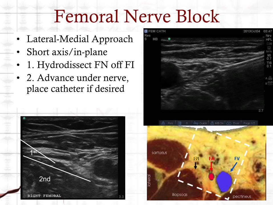
Contraindications?

Lateral Femoral Cutaneous • If missed with FNB
• e.g., skin graft • Start at ASIS with linear
probe and scan area (2 cm down, 2cm medial)

Obturator Nerve • Rarely needed?
• Possible knee innervation (~20%) – 2 studies show decreased pain scores in TKA patients with obturator block.
• Located medial to femoral vein
• Derives from L2-4 • Enters thigh through obturator
foramen
• Motor component of medial thigh (adductors)
• Variable sensory • 57% no sensory • 20% show deficit on medial aspect
of upper thigh (including knee)

Saphenous
• For foot and ankle surgery • Remember, sensory only • Can do field block…
• 33-65% effective
• Better options…

Saphenous
• Sub-sartorial • 5-7 cm proximal to knee • Lateral-medial approach • Fascial plane between
sartorius m. and vastus medialis m.
• Perifemoral • Mid-thigh • Lateral-medial approach • Nerve lateral to superficial
femoral artery and vein
Adductor Canal Block
• Adductor Canal anatomy relevant to knee • Aponeurotic space extending from apex of femoral triangle to the adductor hiatus • Contains:
• Saphenous n. (femoral) • Infrapatellar skin and anterior knee capsule
• Distal branch of the motor n. to vastus medialis (femoral)
• Sensory innervation to superomedial aspect of knee and knee capsule

Adductor Canal Block • Posterior branch of
obturator n. (inconsistently present)
• +/- medial cutaneous and and anterior branches of obturator n.
• Most motor branches to quadriceps m. branch from femoral n. approximately 5 cm below inguinal ligament

Adductor Canal Block • Postop analgesia with motor sparing?
• Effective analgesic adjunct for TKA • Jenstrup, et al. Acta Anaesthes Scand 2011
• AC catheter boluses vs. morphine PCA 2.5 mg bolus, 10 min lockout
• Improved pain, decreased morphine consumption, improved ambulation
• Perlas, et al. RAPM 2013 • ACB + knee infiltration better postop analgesia than fem n. cath
• Mid-thigh ACB is motor-sparing • Kwofie, et al. RAPM 2013
• 95% strength reduction with femoral n. block • 11% strength reduction with ACB (mostly vastus medialis) • No significant reduction in hip adductor strength

Sacral Plexus Anatomy
• 12 branches • 5 in pelvis (gluteal nerves) • 7 emerge (buttock and LE)
• Sciatic is largest • Lumbosacral trunk L4-5 and anterior
divisions of sacral plexus S1-3 form TIBIAL nerve
• Post divisions form PERONEAL nerve
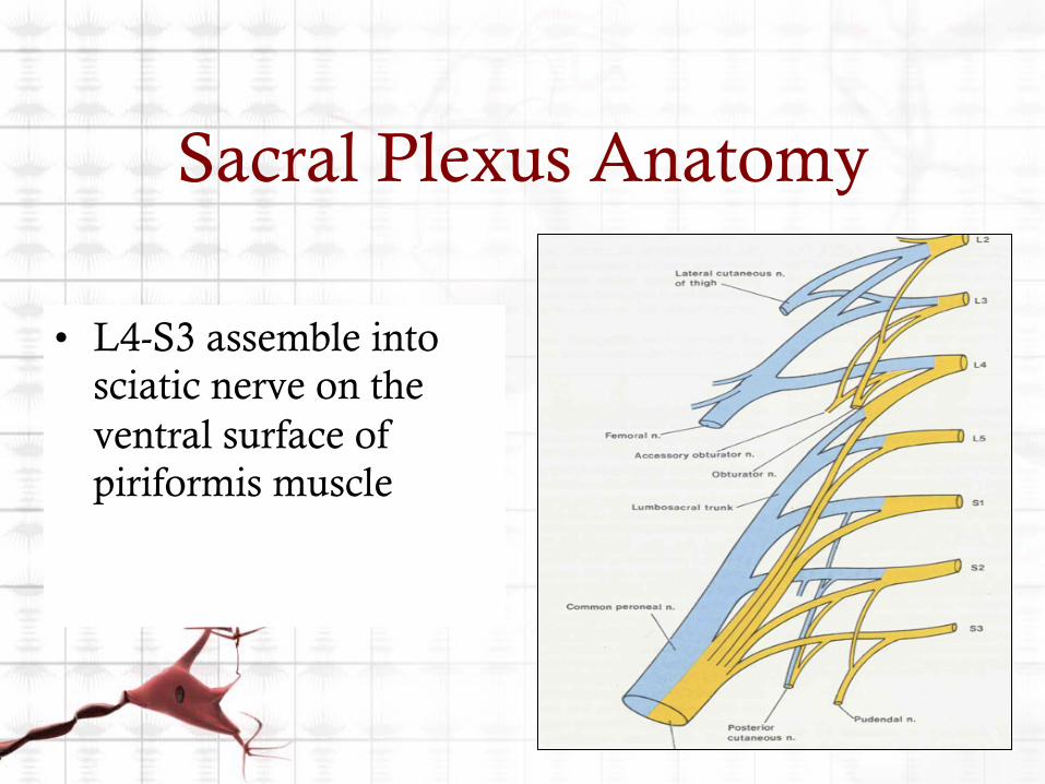
Sacral Plexus Anatomy
• L4-S3 assemble into sciatic nerve on the ventral surface of piriformis muscle

Approaches to Sciatic Nerve Blockade
Parasacral Classic (Labat)
Subgluteal
Popliteal Fossa

Subgluteal • The lowest sciatic block that
reliably picks up the PFC • Ideal • Realistic
• I do this full prone as well • Scan distal-to-proximal can be
helpful

Ultrasound of Sciatic

Sciatic Scan

Popliteal • Great for learning US
• Popliteal artery
• Scan posterolateral from artery
• Prone position easier. • Can flip screen for
supine
• Start at crease and scan cephalad until the two nerves become one (or at least 7- 10 cm)
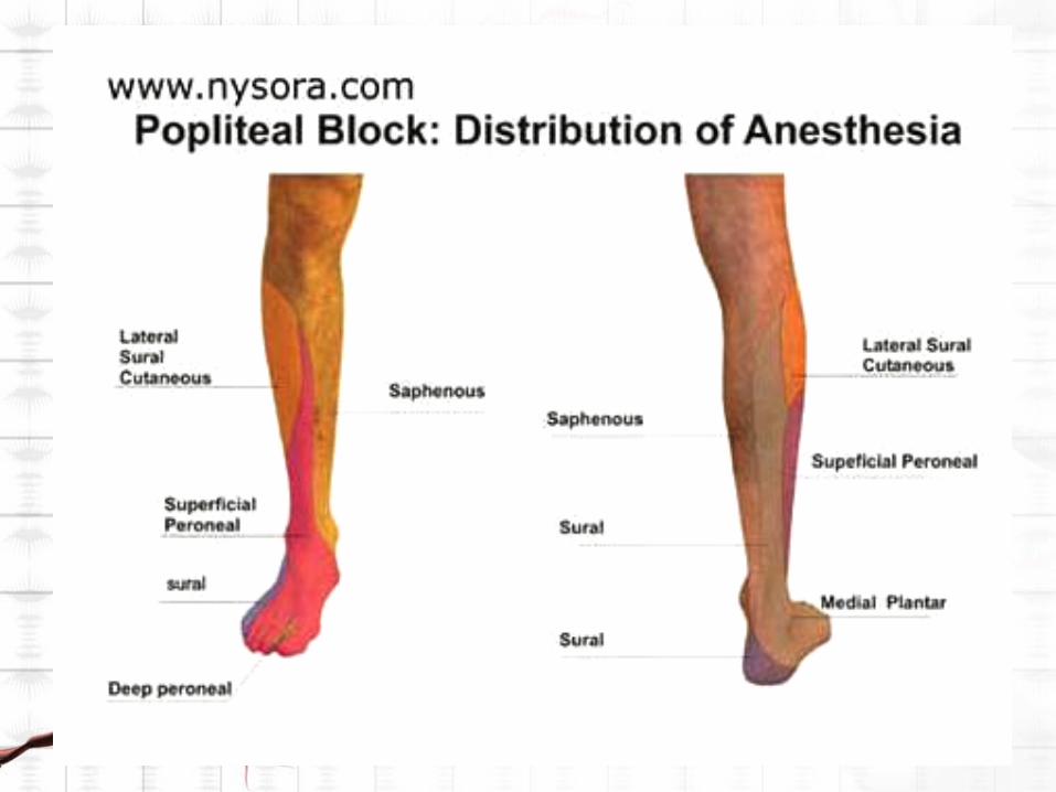

Popliteal
TN PN

Popliteal • A few tips
• Tibial is deeper, more medial,
• Peroneal is more superficial and lateral
• If adding twitch, remember:
• Tibial (Toes) • Plantar flexion and inversion
• Peroneal • Dorsiflexion and eversion

Popliteal • US view in supine
position
• Popliteal Artery
• Nerve is superficial and lateral (almost always)

Popliteal Ultrasound Lateral Approach
• Needle entry point on lateral aspect of leg to ensure parallel path of needle to US probe (spectral reflection) • Increases needle
visualization

Popliteal Lateral Approach with Ultrasound
POSTERIOR
ANTERIOR
PN
TN

Complications

Rectus Sheath Block

Rectus Sheath Block

Rectus Sheath Block • Indications:
• Umbilical hernia repair • Laparoscopy (umbilical port) • Midline incision laparotomy
• TAH • Vascular procedures midline

Rectus Sheath Block Anatomy • Supplied bilaterally by terminal cutaneous branch of T10
spinal (intercostal) nerves • sensory innervation to the skin after passing through the
rectus abdominis muscles. • Rectus sheath (fascia of the external/internal oblique
aponeurosis) envelops the two muscles.
QUESTIONS?