Lower Extremity HIP BIOMECHANICS, LUMBOSACRAL PLEXUS, AND LEG MUSCULATURE.
18
Lower Extremity HIP BIOMECHANICS, LUMBOSACRAL PLEXUS, AND LEG MUSCULATURE
-
Upload
karen-rodgers -
Category
Documents
-
view
239 -
download
2
Transcript of Lower Extremity HIP BIOMECHANICS, LUMBOSACRAL PLEXUS, AND LEG MUSCULATURE.

Lower ExtremityHIP BIOMECHANICS, LUMBOSACRAL PLEXUS, AND LEG MUSCULATURE

Objectives
Describe the gross anatomy for each system (circulatory, muscular, nervous, and skeletal) in the lower extremity.
Integrate the systems to discuss the lower extremity stability and mobility functions.
Analyze common injuries in the lower extremities.
For each muscle, describe how the attachment sites result in an action around a joint.
For each muscle, identify the innervation (peripheral nerve and nerve roots).

Announcements
No office hours Thursday
Lab closed Friday and Monday
Tag Test reminders

Presentations

Which of the following is incorrect pertaining to the patella?
A. It increases the mechanical advantage of the quadriceps femoris for extending the knee.
B. Pain felt deep to it may be associated with a condition known as chondromalacia patella.
C. Testing of its associated tendon reflex is done with the patient sitting and legs "dangling.“
D. It tracks superomedially on the femur. It
increase
s the m
echan
ical a
dv...
Pain fe
lt deep
to it
may be a
sso...
Testi
ng of it
s asso
ciated
tendon ...
It tra
cks s
uperom
edial
ly on th
e...
25% 25%25%25%

Your friend is diagnosed with trochanteric bursitis. You explain to him that this bursa is between the:
A. iliopsoas tendon and the lesser trochanter, allowing the muscle to move freely across the neck of the femur.
B. obturator externus and the lesser trochanter, allowing the muscle to move freely across the trochanter.
C. gluteus maximus and medius at the site of the greater trochanter, allowing the muscles to move freely across one another.
D. skin and the gluteus maximus at the site of the greater trochanter, allowing comfortable sitting by distributing forces across the trochanter.
E. gluteus maximus and the greater trochanter, allowing the muscle to smoothly slide over the trochanter.
iliopso
as tendon and th
e lesse
r ...
obtura
tor e
xtern
us and th
e lesse
...
glute
us maxim
us and m
edius a
t...
skin and th
e gluteus m
axim
us at..
.
glute
us maxim
us and th
e greate
...
20%20% 20%20%20%

In the following popliteal arteriogram, the arrow points to the:
A. popliteal artery.B. anterior tibial artery.C. fibular artery.D. medial genicular artery.E. fibular recurrent artery.
poplitea
l arte
ry.
anter
ior tibial
arter
y.
fibular arte
ry.
medial ge
nicular a
rtery.
fibular recu
rrent a
rtery.
20% 20% 20%20%20%

In the following coronal MRI of the ankle joint, the arrow points to the:A. plantar
calcaneonavicular (spring) ligament.
B. medial (deltoid) ligament.
C. long plantar ligament.D. abductor hallucis
longus.E. flexor hallucis longus.
planta
r calc
aneonavic
ular (sp
ring..
.
medial (d
eltoid) li
gament.
long plan
tar lig
ament.
abducto
r hall
ucis lo
ngus.
flexor h
allucis
longu
s.
20%20% 20%20%20%

Leg Musculature
Anterior Compartment
Lateral Compartment
Posterior Compartments Superficial
Deep
Leg muscles primarily act on the ankle AND foot
Malleoli serve as pulleys for tendons
Tendons wrapped in synovial sheaths
Retinacula prevent bowstringing

Anterior Compartment Tibialis anterior
Extensor hallucis longus
Extensor digitorum longus
Fibularis tertius
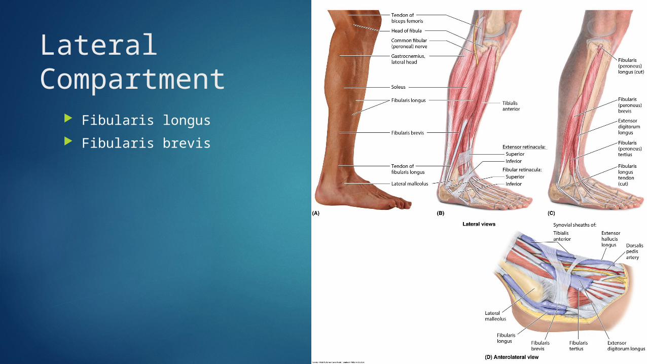
Lateral Compartment
Fibularis longus
Fibularis brevis

Posterior Compartments
Superficial Triceps surae
Plantaris
Deep Tibialis posterior
Flexor digitorum longus
Flexor hallucis longus
Popliteus

Which of the following is incorrect pertaining to the calcaneal (Achilles) tendon?
A. It is the common distal attachment of the gastrocnemius, and soleus muscles.
B. It continues into the foot as the long plantar ligament.
C. It is used to test S1and S2 nerve function.
D. It is separated from the superior part of the posterior surface of the calcaneus by a bursa.
E. It spirals while passing inferiorly to its attachment on the calcaneus.
It is
the c
ommon dist
al attach
me..
It co
ntinues into
the f
oot as t
he...
It is
used to
test
S1and S2
nerve ...
It is
separat
ed fr
om the su
perior..
.
It sp
irals
while pas
sing i
nferiorly
..
20%20% 20%20%20%

The tibialis posterior:
A. everts the foot.B. attaches to the femur.C. supports the medial
longitudinal arch of the foot.
D. is an important swing phase muscle.
E. is innervated by the deep fibular nerve. eve
rts th
e foot.
attac
hes to th
e femur.
supports
the m
edial
longit
udinal...
is an
importa
nt swing p
hase m
u...
is inner
vate
d by the deep
fibula...
20%20% 20%20%20%

Clinical relevance
“Shin splints”
Compartment syndrome
Stress fractures

Clinical relevance cont’d
Foot drop

Your patient is a runner complaining of leg pain. Which of the following can you safely assume is not a likely diagnosis for this patient?
A. Shin splintsB. Tibia stress fractureC. Compartment
syndromeD. Foot drop
Shin sp
lints
Tibia
stress
fractu
re
Compartment s
yndrome
Foot d
rop
25% 25%25%25%

You see a patient in the emergency room with a fibular neck fracture subsequent to an automobile accident. You are fearful that the patient may have severed his common fibular nerve. To determine this, you ask him to walk. Which of the following walking abnormalities would suggest that the nerve has been severed?
A. excessive flexion of the knee at heel-strike
B. shortened stance phaseC. high-stepping (steppage) gaitD. lack of balance (use of a cane)E. lack of effective hallux push-off
exces
sive flexio
n of the k
nee at..
.
shorte
ned stance
phase
high-st
epping (
steppage
) gait
lack of b
alance (u
se of a
cane)
lack of e
ffective
hallux p
ush-o
ff
20%20% 20%20%20%
![Plexus Lumbosacral Ham [Dr. Hasan]](https://static.fdocuments.net/doc/165x107/5571ff0e49795991699c8de4/plexus-lumbosacral-ham-dr-hasan.jpg)