Loss of Protein Structure Stability as a Major Causative...
15
Loss of Protein Structure Stability as a Major Causative Factor in Monogenic Disease Peng Yue 1,2 , Zhaolong Li 1 and John Moult 1 * 1 Center for Advanced Research in Biotechnology, University of Maryland Biotechnology Institute, Rockville, MD 20850 USA 2 Molecular and Cellular Biology Program, University of Maryland, College Park, MD 20742, USA The most common cause of monogenic disease is a single base DNA variant resulting in an amino acid substitution. In a previous study, we observed that a high fraction of these substitutions appear to result in reduction of stability of the corresponding protein structure. We have now investigated this phenomenon more fully. A set of structural effects, such as reduction in hydrophobic area, overpacking, backbone strain, and loss of electrostatic interactions, is used to represent the impact of single residue mutations on protein stability. A support vector machine (SVM) was trained on a set of mutations causative of disease, and a control set of non-disease causing mutations. In jack-knifed testing, the method identifies 74% of disease mutations, with a false positive rate of 15%. Evaluation of a set of in vitro mutagenesis data with the SVM established that the majority of disease mutations affect protein stability by 1 to 3 kcal/mol. The method’s effective distinction between disease and non-disease variants, strongly supports the hypothesis that loss of protein stability is a major factor contributing to monogenic disease. Mutant analysis is available (www.snps3d.org). q 2005 Elsevier Ltd. All rights reserved. Keywords: protein structure; protein stability; monogenic disease; mis-sense mutations; single nucleotide polymorphisms *Corresponding author Introduction Over 1000 human genes have been identified where one or more sequence modifications are directly causative of disease. 1 These mutations may affect protein function through a number of mechanisms, such as changes in transcription, RNA processing, protein expression, folding of the polypeptide chain, stability of the folded state, post- translational modification, interactions with bind- ing partners, and alterations to catalysis. An analysis of the Human Gene Mutation Database (HGMD) 1 has shown that the vast majority of known cases act through changes to the coding sequence, with mis-sense mutations (a single base change resulting in change of a single amino acid) by far the most common effect, accounting for greater than 60% of all monogenic disease mutations. In a previous study, we investigated the relationship between these mis-sense mutations and disease, in terms of the effect of the resulting amino acid change on protein structure and function. 2 In that work, we concluded that the most common mechanism (up to 80% of cases) by which a non-synonymous base change results in disease is destabilization of the protein structure, relative to the unfolded state. A further approxi- mately 10% of mis-sense mutations were seen to affect some known aspect of molecular function, and the remaining 10% to operate by other, unidentified, mechanisms. That study relied primarily on visual inspection of the effect of an amino acid substitution on protein structure and function. Here, we have developed a more objective model, focusing just on the role of destabilization of the folded structure. The primary goals are to more rigorously investigate the extent to which stability is a common factor in causing monogenic disease, and to provide a general and fully automatic stability perturbation model that can be used for analysis of the impact of non-synonymous single nucleotide polymorphisms (SNPs) found in the human population. Two principal strategies have been developed for identifying which mis-sense base changes are most likely to be causative of disease. The most common approach makes use of the fact that the more critical 0022-2836/$ - see front matter q 2005 Elsevier Ltd. All rights reserved. Abbreviations used: SVM, support vector machine; HGMD, Human Gene Mutation Database; PDB, Protein Data Bank. E-mail address of the corresponding author: [email protected] doi:10.1016/j.jmb.2005.08.020 J. Mol. Biol. (2005) 353, 459–473
Transcript of Loss of Protein Structure Stability as a Major Causative...
doi:10.1016/j.jmb.2005.08.020 J. Mol. Biol. (2005) 353, 459–473
Loss of Protein Structure Stability as a Major CausativeFactor in Monogenic Disease
Peng Yue1,2, Zhaolong Li1 and John Moult1*
1Center for Advanced Researchin Biotechnology, University ofMaryland BiotechnologyInstitute, Rockville, MD 20850USA
2Molecular and CellularBiology Program, University ofMaryland, College Park, MD20742, USA
0022-2836/$ - see front matter q 2005 E
Abbreviations used: SVM, suppoHGMD, Human Gene Mutation DaData Bank.E-mail address of the correspond
Themost common cause of monogenic disease is a single base DNAvariantresulting in an amino acid substitution. In a previous study, we observedthat a high fraction of these substitutions appear to result in reduction ofstability of the corresponding protein structure. We have now investigatedthis phenomenon more fully. A set of structural effects, such as reduction inhydrophobic area, overpacking, backbone strain, and loss of electrostaticinteractions, is used to represent the impact of single residue mutations onprotein stability. A support vector machine (SVM) was trained on a set ofmutations causative of disease, and a control set of non-disease causingmutations. In jack-knifed testing, the method identifies 74% of diseasemutations, with a false positive rate of 15%. Evaluation of a set of in vitromutagenesis data with the SVM established that the majority of diseasemutations affect protein stability by 1 to 3 kcal/mol. The method’s effectivedistinction between disease and non-disease variants, strongly supportsthe hypothesis that loss of protein stability is a major factor contributing tomonogenic disease. Mutant analysis is available (www.snps3d.org).
q 2005 Elsevier Ltd. All rights reserved.
Keywords: protein structure; protein stability; monogenic disease; mis-sensemutations; single nucleotide polymorphisms
*Corresponding authorIntroduction
Over 1000 human genes have been identifiedwhere one or more sequence modifications aredirectly causative of disease.1 These mutations mayaffect protein function through a number ofmechanisms, such as changes in transcription,RNA processing, protein expression, folding of thepolypeptide chain, stability of the folded state, post-translational modification, interactions with bind-ing partners, and alterations to catalysis. Ananalysis of the Human Gene Mutation Database(HGMD)1 has shown that the vast majority ofknown cases act through changes to the codingsequence, with mis-sense mutations (a single basechange resulting in change of a single amino acid)by far the most common effect, accounting forgreater than 60% of all monogenic diseasemutations. In a previous study, we investigatedthe relationship between these mis-sense mutationsand disease, in terms of the effect of the resulting
lsevier Ltd. All rights reserve
rt vector machine;tabase; PDB, Protein
ing author:
amino acid change on protein structure andfunction.2 In that work, we concluded that themost common mechanism (up to 80% of cases) bywhich a non-synonymous base change results indisease is destabilization of the protein structure,relative to the unfolded state. A further approxi-mately 10% of mis-sense mutations were seen toaffect some known aspect of molecular function,and the remaining 10% to operate by other,unidentified, mechanisms. That study reliedprimarily on visual inspection of the effect of anamino acid substitution on protein structure andfunction.Here, we have developed amore objective model,
focusing just on the role of destabilization of thefolded structure. The primary goals are to morerigorously investigate the extent to which stabilityis a common factor in causing monogenic disease,and to provide a general and fully automaticstability perturbation model that can be used foranalysis of the impact of non-synonymous singlenucleotide polymorphisms (SNPs) found in thehuman population.Two principal strategies have been developed for
identifying which mis-sense base changes are mostlikely to be causative of disease. The most commonapproach makes use of the fact that the more critical
d.
460 Protein Variants in Monogenic Disease
a position in a protein sequence is to viability, themore restricted are the residue types accepted there.A number of different methods for assessing thesignificance of amino acid conservation have beendeveloped.3–7 Methods that utilize sequence con-servation have the advantage of including all kindsof impact on protein viability. Also, the methods canbe used with any human protein for which asuitable set of sequence relatives is known, and sohave wide applicability. The approach has thedisadvantage that it provides no direct insight intothe underlying mechanism. The second strategy isto make use of knowledge of protein structure andfunction. For instance, recognizing that a changeoccurs in a key catalytic residue, or one involved inligand binding, or a target for post-translationalmodification.
Wang & Moult2 used a structure-based model toidentify amino acid substitutions likely to signifi-cantly affect protein stability as well as othercontributions to function. Stability impact wasassessed using a set of simple rules based onchanges in hydrophobic burial, backbone strain,overpacking, and electrostatic interactions. Thatwork forms the foundation of the present study.Other groups have combined sequence and struc-ture strategies to varying degrees. Sunyaev4,5
predicted the effect of mis-sense mutations usingempirically derived rules which make use of avariety of data, such as functional information,hydrophobic propensity, side-chain volume changeand transmembrane location,8 together withsequence information. The rules were tested againstdisease causing mutations annotated by Swiss-Prot,using the variation between human and otherspecies as a control. In Chasman’s6 method,ANOVA and principal component analysis wereapplied to a series of features that capture aspects ofstructural and sequence context. Data on therelationship between site-directed mutants andchanges in phenotype for a phage protein, T4lysozyme, and a bacterial protein, Lac repressor,were used as training and testing sets. Featuresshowing strong discrimination between mutationsaffecting or not affecting the phenotype, such as therelative residue temperature factor, relative surfaceaccessibility, relative phylogenetic entropy(sequence conservation in the protein family) andburial of charge, were selected, A probability modelwas then constructed based on the selected features,and used to estimate the likelihood that a givenmutation will affect function. A similar probabilityapproach has also been used to include functioneffects.9 Krishnan & Westhead7 used two machinelearning methods, a decision tree and a supportvector machine, to predict the impact of singleamino acid changes based on a set of structural(secondary structure and surface accessibility) andsequence attributes, such as sequence conservationscore calculated using ScoreCons.10 Secondarystructure and surface accessibility data were takenfrom the HSSP database11 or predicted usingPHD.12
The central hypothesis of the present work is thatmoderate loss of stability of the folded state of aprotein molecule is frequently associated withmonogenic disease. To investigate this, we mustidentify significant changes in the free energydifference between the folded and unfolded statesof a protein molecule resulting from an amino acidsubstitution. A theoretically rigorous approachwould be to use an appropriate integration of theenergy change as one amino acid is morphed intoanother in the context of the protein structure. Thesefree energy perturbation techniques13 have beenincorporated in a number of the more widely usedmolecular dynamics software packages. Issues ofconformational sampling, appropriate representationof the unfolded state and force field accuracy havegenerally resulted in poor accuracy.14 Recent resultsshow encouraging improvement, but require careand method optimization in each case,15 restrictinglarge-scale application. Force field deficiencies maybe reduced by parameterizing using free energydifferences obtained from site-directed mutagenesisexperiments.16 The resulting model is effective atpredicting this type of stability change.
We have developed a knowledge-based methodthat estimates whether or not an amino acidsubstitution reduces protein structure stabilitysufficiently to be potentially causative of monogenicdisease. As in the earlier work,2 we make use of theextensive literature on the effect of amino acidsubstitutions on protein stability, as well as knowl-edge of the underlying factors affecting the freeenergy of the folded state. We identify a set of 15such factors that may contribute to a free energydifference, through changes in interaction energybetween amino acids, effects on the entropy of thesystem, and the local rigidity of the structure.A machine learning technique (a support vectormachine, SVM17) is used to partition the 15-dimensional space representing these factors intotwo volumes, in such a way that, as far as possible,disease causing mutations fall in one volume andnon-disease causing ones in the other. Any newmutation may then be assigned a position in thisspace. Mutations falling in one volume are pre-dicted to significantly decrease protein stability, andthus to be potentially disease causing. Those fallingin the other volume are considered non-diseasecausing. Distance from the volume partitioningsurface provides an approximate measure ofconfidence in the assignments.
The model is trained on a set of mis-sensemutations that cause monogenic disease, extractedfrom the HGMD.1 A control set of residuesubstitutions not contributing to disease suscepti-bility was based on inter-species differences.4
Stability effects are analyzed using available experi-mental structures of human proteins, or reliablecomparative models. Jack-knifed testing shows thatthis model does differentiate between disease andnon-disease mutations, validating the hypothesisthat stability effects play a major and quite generalrole in monogenic disease.
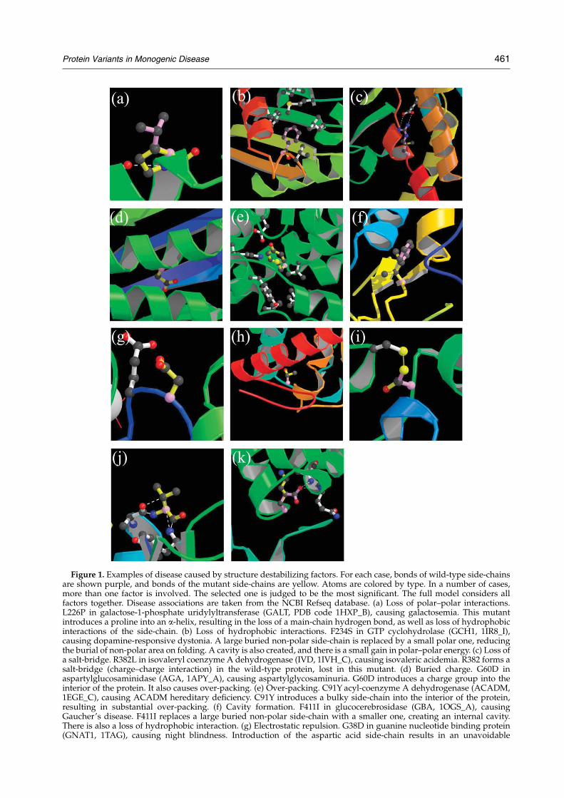
Figure 1. Examples of disease caused by structure destabilizing factors. For each case, bonds of wild-type side-chainsare shown purple, and bonds of the mutant side-chains are yellow. Atoms are colored by type. In a number of cases,more than one factor is involved. The selected one is judged to be the most significant. The full model considers allfactors together. Disease associations are taken from the NCBI Refseq database. (a) Loss of polar–polar interactions.L226P in galactose-1-phosphate uridylyltransferase (GALT, PDB code 1HXP_B), causing galactosemia. This mutantintroduces a proline into an a-helix, resulting in the loss of a main-chain hydrogen bond, as well as loss of hydrophobicinteractions of the side-chain. (b) Loss of hydrophobic interactions. F234S in GTP cyclohydrolase (GCH1, 1IR8_I),causing dopamine-responsive dystonia. A large buried non-polar side-chain is replaced by a small polar one, reducingthe burial of non-polar area on folding. A cavity is also created, and there is a small gain in polar–polar energy. (c) Loss ofa salt-bridge. R382L in isovaleryl coenzyme A dehydrogenase (IVD, 1IVH_C), causing isovaleric acidemia. R382 forms asalt-bridge (charge–charge interaction) in the wild-type protein, lost in this mutant. (d) Buried charge. G60D inaspartylglucosaminidase (AGA, 1APY_A), causing aspartylglycosaminuria. G60D introduces a charge group into theinterior of the protein. It also causes over-packing. (e) Over-packing. C91Y acyl-coenzyme A dehydrogenase (ACADM,1EGE_C), causing ACADM hereditary deficiency. C91Y introduces a bulky side-chain into the interior of the protein,resulting in substantial over-packing. (f) Cavity formation. F411I in glucocerebrosidase (GBA, 1OGS_A), causingGaucher’s disease. F411I replaces a large buried non-polar side-chain with a smaller one, creating an internal cavity.There is also a loss of hydrophobic interaction. (g) Electrostatic repulsion. G38D in guanine nucleotide binding protein(GNAT1, 1TAG), causing night blindness. Introduction of the aspartic acid side-chain results in an unavoidable
Protein Variants in Monogenic Disease 461
462 Protein Variants in Monogenic Disease
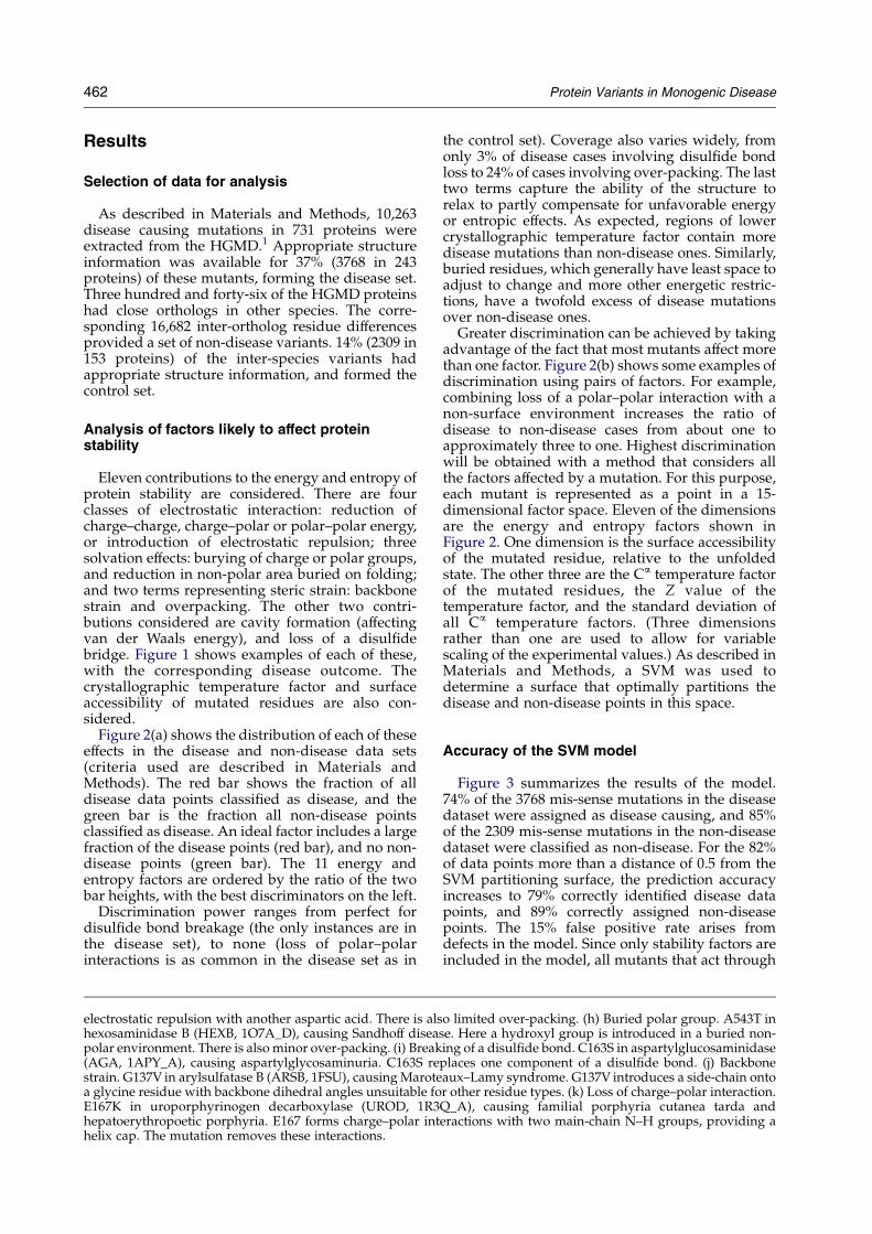
Results
Selection of data for analysis
As described in Materials and Methods, 10,263disease causing mutations in 731 proteins wereextracted from the HGMD.1 Appropriate structureinformation was available for 37% (3768 in 243proteins) of these mutants, forming the disease set.Three hundred and forty-six of the HGMD proteinshad close orthologs in other species. The corre-sponding 16,682 inter-ortholog residue differencesprovided a set of non-disease variants. 14% (2309 in153 proteins) of the inter-species variants hadappropriate structure information, and formed thecontrol set.
Analysis of factors likely to affect proteinstability
Eleven contributions to the energy and entropy ofprotein stability are considered. There are fourclasses of electrostatic interaction: reduction ofcharge–charge, charge–polar or polar–polar energy,or introduction of electrostatic repulsion; threesolvation effects: burying of charge or polar groups,and reduction in non-polar area buried on folding;and two terms representing steric strain: backbonestrain and overpacking. The other two contri-butions considered are cavity formation (affectingvan der Waals energy), and loss of a disulfidebridge. Figure 1 shows examples of each of these,with the corresponding disease outcome. Thecrystallographic temperature factor and surfaceaccessibility of mutated residues are also con-sidered.
Figure 2(a) shows the distribution of each of theseeffects in the disease and non-disease data sets(criteria used are described in Materials andMethods). The red bar shows the fraction of alldisease data points classified as disease, and thegreen bar is the fraction all non-disease pointsclassified as disease. An ideal factor includes a largefraction of the disease points (red bar), and no non-disease points (green bar). The 11 energy andentropy factors are ordered by the ratio of the twobar heights, with the best discriminators on the left.
Discrimination power ranges from perfect fordisulfide bond breakage (the only instances are inthe disease set), to none (loss of polar–polarinteractions is as common in the disease set as in
electrostatic repulsion with another aspartic acid. There is alshexosaminidase B (HEXB, 1O7A_D), causing Sandhoff diseapolar environment. There is also minor over-packing. (i) Break(AGA, 1APY_A), causing aspartylglycosaminuria. C163S restrain. G137V in arylsulfatase B (ARSB, 1FSU), causingMarotea glycine residue with backbone dihedral angles unsuitable foE167K in uroporphyrinogen decarboxylase (UROD, 1R3hepatoerythropoetic porphyria. E167 forms charge–polar inthelix cap. The mutation removes these interactions.
the control set). Coverage also varies widely, fromonly 3% of disease cases involving disulfide bondloss to 24% of cases involving over-packing. The lasttwo terms capture the ability of the structure torelax to partly compensate for unfavorable energyor entropic effects. As expected, regions of lowercrystallographic temperature factor contain moredisease mutations than non-disease ones. Similarly,buried residues, which generally have least space toadjust to change and more other energetic restric-tions, have a twofold excess of disease mutationsover non-disease ones.
Greater discrimination can be achieved by takingadvantage of the fact that most mutants affect morethan one factor. Figure 2(b) shows some examples ofdiscrimination using pairs of factors. For example,combining loss of a polar–polar interaction with anon-surface environment increases the ratio ofdisease to non-disease cases from about one toapproximately three to one. Highest discriminationwill be obtained with a method that considers allthe factors affected by a mutation. For this purpose,each mutant is represented as a point in a 15-dimensional factor space. Eleven of the dimensionsare the energy and entropy factors shown inFigure 2. One dimension is the surface accessibilityof the mutated residue, relative to the unfoldedstate. The other three are the Ca temperature factorof the mutated residues, the Z value of thetemperature factor, and the standard deviation ofall Ca temperature factors. (Three dimensionsrather than one are used to allow for variablescaling of the experimental values.) As described inMaterials and Methods, a SVM was used todetermine a surface that optimally partitions thedisease and non-disease points in this space.
Accuracy of the SVM model
Figure 3 summarizes the results of the model.74% of the 3768 mis-sense mutations in the diseasedataset were assigned as disease causing, and 85%of the 2309 mis-sense mutations in the non-diseasedataset were classified as non-disease. For the 82%of data points more than a distance of 0.5 from theSVM partitioning surface, the prediction accuracyincreases to 79% correctly identified disease datapoints, and 89% correctly assigned non-diseasepoints. The 15% false positive rate arises fromdefects in the model. Since only stability factors areincluded in the model, all mutants that act through
o limited over-packing. (h) Buried polar group. A543T inse. Here a hydroxyl group is introduced in a buried non-ing of a disulfide bond. C163S in aspartylglucosaminidaseplaces one component of a disulfide bond. (j) Backboneaux–Lamy syndrome. G137V introduces a side-chain ontor other residue types. (k) Loss of charge–polar interaction.Q_A), causing familial porphyria cutanea tarda anderactions with two main-chain N–H groups, providing a
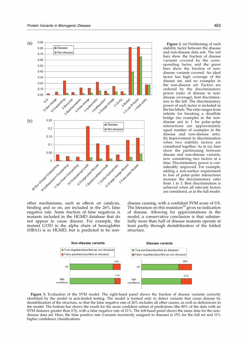
Figure 2. (a) Partitioning of eachstability factor between the diseaseand non-disease data sets. The redbars show the fraction of diseasevariants covered by the corre-sponding factor, and the greenbars show the fraction of non-disease variants covered. An idealfactor has high coverage of thedisease set, and no examples inthe non-disease set. Factors areordered by the discriminatorypower (ratio of disease to non-disease coverage), best discrimina-tors to the left. The discriminatorypower of each factor is included inthe bar labels. The ratio ranges frominfinite for breaking a disulfidebridge (no examples in the non-disease set) to 1 for polar–polarinteractions (an approximatelyequal number of examples in thedisease and non-disease sets).(b) Improvement in discriminationwhen two stability factors areconsidered together. As in (a), barsshow the partitioning betweendisease and non-disease variants,now considering two factors at atime. Discriminatory power is con-siderably improved. For example,adding a non-surface requirementto loss of polar–polar interactionsincrease the discriminatory ratiofrom 1 to 3. Best discrimination isachieved when all relevant factorsare considered, as in the full model.
Protein Variants in Monogenic Disease 463
other mechanisms, such as effects on catalysis,binding and so on, are included in the 26% falsenegative rate. Some fraction of false negatives ismutants included in the HGMD database that donot appear to cause disease. For example, themutant G15D in the alpha chain of hemoglobin(HBA1) is in HGMD, but is predicted to be non-
Figure 3. Evaluation of the SVM model. The right-handidentified by the model in jack-knifed testing. The modeldestabilization of the structure, so that the false negative ratethe model. The bottom bar shows the result for the more conSVM distance greater than 0.5), with a false negative rate of 2disease data set. Here, the false positive rate (variants incorrhigher confidence classifications.
disease causing, with a confident SVM score of 0.8.The literature on this mutation18 gives no indicationof disease. Allowing for approximations in themodel, a conservative conclusion is that substan-tially more than half of disease mutants operate atleast partly through destabilization of the foldedstructure.
panel shows the fraction of disease variants correctlyis trained only to detect variants that cause disease byof 26% includes all other causes, as well as deficiencies infident subset of predictions (the 80% of the data with an1%. The left-hand panel shows the same data for the non-ectly assigned to disease) is 15% for the full set and 11%
464 Protein Variants in Monogenic Disease
Model evaluation using in vitro mutagenesisstability data
The SVM disease model is trained entirely ondisease related mutant data, containing no explicitinformation about stability. Evaluation of themodel’s performance against in vitro mutagenesisfree energy data provides an independent test of thehypothesis that disease is strongly coupled withstructure destabilization. We would expect thatthere should be a strong correlation between apotential disease outcome and the change in the freeenergy difference between the folded and unfoldedstates.
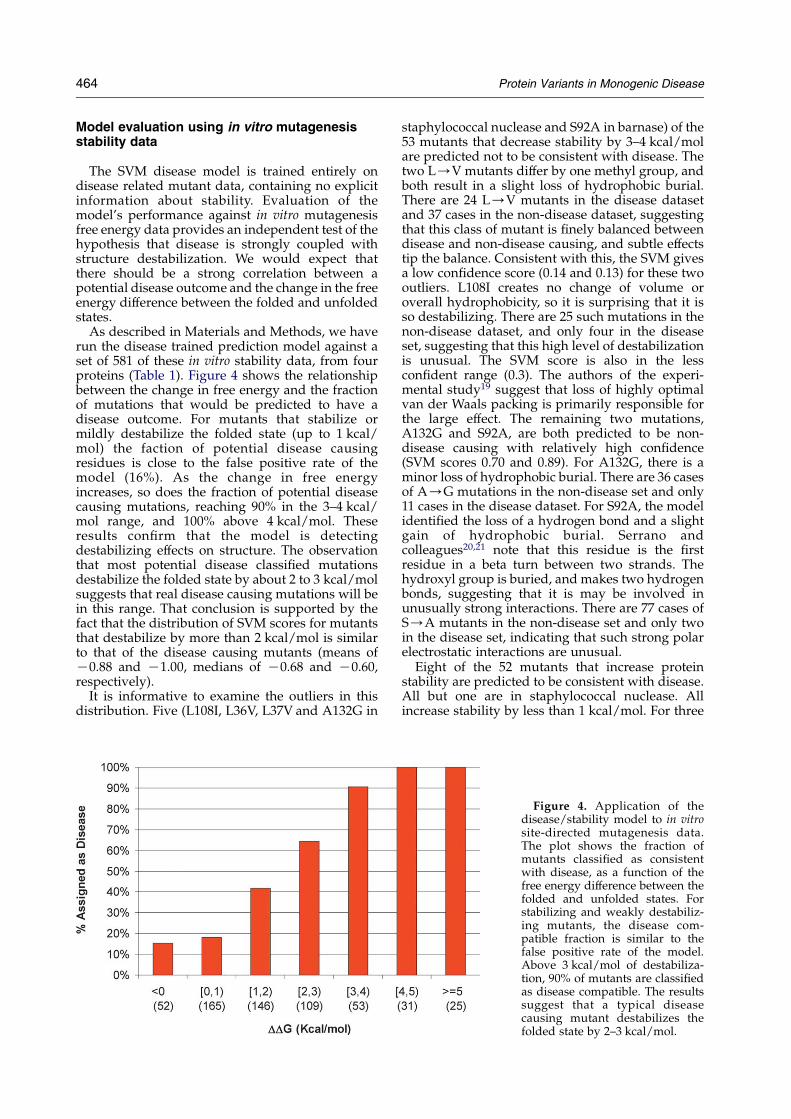
As described in Materials and Methods, we haverun the disease trained prediction model against aset of 581 of these in vitro stability data, from fourproteins (Table 1). Figure 4 shows the relationshipbetween the change in free energy and the fractionof mutations that would be predicted to have adisease outcome. For mutants that stabilize ormildly destabilize the folded state (up to 1 kcal/mol) the faction of potential disease causingresidues is close to the false positive rate of themodel (16%). As the change in free energyincreases, so does the fraction of potential diseasecausing mutations, reaching 90% in the 3–4 kcal/mol range, and 100% above 4 kcal/mol. Theseresults confirm that the model is detectingdestabilizing effects on structure. The observationthat most potential disease classified mutationsdestabilize the folded state by about 2 to 3 kcal/molsuggests that real disease causing mutations will bein this range. That conclusion is supported by thefact that the distribution of SVM scores for mutantsthat destabilize by more than 2 kcal/mol is similarto that of the disease causing mutants (means ofK0.88 and K1.00, medians of K0.68 and K0.60,respectively).
It is informative to examine the outliers in thisdistribution. Five (L108I, L36V, L37V and A132G in
staphylococcal nuclease and S92A in barnase) of the53 mutants that decrease stability by 3–4 kcal/molare predicted not to be consistent with disease. Thetwo L/V mutants differ by one methyl group, andboth result in a slight loss of hydrophobic burial.There are 24 L/V mutants in the disease datasetand 37 cases in the non-disease dataset, suggestingthat this class of mutant is finely balanced betweendisease and non-disease causing, and subtle effectstip the balance. Consistent with this, the SVM givesa low confidence score (0.14 and 0.13) for these twooutliers. L108I creates no change of volume oroverall hydrophobicity, so it is surprising that it isso destabilizing. There are 25 such mutations in thenon-disease dataset, and only four in the diseaseset, suggesting that this high level of destabilizationis unusual. The SVM score is also in the lessconfident range (0.3). The authors of the experi-mental study19 suggest that loss of highly optimalvan der Waals packing is primarily responsible forthe large effect. The remaining two mutations,A132G and S92A, are both predicted to be non-disease causing with relatively high confidence(SVM scores 0.70 and 0.89). For A132G, there is aminor loss of hydrophobic burial. There are 36 casesof A/G mutations in the non-disease set and only11 cases in the disease dataset. For S92A, the modelidentified the loss of a hydrogen bond and a slightgain of hydrophobic burial. Serrano andcolleagues20,21 note that this residue is the firstresidue in a beta turn between two strands. Thehydroxyl group is buried, and makes two hydrogenbonds, suggesting that it is may be involved inunusually strong interactions. There are 77 cases ofS/A mutants in the non-disease set and only twoin the disease set, indicating that such strong polarelectrostatic interactions are unusual.
Eight of the 52 mutants that increase proteinstability are predicted to be consistent with disease.All but one are in staphylococcal nuclease. Allincrease stability by less than 1 kcal/mol. For three
Figure 4. Application of thedisease/stability model to in vitrosite-directed mutagenesis data.The plot shows the fraction ofmutants classified as consistentwith disease, as a function of thefree energy difference between thefolded and unfolded states. Forstabilizing and weakly destabiliz-ing mutants, the disease com-patible fraction is similar to thefalse positive rate of the model.Above 3 kcal/mol of destabiliza-tion, 90% of mutants are classifiedas disease compatible. The resultssuggest that a typical diseasecausing mutant destabilizes thefolded state by 2–3 kcal/mol.
Protein Variants in Monogenic Disease 465
cases: N138G, S128A and H124F, the SVM returns alow confidence score. In none of the other cases is itclear why there is disagreement with experiment.For D21A and D21G, there is a predicted loss ofcharge–charge and charge–polar interactions. Thedistributions of these two mutations between thedisease and non-disease datasets are 8/8 and 57/11,respectively. T41I is predicted to result in a largegain of hydrophobic burial, offset by the loss of acharge–polar and polar–polar interactions in aburied environment. There are 41 cases of T/Imutations in the disease dataset and 18 cases in thenon-disease dataset, most with a predicted largegain of hydrophobic burial and decreased electro-static interactions. G50A is predicted to result inbackbone strain. It is probable that the structure isable to relax to accommodate the change inbackbone angles. The temperature factor is moder-ately high, supporting this possibility. The eighthmutant, N58D, is in barnase. There is a predictedloss of polar–polar interaction and a slight gain ofcharge–polar interaction.
Alternative test sets
This work uses disease and non-disease relateddata for training and testing. Others3,6,7 have useddata on the phenotypic impact of single residuemutants in a bacterial and a phage protein. We haveinvestigated the relationship between our assign-ment of disease potential and phenotypic impact inthese mutagenesis sets. The data are a set of about4000 mutants of the Escherichia coli lac repressor22
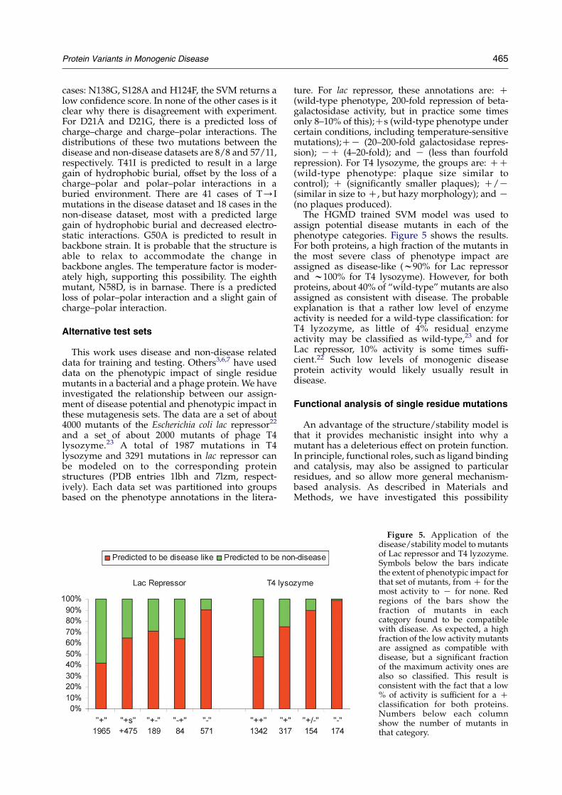
and a set of about 2000 mutants of phage T4lysozyme.23 A total of 1987 mutations in T4lysozyme and 3291 mutations in lac repressor canbe modeled on to the corresponding proteinstructures (PDB entries 1lbh and 7lzm, respect-ively). Each data set was partitioned into groupsbased on the phenotype annotations in the litera-
ture. For lac repressor, these annotations are: C(wild-type phenotype, 200-fold repression of beta-galactosidase activity, but in practice some timesonly 8–10% of this);Cs (wild-type phenotype undercertain conditions, including temperature-sensitivemutations);CK (20–200-fold galactosidase repres-sion); KC (4–20-fold); and K (less than fourfoldrepression). For T4 lysozyme, the groups are: CC(wild-type phenotype: plaque size similar tocontrol); C (significantly smaller plaques); C/K(similar in size to C, but hazy morphology); and K(no plaques produced).The HGMD trained SVM model was used to
assign potential disease mutants in each of thephenotype categories. Figure 5 shows the results.For both proteins, a high fraction of the mutants inthe most severe class of phenotype impact areassigned as disease-like (w90% for Lac repressorand w100% for T4 lysozyme). However, for bothproteins, about 40% of “wild-type” mutants are alsoassigned as consistent with disease. The probableexplanation is that a rather low level of enzymeactivity is needed for a wild-type classification: forT4 lyzozyme, as little of 4% residual enzymeactivity may be classified as wild-type,23 and forLac repressor, 10% activity is some times suffi-cient.22 Such low levels of monogenic diseaseprotein activity would likely usually result indisease.
Functional analysis of single residue mutations
An advantage of the structure/stability model isthat it provides mechanistic insight into why amutant has a deleterious effect on protein function.In principle, functional roles, such as ligand bindingand catalysis, may also be assigned to particularresidues, and so allow more general mechanism-based analysis. As described in Materials andMethods, we have investigated this possibility
Figure 5. Application of thedisease/stability model to mutantsof Lac repressor and T4 lyzozyme.Symbols below the bars indicatethe extent of phenotypic impact forthat set of mutants, fromC for themost activity to K for none. Redregions of the bars show thefraction of mutants in eachcategory found to be compatiblewith disease. As expected, a highfraction of the low activity mutantsare assigned as compatible withdisease, but a significant fractionof the maximum activity ones arealso so classified. This result isconsistent with the fact that a low% of activity is sufficient for a Cclassification for both proteins.Numbers below each columnshow the number of mutants inthat category.
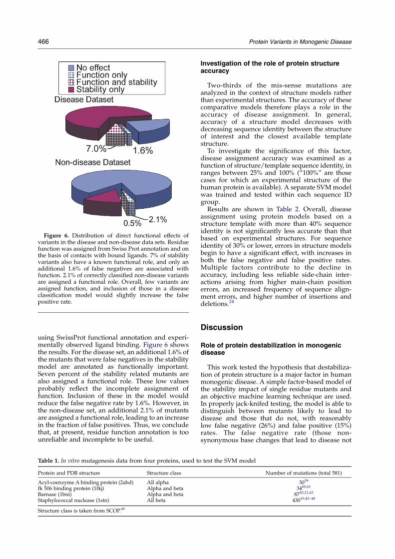
Figure 6. Distribution of direct functional effects ofvariants in the disease and non-disease data sets. Residuefunction was assigned from Swiss Prot annotation and onthe basis of contacts with bound ligands. 7% of stabilityvariants also have a known functional role, and only anadditional 1.6% of false negatives are associated withfunction. 2.1% of correctly classified non-disease variantsare assigned a functional role. Overall, few variants areassigned function, and inclusion of those in a diseaseclassification model would slightly increase the falsepositive rate.
466 Protein Variants in Monogenic Disease
using SwissProt functional annotation and experi-mentally observed ligand binding. Figure 6 showsthe results. For the disease set, an additional 1.6% ofthe mutants that were false negatives in the stabilitymodel are annotated as functionally important.Seven percent of the stability related mutants arealso assigned a functional role. These low valuesprobably reflect the incomplete assignment offunction. Inclusion of these in the model wouldreduce the false negative rate by 1.6%. However, inthe non-disease set, an additional 2.1% of mutantsare assigned a functional role, leading to an increasein the fraction of false positives. Thus, we concludethat, at present, residue function annotation is toounreliable and incomplete to be useful.
Table 1. In vitro mutagenesis data from four proteins, used to
Protein and PDB structure Structure class
Acyl-coenzyme A binding protein (2abd) All alphafk 506 binding protein (1fkj) Alpha and betaBarnase (1bni) Alpha and betaStaphylococcal nuclease (1stn) All beta
Structure class is taken from SCOP.49
Investigation of the role of protein structureaccuracy
Two-thirds of the mis-sense mutations areanalyzed in the context of structure models ratherthan experimental structures. The accuracy of thesecomparative models therefore plays a role in theaccuracy of disease assignment. In general,accuracy of a structure model decreases withdecreasing sequence identity between the structureof interest and the closest available templatestructure.
To investigate the significance of this factor,disease assignment accuracy was examined as afunction of structure/template sequence identity, inranges between 25% and 100% (“100%” are thosecases for which an experimental structure of thehuman protein is available). A separate SVMmodelwas trained and tested within each sequence IDgroup.
Results are shown in Table 2. Overall, diseaseassignment using protein models based on astructure template with more than 40% sequenceidentity is not significantly less accurate than thatbased on experimental structures. For sequenceidentity of 30% or lower, errors in structure modelsbegin to have a significant effect, with increases inboth the false negative and false positive rates.Multiple factors contribute to the decline inaccuracy, including less reliable side-chain inter-actions arising from higher main-chain positionerrors, an increased frequency of sequence align-ment errors, and higher number of insertions anddeletions.24
Discussion
Role of protein destabilization in monogenicdisease
This work tested the hypothesis that destabiliza-tion of protein structure is a major factor in humanmonogenic disease. A simple factor-based model ofthe stability impact of single residue mutants andan objective machine learning technique are used.In properly jack-knifed testing, the model is able todistinguish between mutants likely to lead todisease and those that do not, with reasonablylow false negative (26%) and false positive (15%)rates. The false negative rate (those non-synonymous base changes that lead to disease not
test the SVM model
Number of mutations (total 581)
3059
3460,61
8720,21,62
43019,42–48
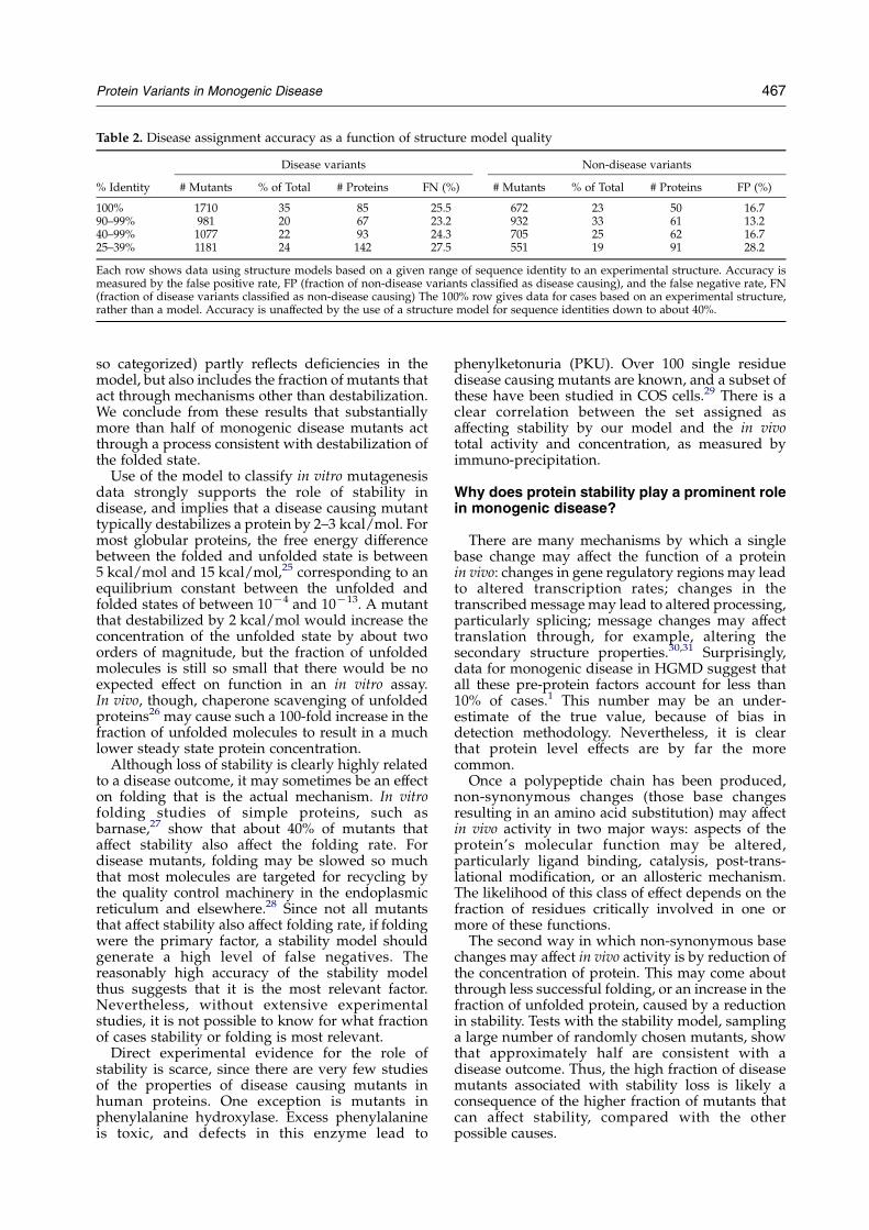
Table 2. Disease assignment accuracy as a function of structure model quality
Disease variants Non-disease variants
% Identity # Mutants % of Total # Proteins FN (%) # Mutants % of Total # Proteins FP (%)
100% 1710 35 85 25.5 672 23 50 16.790–99% 981 20 67 23.2 932 33 61 13.240–99% 1077 22 93 24.3 705 25 62 16.725–39% 1181 24 142 27.5 551 19 91 28.2
Each row shows data using structure models based on a given range of sequence identity to an experimental structure. Accuracy ismeasured by the false positive rate, FP (fraction of non-disease variants classified as disease causing), and the false negative rate, FN(fraction of disease variants classified as non-disease causing) The 100% row gives data for cases based on an experimental structure,rather than a model. Accuracy is unaffected by the use of a structure model for sequence identities down to about 40%.
Protein Variants in Monogenic Disease 467
so categorized) partly reflects deficiencies in themodel, but also includes the fraction of mutants thatact through mechanisms other than destabilization.We conclude from these results that substantiallymore than half of monogenic disease mutants actthrough a process consistent with destabilization ofthe folded state.
Use of the model to classify in vitro mutagenesisdata strongly supports the role of stability indisease, and implies that a disease causing mutanttypically destabilizes a protein by 2–3 kcal/mol. Formost globular proteins, the free energy differencebetween the folded and unfolded state is between5 kcal/mol and 15 kcal/mol,25 corresponding to anequilibrium constant between the unfolded andfolded states of between 10K4 and 10K13. A mutantthat destabilized by 2 kcal/mol would increase theconcentration of the unfolded state by about twoorders of magnitude, but the fraction of unfoldedmolecules is still so small that there would be noexpected effect on function in an in vitro assay.In vivo, though, chaperone scavenging of unfoldedproteins26 may cause such a 100-fold increase in thefraction of unfolded molecules to result in a muchlower steady state protein concentration.
Although loss of stability is clearly highly relatedto a disease outcome, it may sometimes be an effecton folding that is the actual mechanism. In vitrofolding studies of simple proteins, such asbarnase,27 show that about 40% of mutants thataffect stability also affect the folding rate. Fordisease mutants, folding may be slowed so muchthat most molecules are targeted for recycling bythe quality control machinery in the endoplasmicreticulum and elsewhere.28 Since not all mutantsthat affect stability also affect folding rate, if foldingwere the primary factor, a stability model shouldgenerate a high level of false negatives. Thereasonably high accuracy of the stability modelthus suggests that it is the most relevant factor.Nevertheless, without extensive experimentalstudies, it is not possible to know for what fractionof cases stability or folding is most relevant.
Direct experimental evidence for the role ofstability is scarce, since there are very few studiesof the properties of disease causing mutants inhuman proteins. One exception is mutants inphenylalanine hydroxylase. Excess phenylalanineis toxic, and defects in this enzyme lead to
phenylketonuria (PKU). Over 100 single residuedisease causing mutants are known, and a subset ofthese have been studied in COS cells.29 There is aclear correlation between the set assigned asaffecting stability by our model and the in vivototal activity and concentration, as measured byimmuno-precipitation.
Why does protein stability play a prominent rolein monogenic disease?
There are many mechanisms by which a singlebase change may affect the function of a proteinin vivo: changes in gene regulatory regions may leadto altered transcription rates; changes in thetranscribedmessage may lead to altered processing,particularly splicing; message changes may affecttranslation through, for example, altering thesecondary structure properties.30,31 Surprisingly,data for monogenic disease in HGMD suggest thatall these pre-protein factors account for less than10% of cases.1 This number may be an under-estimate of the true value, because of bias indetection methodology. Nevertheless, it is clearthat protein level effects are by far the morecommon.Once a polypeptide chain has been produced,
non-synonymous changes (those base changesresulting in an amino acid substitution) may affectin vivo activity in two major ways: aspects of theprotein’s molecular function may be altered,particularly ligand binding, catalysis, post-trans-lational modification, or an allosteric mechanism.The likelihood of this class of effect depends on thefraction of residues critically involved in one ormore of these functions.The second way in which non-synonymous base
changes may affect in vivo activity is by reduction ofthe concentration of protein. This may come aboutthrough less successful folding, or an increase in thefraction of unfolded protein, caused by a reductionin stability. Tests with the stability model, samplinga large number of randomly chosen mutants, showthat approximately half are consistent with adisease outcome. Thus, the high fraction of diseasemutants associated with stability loss is likely aconsequence of the higher fraction of mutants thatcan affect stability, compared with the otherpossible causes.
†www.snps3d.org
468 Protein Variants in Monogenic Disease
Distinguishing properties of monogenic diseaseproteins
For the 1000 or so monogenic disease proteins inHGMD, the average number of known singleresidue mutants leading to disease is just over 10.1
Yet no mutants directly causative of monogenicdisease are known in the remaining approximately22,000 human proteins. What is the differencebetween these two sets of proteins? First, mono-genic disease proteins may be abnormally unstableor have abnormally fragile folding behavior. Thereis very little data with which to address thispossibility, but many are relatively simple metabolicenzymes, and compared with most humanproteins, the least likely to exhibit this sort offragility. A second possibility is that mutants inmany of the other proteins lead to a non-viablefetus, and so are never classified as disease causing.Gene suppression in Caenorhabditis elegans32 andSaccharomyces,33,34 as well as limited mouse knock-out data all suggest that only 10–20% of proteins areessential in this sense, and so that is unlikelyexplanation. Third, and most probable, monogenicdisease proteins may be the subset to which thesystem is least robust to component failure.Analysis of non-synonymous single nucleotidepolymorphisms in the human population shows asignificant fraction that appear to be as deleteriousto protein structure and function as those found inmonogenic disease genes,3,5,6,35 but with no diseaseoutcome. Limited knowledge of human proteinnetworks makes it difficult to rigorously test thispossibility. Nevertheless, inspection of the pathwaycontext of monogenic disease proteins supports thisexplanation. Many, such as phenylalaninehydroxylase, appear to perform unique roles, withno redundancy alternative pathways. In contrast,inspection of the pathway context of proteinscontaining SNPs that destabilize protein structuresignificantly, such as the T cell receptors,36 usuallysuggests a mechanism that makes the system robustto failure of a protein component. Many different Tcell receptors are involved in an antigenic response,so that reduced effectiveness of some will not haveobvious disease consequences, although it mayinfluence resistance to particular infections in subtlebut significant ways.
Advantages and disadvantages of a proteinstructure-based approach
An advantage of the structure-based approach isthat it provides a detailed atomic level model of theprecise mechanism by which an amino acid changeresults in a change in protein properties.A disadvantage is that it is limited to stabilityeffects, and to cases where structure is available.Use of comparative modeling allowed us to extendthe number of mutants that can be analyzed. Testsshowed that disease prediction accuracy isunaffected by the use of a model, down to 40%sequence identity to a known structure. This is in
keeping with studies of the accuracy of structuremodelingmethods,24 and also partly reflects the factthat the method does not depend on very accuratestructures. Even so, only about 10% of humanprotein domains can currently be analyzed. Therapid advance of structural genomics37 may quicklyreduce this limitation.
Access to results
Analysis results for all missense disease andcontrol mutations is available†.
Materials and Methods
Identification of single residue variants related tomonogenic disease
Genes associated with monogenic disease were identi-fied by checking all 16,220 human gene names in theNCBI Locuslink38 database (as of 04/26/2002) against theHuman Gene Mutation Database1 (HGMD) (as of 02/09/2002). HGMD contains the most comprehensive collec-tion of mutations related to monogenic disease. Most arecausative of monogenic disease, although a few may beassociated with disease as a result of linkage dis-equilibrium rather than directly causative, or contributeto complex trait disease. Later versions of HGMD includemore of the latter class, and so the earlier version waspreferred. A total of 731 genes containing 10,263 singleresidue variations were identified.
Identification of a set of single residue variants notrelated to disease
We also required a control set of mutants, not causativeof disease. It is not known which base variants in thehuman population contribute to complex trait disease,and so it is not possible to use these. Following others,4
we used non-synonymous base differences betweenhuman proteins and closely related proteins in othermammals. The justification here is that almost all variantsthat are fixed between species are essentially neutral andnon-deleterious. To maintain compatibility between thedisease and control sets, the same 731 monogenic diseaseproteins were used. The protein sequences of these geneswere compared to all other mammalian protein sequencesin SWISS-PROT,39 using BLAST.40 Proteins with at least90% sequence identity over at least 80% of the full lengthwere selected. Single residue differences in these align-ments were used as a set of pseudo “mutations”,providing the non-disease set. A total of 348 proteinscontaining 16,682 such single-residue differences to thehuman disease set were obtained.
Selection of sets of mutants with protein structure
Each of the 731 human proteins was checked for entriesin the Protein Data Bank (as of 7/26/2004).41 Templatesfor models of human proteins were taken from the PDBfor cases where there was no human structure available,and there was a PDB entry for an X-ray structure at least
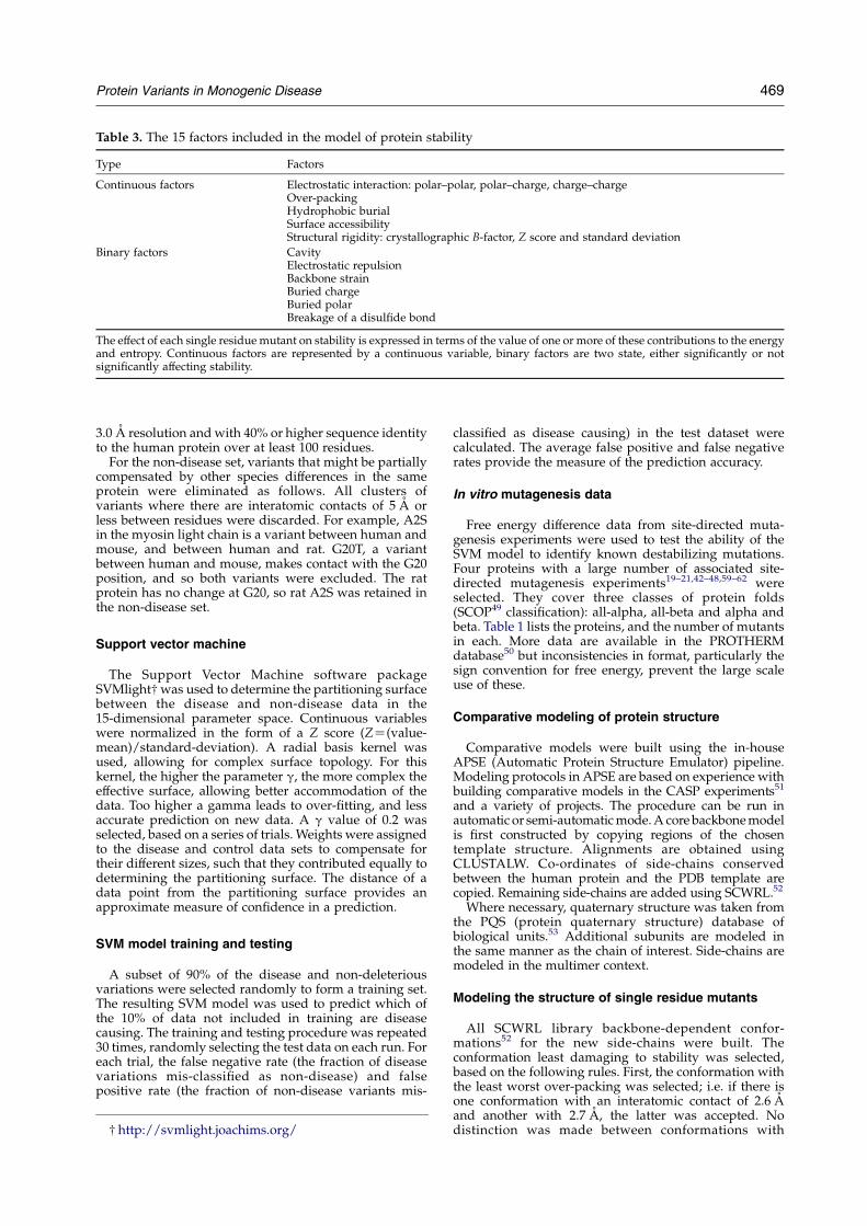
Table 3. The 15 factors included in the model of protein stability
Type Factors
Continuous factors Electrostatic interaction: polar–polar, polar–charge, charge–chargeOver-packingHydrophobic burialSurface accessibilityStructural rigidity: crystallographic B-factor, Z score and standard deviation
Binary factors CavityElectrostatic repulsionBackbone strainBuried chargeBuried polarBreakage of a disulfide bond
The effect of each single residuemutant on stability is expressed in terms of the value of one or more of these contributions to the energyand entropy. Continuous factors are represented by a continuous variable, binary factors are two state, either significantly or notsignificantly affecting stability.
Protein Variants in Monogenic Disease 469
3.0 A resolution andwith 40% or higher sequence identityto the human protein over at least 100 residues.For the non-disease set, variants that might be partially
compensated by other species differences in the sameprotein were eliminated as follows. All clusters ofvariants where there are interatomic contacts of 5 A orless between residues were discarded. For example, A2Sin the myosin light chain is a variant between human andmouse, and between human and rat. G20T, a variantbetween human and mouse, makes contact with the G20position, and so both variants were excluded. The ratprotein has no change at G20, so rat A2S was retained inthe non-disease set.
Support vector machine
The Support Vector Machine software packageSVMlight†was used to determine the partitioning surfacebetween the disease and non-disease data in the15-dimensional parameter space. Continuous variableswere normalized in the form of a Z score (ZZ(value-mean)/standard-deviation). A radial basis kernel wasused, allowing for complex surface topology. For thiskernel, the higher the parameter g, the more complex theeffective surface, allowing better accommodation of thedata. Too higher a gamma leads to over-fitting, and lessaccurate prediction on new data. A g value of 0.2 wasselected, based on a series of trials. Weights were assignedto the disease and control data sets to compensate fortheir different sizes, such that they contributed equally todetermining the partitioning surface. The distance of adata point from the partitioning surface provides anapproximate measure of confidence in a prediction.
SVM model training and testing
A subset of 90% of the disease and non-deleteriousvariations were selected randomly to form a training set.The resulting SVM model was used to predict which ofthe 10% of data not included in training are diseasecausing. The training and testing procedure was repeated30 times, randomly selecting the test data on each run. Foreach trial, the false negative rate (the fraction of diseasevariations mis-classified as non-disease) and falsepositive rate (the fraction of non-disease variants mis-
† http://svmlight.joachims.org/
classified as disease causing) in the test dataset werecalculated. The average false positive and false negativerates provide the measure of the prediction accuracy.
In vitro mutagenesis data
Free energy difference data from site-directed muta-genesis experiments were used to test the ability of theSVM model to identify known destabilizing mutations.Four proteins with a large number of associated site-directed mutagenesis experiments19–21,42–48,59–62 wereselected. They cover three classes of protein folds(SCOP49 classification): all-alpha, all-beta and alpha andbeta. Table 1 lists the proteins, and the number of mutantsin each. More data are available in the PROTHERMdatabase50 but inconsistencies in format, particularly thesign convention for free energy, prevent the large scaleuse of these.
Comparative modeling of protein structure
Comparative models were built using the in-houseAPSE (Automatic Protein Structure Emulator) pipeline.Modeling protocols in APSE are based on experience withbuilding comparative models in the CASP experiments51
and a variety of projects. The procedure can be run inautomatic or semi-automaticmode.A core backbonemodelis first constructed by copying regions of the chosentemplate structure. Alignments are obtained usingCLUSTALW. Co-ordinates of side-chains conservedbetween the human protein and the PDB template arecopied. Remaining side-chains are added using SCWRL.52
Where necessary, quaternary structure was taken fromthe PQS (protein quaternary structure) database ofbiological units.53 Additional subunits are modeled inthe same manner as the chain of interest. Side-chains aremodeled in the multimer context.
Modeling the structure of single residue mutants
All SCWRL library backbone-dependent confor-mations52 for the new side-chains were built. Theconformation least damaging to stability was selected,based on the following rules. First, the conformation withthe least worst over-packing was selected; i.e. if there isone conformation with an interatomic contact of 2.6 Aand another with 2.7 A, the latter was accepted. Nodistinction was made between conformations with
470 Protein Variants in Monogenic Disease
contacts 3.0 A or longer. If more than one conformationremained, the one with the least loss of hydrophobic areawas selected. In cases where there is no loss ofhydrophobic area, conformations with loss of a salt-bridge were next eliminated, then those with electrostaticrepulsion, hydrogen bond loss, cavity formation, back-bone strain, introduction of a buried charge, and finally,introduction of a buried polar group.
Modeling the stability impact of a single residuemutant
Table 3 lists the stability factors that provide the 15dimensions used in assessing the impact of each mutanton protein stability. These are divided into those factorstreated as continuous variables, and those treated as twostate variables (significantly destabilizing or not).
Continuous factors
(1) Electrostatic interactions. The difference in electrostaticenergy between a wild-type protein and its corre-sponding mutant was calculated using a simpleCoulomb’s law treatment, with no solvent model.The partial electrostatic interaction energy between apair of polar or charged groups i and j is calculated inthe usual manner as:
Eij ZKX
k
X
l
qkql=rlk
where the sums are over all atoms k of group i andatoms l of group j, the qs are the partial atomic chargesin electrons, and rlk is the distance between atoms land k, in A. K is the scaling constant (332) nominallyconverting energies to kcal/mol. (Absolute scale isnot significant here, because of the Z scorenormalization.) Interactions between a pair of groupsare included if the centers of charge are less than acutoff distance dc apart. The center of charge of agroup rc is defined as:
rc ZX
k
jqkjrk=X
k
jqkj
where the sum is over all atoms in the group.Electrostatic group definitions and partial atomiccharges are as defined by Pedersen & Moult.54 Thethreshold for group–group interactions, dc, is 5 A.This protocol for electrostatic calculations has beenshown to be effective at identity incorrect structuralfeatures in experimental structures.55
(2) Overpacking. For each mutant, the closest inter-atomicdistance between the mutant residue and anyneighboring residue was used.
(3) Relative surface accessibility. Solvent accessible sur-face56 was calculated with in-house software. Therelative surface accessibility of a residue is defined asthe surface area of the side-chain in the folded statedivided by an estimate of the average surface area inthe unfolded state.57
(4) Hydrophobic burial change. The change in buried non-polar area DANP resulting from a single residuemutation is defined as:
DANP ZX
i
DaiKX
j
Daj
where the first sum i is the change in non-polar areaon folding for all non-polar atoms in the mutant
structure and the second sum j is over all non-polaratoms in the wild-type structure. The change inatomic non-polar area in folding is given by:
DaZ auKaf
where au is the estimate of the average atomic surfacearea in the unfolded state57 for that atom, and af is thecalculated atomic area in the folded structure. Non-polar atoms are those assigned zero charge.
(5) Crystallographic temperature factors. For each experi-mental structure used directly or as a model, theaverage temperature factor hBi, and standarddeviation s(B) over all Ca atoms was calculated, andused to obtain a temperature factor Z score for eachCa: ZiZ ðBiKhBiÞ=sðBÞ. Bi, Zi, and s(B) were used asparameters in the SVM.
Binary factors
(6) A cavity is assigned to any mutation resulting in theloss of volume of an aliphatic carbon group or greaterat a zero solvent accessibility position. For example,Ala mutated to Gly, where the wild-type Cb atom haszero solvent accessibility.
(7) Electrostatic repulsion is assigned to any mutationwhich results in two like charged groups with anunavoidable atomic contact of less than 4.5 A.
(8) Backbone strain is assigned to any mutation if one ofthe following conditions is met. (A) Replacement of aglycine residue with f/j angles in a non-allowedregion for other residue types. Allowed regions werethose covering 90% of observed f/j values, asprovided in PROCHECK.58 (B) Replacement of a cis-proline (uZ0(G60)8) with another residue. (C)Replacement of another residue by proline,where the f value is inappropriate (permitted f forProZK60(G15)8).
(9) Buried charge is assigned to any mutation that resultsin a zero solvent accessibility, electrostaticallyisolated, charge group.
(10)Buried polar is assigned to any mutation that resultsin a zero solvent accessibility polar group with nohydrogen bond. A hydrogen bond is defined as adonor to acceptor distance %2.5 A, and an angle atthe acceptor R90.08.
(11)Breakage of a disulfide bond is assigned to anymutation that replaces a cysteine residue in an S–Sbond with a non-cysteine residue.
Evaluation of discrimination power of each stabilityfactor
The frequencies of each stability factor in the diseaseand non-disease datasets were calculated. The ratio of thetwo frequencies defines a discrimination power. For thispurpose, a threshold was chosen for each of thecontinuous factors. Any mutation with a value higherthan the threshold was considered to destabilize proteinstructure. Thresholds were chosen by inspection of thedistribution of values for the disease and non-disease sets,selecting levels that provide a high fraction of truepositives and true negatives, while minimizing falsenegatives and false positives. The following values wereused:
(1) Overpacking: at least one unavoidable atomic contact
Protein Variants in Monogenic Disease 471
of 2.5 A or less of the mutated residue to aneighboring one.
(2) Hydrophobic burial: loss of hydrophobic burial ofmore than 50 A2.
(3) Electrostatic interaction: any reduction in electrostaticinteraction energy, for polar–polar, charge–charge andcharge–polar interactions.
(4) Buried residue: relative residue accessibility of lessthan 20% (i.e. the wild-type side-chain accessibility isless than 20% of the estimated average unfolded stateaccessibility).
(5) Moderate crystallographic temperature factor: the Ca
temperature factor of the mutated residue has a Zscore of less thanC1 (i.e. the temperature factor is lessthan one standard deviation above the mean for theprotein).
Identification of residues with a role in molecularfunction
Each mutated residue, and all residues with one ormore atomic contacts of 6 A or less to it, was checkedagainst the SWISS-PROT feature annotation table forpossible functional effects. Additionally, a check wasmade for atomic contacts of the mutated residue of 6 A orless to any ligand atom in PDB entries for that protein andother X-ray structures with at least 40% sequence identityover at least 100 amino acid residues, and at 3.0 A orbetter resolution.
Acknowledgements
This work was supported by grant LM07174 fromthe National Library of Medicine.
References
1. Stenson, P. D., Ball, E. V., Mort, M., Phillips, A. D.,Shiel, J. A., Thomas, N. S. et al. (2003). Human GeneMutation Database (HGMD): 2003 update. Hum.Mutat. 21, 577–581.
2. Wang, Z. & Moult, J. (2001). SNPs, protein structure,and disease. Hum. Mutat. 17, 263–270.
3. Ng, P. C. & Henikoff, S. (2003). SIFT: predicting aminoacid changes that affect protein function. Nucl. AcidsRes. 31, 3812–3814.
4. Sunyaev, S., Ramensky, V., Koch, I., Lathe, W., 3rd,Kondrashov, A. S. & Bork, P. (2001). Prediction ofdeleterious human alleles. Hum. Mol. Genet. 10,591–597.
5. Ramensky, V., Bork, P. & Sunyaev, S. (2002). Humannon-synonymous SNPs: server and survey. Nucl.Acids Res. 30, 3894–3900.
6. Chasman, D. & Adams, R. M. (2001). Predicting thefunctional consequences of non-synonymous singlenucleotide polymorphisms: structure-basedassessment of amino acid variation. J. Mol. Biol. 307,683–706.
7. Krishnan, V. G. & Westhead, D. R. (2003). Acomparative study of machine-learning methods topredict the effects of single nucleotidepolymorphisms on protein function. Bioinformatics,19, 2199–2209.
8. Ng, P. C., Henikoff, J. G. &Henikoff, S. (2000). PHAT: atransmembrane-specific substitution matrix.Predicted hydrophobic and transmembrane.Bioinformatics, 16, 760–766.
9. Lau, A. Y. & Chasman, D. I. (2004). Functionalclassification of proteins and protein variants. Proc.Natl Acad. Sci. USA, 101, 6576–6581.
10. Valdar, W. S. & Thornton, J. M. (2001). Conservationhelps to identify biologically relevant crystal contacts.J. Mol. Biol. 313, 399–416.
11. Dodge, C., Schneider, R., Sander, C. & The,H. S. S. P. (1998). database of protein structure-sequence alignments and family profiles. Nucl.Acids Res. 26, 313–315.
12. Przybylski, D. & Rost, B. (2002). Alignments grow,secondary structure prediction improves. Proteins:Struct. Funct. Genet. 46, 197–205.
13. Beveridge, D. L. & DiCapua, F. M. (1989). Free energyvia molecular simulation: applications to chemicaland biomolecular systems. Annu. Rev. Biophys.Biophys. Chem. 18, 431–492.
14. Mark, A. E. & van Gunsteren, W. F. (1994). Decompo-sition of the free energy of a system in terms ofspecific interactions. Implications for theoretical andexperimental studies. J. Mol. Biol. 240, 167–176.
15. Pan, Y. & Daggett, V. (2001). Direct comparison ofexperimental and calculated folding free energies forhydrophobic deletion mutants of chymotrypsininhibitor 2: free energy perturbation calculationsusing transition and denatured states from moleculardynamics simulations of unfolding. Biochemistry, 40,2723–2731.
16. Guerois, R., Nielsen, J. E. & Serrano, L. (2002).Predicting changes in the stability of proteins andprotein complexes: a study of more than 1000mutations. J. Mol. Biol. 320, 369–387.
17. Vapnik, V. N. (1995). The Nature of Statistical LearningTheory, Springer, New York.
18. Molchanova, T. P., Pobedimskaya, D. D. & Postnikov,Yu. V. (1994). A simplified procedure for sequencingamplified DNA containing the alpha 2- or alpha1-globin gene. Hemoglobin, 18, 251–255.
19. Holder, J. B., Bennett, A. F., Chen, J., Spencer, D. S.,Byrne, M. P. & Stites, W. E. (2001). Energetics of sidechain packing in staphylococcal nuclease assessed byexchange of valines, isoleucines, and leucines.Biochemistry, 40, 13998–14003.
20. Serrano, L., Kellis, J. T., Jr, Cann, P., Matouschek, A. &Fersht, A. R. (1992). The folding of an enzyme. II.Substructure of barnase and the contribution ofdifferent interactions to protein stability. J. Mol. Biol.224, 783–804.
21. Serrano, L., Matouschek, A. & Fersht, A. R. (1992). Thefolding of an enzyme. III. Structure of the transitionstate for unfolding of barnase analysed by a proteinengineering procedure. J. Mol. Biol. 224, 805–818.
22. Markiewicz, P., Kleina, L. G., Cruz, C., Ehret, S. &Miller, J. H. (1994). Genetic studies of the lac repressor.XIV. Analysis of 4000 altered Escherichia coli lacrepressors reveals essential and non-essential resi-dues, as well as “spacers” which do not require aspecific sequence. J. Mol. Biol. 240, 421–433.
23. Rennell, D., Bouvier, S. E., Hardy, L. W. & Poteete,A. R. (1991). Systematic mutation of bacteriophage T4lysozyme. J. Mol. Biol. 222, 67–88.
24. Tramontano, A. & Morea, V. (2003). Assessment ofhomology-based predictions in CASP5. Proteins:Struct. Funct. Genet. 53, 352–368. (Suppl. 6).

472 Protein Variants in Monogenic Disease
25. Privalov, P. L. (1979). Stability of proteins: smallglobular proteins. Advan. Protein Chem. 33, 167–241.
26. Hohfeld, J., Cyr, D. M. & Patterson, C. (2001). Fromthe cradle to the grave: molecular chaperones thatmay choose between folding and degradation. EMBORep. 2, 885–890.
27. Serrano, L., Matouschek, A. & Fersht, A. R. (1992). Thefolding of an enzyme. VI. The folding pathway ofbarnase: comparison with theoretical models. J. Mol.Biol. 224, 847–859.
28. Plemper, R. K. & Wolf, D. H. (1999). Retrogradeprotein translocation: ERADication of secretory pro-teins in health and disease. Trends Biochem. Sci. 24,266–270.
29. Scriver, C. R., Hurtubise, M., Konecki, D.,Phommarinh, M., Prevost, L., Erlandsen, H. et al.(2003). PAHdb 2003: what a locus-specific knowl-edgebase can do. Hum. Mutat. 21, 333–344.
30. Shen, L. X., Basilion, J. P. & Stanton, V. P., Jr (1999).Single-nucleotide polymorphisms can cause differentstructural folds of mRNA. Proc. Natl Acad. Sci. USA,96, 7871–7876.
31. Pelletier, J. & Sonenberg, N. (1987). The involvementof mRNA secondary structure in protein synthesis.Biochem. Cell. Biol. 65, 576–581.
32. Kamath, R. S., Fraser, A. G., Dong, Y., Poulin, G.,Durbin, R., Gotta, M. et al. (2003). Systematicfunctional analysis of the Caenorhabditis elegansgenome using RNAi. Nature, 421, 231–237.
33. Cliften, P., Sudarsanam, P., Desikan, A., Fulton, L.,Fulton, B., Majors, J. et al. (2003). Finding functionalfeatures in Saccharomyces genomes by phylogeneticfootprinting. Science, 301, 71–76.
34. Rubin, G. M., Yandell, M. D., Wortman, J. R., GaborMiklos, G. L., Nelson, C. R., Hariharan, I. K. et al.(2000). Comparative genomics of the eukaryotes.Science, 287, 2204–2215.
35. Yue, P. & Moult, J. (2005). Identification and analysisof deleterious human SNPs. J. Mol. Biol. In the press.
36. Wang, Z. & Moult, J. (2003). Three-dimensionalstructural location and molecular functional effectsof missense SNPs in the Tcell receptor Vbeta domain.Proteins: Struct. Funct. Genet. 53, 748–757.
37. Service, R. (2005). Structural biology. Structuralgenomics, round 2. Science, 307, 1554–1558.
38. Wheeler, D. L., Church, D. M., Edgar, R., Federhen, S.,Helmberg, W., Madden, T. L. et al. (2004). Databaseresources of the National Center for BiotechnologyInformation: update. Nucl. Acids Res. 32, D35–D40.
39. Boeckmann, B., Bairoch, A., Apweiler, R., Blatter,M. C., Estreicher, A., Gasteiger, E. et al. (2003). TheSWISS-PROT protein knowledgebase and its sup-plement TrEMBL in 2003. Nucl. Acids Res. 31, 365–370.
40. Altschul, S. F., Madden, T. L., Schaffer, A. A., Zhang, J.,Zhang, Z., Miller, W. & Lipman, D. J. (1997). GappedBLAST and PSI-BLAST: a new generation of proteindatabase search programs. Nucl. Acids Res. 25,3389–3402.
41. Deshpande, N., Addess, K. J., Bluhm, W. F., Merino-Ott, J. C., Townsend-Merino, W., Zhang, Q. et al. (2005).The RCSB Protein Data Bank: a redesigned querysystem and relational database based on the mmCIFschema.Nucl. Acids Res. 33, D233–D237.
42. Shortle, D., Stites, W. E. & Meeker, A. K. (1990).Contributions of the large hydrophobic amino acidsto the stability of staphylococcal nuclease.Biochemistry, 29, 8033–8041.
43. Green, S. M. & Shortle, D. (1993). Patterns of
nonadditivity between pairs of stability mutations instaphylococcal nuclease. Biochemistry, 32,10131–10139.
44. Green, S. M., Meeker, A. K. & Shortle, D. (1992).Contributions of the polar, uncharged amino acids tothe stability of staphylococcal nuclease: evidence formutational effects on the free energy of the denaturedstate. Biochemistry, 31, 5717–5728.
45. Meeker, A. K., Garcia-Moreno, B. & Shortle, D. (1996).Contributions of the ionizable amino acids to thestability of staphylococcal nuclease. Biochemistry, 35,6443–6449.
46. Stites, W. E., Meeker, A. K. & Shortle, D. (1994).Evidence for strained interactions between side-chains and the polypeptide backbone. J. Mol. Biol.235, 27–32.
47. Schwehm, J. M., Kristyanne, E. S., Biggers, C. C. &Stites, W. E. (1998). Stability effects of increasing thehydrophobicity of solvent-exposed side chains instaphylococcal nuclease. Biochemistry, 37, 6939–6948.
48. Byrne, M. P., Manuel, R. L., Lowe, L. G. & Stites, W. E.(1995). Energetic contribution of side chain hydrogenbonding to the stability of staphylococcal nuclease.Biochemistry, 34, 13949–13960.
49. Andreeva, A., Howorth, D., Brenner, S. E.,Hubbard, T. J., Chothia, C. & Murzin, A. G.(2004). SCOP database in 2004: refinements inte-grate structure and sequence family data. Nucl.Acids Res. 32, D226–D229.
50. Bava, K. A., Gromiha, M. M., Uedaira, H., Kitajima, K.& Sarai, A. (2004). ProTherm, version 4.0: thermo-dynamic database for proteins and mutants. Nucl.Acids Res. 32, D120–D121.
51. Samudrala, R. & Moult, J. (1997). Handling context-sensitivity in protein structures using graph theory:bona fide prediction. Proteins: Struct. Funct. Genet. 1,43–49.
52. Canutescu, A. A., Shelenkov, A. A. & Dunbrack, R. L.,Jr (2003). A graph-theory algorithm for rapid proteinside-chain prediction. Protein Sci. 12, 2001–2014.
53. Henrick, K. & Thornton, J. M. (1998). PQS: a proteinquaternary structure file server. Trends Biochem. Sci.23, 358–361.
54. Pedersen, J. T. & Moult, J. (1997). Protein foldingsimulations with genetic algorithms and a detailedmolecular description. J. Mol. Biol. 269, 240–259.
55. Oliva, M. T. & Moult, J. (1999). Local electrostaticoptimization in proteins. Protein Eng. 12, 727–735.
56. Lee, B. & Richards, F. M. (1971). The interpretation ofprotein structures: estimation of static accessibility.J. Mol. Biol. 55, 379–400.
57. Shrake, A. & Rupley, J. A. (1973). Environment andexposure to solvent of protein atoms. Lysozyme andinsulin. J. Mol. Biol. 79, 351–371.
58. Laskowski, R. A. M. M., Moss, D. S. & Thornton, J. M.(1993). PROCHECK: a program to check the stereo-chemical quality of protein structures. J. Appl.Crystallog. 26, 283–291.
59. Kragelund, B. B., Osmark, P., Neergaard, T. B.,Schiodt, J., Kristiansen, K., Knudsen, J. & Poulsen,F. M. (1999). The formation of a native-like structurecontaining eight conserved hydrophobic residues israte limiting in two-state protein folding of ACBP.Nature Struct. Biol. 6, 594–601.
60. Main, E. R., Fulton, K. F. & Jackson, S. E. (1998).Context-dependent nature of destabilizing mutationson the stability of FKBP12. Biochemistry, 37, 6145–6153.
61. Fulton, K. F., Main, E. R., Daggett, V. & Jackson, S. E.
Protein Variants in Monogenic Disease 473
(1999). Mapping the interactions present in thetransition state for unfolding/folding of FKBP12.J. Mol. Biol. 291, 445–461.
62. Serrano, L., Sancho, J., Hirshberg, M. & Fersht, A. R.(1992). Alpha-helix stability in proteins. I. Empirical
correlations concerning substitution of side-chains atthe N and C-caps and the replacement of alanine byglycine or serine at solvent-exposed surfaces. J. Mol.Biol. 227, 544–559.
Edited C. R. Matthews
(Received 7 May 2005; received in revised form 8 August 2005; accepted 10 August 2005)Available online 31 August 2005