Lopez Circulation and Gas Exchange Louie - Weebly
75
Lectures by Chris Romero Circulation and Gas Exchange Mark Louie D. Lopez Department of Biology Polytechnic University of the Philippines ecture of Mark Louie D. Lop
Transcript of Lopez Circulation and Gas Exchange Louie - Weebly

Copyright © 2005 Pearson Education, Inc. publishing as Benjamin Cummings
PowerPoint Lectures for
Biology, Seventh Edition
Neil Campbell and Jane Reece
Lectures by Chris Romero
Circulation and
Gas Exchange
Mark Louie D. Lopez
Department of Biology
Polytechnic University of the Philippines
Lectu
re of
Mark Lo
uie D
. Lop
ez
Copyright © 2005 Pearson Education, Inc. publishing as Benjamin Cummings
EXCHANGE OF MATERIALS
• Overview: Trading with the Environment
• Every organism must exchange materials with
its environment
– And this exchange ultimately occurs at the
cellular level
Lectu
re of
Mark Lo
uie D
. Lop
ez

Copyright © 2005 Pearson Education, Inc. publishing as Benjamin Cummings
EXCHANGE OF MATERIALS
• In unicellular organisms
– These exchanges occur directly with the
environment
• For most of the cells making up multicellular
organisms
– Direct exchange with the environment is not
possible
Lectu
re of
Mark Lo
uie D
. Lop
ez
Copyright © 2005 Pearson Education, Inc. publishing as Benjamin Cummings
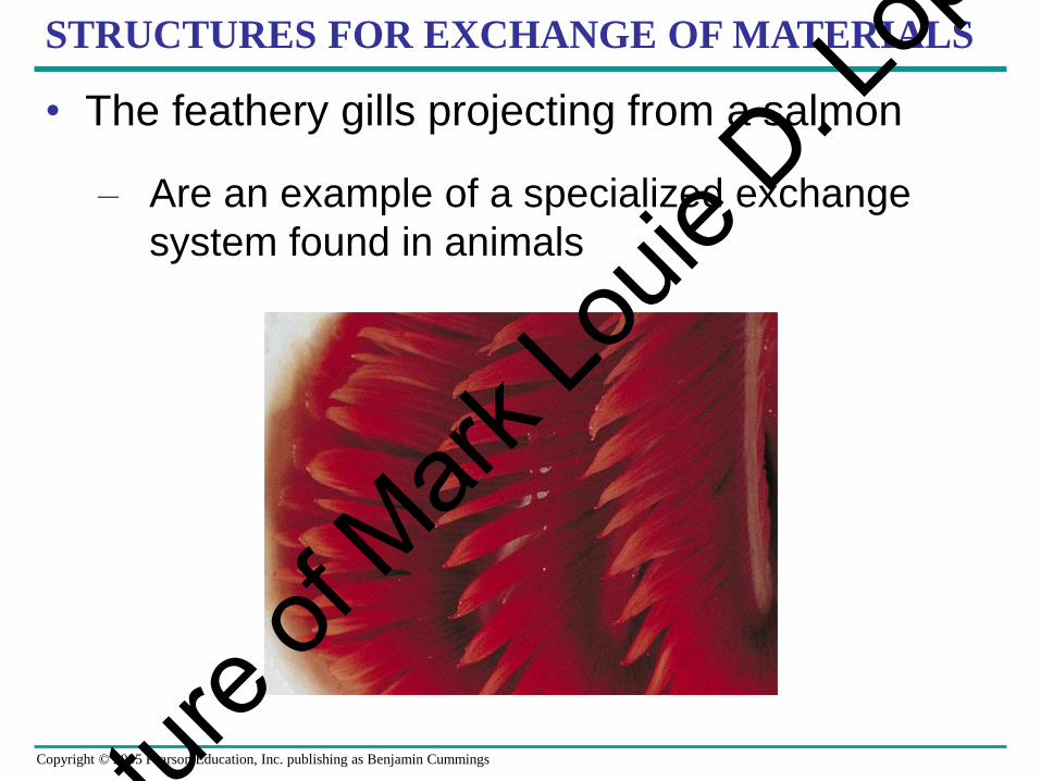
STRUCTURES FOR EXCHANGE OF MATERIALS
• The feathery gills projecting from a salmon
– Are an example of a specialized exchange
system found in animals
Lectu
re of
Mark Lo
uie D
. Lop
ez

Copyright © 2005 Pearson Education, Inc. publishing as Benjamin Cummings
CIRCULATORY SYSTEM
• Circulatory systems reflect phylogeny
• Transport systems
– Functionally connect the organs of exchange
with the body cells
Lectu
re of
Mark Lo
uie D
. Lop
ez
Copyright © 2005 Pearson Education, Inc. publishing as Benjamin Cummings
CIRCULATORY SYSTEM
• Most complex animals have internal transport
systems
– That circulate fluid, providing a lifeline between
the aqueous environment of living cells and
the exchange organs, such as lungs, that
exchange chemicals with the outside
environment
Lectu
re of
Mark Lo
uie D
. Lop
ez

Copyright © 2005 Pearson Education, Inc. publishing as Benjamin Cummings
INVERTEBRATE CIRCULATION
• The wide range of invertebrate body size and
form
– Is paralleled by a great diversity in
circulatory systems
Lectu
re of
Mark Lo
uie D
. Lop
ez

Copyright © 2005 Pearson Education, Inc. publishing as Benjamin Cummings
GASTROVASCULAR CAVITIES
• Simple animals, such as cnidarians
– Have a body wall only two cells thick that
encloses a gastrovascular cavity
• The gastrovascular cavity
– Functions in both digestion and distribution of
substances throughout the body
Lectu
re of
Mark Lo
uie D
. Lop
ez
Copyright © 2005 Pearson Education, Inc. publishing as Benjamin Cummings
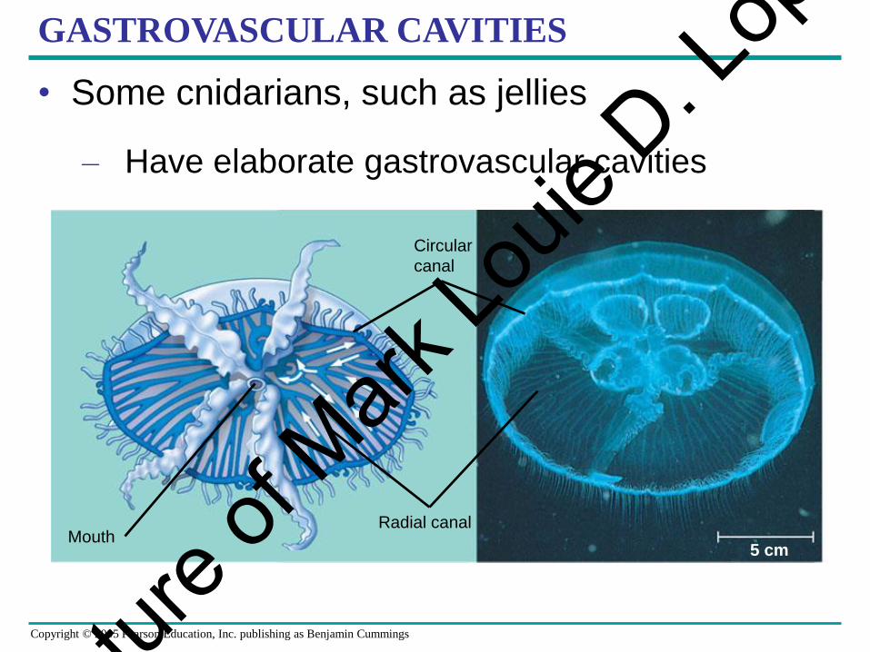
GASTROVASCULAR CAVITIES
• Some cnidarians, such as jellies
– Have elaborate gastrovascular cavities
Circular
canal
Radial canal
5 cmMouth
Lectu
re of
Mark Lo
uie D
. Lop
ez

Copyright © 2005 Pearson Education, Inc. publishing as Benjamin Cummings
OPEN AND CLOSED CIRCULATORY SYSTEMS
• More complex animals
– Have one of two types of circulatory systems:
open or closed
• Both of these types of systems have three
basic components
– A circulatory fluid (blood)
– A set of tubes (blood vessels)
– A muscular pump (the heart)
Lectu
re of
Mark Lo
uie D
. Lop
ez

Copyright © 2005 Pearson Education, Inc. publishing as Benjamin Cummings
OPEN CIRCULATORY SYSTEM
• In insects, other arthropods, and most molluscs
– Blood bathes the organs directly in an open
circulatory system
Heart
Hemolymph in sinuses
surrounding ograns
Anterior
vessel
Tubular heart
Lateral
vessels
Ostia
(a) An open circulatory systemFigure 42.3a
Lectu
re of
Mark Lo
uie D
. Lop
ez

Copyright © 2005 Pearson Education, Inc. publishing as Benjamin Cummings
CLOSED CIRCULATORY SYSTEM
• In a closed circulatory system
– Blood is confined to vessels and is distinct
from the interstitial fluid
Interstitial
fluid
Heart
Small branch vessels
in each organ
Dorsal vessel
(main heart)
Ventral vesselsAuxiliary hearts
(b) A closed circulatory system
Lectu
re of
Mark Lo
uie D
. Lop
ez

Copyright © 2005 Pearson Education, Inc. publishing as Benjamin Cummings
SURVEY OF VERTEBRATE CIRCULATION
• Humans and other vertebrates have a closed
circulatory system
– Often called the cardiovascular system
• Blood flows in a closed cardiovascular system
– Consisting of blood vessels and a two- to four-
chambered heart
Lectu
re of
Mark Lo
uie D
. Lop
ez

Copyright © 2005 Pearson Education, Inc. publishing as Benjamin Cummings
BLOOD VESSELS
• Arteries carry blood to capillaries
– The sites of chemical exchange between the
blood and interstitial fluid
• Veins
– Return blood from capillaries to the heart
Lectu
re of
Mark Lo
uie D
. Lop
ez

Copyright © 2005 Pearson Education, Inc. publishing as Benjamin Cummings
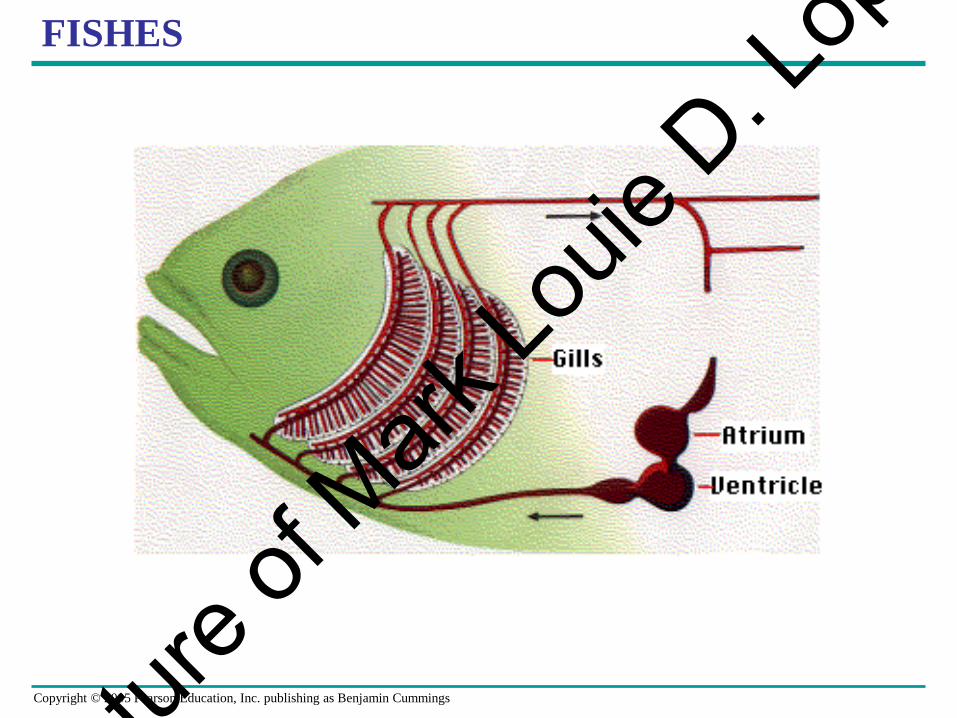
FISHES
• A fish heart has two main chambers
– One ventricle and one atrium
• Blood pumped from the ventricle
– Travels to the gills, where it picks up O2 and
disposes of CO2
Lectu
re of
Mark Lo
uie D
. Lop
ez
Copyright © 2005 Pearson Education, Inc. publishing as Benjamin Cummings
FISHES
Lectu
re of
Mark Lo
uie D
. Lop
ez

Copyright © 2005 Pearson Education, Inc. publishing as Benjamin Cummings
AMPHIBIANS
• Frogs and other amphibians
– Have a three-chambered heart, with two atria
and one ventricle
• The ventricle pumps blood into a forked artery
– That splits the ventricle’s output into the
pulmocutaneous circuit and the systemic
circuit
Lectu
re of
Mark Lo
uie D
. Lop
ez

Copyright © 2005 Pearson Education, Inc. publishing as Benjamin Cummings
AMPHIBIANS
Lectu
re of
Mark Lo
uie D
. Lop
ez

Copyright © 2005 Pearson Education, Inc. publishing as Benjamin Cummings
REPTILES (EXCEPT BIRDS)
• Reptiles have double circulation
– With a pulmonary circuit (lungs) and a
systemic circuit
• Turtles, snakes, and lizards
– Have a three-chambered heart
Lectu
re of
Mark Lo
uie D
. Lop
ez

Copyright © 2005 Pearson Education, Inc. publishing as Benjamin Cummings
MAMMALS AND BIRDS
• In all mammals and birds
– The ventricle is completely divided into
separate right and left chambers
• The left side of the heart pumps and receives
only oxygen-rich blood
– While the right side receives and pumps only
oxygen-poor blood
Lectu
re of
Mark Lo
uie D
. Lop
ez

Copyright © 2005 Pearson Education, Inc. publishing as Benjamin Cummings
VARIATION IN ANIMAL CIRCULATORY SYSTEM
FISHES AMPHIBIANS REPTILES (EXCEPT BIRDS) MAMMALS AND BIRDS
Systemic capillaries Systemic capillaries Systemic capillaries Systemic capillaries
Lung capillaries Lung capillariesLung and skin capillariesGill capillaries
Right Left Right Left Right Left
Systemic
circuitSystemic
circuit
Pulmocutaneous
circuit
Pulmonary
circuitPulmonary
circuit
Systemic
circulationVein
Atrium (A)
Heart:
ventricle (V)
Artery Gill
circulation
A
V VV VV
A A A AALeft
Systemic
aorta
Right
systemic
aorta
• Vertebrate circulatory systems
Lectu
re of
Mark Lo
uie D
. Lop
ez

Copyright © 2005 Pearson Education, Inc. publishing as Benjamin Cummings
MAMMALIAN CIRCULATORY SYSTEM
• Double circulation in mammals depends on the
anatomy and pumping cycle of the heart
• The structure and function of the human
circulatory system
– Can serve as a model for exploring
mammalian circulation in general
Lectu
re of
Mark Lo
uie D
. Lop
ez
Copyright © 2005 Pearson Education, Inc. publishing as Benjamin Cummings
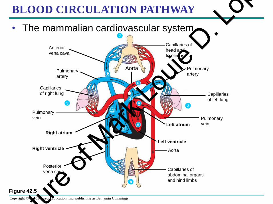
BLOOD CIRCULATION PATHWAY
• The mammalian cardiovascular system
Pulmonary
vein
Right atrium
Right ventricle
Posterior
vena cavaCapillaries of
abdominal organs
and hind limbs
Aorta
Left ventricle
Left atrium
Pulmonary
vein
Pulmonary
artery
Capillaries
of left lung
Capillaries of
head and
forelimbs
Anterior
vena cava
Pulmonary
artery
Capillaries
of right lung
Aorta
Figure 42.5
1
10
11
5
4
6
2
9
33
7
8
Lectu
re of
Mark Lo
uie D
. Lop
ez
Copyright © 2005 Pearson Education, Inc. publishing as Benjamin Cummings
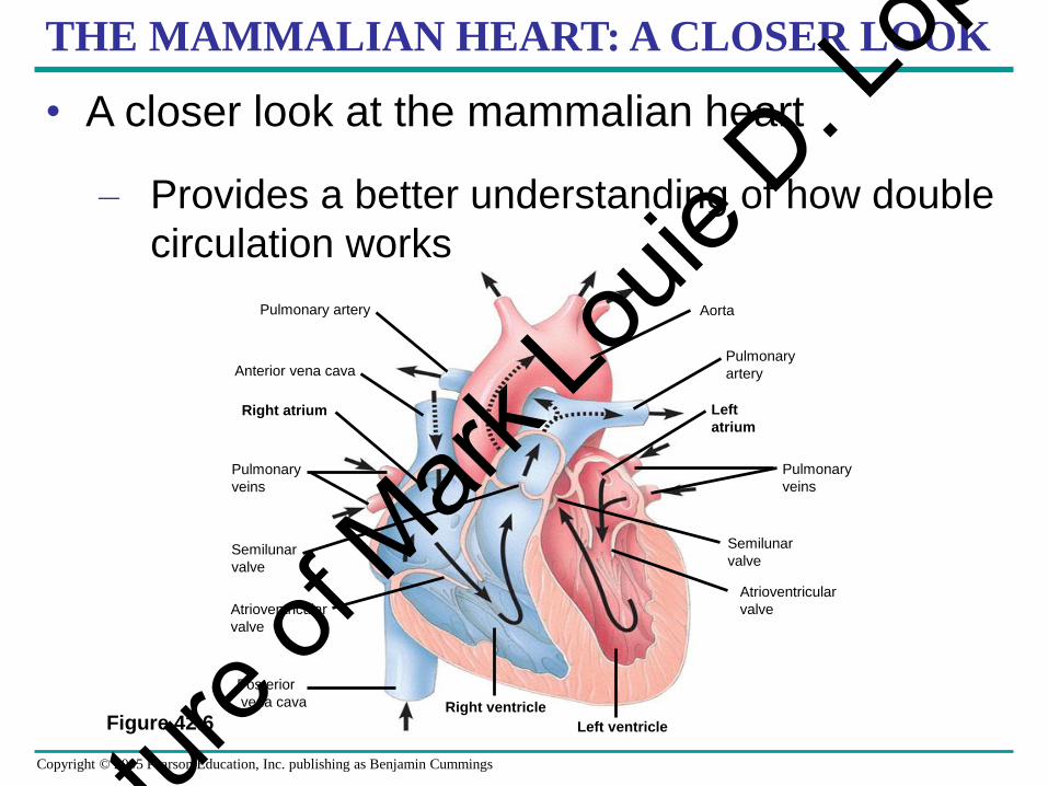
THE MAMMALIAN HEART: A CLOSER LOOK
• A closer look at the mammalian heart
– Provides a better understanding of how double
circulation works
Figure 42.6
Aorta
Pulmonary
veins
Semilunar
valve
Atrioventricular
valve
Left ventricle
Right ventricle
Anterior vena cava
Pulmonary artery
Semilunar
valve
Atrioventricular
valve
Posterior
vena cava
Pulmonary
veins
Right atrium
Pulmonary
artery
Left
atrium
Lectu
re of
Mark Lo
uie D
. Lop
ez

Copyright © 2005 Pearson Education, Inc. publishing as Benjamin Cummings
CARDIAC CYCLE
• The heart contracts and relaxes
– In a rhythmic cycle called the cardiac cycle
• The contraction, or pumping, phase of the
cycle
– Is called systole
• The relaxation, or filling, phase of the cycle
– Is called diastole
Lectu
re of
Mark Lo
uie D
. Lop
ez

Copyright © 2005 Pearson Education, Inc. publishing as Benjamin Cummings
CARDIAC CYCLE
Figure 42.7
Semilunar
valves
closed
AV valves
open
AV valves
closed
Semilunar
valves
open
Atrial and
ventricular
diastole
1
Atrial systole;
ventricular
diastole
2
Ventricular systole;
atrial diastole
3
0.1 sec
0.3 sec0.4 sec
Lectu
re of
Mark Lo
uie D
. Lop
ez

Copyright © 2005 Pearson Education, Inc. publishing as Benjamin Cummings
BLOOD CIRCULATION
• The same physical principles that govern the
movement of water in plumbing systems
– Also influence the functioning of animal
circulatory systems
Lectu
re of
Mark Lo
uie D
. Lop
ez

Copyright © 2005 Pearson Education, Inc. publishing as Benjamin Cummings
BLOOD VESSEL STRUCTURE AND FUNCTION
• The “infrastructure” of the circulatory system
– Is its network of blood vessels
Lectu
re of
Mark Lo
uie D
. Lop
ez

Copyright © 2005 Pearson Education, Inc. publishing as Benjamin Cummings
BLOOD VESSEL STRUCTURE
• Structural differences in arteries, veins, and
capillaries
– Correlate with their different functions
• Arteries have thicker walls
– To accommodate the high pressure of blood
pumped from the heart
Lectu
re of
Mark Lo
uie D
. Lop
ez

Copyright © 2005 Pearson Education, Inc. publishing as Benjamin Cummings
BLOOD VESSEL STRUCTURE
• In the thinner-walled veins
– Blood flows back to the heart mainly as a
result of muscle action
Figure 42.10
Direction of blood flow
in vein (toward heart)
Valve (open)
Skeletal muscle
Valve (closed)
Lectu
re of
Mark Lo
uie D
. Lop
ez

Copyright © 2005 Pearson Education, Inc. publishing as Benjamin Cummings
CAPILLARY FUNCTION
• Capillaries in major organs are usually filled to
capacity
– But in many other sites, the blood supply
varies
Lectu
re of
Mark Lo
uie D
. Lop
ez

Copyright © 2005 Pearson Education, Inc. publishing as Benjamin Cummings
CAPILLARY FUNCTION
• Two mechanisms
– Regulate the distribution of blood in capillary
beds
• In one mechanism
– Contraction of the smooth muscle layer in the
wall of an arteriole constricts the vessel
Lectu
re of
Mark Lo
uie D
. Lop
ez

Copyright © 2005 Pearson Education, Inc. publishing as Benjamin Cummings
CAPILLARY FUNCTION
• In a second mechanism
– Precapillary sphincters control the flow of
blood between arterioles and venules
Figure 42.13 a–c
Precapillary sphincters Thoroughfare
channel
ArterioleCapillaries
Venule(a) Sphincters relaxed
(b) Sphincters contracted
VenuleArteriole
(c) Capillaries and larger vessels (SEM)
20 m
Lectu
re of
Mark Lo
uie D
. Lop
ez

Copyright © 2005 Pearson Education, Inc. publishing as Benjamin Cummings
CAPILLARY FUNCTION
• The critical exchange of substances between
the blood and interstitial fluid
– Takes place across the thin endothelial walls of
the capillaries
Lectu
re of
Mark Lo
uie D
. Lop
ez

Copyright © 2005 Pearson Education, Inc. publishing as Benjamin Cummings
BLOOD
• Blood is a connective tissue with cells
suspended in plasma
• Blood in the circulatory systems of vertebrates
– Is a specialized connective tissue
Lectu
re of
Mark Lo
uie D
. Lop
ez

Copyright © 2005 Pearson Education, Inc. publishing as Benjamin Cummings
BLOOD COMPOSITION AND FUNCTION
• Blood consists of several kinds of cells
– Suspended in a liquid matrix called plasma
• The cellular elements
– Occupy about 45% of the volume of blood
Lectu
re of
Mark Lo
uie D
. Lop
ez
Copyright © 2005 Pearson Education, Inc. publishing as Benjamin Cummings
PLASMA
• Blood plasma is about 90% water
• Among its many solutes are
– Inorganic salts in the form of dissolved ions,
sometimes referred to as electrolytes
Lectu
re of
Mark Lo
uie D
. Lop
ez

Copyright © 2005 Pearson Education, Inc. publishing as Benjamin Cummings
PLASMA COMPOSITION
• The composition of mammalian plasmaPlasma 55%
Constituent Major functions
Water Solvent for
carrying other
substances
Sodium
Potassium
Calcium
Magnesium
Chloride
Bicarbonate
Osmotic balance
pH buffering, and
regulation of
membrane
permeability
Albumin
Fibringen
Immunoglobulins
(antibodies)
Plasma proteins
Icons (blood electrolytes
Osmotic balance,
pH buffering
Substances transported by bloodNutrients (such as glucose, fatty acids, vitamins)
Waste products of metabolism
Respiratory gases (O2 and CO2)
Hormones
Defense
Figure 42.15
Separated
blood
elements
Clotting
Lectu
re of
Mark Lo
uie D
. Lop
ez

Copyright © 2005 Pearson Education, Inc. publishing as Benjamin Cummings
CELLULAR ELEMENTS
• Suspended in blood plasma are two classes of
cells
– Red blood cells, which transport oxygen
– White blood cells, which function in defense
• A third cellular element, platelets
– Are fragments of cells that are involved in
clotting
Lectu
re of
Mark Lo
uie D
. Lop
ez

Copyright © 2005 Pearson Education, Inc. publishing as Benjamin Cummings
Figure 42.15
Cellular elements 45%
Cell type Number
per L (mm3) of blood
Functions
Erythrocytes
(red blood cells) 5–6 million Transport oxygen
and help transport
carbon dioxide
Leukocytes
(white blood cells)5,000–10,000 Defense and
immunity
Eosinophil
Basophil
Platelets
NeutrophilMonocyte
Lymphocyte
250,000
400,000 Blood clotting
CELLULAR ELEMENTS
• The cellular elements of mammalian blood
Separated
blood
elements
Lectu
re of
Mark Lo
uie D
. Lop
ez

Copyright © 2005 Pearson Education, Inc. publishing as Benjamin Cummings
ERYTHROCYTES
• Red blood cells, or erythrocytes
– Are by far the most numerous blood cells
– Transport oxygen throughout the body
Lectu
re of
Mark Lo
uie D
. Lop
ez

Copyright © 2005 Pearson Education, Inc. publishing as Benjamin Cummings
LEUKOCYTES
• The blood contains five major types of white
blood cells, or leukocytes
– Monocytes, neutrophils, basophils,
eosinophils, and lymphocytes, which function
in defense by phagocytizing bacteria and
debris or by producing antibodies
Lectu
re of
Mark Lo
uie D
. Lop
ez

Copyright © 2005 Pearson Education, Inc. publishing as Benjamin Cummings
PLATELETS
• Platelets function in blood clotting
Lectu
re of
Mark Lo
uie D
. Lop
ez

Copyright © 2005 Pearson Education, Inc. publishing as Benjamin Cummings
BLOOD CLOTTING
• A cascade of complex reactions
– Converts fibrinogen to fibrin, forming a clot
Platelet
plug
Collagen fibers
Platelet releases chemicals
that make nearby platelets sticky
Clotting factors from:
Platelets
Damaged cells
Plasma (factors include calcium, vitamin K)
Prothrombin Thrombin
Fibrinogen Fibrin5 µm
Fibrin clotRed blood cell
The clotting process begins
when the endothelium of a
vessel is damaged, exposing
connective tissue in the
vessel wall to blood. Platelets
adhere to collagen fibers in
the connective tissue and
release a substance that
makes nearby platelets sticky.
1 The platelets form a
plug that provides
emergency protection
against blood loss.
2 This seal is reinforced by a clot of fibrin when
vessel damage is severe. Fibrin is formed via a
multistep process: Clotting factors released from
the clumped platelets or damaged cells mix with
clotting factors in the plasma, forming an
activation cascade that converts a plasma protein
called prothrombin to its active form, thrombin.
Thrombin itself is an enzyme that catalyzes the
final step of the clotting process, the conversion of
fibrinogen to fibrin. The threads of fibrin become
interwoven into a patch (see colorized SEM).
3
Figure 42.17
Lectu
re of
Mark Lo
uie D
. Lop
ez

Copyright © 2005 Pearson Education, Inc. publishing as Benjamin Cummings
REPLACEMENT OF CELLULAR ELEMENTS
• The cellular elements of blood wear out
– And are replaced constantly throughout a
person’s life
Lectu
re of
Mark Lo
uie D
. Lop
ez

Copyright © 2005 Pearson Education, Inc. publishing as Benjamin Cummings
BLOOD FORMATION
• Erythrocytes, leukocytes, and platelets all
develop from a common source
– A single population of cells called pluripotent
stem cells in the red marrow of bones
B cells T cells
Lymphoid
stem cells
Pluripotent stem cells
(in bone marrow)
Myeloid
stem cells
Erythrocytes
Platelets Monocytes
Neutrophils
Eosinophils
Basophils
Lymphocytes
Figure 42.16
Lectu
re of
Mark Lo
uie D
. Lop
ez

Copyright © 2005 Pearson Education, Inc. publishing as Benjamin Cummings
GAS EXCHANGE
• Gas exchange occurs across specialized
respiratory surfaces
• Gas exchange
– Supplies oxygen for cellular respiration and
disposes of carbon dioxide
Figure 42.19
Organismal
level
Cellular level
Circulatory system
Cellular respiration ATPEnergy-rich
molecules
from food
Respiratory
surface
Respiratory
medium
(air of water)
O2 CO2
Lectu
re of
Mark Lo
uie D
. Lop
ez
Copyright © 2005 Pearson Education, Inc. publishing as Benjamin Cummings
GAS EXCHANGE
• Animals require large, moist respiratory
surfaces for the adequate diffusion of
respiratory gases
– Between their cells and the respiratory
medium, either air or water
Lectu
re of
Mark Lo
uie D
. Lop
ez

Copyright © 2005 Pearson Education, Inc. publishing as Benjamin Cummings
GILLS IN AQUATIC ANIMALS
• Gills are outfoldings of the body surface
– Specialized for gas exchange
Lectu
re of
Mark Lo
uie D
. Lop
ez

Copyright © 2005 Pearson Education, Inc. publishing as Benjamin Cummings
GILLS WITHIN ANIMAL KINGDOM
• In some invertebrates
– The gills have a simple shape and are
distributed over much of the body
(a) Sea star. The gills of a sea
star are simple tubular
projections of the skin.
The hollow core of each gill
is an extension of the coelom
(body cavity). Gas exchange
occurs by diffusion across the
gill surfaces, and fluid in the
coelom circulates in and out of
the gills, aiding gas transport.
The surfaces of a sea star’s
tube feet also function in
gas exchange.
Gills
Tube foot
Coelom
Figure 42.20a
Lectu
re of
Mark Lo
uie D
. Lop
ez

Copyright © 2005 Pearson Education, Inc. publishing as Benjamin Cummings
GILLS WITHIN ANIMAL KINGDOM
• Many segmented worms have flaplike gills
– That extend from each segment of their body
Figure 42.20b
(b) Marine worm. Many
polychaetes (marine
worms of the phylum
Annelida) have a pair
of flattened appendages
called parapodia on
each body segment. The
parapodia serve as gills
and also function in
crawling and swimming.
Gill
Parapodia
Lectu
re of
Mark Lo
uie D
. Lop
ez

Copyright © 2005 Pearson Education, Inc. publishing as Benjamin Cummings
GILLS WITHIN ANIMAL KINGDOM
• The gills of clams, crayfish, and many other
animals
– Are restricted to a local body region
Figure 42.20c, d
(d) Crayfish. Crayfish and
other crustaceans
have long, feathery
gills covered by the
exoskeleton. Specialized
body appendages
drive water over
the gill surfaces.
(c) Scallop. The gills of a
scallop are long,
flattened plates
that project from the
main body mass
inside the hard shell.
Cilia on the gills
circulate water around
the gill surfaces.
Gills
Gills
Lectu
re of
Mark Lo
uie D
. Lop
ez

Copyright © 2005 Pearson Education, Inc. publishing as Benjamin Cummings
GILLS WITHIN ANIMAL KINGDOM
• The effectiveness of gas exchange in some
gills, including those of fishes
– Is increased by ventilation and countercurrent
flow of blood and water
Countercurrent exchange
Figure 42.21
Gill arch
Water
flow Operculum
Gill
arch
Blood
vessel
Gill
filaments
Oxygen-poor
blood
Oxygen-rich
blood
Water flow
over lamellae
showing % O2
Blood flow
through capillaries
in lamellae
showing % O2
Lamella
O2
Lectu
re of
Mark Lo
uie D
. Lop
ez

Copyright © 2005 Pearson Education, Inc. publishing as Benjamin Cummings
Figure 42.22a
Tracheae
Air sacs
Spiracle
(a) The respiratory system of an insect consists of branched internal
tubes that deliver air directly to body cells. Rings of chitin reinforce
the largest tubes, called tracheae, keeping them from collapsing.
Enlarged portions of tracheae form air sacs near organs that require
a large supply of oxygen. Air enters the tracheae through openings
called spiracles on the insect’s body surface and passes into smaller
tubes called tracheoles. The tracheoles are closed and contain fluid
(blue-gray). When the animal is active and is using more O2, most of
the fluid is withdrawn into the body. This increases the surface area
of air in contact with cells.
TRACHEAL SYSTEMS IN INSECTS
• The tracheal system of insects
– Consists of tiny branching tubes that penetrate
the body
Lectu
re of
Mark Lo
uie D
. Lop
ez

Copyright © 2005 Pearson Education, Inc. publishing as Benjamin Cummings
GAS EXCHANGE IN TRACHEAL TUBES
• The tracheal tubes
– Supply O2 directly to body cells
Air
sac
Body
cell
Trachea
Tracheole
TracheolesMitochondria
Myofibrils
Body wall
(b) This micrograph shows cross
sections of tracheoles in a tiny
piece of insect flight muscle (TEM).
Each of the numerous mitochondria
in the muscle cells lies within about
5 µm of a tracheole.
Figure 42.22b 2.5 µm
Air
Lectu
re of
Mark Lo
uie D
. Lop
ez

Copyright © 2005 Pearson Education, Inc. publishing as Benjamin Cummings
LUNGS
• Spiders, land snails, and most terrestrial
vertebrates
– Have internal lungs
Lectu
re of
Mark Lo
uie D
. Lop
ez

Copyright © 2005 Pearson Education, Inc. publishing as Benjamin Cummings
MAMMALIAN RESPIRATORY SYSTEMS
• A system of branching ducts
– Conveys air to the lungsBranch
from the
pulmonary
vein
(oxygen-rich
blood)
Terminal
bronchiole
Branch
from the
pulmonary
artery
(oxygen-poor
blood)
Alveoli
Colorized SEMSEM
50 µ
m
50 µ
m
Heart
Left
lung
Nasal
cavity
Pharynx
Larynx
Diaphragm
Bronchiole
Bronchus
Right lung
Trachea
Esophagus
Figure 42.23
Lectu
re of
Mark Lo
uie D
. Lop
ez

Copyright © 2005 Pearson Education, Inc. publishing as Benjamin Cummings
MAMMALIAN RESPIRATORY SYSTEMS
• In mammals, air inhaled through the nostrils
– Passes through the pharynx into the trachea,
bronchi, bronchioles, and dead-end alveoli,
where gas exchange occurs
Lectu
re of
Mark Lo
uie D
. Lop
ez
Copyright © 2005 Pearson Education, Inc. publishing as Benjamin Cummings
HOW AN AMPHIBIAN BREATHES
• An amphibian such as a frog
– Ventilates its lungs by positive pressure
breathing, which forces air down the trachea
Lectu
re of
Mark Lo
uie D
. Lop
ez

Copyright © 2005 Pearson Education, Inc. publishing as Benjamin Cummings
HOW A MAMMAL BREATHES
• Mammals ventilate their lungs
– By negative pressure breathing, which pulls air
into the lungs
Air inhaled Air exhaled
INHALATION
Diaphragm contracts
(moves down)
EXHALATION
Diaphragm relaxes
(moves up)
Diaphragm
Lung
Rib cage
expands as
rib muscles
contract
Rib cage gets
smaller as
rib muscles
relax
Figure 42.24
Lectu
re of
Mark Lo
uie D
. Lop
ez

Copyright © 2005 Pearson Education, Inc. publishing as Benjamin Cummings
HOW A BIRD BREATHES
• Besides lungs, bird have eight or nine air sacs
– That function as bellows that keep air flowing
through the lungs
INHALATION
Air sacs fill
EXHALATION
Air sacs empty; lungs fill
Anterior
air sacs
Trachea
LungsLungsPosterior
air sacs
Air Air
1 mm
Air tubes
(parabronchi)
in lung
Figure 42.25
Lectu
re of
Mark Lo
uie D
. Lop
ez

Copyright © 2005 Pearson Education, Inc. publishing as Benjamin Cummings
THE ROLE OF PARTIAL PRESSURE GRADIENTS
• Gases diffuse down pressure gradients
– In the lungs and other organs
• Diffusion of a gas
– Depends on differences in a quantity called
partial pressure
Lectu
re of
Mark Lo
uie D
. Lop
ez

Copyright © 2005 Pearson Education, Inc. publishing as Benjamin Cummings
• A gas always diffuses from a region of
higher partial pressure
– To a region of lower partial pressure
Lectu
re of
Mark Lo
uie D
. Lop
ez

Copyright © 2005 Pearson Education, Inc. publishing as Benjamin Cummings
GAS EXCHANGE IN THE LUNGS
• In the lungs and in the tissues
– O2 and CO2 diffuse from where their partial pressures are higher to where they are lower
Lectu
re of
Mark Lo
uie D
. Lop
ez

Copyright © 2005 Pearson Education, Inc. publishing as Benjamin Cummings
GAS EXCHANGE IN THE LUNGSInhaled air Exhaled air
160 0.2
O2 CO2
O2 CO2
O2 CO2
O2 CO2 O2 CO2
O2 CO2 O2 CO2
O2 CO2
40 45
40 45
100 40
104 40
104 40
120 27
CO2O2
Alveolar
epithelial
cells
Pulmonary
arteries
Blood
entering
alveolar
capillaries
Blood
leaving
tissue
capillaries
Blood
entering
tissue
capillaries
Blood
leaving
alveolar
capillaries
CO2O2
Tissue
capillaries
Heart
Alveolar
capillaries
of lung
<40 >45
Tissue
cells
Pulmonary
veins
Systemic
arteriesSystemic
veins
O2CO2
O2
Alveolar spaces
12
43
Figure 42.27
Lectu
re of
Mark Lo
uie D
. Lop
ez

Copyright © 2005 Pearson Education, Inc. publishing as Benjamin Cummings
RESPIRATORY PIGMENTS
• Respiratory pigments
– Are proteins that transport oxygen
– Greatly increase the amount of oxygen that
blood can carry
Lectu
re of
Mark Lo
uie D
. Lop
ez

Copyright © 2005 Pearson Education, Inc. publishing as Benjamin Cummings
OXYGEN TRANSPORT
• The respiratory pigment of almost all
vertebrates
– Is the protein hemoglobin, contained in the
erythrocytes
Lectu
re of
Mark Lo
uie D
. Lop
ez

Copyright © 2005 Pearson Education, Inc. publishing as Benjamin Cummings
HEMOGLOBIN
• Like all respiratory pigments
– Hemoglobin must reversibly bind O2, loading
O2 in the lungs and unloading it in other parts
of the body
Heme group Iron atom
O2 loaded
in lungs
O2 unloaded
In tissues
Polypeptide chain
O2
O2
Figure 42.28
Lectu
re of
Mark Lo
uie D
. Lop
ez

Copyright © 2005 Pearson Education, Inc. publishing as Benjamin Cummings
CARBON DIOXIDE TRANSPORT
• Hemoglobin also helps transport CO2
– And assists in buffering
Lectu
re of
Mark Lo
uie D
. Lop
ez

Copyright © 2005 Pearson Education, Inc. publishing as Benjamin Cummings
CARBON DIOXIDE TRANSPORT
• Carbon from respiring cells
– Diffuses into the blood plasma and then into
erythrocytes and is ultimately released in the
lungs
Lectu
re of
Mark Lo
uie D
. Lop
ez

Copyright © 2005 Pearson Education, Inc. publishing as Benjamin Cummings
CARBON DIOXIDE TRANSPORT
Figure 42.30
Tissue cell
CO2Interstitial
fluid
CO2 producedCO2 transport
from tissues
CO2
CO2
Blood plasma
within capillaryCapillary
wall
H2O
Red
blood
cellHbCarbonic acid
H2CO3
HCO3–
H++Bicarbonate
HCO3–
Hemoglobin
picks up
CO2 and H+
HCO3–
HCO3– H++
H2CO3Hb
Hemoglobin
releases
CO2 and H+
CO2 transport
to lungs
H2O
CO2
CO2
CO2
CO2
Alveolar space in lung
2
1
34
56
7
8
9
10
11
To lungs
Carbon dioxide produced by
body tissues diffuses into the
interstitial fluid and the plasma.
Over 90% of the CO2 diffuses
into red blood cells, leaving only 7%
in the plasma as dissolved CO2.
Some CO2 is picked up and
transported by hemoglobin.
However, most CO2 reacts with water
in red blood cells, forming carbonic
acid (H2CO3), a reaction catalyzed by
carbonic anhydrase contained. Within
red blood cells.
Carbonic acid dissociates into a
biocarbonate ion (HCO3–) and a
hydrogen ion (H+).
Hemoglobin binds most of the
H+ from H2CO3 preventing the H+
from acidifying the blood and thus
preventing the Bohr shift.
CO2 diffuses into the alveolar
space, from which it is expelled
during exhalation. The reduction
of CO2 concentration in the plasma
drives the breakdown of H2CO3
Into CO2 and water in the red blood
cells (see step 9), a reversal of the
reaction that occurs in the tissues
(see step 4).
Most of the HCO3– diffuse
into the plasma where it is
carried in the bloodstream to
the lungs.
In the HCO3– diffuse
from the plasma red blood cells,
combining with H+ released from
hemoglobin and forming H2CO3.
Carbonic acid is converted back
into CO2 and water.
CO2 formed from H2CO3 is unloaded
from hemoglobin and diffuses into the
interstitial fluid.
1
2
3
4
5
6
7
8
9
10
11
Lectu
re of
Mark Lo
uie D
. Lop
ez

Copyright © 2005 Pearson Education, Inc. publishing as Benjamin Cummings
ELITE ANIMAL ATHLETES
• Migratory and diving mammals
– Have evolutionary adaptations that allow them
to perform extraordinary feats
Lectu
re of
Mark Lo
uie D
. Lop
ez

Copyright © 2005 Pearson Education, Inc. publishing as Benjamin Cummings
THE ULTIMATE ENDURANCE RUNNER
• The extreme O2 consumption of the antelope-
like pronghorn
– Underlies its ability to run at high speed over
long distances
Figure 42.31
Lectu
re of
Mark Lo
uie D
. Lop
ez
Copyright © 2005 Pearson Education, Inc. publishing as Benjamin Cummings
DIVING MAMMALS
• Deep-diving air breathers
– Stockpile O2 and deplete it slowly
Lectu
re of
Mark Lo
uie D
. Lop
ez

Copyright © 2005 Pearson Education, Inc. publishing as Benjamin Cummings
Lectu
re of
Mark Lo
uie D
. Lop
ez