Long-term stability of bevacizumab repackaged in 1mL polypropylene
16
Please cite this article in press as: Paul M, et al. Long-term stability of bevacizumab repackaged in 1 mL polypropylene syringes for intravitreal administration. Ann Pharm Fr (2012), doi:10.1016/j.pharma.2012.03.006 ARTICLE IN PRESS +Model PHARMA-232; No. of Pages 16 Annales Pharmaceutiques Françaises (2012) xxx, xxx—xxx Disponible en ligne sur www.sciencedirect.com ORIGINAL ARTICLE Long-term stability of bevacizumab repackaged in 1 mL polypropylene syringes for intravitreal administration Stabilité au long terme du bevacizumab conditionné en seringue de polypropylène de 1 mL pour administration intravitréenne M. Paul a,∗ , V. Vieillard a , E. Roumi a , A. Cauvin a , M.C. Despiau b , M. Laurent b , A. Astier a a Unité pharmaceutique de recherche en essais cliniques, département de pharmacie, CHU Henri-Mondor, AP—HP, 51, avenue du Maréchal-de-Lattre-de-Tassigny, 94010 Créteil, France b Service de pharmacie, centre hospitalier national d’ophtalmologie des Quinze-Vingt, 75012 Paris, France Received 13 February 2012; accepted 29 March 2012 KEYWORDS Bevacizumab; Intravitreal administration; Off-label use; Stability; Ophthalmology Summary Introduction. — The anti-angiogenic monoclonal antibody, bevacizumab, is currently used by intravitreal administration as off-label drug to treat age-related macular degeneration or other ophthalmologic diseases. For this purpose, commercial bevacizumab is repackaged in 1 mL polypropylene syringes under sterile conditions. However, no complete study on the stability of this hospital-based preparation is available. Methods. — Commercial bevacizumab (25 mg/mL; Avastin ® ) was aseptically repackaged in 1 mL polypropylene syringes, stored at 4 ◦ C, and analyzed within the preparation day (D0), after 30 days (D30) and 90 days (D90). Some syringes were kept for up to 8 months to observe possi- ble instability. Several complementary and stability-indicating analytical methods were used to assess in details the primary, secondary and tertiary structure of the antibody during its conser- vation: ionic chromatography, size-exclusion chromatography, peptide mapping, 2nd derivative UV and IR spectroscopy, turbidimetry, diffraction laser spectroscopy, thermal denaturation curves, microscopic examination and image analysis. Results. — We clearly demonstrate that the commercial solution of bevacizumab can be safely repackaged in polypropylene syringes and stored up to 3 months at 4 ◦ C without alteration of its primary, secondary and tertiary structure. The only difference observed is the contamination of the syringe content by silicone oil microdroplets, which is quite immediate and does not change significantly during the storage in terms of number and size. ∗ Corresponding author. E-mail address: [email protected] (M. Paul). 0003-4509/$ — see front matter © 2012 Published by Elsevier Masson SAS. doi:10.1016/j.pharma.2012.03.006
Transcript of Long-term stability of bevacizumab repackaged in 1mL polypropylene

ARTICLE IN PRESS+ModelPHARMA-232; No. of Pages 16
Annales Pharmaceutiques Françaises (2012) xxx, xxx—xxx
Disponible en ligne sur
www.sciencedirect.com
ORIGINAL ARTICLE
Long-term stability of bevacizumab repackagedin 1 mL polypropylene syringes for intravitrealadministration
Stabilité au long terme du bevacizumab conditionné en seringue depolypropylène de 1 mL pour administration intravitréenne
M. Paula,∗, V. Vieillarda, E. Roumia, A. Cauvina,M.C. Despiaub, M. Laurentb, A. Astiera
a Unité pharmaceutique de recherche en essais cliniques, département de pharmacie, CHUHenri-Mondor, AP—HP, 51, avenue du Maréchal-de-Lattre-de-Tassigny, 94010 Créteil, Franceb Service de pharmacie, centre hospitalier national d’ophtalmologie des Quinze-Vingt,75012 Paris, France
Received 13 February 2012; accepted 29 March 2012
KEYWORDSBevacizumab;Intravitrealadministration;Off-label use;Stability;Ophthalmology
SummaryIntroduction. — The anti-angiogenic monoclonal antibody, bevacizumab, is currently used byintravitreal administration as off-label drug to treat age-related macular degeneration or otherophthalmologic diseases. For this purpose, commercial bevacizumab is repackaged in 1 mLpolypropylene syringes under sterile conditions. However, no complete study on the stability ofthis hospital-based preparation is available.Methods. — Commercial bevacizumab (25 mg/mL; Avastin®) was aseptically repackaged in 1 mLpolypropylene syringes, stored at 4 ◦C, and analyzed within the preparation day (D0), after30 days (D30) and 90 days (D90). Some syringes were kept for up to 8 months to observe possi-ble instability. Several complementary and stability-indicating analytical methods were used toassess in details the primary, secondary and tertiary structure of the antibody during its conser-vation: ionic chromatography, size-exclusion chromatography, peptide mapping, 2nd derivativeUV and IR spectroscopy, turbidimetry, diffraction laser spectroscopy, thermal denaturationcurves, microscopic examination and image analysis.
Results. — We clearly demonstrate that the commercial solution of bevacizumab can be safelye syringes and stored up to 3 months at 4 ◦C without alteration of its
repackaged in polypropylenPlease cite this article in press as: Paul M, et al. Long-term stability of bevacizumab repackaged in 1 mL polypropylenesyringes for intravitreal administration. Ann Pharm Fr (2012), doi:10.1016/j.pharma.2012.03.006
primary, secondary and tertiary structure. The only difference observed is the contaminationof the syringe content by silicone oil microdroplets, which is quite immediate and does notchange significantly during the storage in terms of number and size.
∗ Corresponding author.E-mail address: [email protected] (M. Paul).
0003-4509/$ — see front matter © 2012 Published by Elsevier Masson SAS.doi:10.1016/j.pharma.2012.03.006

ARTICLE IN PRESS+ModelPHARMA-232; No. of Pages 16
2 M. Paul et al.
Conclusion. — Our results support the off-label use of repackaged bevacizumab by intravit-real administration, at least from a pharmaceutical point of view, with a validated stabilityof 3 months. This stability period is largely enough to practical situations and support currentpractices, such as in advance or batch preparations, which present major advantages in termsof GMP respect, workload optimization and financial savings.© 2012 Published by Elsevier Masson SAS.
MOTS CLÉSBevacizumab ;Administrationintravitréale ;Utilisation hors AMM ;Stabilité ;Ophtalmologie
RésuméIntroduction. — L’anticorps anti-angiogénique bevacizumab est couramment utilisé par voieintravitréale, hors AMM, pour traiter la dégénération maculaire liée à l’âge ou d’autres patholo-gies ophtalmiques. Dans ce but, le bevacizumab commercial est reconditionné, en conditionsaseptiques, dans des seringues de 1 mL en polypropylène. Cependant, aucune étude complètesur la stabilité de cette préparation hospitalière n’est actuellement disponible.Methodes. — Le bevacizumab commercial (25 mg/mL ; Avastin®) a été reconditionné asep-tiquement en seringues polypropylène de 1 mL, stocké à 4 ◦C et analysé le jour même dela préparation (D0), 30 jours (D30) et 90 jours (D90). Des seringues ont été conservées pen-dant plus de huit mois pour observer de possibles signes d’instabilité. Plusieurs méthodescomplémentaires et indicatrices de stabilité ont été utilisées pour étudier en détail la structureprimaire, secondaire et tertiaire de l’anticorps durant la conservation : chromatographies ion-ique et d’exclusion de taille, carte peptidique, spectroscopie UV et IR en dérivée seconde,turbidimétrie, spectrométrie de diffraction laser, courbes d’agrégation thermique, examenmicroscopique avec analyse d’image.Résultats. — Nous démontrons clairement que la solution commerciale de bevacizumab(25 mg/mL) peut être reconditionnée en toute sécurité en seringue polypropylène et stockéeà + 4 ◦C pendant trois mois sans altération de sa structure primaire, secondaire et tertiaire. Laseule différence observée est la contamination du contenu par des microgouttelettes d’huile desilicone, qui est immédiate et n’évolue pas en termes de taille et de nombre durant le stockage.Conclusion. — Nos résultats supportent, du moins d’un point de vue pharmaceutique,l’utilisation hors AMM par voie intravitréale du bevacizumab reconditionné, avec une duréede stabilité validée de trois mois. Cette période de stabilité est largement suffisante dans lessituations rencontrées en routine et supporte les pratiques courantes, comme les préparationsà l’avance ou par lots, qui présentent des avantages majeurs en termes de respect des bonnespratiques de fabrication, d’optimisation de la charge de travail et d’économies.
asso
I
AtaTaTvmrrabiarpcplta
wh2tNtbMbfipiyMscatC
© 2012 Publie par Elsevier M
ntroduction
healthy cornea is devoid of blood vessels but inflamma-ory states such as chemical burns or infections can lead to
breakdown of this so-called ‘‘angiogenic privilege’’ [1,2].his corneal neovascularisation occurring prior or after ker-toplasty significantly increases the risk of graft rejection.ill now, no specific treatment was available for corneal neo-ascularisation. However, recently, some progresses wereade in antiangiogenesis research and new drugs are cur-
ently used in oncology and against neovascular diseases ofetina and choroid. Bevacizumab (Avastin®) is a humanizednti-VEGF monoclonal antibody currently used in colorectal,reast and lung cancers. In addition, it is used off-label asntravitreal injections to treat age-related macular degener-tion (AMD) [3,4], choroidal neovascularisation [5], diabeticetinopathies [6] and neovascular glaucoma [7]. In theserevious indications, bevacizumab has demonstrated its effi-iency and its short-term safety [8]. For the treatment of
Please cite this article in press as: Paul M, et al. Long-term stsyringes for intravitreal administration. Ann Pharm Fr (2012), d
roliferative eyes diseases such as the choroidal neovascu-ar membrane observed in AMD, ranibizumab (Lucentis®) ishe only drug approved in some countries such as the USAnd France. This drug is a Fab fragment of bevacizumab,
Oa
u
n SAS.
hich has proven efficacy. However, considering the veryigh cost of this drug, intravitreal injection of 1.25 to.5 mg of bevacizumab is currently performed by many oph-halmologists. In 2006, the National Eye Institute, of theational Institutes of Health (NIH), launched a compara-ive phase III study to assess the efficacy and safety ofoth drugs in AMD (CATT study: Comparison of Age-Relatedacular Degeneration Treatment; ranibizumab: 0.05 mg,evacizumab 1.25 mg both in 0.05 mL intravitreal). Thenal results of this study, which had enrolled about 1200atients, were published in 2011 and showed no differencen the effectiveness (visual acuity) and tolerance after one-ear treatment between bevacizumab and ranibizumab [9].oreover, a very interesting consequence of this clinical
imilarity was the cost per treatment, which was 40-foldheaper for bevacizumab vs. ranibizumab. These resultsppeared so convincing that the American Academy of Oph-halmology claimed very recently: ‘‘The initial results of theATT study affirm the position of the American Academy of
® ®
ability of bevacizumab repackaged in 1 mL polypropyleneoi:10.1016/j.pharma.2012.03.006
phthalmology that both Lucentis and Avastin should bevailable for the treatment of AMD’’ [10].
In its main off-label indications, bevacizumab is usedndiluted (25 mg/mL). However, there truly is an economic

IN+Model
prop
sdw2abf
P
TtE(PccaoSosindsTctsissofp
M
Fa2s(tttutfawsstp
P
ARTICLEPHARMA-232; No. of Pages 16
Long-term stability of bevacizumab repackaged in 1 mL poly
incentive to use bevacizumab instead of ranibizumab onlyif the whole content of a commercial vial of bevacizumab(100 mg; 4 mL of 25 mg/mL solution) can be repackaged inready to use syringes under sterile conditions. Since eachsyringe contents 150 �L of 25 mg/mL solution, about 25syringes can be prepared from a 4-mL vial and 100 froma 16-mL vial. Moreover, to realize the preparation undergood hospital manufacturing practices, batch preparationis mandatory and, even in large hospitals, the preparedsyringes must be stored for various periods until use. There-fore, the long-term stability of the repackaged bevacizumabhas to be considered to ensure adequate quality of thepreparation.
Until now, the available stability data of reconditionedbevacizumab are scarce. The biological activity of beva-cizumab on VEGF has been studied over 6 months afterhaving withdrawn the concentrated solution (25 mg/mL)from a vial and stored it at 4 ◦C in latex-free syringes [11].Bevacizumab (control and stored solutions) was incubatedwith VEGF immobilized on plates designed for luminescentdetection and VEGF-bound bevacizumab was then deter-mined by luminescence assay using a labelled antihumanIgG antibody. The anti-VEGF activity of bevacizumab seemedto remain stable over 3 months (< 10% loss of activity) at4 ◦C. However, this study did not consider the physico-chemical stability of the protein, but only the interactionVEGF/bevacizumab (biological activity) and very recentstudies were focused on the particulate pollution duringmanufacturing and shipping steps of syringe-repacked beva-cizumab [12,13].
We performed an extensive physicochemical stabilitystudy of bevacizumab repackaged in 1-mL polypropylenesyringes. Various protein characterization methods wereused to determine changes in physicochemical propertiesof bevacizumab, including size-exclusion chromatogra-phy (SEC), dynamic light scattering (DLS) and turbidity,cation exchange chromatography (CEX), second derivativeultraviolet spectroscopy and Fourier transform infraredspectroscopy (FT-IR), and peptide mapping by gradient HPLCafter trypsin and endopeptidase digestion (PM).
Materials and methods
Reagents
Disodium hydrogen phosphate, sodium azide, potassiumdihydrogen phosphate and trifluoroacetic acid (TFA) werepurchased from Merck (Darmstadt, Germany). Ammoniumbicarbonate, acetonitrile, dithiothreitol (DTT), guanidinehydrochloride (GnHCl) and iodoacetic acid were obtainedfrom Sigma-Aldrich (Saint-Louis, MO, USA). Formic acid waspurchased from Prolabo (Fontenay-sous-Bois, France). Sil-icone oil 200 Fluid® (10 cPs) was from Aldrich. Isotonicsaline was provided by Fresenius Kabi (Louviers, France).All reagents used were of analytical grade.
Materials
Please cite this article in press as: Paul M, et al. Long-term stsyringes for intravitreal administration. Ann Pharm Fr (2012), d
Bevacizumab was obtained from the commercially availableproduct Avastin® (Roche, Switzerland; 100 mg in 4 mL; batchNo. H0105; exp date: 31/05/2012), formulated in 50 mmol/L
Udo
PRESSylene syringes for intravitreal administration 3
odium phosphate, pH 6.25, 60 mg/mL �-trehalose dihy-rate, and 0.4 mg/mL polysorbate 20. The formulationas presented in glass vials as a concentrated solution of5 mg/mL, ready to dilute in 0.9% sodium chloride solution,ccording to the manufacturer’s instructions. Reconstituteduffer (placebo bevacizumab) was prepared with the sameormulation of excipients but without bevacizumab.
reparation of syringes
he pre-filled syringes were routinely prepared byhe Department of Pharmacy, Quinze-Vingt hospital.ach syringe contained 150 �L of undiluted bevacizumab25 mg/mL) according to the Good Hospital Manufacturingractices. All syringes were issued from the same commer-ial batch of Avastin®. Under laminar flow, the wholeontent of a vial was taken with a 5-mL disposable syringe,nd then filtered at 0.22 �m in another syringe by the meanf a luer—lock/luer—lock transfer set (disc filter ref 15532,artorius, Germany). Using a 3-way connector, about 150 �Lf solution were withdrawn by 1-mL disposable tuberculineyringes (body and plunger in polypropylene, plunger-sealn isoprene covered by silicone oil as lubricant; batcho 1103029; Beckton-Dinckinson). A 22-gauge capped nee-le was fitted to the syringe, which was then inserted into aterile sterilization bag (paper-plastic), sealed and labeled.he entire process was performed following pre-defined pro-edures to avoid introduction of air and bubbling duringhe transfer step and the syringes were filled without head-pace in order to minimize gas-liquid interfaces which mightnduce aggregation. The sealed bag was then packed in aecondary bag. This procedure was used to maintain the out-ide sterility of the primary packaging until opening in theperating room. For each vial, four syringes were kept asideor sterility testing. Therefore, 20 syringes of 150 �L can berepared from one vial of 4 mL.
ethods
or the stability assessment at 4 ◦C (2—8 ◦C), three stor-ge groups of 20 syringes were constituted: two groups of0 syringes stored in a windowed-door refrigerator (expo-ure to light) and one group in a opaque-door refrigeratorno light exposure). Our results from previous studies onhe stability of diluted bevacizumab at higher tempera-ure to stress the antibody (40 ◦C) and on the stability ofhe another mAb cetuximab under mechanical stress weresed to demonstrate the good ‘‘stability-indicating charac-er’’ of the analytical methods [14,15]. Vials of bevacizumabrom the same batch were used as controls before repack-ging in syringes. In some experiments, a D0 time sampleas used, consisting of freshly repackaged bevacizumab in
yringes analyzed within the day after preparation. Syringestored for more than 8 months were also used to ascertainhe relevance of some analytical methods to demonstrate aarticular physical instability.
reparation of analytical samples
ability of bevacizumab repackaged in 1 mL polypropyleneoi:10.1016/j.pharma.2012.03.006
nder laminar flow hood, for each analysis time (D0, D14: 14ays of storage, D30: 1 month of storage and D90: 3 monthsf storage) and storage group, the content of the syringes

IN+ModelP
4
wppswtegcbma0v
T
TacncuIsiTf
T
Il(
wr
Dd
Duommafec
D
d(tNywbtr
hgsi
T
Wocwi
N
wibatmdarrl
cb[dgiccTmmsieafpptdoosptw
�
ARTICLEHARMA-232; No. of Pages 16
as expelled into a 2-mL Eppendorf tube by pushing thelunger. A 100-�L sample was taken by a micropipette,oured into a disposable glass tube and 2.4 mL of potas-ium phosphate buffer (20 mM, pH 6.00, filtered at 0.22 �m)ere added to obtain a 1-mg/mL solution. This concen-
ration is used for SEC and CEX analysis and for the DLSxperiments (hydrodynamic diameter and thermal aggre-ation curves). Samples for chromatographic analysis wereentrifuged in 2-mL Eppendorf tubes to remove insolu-le material (11000 × g, 5 min). For spectrophotometriceasurements (turbidity, tertiary structure) and thermal
ggregation curve, the solution at 1 mg/mL was diluted at.25 mg/mL (1.25 mL of 1 mg/mL completed at 5 mL using aolumetric flask).
urbidity analysis
he turbidimetric method is often used to estimate themount of visible protein aggregates by measuring the opti-al density of samples based on light scattering in theear-UV or visible regions [15—19]. Aggregation of beva-izumab was monitored by measuring absorbance at 350 nmsing a UV-VIS spectrophotometer (Cary 50 Probe, Varian,nc., Palo Alto, USA). Here, turbidance (T�) was mea-ured like absorbance at 350 nm, where none of the knownntrinsic chromophores in the protein formulation absorb.urbidity is a function of the wavelength according to theollowing equation, similar to the Beer-Lambert law:
� = log I0/Is = KtIN (1)
s being the light transmitted through the sample and I0 theight transmitted through the solvent for a path length l1 cm).
The mean value of each experimental sample absorbanceas calculated and compared to the mean value of the refe-
ence sample.
etermination of molecular hydrodynamiciameters
ynamic light scattering (DLS) is a sensitive method to eval-ate the size of aggregate populations containing aggregatesf 1 nm to 6 �m [17]. The analysis is based on the measure-ent of fluctuating scattered light intensity due to Brownianotion of particles, which also defines diffusion D, which
lso depends on viscosity (�), temperature (T) and a dif-usion constant (k). Thus, according to the Stokes-Einsteinquation, the hydrodynamic radius of a particle (R) may bealculated as follows:
= kT/6��R (2)
Therefore, an estimation of the particles size andistribution can be done. A Malvern Zetasier Nano ZSWorcestershire, UK) laser light scattering system was usedo obtain DLS measurements with a 633 nm laser source. TheanoZS® software was used for data acquisition and anal-sis. A total of 400 �L of each sample at about 1 mg/mL
Please cite this article in press as: Paul M, et al. Long-term stsyringes for intravitreal administration. Ann Pharm Fr (2012), d
as added in a 1-cm path length quartz microcuvette rinsedefore use with 0.22 �m of filtered water. All the opera-ions were performed under laminar airflow conditions toeduce the risk of external contamination. The size and the
a
wa
PRESSM. Paul et al.
ydrodynamic diameter distributions of the detected aggre-ates were obtained by measuring and integrating the lightcattering intensity. Parameters were calculated both byntensity and volume analysis.
hermal denaturation curves
ith increases in temperature or simply with time, the sec-ndary, tertiary and quaternary structures of a protein mayhange and lead to protein unfolding and/or aggregation,hich are critical factors of physical instability [19—23]. The
rreversible denaturation transition can be represented by
Kunfol�Kfol
U−→Kagg
D
ere N is the native form of the protein (folded), Us its unfolded form in reversible equilibrium describedy the constants Kunfol (unfolding) and Kfol (folding),nd D is the irreversible aggregated form resulting fromhe irreversible hydrophobic interactions between unfoldedolecules followed by Kagg which is the temperature-ependent first-order kinetic constant. If Kagg » Kfol,ll unfolded species are immediately aggregated withouteturning to their native state. Therefore, the aggregationate is directly representative of the folding/unfolding equi-ibrium of the protein.
These events can be monitored because dramatichanges in protein size during denaturation can easilye identified by DLS or spectrophotometric techniques21—24]. In this study, we followed the increase in opticalensity at 350 nm vs. temperature to assess the aggre-ation of bevacizumab. The diluted sample (0.4 mg/mLn 20 mM phosphate buffer) was introduced into a quartzuvette positioned into a thermostated holder linked to airculating-water bath (Polystat, Thermoscientific, France).he content of the cuvette was stirred (100 rpm) by theean of a 8 mm length Teflon barrel placed on a Variomagini stirrer to ensure homogeneity (Thermo Scientific). We
elected a heating rate of about 1 ◦C/min to draw the heat-ng curves, allowing the system to be a quasi-thermodynamicquilibrium showing good repetition of the results. So, theutomated D0 measurements were collected each minuterom 25 ◦C to 80 ◦C, the corresponding temperature beingrecisely recorded each time by the mean of a thermorobe (1 mm diameter) inserted into the solution. Fromhe curve generated by the thermal aggregation (as theirect consequence of the protein unfolding), the midpointf the thermal transition Tm estimated by the midpointf aggregation was determined using the nonlinear leastquares method to adjust the D0 dependence on the tem-erature. Moreover, the standard Gibbs’ free energy �G◦ ofhe aggregation of bevacizumab at different temperaturesas calculated according to the following equation [25]:
G◦ = −RT ln Keq where Keq = fagggregated
1 − faggregated
(3)
D0T − D025◦C
ability of bevacizumab repackaged in 1 mL polypropyleneoi:10.1016/j.pharma.2012.03.006
nd faggregated =D0max − D025◦C
(4)
here D025 ◦C corresponds to the D0 at 25 ◦C (100% non-ggregated state), D0T the absorbance at T temperature
IN+Model
prop
tw
C
ASsssa
CTo(Dpw0wtBt2ar
SStsi7scAefata
%
wto
amcb
PAUf
ARTICLEPHARMA-232; No. of Pages 16
Long-term stability of bevacizumab repackaged in 1 mL poly
and D0max the highest absorbance reached at about 74—75 ◦C(100% aggregated state). Using the �G◦ vs. T curves, theslope (mT) was calculated around Tm using linear regression.The mT parameter is related to the difference in protein-solvent interaction between the folding and unfolding forms[25]. The enthalpy of denaturation �H◦ was calculated byplotting −Ln Keq vs. 1/T (Van’t Hoff plot) [19,24,26].
Since denaturation curves represent the conformational(thermodynamic) stability of a protein, the modificationof this parameter can indicate subtle destabilizations(‘‘weaknesses’’) that occur during storage, but withoutgross impact on the secondary or tertiary structure of theprotein, thus poorly detectable by classical methods. There-fore, the resistance to thermal denaturation is a usefulmethod to estimate structural modifications of a protein inthe presence of various excipients (formulation purpose) orafter exposure to various stresses (stability purpose) [18,27].
Tertiary structure analysis
The tertiary structure of bevacizumab was examined usingsecond derivative UV spectroscopy. This method allows foran evaluation of the impact of dilution and storage on thearomatic amino acids of the mAb [28—31]. Indeed, the posi-tion of second derivative UV absorbance peaks is sensitiveto the polarity of the microenvironment of the aromaticamino acids and therefore can be used to provide a globalpicture of the tertiary structure of a protein. The spectrawere collected in the near-UV region from 250 to 320 nmin a 1 cm path length quartz cuvette using the previouslydescribed spectrophotometer for turbidimetry experiments.The samples were diluted to 0.4 mg/mL in phosphate bufferas previously described. Second derivative spectra wereobtained using a third Savitzky-Golay polynomial algo-rithm. In these conditions, the second derivative spectrumobtained had effective peak resolutions of 0.01 nm. Fivemajor peaks were studied (252-Phe, 258-Phe, 275-Tyr, 284-Thy/Trp and 292-Trp), as previously reported for IgG2, andalso two minima at 287 and 295 nm [28,31]. The spectra ofall samples (D30, D90 and D180), including the referencesample, were compared to each other.
Secondary structure analysis
The secondary structure of bevacizumab was moni-tored using second derivative FT-IR spectroscopy in theconformation-sensitive amide I band region (1600 and1700 cm−1), which is almost entirely due to C = O stretchvibrations of peptide linkages [32]. This well-establishedmethod has been extensively used in protein denat-uration/aggregation studies [19,33—35]. IR spectra ofbevacizumab samples were recorded with a 2000 FT-IR sys-tem (Perkin Elmer, les Ulis, France). For each spectrum, a256-scan interferogram was collected in single-beam mode,with a 4 cm—1 resolution. The sample cell consisted of 25.4-mm diameter CaF2 windows separated by a Teflon spacerof 15 �m. The spectrum of reconstituted buffer (placebobevacizumab) was obtained under identical conditions. For
Please cite this article in press as: Paul M, et al. Long-term stsyringes for intravitreal administration. Ann Pharm Fr (2012), d
the analysis, spectra were initially converted to absorbancefollowed by subtraction of the corresponding placebo spec-trum. Second derivative spectra were calculated with theSpectrum® software to calculate peak areas and determine
pp2a
PRESSylene syringes for intravitreal administration 5
he percentage area under each peak. All spectra obtainedere the mean of three separate scans.
hromatographic analysis
ll chromatographic analyses were realized with a Dionextation consisting of a Dionex Ultimate 300 chromatographicystem coupled with a Chromeleon® chromatography work-tation (Dionex Corporation, Sunnyvale, CA, USA). Theystem is equipped with a gradient pump with degas optionnd gradient mixer, UV detector and autosampler.
ation exchange chromatography (CEX)he different ionic species of RTX were separatedn a Dionex Propac® WCX-10 cation exchange column4 × 250 mm) with a WCX-10 guard column (4 × 50 mm,ionex, Voisins le Bretonneux, France). Buffer A was pre-ared with 50 mM KH2PO4, 0.05% NaN3 pH 6.0, and buffer Bas prepared with 50 mM KH2PO4, 1 M sodium chloride and.05% NaN3 (w/v), both in deionized water. The flow rateas 1 mL/min, and the injected volume was 100 �L. The elu-
ion gradient started with 100% of A to achieve 50% of A and after 20 min, followed by an equilibrium phase of 5 mino obtain 100% of buffer A. The elution was monitored at80 nm. The mean value of the area under the curve (AUC)nd the retention time (Tr) were compared to those of theeference sample.
ize-exclusion chromatography (SEC)EC analysis is a classical method used in protein studieso detect potential fragmentation and aggregation (smallize aggregates or non-visible aggregates), correspond-ng respectively to chemical and physical instabilities. A.8 mm × 300 nm TSK-GEL G4000 SWXL column (Tosoh Bio-ciences Corporation, Montgomeryville, PA) with a guardolumn was used, and the elution was monitored at 280 nm.
total of 100 �L of each centrifuged sample (1 mg/mL) wereluted isocratically with a pH 7.0 buffer (0.1 M sodium sul-ate, 0.1 M sodium hydrogen phosphate and 0.05% sodiumzide [w/v]), at a flow rate of 0.6 mL/min. The tempera-ure was set at 25 ◦C. The studied parameters were AUC, Tr,nd the percentage of aggregation according to the formula
of aggregation = [1 − (AUCt/AUCt0)] × 100 (5)
here AUC t is the AUC of the main peak corresponding tohe mAb monomer (about 150 kDa) obtained at T, and AUCt0
btained at T0.The molecular weight of the peaks was calculated using
standard solution including several molecular weightarkers ranging from 1.2 to 150 kDa (hydroxocobalamin,
ytochrome C, albumin and cetuximab), and using a cali-ration curve fitted as a third-degree polynomial function.
eptide mappingll samples were concentrated at 16 mg/mL using Amicon®
ltra-10 centrifugal filter units (centrifugation at 4000 × gor 20 min). We note that 1 mL of the concentrated sam-
ability of bevacizumab repackaged in 1 mL polypropyleneoi:10.1016/j.pharma.2012.03.006
le was withdrawn for the next steps. Denaturation waserformed with a final concentration of 6 M GnHCl and50 �L of 50 mM ammonium bicarbonate at room temper-ture for 15 min. Reduction was achieved with 1250 �L of
IN+ModelP
6
2oi
urTs35
wrftsacFteTsspts
M
PbT(CJMtwcbTwseIrrwaseahpc
S
Tda
d22ats
R
C
Tc
CAoapabssiat
STpm8rteiagttAtcoMtnoMts
PNo
ARTICLEHARMA-232; No. of Pages 16
0 mM dithiothreitol (DTT) in a water bath at 37 ◦C forne hour. Alkylation was performed with 500 �L of 300 mModoacetic acid (pH 7) in a water bath at 37 ◦C for 30 min.
A 10 mM bicarbonate ammonium buffer (pH 7.6) wassed for the desalting step with Econo-pac® PG10 Bio-ad columns, according to the manufacturer’s instructions.he eluate was then digested with 100 �L of TPCK trypsinolution (8 mg/mL at a Trypsin/IgG ratio of 1:20 w/w) at7 ◦C overnight, followed by a four-hour digestion step with
�g/mL of endopeptidase (at 37 ◦C).Bond Elut® SPE C18 columns (Varian, Les Ulis, France)
ere used to concentrate the digested samples using theinsing solution A (sterile water with 0.02% TFA and 0.08%ormic acid) and the eluting solution B (acetonitrile con-aining 0.02% TFA and 0.08% formic acid). The final elutedamples were dried under nitrogen, and 120 �L of 10 mMmmonium bicarbonate buffer were added. A C18 Nucleosilolumn (250 mm × 3 mm × 5 �m; Macherey Nagel, Duren,rance) associated with a guard column was used. Detec-ion was performed at 280 and 214 nm. One hundred �l ofach sample was eluted with the following elution gradient:
(0 min): 98% solution A/2% solution B. T (140 min): 75%olution A/25% solution B. T (151 min): 98% solution A/2%olution B. The flow rate was 1 mL/min. The column tem-erature was maintained at 60 ◦C. The mapping profiles ofhe stored samples were compared to those of the referenceamples.
icroscopic evaluation and image analysis
articulate contamination of the solutions was assessedy microscopic examination and digital image capture.he imaging platform consisted in an inverted microscopeOlympus IM; Olympus SA, Rungis, France) equipped with aCD color camera (XCD-U1000CR; 1600 × 1200 pixels; Sony,apan) and linked to an image analysis software (Ellix®,icrovision Instruments, Evry, France). Under laminar flow
o reduce external particle contamination, samples (40 �L)ere introduced in a chamber constituted by a Malassezell (200 �m depth) covered by a glass slide (both washedy 0.22 �m of filtered water and dried under laminar flow).he chamber was observed by the CCD camera under normalhite light illumination through a 20X objective (both corre-
ponding to a final magnification of X924) on a 345 × 460 �mxamination field and a counted volume of 0.03174 mm3.mages were generated in adaptive dark mode on a sizeange from 1 to 60 �m, a brightness of 15, no filter algo-ithm and a standard object separation mode. Backgroundas acquired using placebo bevacizumab previously filteredt 0.22 �m. For each measurement, eight randomly cho-en fields were automatically analyzed by the software tostimate the number of particles per mL, their diameternd shape factor and the corresponding size distributionistogram. The estimations of the characteristics of thearticle population were given with their respective 95%onfidence intervals.
tatistical analysis
Please cite this article in press as: Paul M, et al. Long-term stsyringes for intravitreal administration. Ann Pharm Fr (2012), d
he results are generally presented as mean ± standardeviation (SD) obtained from three separate samplesnd for each storage condition and concentration. All
3tfs
PRESSM. Paul et al.
ata were analyzed with the statistical pack PAST V.01 software (Hammer O, Harper DAT and Ryan PD,001; http//folk.uio.no/ohammer/past). Analyses of vari-nce (two-ways Anova) or non-parametric Mann-Whitneyests, if required, were used to compare data. Statisticalignificance was defined as P < 0.05.
esults
hemical stability
he chemical stability was estimated by three orthogonalhromatographic methods.
ation exchange chromatography (CEX) major peak with a retention time at 4.52 ± 0.02 min wasbserved for the control sample (n = 3). In the optimal stor-ge conditions (4 ◦C), no significant change in chromatogramrofiles was detected for repackaged bevacizumab evenfter 3 months (Fig. 1). There was no significant differenceetween the AUC of the control sample and the repackagedamples stored at 4 ◦C up to 3 months. In a previous study fortressed samples of diluted bevacizumab [14,15], a decreasen AUC was observed at 40 ◦C after 3 months and reached 45%t 6 months and two additional peaks were found just beforehe main peak after 6 months at 40 ◦C (Data not shown).
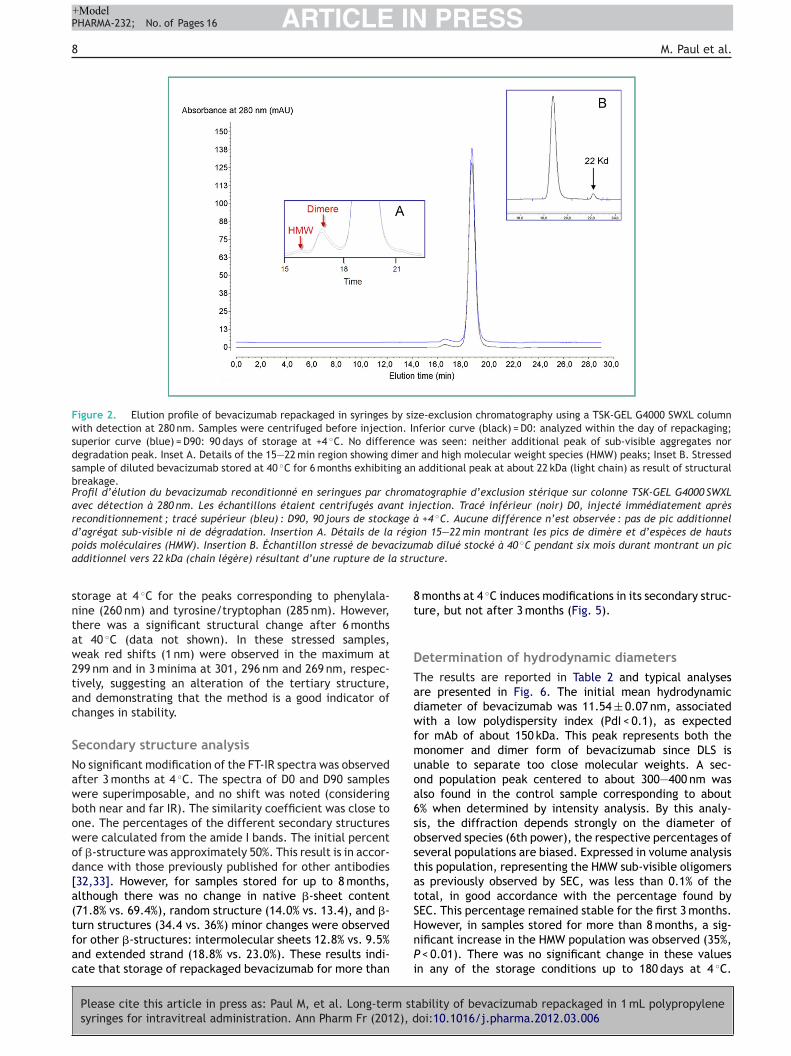
ize-exclusion chromatography (SEC)hree peaks for the bevacizumab control sam-le were found (Fig. 2). The retention time of theonomer was 19.03 ± 0.09 min, and the mean AUC was
0.98 ± 3.93 mAU*min. A second peak, at 16.91 ± 0.29 min,epresenting 1.56% of the protein peaks eluting from 15o 22 min, was observed. Using a calibration curve, itsstimated molecular weight (MW) was about 300—400 kDa,n good accordance with a dimer. A smaller peak elutinground 16 min was considered as a mixture of several aggre-ated molecules (n) of the monomer: (monomer)n, referredo high molecular weight species (HMW) and correspondingo oligomers with n ranging from 6 to 12 (Fig. 1, inset). For repackaged bevacizumab, the respective retentionimes and percentages from these three peaks did nothange up to 3 months (Table 1) and no additional peak wasbserved neither in the high MW region nor in the lowerW region of the chromatogram. Therefore, according
o the equation (5), the percentage of aggregation wasot different from zero. In our previous accelerated studyf diluted bevacizumab at 40 ◦C for 6 months, a lowerW peak (about 22 kDa) was observed corresponding to
he light chains and demonstrating that the method istability-indicating (Fig. 2; inset B).
eptide mappingo significant change in chromatographic profile wasbserved for repackaged bevacizumab stored at 4 ◦C for
ability of bevacizumab repackaged in 1 mL polypropyleneoi:10.1016/j.pharma.2012.03.006
months in syringes (Fig. 3). In our previous studies onhermal stability of bevacizumab, small but significant dif-erences appeared in the peptide profile of stressed samplestored at 40 ◦C, indicating a chemical degradation of the

ARTICLE IN PRESS+ModelPHARMA-232; No. of Pages 16
Long-term stability of bevacizumab repackaged in 1 mL polypropylene syringes for intravitreal administration 7
Figure 1. Cation exchange chromatographic profile of repackaged bevacizumab using a WCX-10 column at pH 6.0 eluted with a linear saltgradient from 0 to 50% of 1 M NaCl (dashed line) and detection at 280 nm. Inferior curve (black) = D0: analyzed within the day of repackaging;superior curve (blue) = D90: 90 days of storage at +4 ◦C. No difference was seen, demonstrating the absence of chemical alteration duringthe storage modifying the isoelectric point of bevacizumab.Profil chromatographique d’échange cationique sur colonne WCX-10 à pH 6,0, éluée par un gradient linéaire salin de 0 à 50 % en NaCl
D0, al’abs
wHde
1 M (ligne pointillée) et détection à 280 nm. Tracé inférieur (noir)
stockage à +4 ◦C. Aucune différence n’est constatée, démontrant
isoélectrique du bevacizumab.
mAb primary structure and demonstrating that this methodwas stability-indicating (data not shown) [14,15].
Physical stability
Please cite this article in press as: Paul M, et al. Long-term stsyringes for intravitreal administration. Ann Pharm Fr (2012), d
TurbidityThe initial absorbance at 350 nm (control) was0.0080 ± 0.0010 D0. During the storage, the optical density
TAd
Table 1 Retention times (RT) and relative percentages of monbevacizumab, estimated by SEC, directly from vial (control) ansyringes (mean ± SD, n = 3); The percentages were calculated
three peaks and multiplying by 100.Temps de rétention (RT) et pourcentages relatifs du monomère, du dicizumab, estimés par SEC (chromatographie d’exclusion de taille), dstockage à +4 ◦C dans des seringues de 1 mL en polypropylène (moyenndivisant les pics respectifs par la somme des trois pics et en multiplia
Time (days) HMW Dimer
RT (min) % RT (min)
Control 15.83 ± 0.21 0.12 ± 0.005 16.91 ± 030a 15.81 ± 0.30 0.18 ± 0.003 16.88 ± 090a 16,14 ± 0.30 0.06 ± 0.011 17.13 ± 0
a All values at 30 and 90 days are not different from control.
près reconditionnement ; tracé supérieur (bleu) : D90, 90 jours deence d’altération chimique durant le stockage modifiant le point
as not modified for the three batches after 1 month.owever, at 3 months, a significant increase in opticalensity was found for all batches regardless of the lightxposure, but remained low (0.042 ± 0.091 D0).
ability of bevacizumab repackaged in 1 mL polypropyleneoi:10.1016/j.pharma.2012.03.006
ertiary structure analysiss shown in Fig. 4, no modification or shift of 2nd ordererivative UV spectrum was observed during the 3-month
omer, dimer and high molecular weight species (HMW) ofd after 30 and 90 days of storage into 1-mL polypropyleneby dividing the respective peak areas by the sum of the
mère et des HMW (espèces de haut poids moléculaire) du beva-irectement issu du flacon (contrôle) et après 30 et 90 jours dee ± déviation standard ; n = 3). Les pourcentages sont calculés ennt par 100.
Monomer
% RT (min) %
.29 1.56 ± 0.21 19.03 ± 0.09 98.32 ± 2.11
.30 1.52 ± 0.05 19.03 ± 0.08 98.36 ± 3.08
.47 1.81 ± 0.34 19.27 ± 0.35 98.35 ± 5.31
ARTICLE IN PRESS+ModelPHARMA-232; No. of Pages 16
8 M. Paul et al.
Figure 2. Elution profile of bevacizumab repackaged in syringes by size-exclusion chromatography using a TSK-GEL G4000 SWXL columnwith detection at 280 nm. Samples were centrifuged before injection. Inferior curve (black) = D0: analyzed within the day of repackaging;superior curve (blue) = D90: 90 days of storage at +4 ◦C. No difference was seen: neither additional peak of sub-visible aggregates nordegradation peak. Inset A. Details of the 15—22 min region showing dimer and high molecular weight species (HMW) peaks; Inset B. Stressedsample of diluted bevacizumab stored at 40 ◦C for 6 months exhibiting an additional peak at about 22 kDa (light chain) as result of structuralbreakage.Profil d’élution du bevacizumab reconditionné en seringues par chromatographie d’exclusion stérique sur colonne TSK-GEL G4000 SWXLavec détection à 280 nm. Les échantillons étaient centrifugés avant injection. Tracé inférieur (noir) D0, injecté immédiatement aprèsreconditionnement ; tracé supérieur (bleu) : D90, 90 jours de stockage à +4 ◦C. Aucune différence n’est observée : pas de pic additionneld’agrégat sub-visible ni de dégradation. Insertion A. Détails de la région 15—22 min montrant les pics de dimère et d’espèces de hautsp cizuma a stru
sntaw2tac
SNawbowod[a(tfac
8t
DTadwfmuoa6sostatS
oids moléculaires (HMW). Insertion B. Échantillon stressé de bevadditionnel vers 22 kDa (chain légère) résultant d’une rupture de l
torage at 4 ◦C for the peaks corresponding to phenylala-ine (260 nm) and tyrosine/tryptophan (285 nm). However,here was a significant structural change after 6 monthst 40 ◦C (data not shown). In these stressed samples,eak red shifts (1 nm) were observed in the maximum at99 nm and in 3 minima at 301, 296 nm and 269 nm, respec-ively, suggesting an alteration of the tertiary structure,nd demonstrating that the method is a good indicator ofhanges in stability.
econdary structure analysiso significant modification of the FT-IR spectra was observedfter 3 months at 4 ◦C. The spectra of D0 and D90 samplesere superimposable, and no shift was noted (consideringoth near and far IR). The similarity coefficient was close tone. The percentages of the different secondary structuresere calculated from the amide I bands. The initial percentf �-structure was approximately 50%. This result is in accor-ance with those previously published for other antibodies32,33]. However, for samples stored for up to 8 months,lthough there was no change in native �-sheet content71.8% vs. 69.4%), random structure (14.0% vs. 13.4), and �-
Please cite this article in press as: Paul M, et al. Long-term stsyringes for intravitreal administration. Ann Pharm Fr (2012), d
urn structures (34.4 vs. 36%) minor changes were observedor other �-structures: intermolecular sheets 12.8% vs. 9.5%nd extended strand (18.8% vs. 23.0%). These results indi-ate that storage of repackaged bevacizumab for more than
HnPi
ab dilué stocké à 40 ◦C pendant six mois durant montrant un piccture.
months at 4 ◦C induces modifications in its secondary struc-ure, but not after 3 months (Fig. 5).
etermination of hydrodynamic diametershe results are reported in Table 2 and typical analysesre presented in Fig. 6. The initial mean hydrodynamiciameter of bevacizumab was 11.54 ± 0.07 nm, associatedith a low polydispersity index (PdI < 0.1), as expected
or mAb of about 150 kDa. This peak represents both theonomer and dimer form of bevacizumab since DLS is
nable to separate too close molecular weights. A sec-nd population peak centered to about 300—400 nm waslso found in the control sample corresponding to about% when determined by intensity analysis. By this analy-is, the diffraction depends strongly on the diameter ofbserved species (6th power), the respective percentages ofeveral populations are biased. Expressed in volume analysishis population, representing the HMW sub-visible oligomerss previously observed by SEC, was less than 0.1% of theotal, in good accordance with the percentage found byEC. This percentage remained stable for the first 3 months.
ability of bevacizumab repackaged in 1 mL polypropyleneoi:10.1016/j.pharma.2012.03.006
owever, in samples stored for more than 8 months, a sig-ificant increase in the HMW population was observed (35%,
< 0.01). There was no significant change in these valuesn any of the storage conditions up to 180 days at 4 ◦C.

ARTICLE IN PRESS+ModelPHARMA-232; No. of Pages 16
Long-term stability of bevacizumab repackaged in 1 mL polypropylene syringes for intravitreal administration 9
Figure 3. Peptide map of bevacizumab. Samples were digested by trypsin and the generated peptides were analyzed by HPLC on a C18column in gradient mode at 280 nm. Inferior trace (blue) = control: bevacizumab directly taken from vial; superior trace (black) = D90: 90 daysof storage at +4 ◦C in syringes. No significant difference was noticed.Carte peptidique du bevacizumab. Les échantillons ont été digérés par de la trysine et les peptides obtenus ont été analysés par CLHP
(bleudiffé
ts
sur une colonne C18 en mode gradient à 280 nm. Tracé inférieur
supérieur = D90 : 90 jours de stockage en seringues à +4 ◦C. Aucune
These results strongly suggest that repackaged bevacizumabremained in monomeric form during storage at 4 ◦C.
Please cite this article in press as: Paul M, et al. Long-term stsyringes for intravitreal administration. Ann Pharm Fr (2012), d
Thermal denaturationThe Fig. 7 presents the evolution of aggregated fractionsof bevacizumab, as estimated by the determination of
cafD
Table 2 Mean hydrodynamic diameter of monomer/dimer anrepackaged in syringes at various storage time determined byobserved between batches (i.e. two protected from light anpooled. Thus, each value represents the mean ± SD of 3 × 3 sewas calculated both by intensity and volume analysis. Control:
hydrodynamic diameters of both populations vs. control.Diamètre hydrodynamique moyen des espèces monomères/dimère eseringues à des temps variables de stockage déterminé par spectroobservée entre les lots et le contrôle pour chaque temps (deux protégéregroupés. Ainsi, chaque valeur représente la moyenne ± déviation std’espèces de haut poids moléculaire a été calculé par analyse d’intensdu flacon. Aucune différence n’est observée pour les diamètres hydro
Control D0
Size ± SD (nm)Monomer/dimer 11.54 ± 0.07 11.50 ±HMW oligomers 360 ± 56 317 ±
% of HMWBy intensity 5.50 ± 1.44 6.26 ±By volume < 0.1 < 0.1
a P < 0.01 vs. control, D0 and D90.
) = contrôle : bevacizumab directement prélevé du flacon ; tracérence significative n’est notée.
urbidance at 350 nm, in function of temperature for eachtorage time. A typical sigmoidal plot was obtained in allases (r2 = 0.99). The Tm values for control bevacizumab
ability of bevacizumab repackaged in 1 mL polypropyleneoi:10.1016/j.pharma.2012.03.006
nd repackaged bevacizumab stored at 4 ◦C, determinedrom these curves were not different between D0 and90 (72.33 ± 0.15 ◦C, 72.37 ± 0.12 ◦C and. 72.48 ± 0.18 ◦C;
d high molecular weight species (HMW) in bevacizumab diffraction laser spectroscopy. Since no difference wasd one exposed to light) at each time, the results wereparate determinations. The percentage of HMW species
bevacizumab from vial. No difference was noticed for the
t de haut poids moléculaire du bevacizumab reconditionné enscopie de diffraction laser. Comme aucune différence n’a étés de la lumière) et un exposé à la lumière), les résultats ont étéandard de trois déterminations indépendantes. Le pourcentageité et de volume. Contrôle : bevacizumab provenant directementdynamiques des deux populations vs le contrôle.
D90 > 8 months
0.36 11.56 ± 0,03 11.53 ± 0.0131 290 ± 42 333 ± 56
1.88 5.99 ± 1.20 35.66 ± 1.01a
< 0.1 0.1a

Please cite this article in press as: Paul M, et al. Long-term stability of bevacizumab repackaged in 1 mL polypropylenesyringes for intravitreal administration. Ann Pharm Fr (2012), doi:10.1016/j.pharma.2012.03.006
ARTICLE IN PRESS+ModelPHARMA-232; No. of Pages 16
10 M. Paul et al.
-0.05
-0.04
-0.03
-0.02
-0.01
0
0.01
0.02
300290280270260250
ControlD90
2th
ord
er
de
riva
tive
ab
so
rba
nce
Wavelength (nm)
252
258
284
292
272
287 295
Figure 4. Second derivative UV spectra of bevacizumab repack-aged in syringe. D0: recorded within the day of repackaging; D90:90 days of storage at +4 ◦C. No difference was observed in the max-ima and minima corresponding to the aromatic amino acids (Tyr, Pheand Tryp), thus indicating no modification of the tertiary structureof the antibody.Spectre UV en dérivé seconde du bevacizumab reconditionné enseringues. D0 : enregistré dans le jour du reconditionnement ; D90 :90 jours de stockage à +4 ◦C. Aucune différence n’est observée dansles maxima et les minima correspondant aux acides aminés aroma-tiques (Tyr, Phe et Tryp), indiquant l’absence de modification de lastructure tertiaire de l’anticorps.
0
0,5
1
1,5
45 50 55 60 65 70 75 80
DOD90
Abso
rba
nce
(3
50
nm
)
Temperature (°C)
Figure 6. Thermal denaturation curves of bevacizumab repack-aged in syringes. The evolution of aggregated fractions ofbevacizumab in function of temperature was followed by the deter-mination of turbidance at 350 nm. No difference was observedbetween curves at D0 (analyzed within the day of repackaging) andD90 (3 months of storage at 4 ◦C). The Tm and the derived ther-modynamic parameters of the transition were not different. Eachcurve represents the mean ± SD of three separate experiments.Courbes de denaturation thermique du bevacizumab reconditionnéen seringues. L’ évolution des fractions agrégées a été suivie parle détermination de la turbidance à 350 nm. Aucune différence n’aété observée entre les courbes D0 (analyse dans la journée de pré-paration) et D90 (trois mois de stockage à 4 ◦C). Les Tm et lesparamètres thermodynamiques dérivés de la transition n’étaientpas différents. Chaque courbe représente la moyenne ± écart-typede trois expérimentations séparées.
Figure 5. Second derivative FT-IR spectra of bevacizumab repackaged in syringes in the 1600—1700 cm—1 region (amide I bands). Spectraat D0 (analyzed within the day of repackaging) and D90 (3 months of storage at 4 ◦C) are superimposable (trace in blue). In the 8 monthssamples modifications were observed in the position of maxima and minima and in the areas of peaks, reflecting alteration of the secondarystructure (black trace).Spectres FT-IR en dérivée seconde du bevacizumab reconditionnée en seringues dans le région 1600—1700 cm—1 (bandes amide I). Lesspectres à D0 (enregistré dans le jour du reconditionnement) et D90 (90 jours de stockage à +4 ◦C) sont superposables (tracé en bleu). Dansles échantillons conservés plus de huit mois, des modifications sont observées dans la position des minima et des maxima et dans l’airedes pics reflétant l’altération de la structure secondaire (tracé en noir).

ARTICLE IN PRESS+ModelPHARMA-232; No. of Pages 16
Long-term stability of bevacizumab repackaged in 1 mL polypropylene syringes for intravitreal administration 11
Figure 7. Size analysis of sub-micronic molecular populations of bevacizumab by diffraction laser spectroscopy. Results are presentedby intensity with the size in log scale (representative traces of three independent experiments each in triplicate). Two populations ofbevacizumab molecules were observed with hydrodynamic diameters of 11.5 nm (monomer and dimer) and about 300—400 nm (oligomers)for control (bevacizumab directly taken from the vial) and D0 (analyzed within the day of repackaging) (up trace). No difference wasobserved after 3 months of storage (median trace) Samples stored for up to 8 months showed an increase of the percentage of the oligomericpopulation; indicating a trend to aggregation (bottom trace).Analyse de taille des populations moléculaires sub-microniques du bevacizumab par spectrométrie de diffraction laser. Les résultats sontprésentés en intensité avec la taille en échelle logarithmique (tracés représentatifs de trois déterminations indépendantes mesurées entriplicate) Deux populations moléculaires ont été observées avec des diamètres hydrodynamiques de 11,5 nm (monomère et dimère) etenviron 300—400 nm (oligomères) pour le contrôle (bevacizumab prélevé directement dans le flacon) et D0 (analysé le jour du recondition-nement) (image supérieure). Aucune différence n’est notée après trois mois de stockage (image médiane). Les échantillons stockés pendantplus de huit mois montraient une augmentation du pourcentage de la population d’oligomères, indiquant une tendance à l’agrégation (image
concwdwi
inférieure).
respectively) (Table 3). The derived thermodynamic param-eters mT, �H◦ and �S were not different (Table 2). Theseresults demonstrate that repackaging of bevacizumab insyringes and subsequent storage at 4 ◦C did not induceappreciable destabilization of the antibody structure until3 months.
Please cite this article in press as: Paul M, et al. Long-term stsyringes for intravitreal administration. Ann Pharm Fr (2012), d
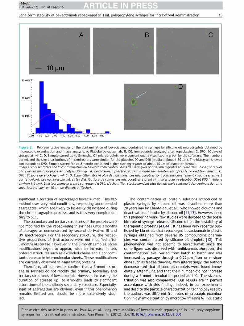
Microscopic examinationMicroscopic examination and subsequent image analysis ofbevacizumab solutions showed the presence of globular andrefringent ring-shaped objects (size ranging from 1 to 15 �m;
wtii
entered to about 2 �m). The mean intensity level of thesebjects, as determined by the software, was very homoge-eous (106.6 ± 7.2 arbitrary units), suggesting that they arelosely similar in composition. Consequently, these objectsere identified as silicone oil microdroplets, as previouslyescribed by several authors [12,13]. These oil dropletsere found in all tested samples, even in D0 samples and
ndependently of storage duration. However, no oil droplet
ability of bevacizumab repackaged in 1 mL polypropyleneoi:10.1016/j.pharma.2012.03.006
as found in bevacizumab directly taken from its vial (con-rol). This finding strongly suggested that oil droplets werentroduced during the repackaging process. Moreover, exper-ments performed after preparing syringes using placebo

ARTICLE IN PRESS+ModelPHARMA-232; No. of Pages 16
12 M. Paul et al.
Table 3 Thermodynamic functions calculated from thermal aggregation curves. Each value represents the mean ± SDof three separate determinations; No difference was observed between control and repackaged bevacizumab stored for3 months at 4 ◦C in polypropylene syringes.Fonctions thermodynamiques calculées à partir des courbes d’agrégation thermique. Chaque valeur représente la moyenne SD de troisessais indépendants. Aucune différence n’a été observée entre le contrôle et le bevacizumab reconditionné stocké durant trois mois à4 ◦C en seringues polypropylène ?
Sample mT (cal mol−1 K) �S (cal mol−1 K−1) �H (kcal mol−1)
D0 2190 ± 195 139 ± 13 369 ± 42◦
btwtpomandtpdaftb2i8
acids
D
Tbsaoabterdb
tfFimb
ncIamabsac
tiirdT7Msio41t[bitbbcooruab
s(ocrt
D3 months 4 C 897 ± 123
evacizumab showed a similar contamination of the solu-ion by oil droplets, even immediately after preparationithout storage period. The mean number and its respec-
ive 95% CI and size distribution of the oil droplets in thelacebo solution was 536 200 ± 147 968 per mL. Examinationf pure water stored in syringes also showed a conta-ination by oil droplets, indicating that the presence of
tensioactive agent as in the placebo bevacizumab wasot a prerequisite to the release of silicone oil from theevice. These results confirmed that the oily contamina-ion was issued from syringes and was independent of therotein presence. Interestingly, the contamination was notifferent from D0 samples (bevacizumab freshly repack-ged): 1 099 181 ± 277 543 per mL, and samples stored evenor 3 months: 812 854 ± 87 776 per mL. The size distribu-ion of the droplet population was similar between D0 andevacizumab stored for 3 months: mean diameter (± CI):.58 ± 0.37 �m with more than 98% of the droplets exhibit-ng sizes less than 10 �m. Moreover, a sample stored for
months showed quite identical level of contamination.Our findings demonstrate that even a long-term stor-
ge of bevacizumab in syringes did not modify the initialharacteristics of the contamination by silicone oil dropletsssued from these syringes. Representative images of the oilroplets and the respective size distribution histogram arehowed in Fig. 8.
iscussion
he main result from our study is that commercial 25 mg/mLevacizumab solution, repackaged in 1-mL polypropyleneyringe, is not significantly altered until 3 months of storaget 4 ◦C. Indeed, using several complementary and orthog-nal stability-indicating methods, no modification of thentibody structure was observed, in terms of chemical sta-ility (primary structure) or physical stability (secondary andertiary structure). The only difference we found is the pres-nce of silicone oil droplets issued from the syringes in theepackaged bevacizumab. However, the increase in storageuration leads to significant signs of alteration of the anti-ody, as demonstrated using 8 months stored syringes.
SEC did not show sub-visible aggregates or fragmenta-ion. Retention time and AUC of eluted peaks were identicalor the control and the solutions stored for 3 months at 4 ◦C.
Please cite this article in press as: Paul M, et al. Long-term stsyringes for intravitreal administration. Ann Pharm Fr (2012), d
urthermore, the native percentage of dimer was not mod-fied. Moreover, the percentage of HMW species was notodified. No additional peak or decrease in the AUC of theevacizumab peak was found by CEX, demonstrating that
gfat
145 ± 15 352 ± 39
o desamidation or chemical alteration, leading to modifi-ations of the isoelectric point of the protein, occurred [19].ndeed, CEX is a useful tool to detect subtle protein alter-tions such as deamidation or oxidation [36]. Finally, peptideapping confirms the absence of chemical alteration of the
ntibody. This excellent chemical stability of repackagedevacizumab was expected since it was unlikely that theimple reconditioning of the initial solution of bevacizumabnd subsequent storage at the recommended temperatureould induce chemical degradation.
Moreover, these results are not surprising consideringhe presence of a significant number of disulfide bonds,ntimate domain-domain interactions and the high contentn �-sheets in IgG. This gives mAbs a better stability andesistance to moderate thermal stress in non-enzymatic con-itions, as compared to other proteins and revealed bym or Tagg higher values than in other proteins (range of0—80 ◦C vs. range of 40—65 ◦C for globular protein) [19].oreover, several previous studies demonstrated the strong
tability of various mAbs. Liu et al. showed that a long-termncubation at relatively high temperature was necessary tobserve clear physicochemical instability (i.e. 6 months at0 ◦C) [37]. Usami et al. found that storage at 37 ◦C for4 days did not alter the tested mAb [38]. Cordoba showedhat four humanized IgG1 were stable for 1 month at 5 ◦C39]. Finally, Bakri claimed a 6-month stability at 4 ◦C forevacizumab [11]. However, this study was only based onmmunoassay methods to measure bevacizumab concentra-ion, and very recently, it has been suggested that theseiological methods alone are insufficient to prove the sta-ility of these complex molecules [40]. Therefore, it wasritical to use several orthogonal chromatographic meth-ds to demonstrate unambiguously the chemical stabilityf repackaged bevacizumab. These methods were currentlyecommended in antibody stability studies [18—20], largelysed by the other previously cited authors and we have alsoscertained that our methods were stability-indicating forevacizumab by using stressed conditions.
No significant difference was found by DLS con-idering the hydrodynamic diameter of bevacizumabmonomer/dimer peak) up to 3 months. A small populationf high molecular size, initially present in the commer-ial vials, was observed but its percentage (about 0.1%)emained constant up to 3 months. No additional popula-ion, corresponding to sub-visible aggregates of diameter
ability of bevacizumab repackaged in 1 mL polypropyleneoi:10.1016/j.pharma.2012.03.006
reater than 1 �m, was observed, confirming the resultsrom SEC. However, an increase in this HMW populationppeared in tested samples, which had been stored for morehan 8 months, suggesting that a too long storage can induce
ARTICLE IN PRESS+ModelPHARMA-232; No. of Pages 16
Long-term stability of bevacizumab repackaged in 1 mL polypropylene syringes for intravitreal administration 13
Figure 8. Representative images of the contamination of bevacizumab contained in syringes by silicone oil microdroplets obtained bymicroscopic examination and image analysis. A. Placebo bevacizumab. B. D0: immediately analyzed after repackaging. C. D90: 90 days ofstorage at +4 ◦C. D. Sample stored up to 8 months. Oil microdroplets were conventionally visualized in green by the software. The numbersper mL and the size distributions of microdroplets were similar for the placebo, D0 and D90 (median: about 1.50 �m). The histogram showedcorresponds to D90. Sample stored for up 8 months contained higher size aggregates of about 10 �m of diameter (arrow).Images représentatives de la contamination du bevacizumab contenu dans des seringues par des microgouttes d’huile de silicone ; obtenuespar examen microscopique et analyse d’image. A. Bevacizumab placebo. B. D0 : analysé immédiatement après le reconditionnement. C.D90 : 90 jours de stockage à +4 ◦C. D. Échantillon stocké plus de huit mois. Les microgouttes sont conventionnellement visualisées en vertpar le logiciel. Les nombres par mL et les distributions de tailles des microgouttes étaient similaires pour le placebo, D0 et D90 (médiane
tillo
p2dtbtlscpsciddddt
environ 1,5 �m). L’histogramme présenté correspond à D90. L’échansupérieure d’environ 10 �m de diamètre (flèche).
significant alteration of repackaged bevacizumab. This DLSmethod uses very mild conditions, respecting loose-bondedaggregates, which are likely to be easily dissociated duringthe chromatographic process, and is thus very complemen-tary to SEC.
The secondary and tertiary structures of the protein werenot modified by the repackaging in syringes until 3 monthsof storage, as demonstrated by second derivative IR andUV spectroscopy. For the secondary structure, the respec-tive proportions of �-structures were not modified after3 months of storage. However, in the 8-month samples, somemodifications began to appear, with an increase in lessordered structures such as extended chains and a concomi-tant decrease in intermolecular sheets. These modificationsare currently observed in aggregating proteins.
Therefore, all our results confirm that a 3-month stor-age in syringes do not modify the primary, secondary andtertiary structures of bevacizumab. However, increasing theduration of storage up, to 8 months leads to significant
Please cite this article in press as: Paul M, et al. Long-term stsyringes for intravitreal administration. Ann Pharm Fr (2012), d
alterations of the antibody secondary structure. Especially,signs of aggregation are obvious, even if this phenomenonremains limited and should be more extensively stud-ied.
aatt
n stocké pendant plus de huit mois contenait des agrégats de taille
The contamination of protein solutions introduced inlastic syringes by silicone oil was described more than0 years ago by Chanteleau et al., who showed clouding andeactivation of insulin by silicone oil [41,42]. However, sincehis pioneering work, few studies were devoted to the possi-le role of syringe-released silicone oil on the instability ofherapeutic proteins [43,44]. It has been very recently pub-ished by Liu et al. that repackaged bevacizumab in plasticyringes obtained from several US compounding pharma-ies was contaminated by silicone oil droplets [12]. Thishenomenon was not specific to bevacizumab since theame figure was observed with ranibizumab. Moreover, theontamination level varied from batch to batch and wasncreased by passage through a 0.22 �m filter or mishan-ling such as freeze-thawing. Very interestingly, the authorsemonstrated that silicone oil droplets were found imme-iately after filling and that their number did not increaseuring a 3-month incubation period at 4 ◦C. The size dis-ribution was also comparable. Our results are in perfect
ability of bevacizumab repackaged in 1 mL polypropyleneoi:10.1016/j.pharma.2012.03.006
ccordance with this finding. Indeed, in our experimentsnd despite the particle characterization technology used byhe authors was different from ours (microscopic examina-ion in dynamic situation by microflow imaging MFI vs. static

IN+ModelP
1
imcaetm(dtvaicmAcmcocrtcaa
aaratt3imaposi
cmtrttrposIbfeoiasts
aeiWbiisesUu
di(tapbtaspiadaebwe
C
OicsusosdnricpowduCaos
ARTICLEHARMA-232; No. of Pages 16
4
maging, respectively), the number of microdroplets perL were very similar. For example, for the placebo beva-
izumab, we found 536 200 ± 147 968 microdroplets per mLs compared to 492 314 ± 396 361 per mL in the study of Liut al. In this study, the authors pointed out the possibilityo find also, in the tested batches from compounding phar-acies, protein aggregates with significant loss of monomer
up to 10%) and subsequent increase in HMW oligomers, asetermined by SEC. However, in their controlled labora-ory samples of repackaged bevacizumab, neither significantariation in the monomer percentage nor apparition ofggregates was seen. The authors attributed these find-ngs to a mishandling of syringes such as freezing by directontact of syringes against the frozen gel packs used toaintain the temperature around 4 ◦C during the shipping.ccordingly, our results showed that the respective per-entages of monomer, dimer and HMW oligomers were notodified by the presence of silicone oil microdroplets at
oncentration less than 106/mL, confirming that this levelf contamination could not lead to aggregation of beva-izumab, at least for short-term storage. Indeed, it has beenecently demonstrated that a monoclonal antibody exposedo a very high level of silicone oil microdroplets in staticondition did not tend to aggregate. However, even a middlegitation, induced the appearance of sub-visible and visibleggregates [44].
A slight increase in the absorbance at 350 nm of repack-ged solutions was observed in our turbidimetry experimentsfter 3 months of storage. Since this increase was not cor-elated with other aggregation markers such as SEC, DLSnd microscopic examination, which remained, unchanged,his result remains unclear and could suggest an experimen-al bias. Indeed, a possible explanation could be that after
months, more antibody molecules would be present at thenterface water/oil. Since the dilution step prior measure-ent is performed with simple phosphate buffer, it leads to
decrease in the concentration of the tensioactive agentolysorbate. Thus, a destabilization of the emulsion canccur, with a coalescence of the microdroplets and sub-equent formation of large size aggregates leading to anncrease in OD.
Despite it has been claimed that the presence of sili-one microdroplets had no consequence on the stability ofAb, we tried to ascertain that more subtle destabiliza-
ion did not occur. For this purpose, we tested the thermalesistance of repackaged bevacizumab by the determina-ion of the thermal aggregation curves and their derivedhermodynamic parameters. Our results showed that forepackaged bevacizumab as compared to control, thesearameters remained unchanged, confirming the absencef intra- or intermolecular interactions, and a conservedtructural integrity without appreciable destabilization.ndeed, the stability of a protein results from equilibriumetween destabilizing and stabilizing forces. Destabilizingorces are mainly due to a large increase in unfoldingntropy (dissipative forces, loss of conformational entropyf native state), and stabilizing forces (intra-molecularnteractions), which are mainly due to non-covalent inter-
Please cite this article in press as: Paul M, et al. Long-term stsyringes for intravitreal administration. Ann Pharm Fr (2012), d
ctions. Many factors can disrupt this delicate balance,uch as temperature, which is the most important fac-or influencing protein stability (�G < 0), added chemicalsuch as excipients or contaminants [18—22]. Many forces
waso
PRESSM. Paul et al.
re involved in protein folding, including hydrophobic andlectrophobic interactions (charge repulsion and ion pair-ng), hydrogen bonding, intrinsic propensities and Van deraals forces, but the apparent dominant force is hydropho-ic interaction. Hydrophobic interactions increase with thencrease in temperature. In fact, proteins aggregate to min-mize thermodynamically unfavorable interactions betweenolvent and exposed hydrophobic residues. Temperaturenhances the hydrophobic surface area and the flexibility ofolvent-accessible amino acid residues, causing aggregation.sually, thermal-induced denaturation is irreversible, as thenfolded molecules rapidly associate to form aggregates.
Different parameters can be studied to estimate theegree of internal or external stabilization, such as changesn unfolding temperature (Tm) or its surrogate, aggregationTagg) temperature. Generally speaking, weak alterations ofhe secondary and tertiary structures are characterized by
negative shift of the Tm of about 0.5 ◦C and, when therotein integrity is strongly altered (breakdown of disulfideonds), the decrease in this parameter is more impor-ant (> 1 ◦C) [19,20,23—26]. As expected, we observed thatll thermodynamic parameters were positive, thus corre-ponding to an endothermic process as described for otherroteins. The denaturation enthalpies we founded weren the same order of magnitude that those described fornother IgG if we consider the sum of the two-denaturationomains (Fab and Fc), as previously described by Vermeernd Norde using differential scanning calorimetry [45]. Nev-rtheless, we demonstrated that the thermal resistance ofevacizumab and its derived thermodynamic parametersere unaltered by storage in syringes for up to 3 months,ven contaminated by silicone oil microdroplets.
onclusion
ur results using several complementary and stability-ndicating analytical methods demonstrate that theommercial solution of bevacizumab (25 mg/mL) can beafely repackaged in polypropylene syringes and storedp to 3 months at 4 ◦C without alteration of its primary,econdary and tertiary structure. The only differencebserved is the contamination of the syringe content byilicone oil microdroplets, which is quite immediate andoes not change significantly during the storage in terms ofumber and size. Since it has been suggested that recenteports of sustained elevation of intraocular pressure andnflammation after intravitreal administration of both beva-izumab and ranibizumab could be due to contamination byarticulates material, it is crucial to minimize the formationf these particles. Our results confirm those from Liu et al.,hich demonstrated that the presence of silicone oil micro-roplets in repackaged bevacizumab was inherent to these of plastic syringes, thus independent of the used mAb.onsidering the off-label use of bevacizumab in intravitrealdministration as compared to ranibizumab, this finding isf paramount importance to support its use. Finally, sinceilicone oil can also be found in simple aqueous solutions
ability of bevacizumab repackaged in 1 mL polypropyleneoi:10.1016/j.pharma.2012.03.006
ithout extracting properties (i.e. without tensioactivegents) in contact with siliconized syringes, even after ahort time contact, this finding clearly opens the questionf the contamination of all solutions currently administered
IN+Model
prop
[
[
[
[
[
[
[
[
[
[
[
[
[
[
[
[
[
[
[
[
[
[
ARTICLEPHARMA-232; No. of Pages 16
Long-term stability of bevacizumab repackaged in 1 mL poly
by intravitreal routes such as antibiotics. In conclusion,our results support the off-label use of repackaged beva-cizumab by intravitreal administration, at least from apharmaceutical point of view, since we demonstrated thatits stability remains excellent for 3 months. This period islargely enough to many practical situations and supportcurrent practices, such as in advance or batch preparations,which present major advantages in terms of GMP respect,workload optimization and financial savings. However, itshould be emphasized that particular care should be takento avoid mishandling during manufacturing and shipping, asrecently suggested by Liu et al. Therefore, it seems impor-tant to develop consensual standard operating proceduresto prepare, handle and ship bevacizumab repackaged insyringe to ensure its safest use for patients.
References
[1] Cursiefen C, Maruyama K, Liu Y, Chen L, Jackson D, Wie-gand S, et al. Inhibition of hem- and lymphangiogenesisafter normal-risk corneal transplantation by neutralizing VEGFpromotes graft survival. Invest Ophthalmol Vis Sci 2004;45:2666—73.
[2] Bock F, Onderka J, Dietrich T, Bachmann B, Kruse FE, PaschkeM, et al. Bevacizumab as a potent inhibitor of inflammatorycorneal angiogenesis and lymphangiogenesis. Br J Ophthalmol2007;91:804—7.
[3] Bressler NM. Anti-angiogenic approaches to age-related macu-lar degeneration today. Ophtalmology 2009;116(10):15—23.
[4] Hubschman JP, Reddy S, Schwartz S. Age-related mac-ular degeneration: current treatments. Clin Ophthalmol2009;3:155—66.
[5] Cohen SY. Anti-VEGF drugs as the 2009 first-line therapyfor choroidal neovacularization in pathologic myopia. Retina2009;29(8):1062—6.
[6] Schwartz SG, Flynn Jr HW. Pharmacotherapies for diabeticretinopathy: present and future. Exp Diabetes Res; 2007, Arti-cle ID 52487, doi:10.1155/2007/52487.
[7] Brouzas D, Charakidas A, Moschos M, koutsandrea C, Apos-tolopoulos M, Baltazis S. Bevacizumab (Avastin®) for themanagement of anterior chamber neovascularisation and neo-vascular glaucoma. Clin Ophthalmol 2009;3:685—8.
[8] Fung AE, Rosenfeld PJ, Reichel E. The international intravitrealbevacizumab safety survey: using the Internet to assess drugsafety worldwide. Br J Ophtalmol 2006;90:1344—9.
[9] The CATT group. Ranibizumab and bevacizumab for neo-vascular age-related macular degeneration. New Eng J Med2011;364:1897—908.
[10] http://www.aao.org/newsroom/release/20110428.cfm.Accédé le 3 janvier 2012.
[11] Bakri SJ, Snyder MR, Pulido JS, McCannel CA, Weiss WT, SinghRJ. Six-month stability of bevacizumab (Avastin®) binding tovascular endothelial growth factor after withdrawal into asyringe and refrigeration or freezing. Retina 2006;26:519—22.
[12] Liu L, Ammar DA, Ross LA, Mandava N, Kahook NY, Carpen-ter JF. Silicone oil microdroplets and protein aggregates inrepackaged bevacizumab and ranibizumab: effects of long-term storage and product mishandling. Invest Ophtalmol VisSci 2011;52:1023—34.
[13] Kahook MY, Liu L, Ruzycki P, Mandava N, Carpenter JF, PetrashJM, et al. High molecular weight aggregates in repackaged
Please cite this article in press as: Paul M, et al. Long-term stsyringes for intravitreal administration. Ann Pharm Fr (2012), d
bevacizumab. Retina 2010;30:887—92.[14] Paul M, Lahlou A, Carvalho M, Blanchet B, Astier A. Thermal
stability of two monoclonal antibodies: cetuximab and beva-cizumab. Eur J Oncol Pharm 2008;2(1):37.
PRESSylene syringes for intravitreal administration 15
15] Lahlou A, Blanchet B, Carvalho M, Paul M, Astier A.Mechanically-induced aggregation of the monoclonal antibodycetuximab. Ann Pharm Fr 2009;67:340—52.
16] Mahler C, et al. Induction and analysis of aggregates ina liquid igG1-antibody formulation. Eur J Pharm Biopharm2005;59:407—17.
17] Bloomfield VA. Static and dynamic light scattering from aggre-gating particles. Biopolymers 2000;54:178—82.
18] Wang W. Protein aggregation and its inhibition in biopharma-ceutics. Int J Pharm 2005;289:1—30.
19] Wang W. Instability, stabilization, and formulation of liquidprotein pharmaceuticals. Int J Pharm 1999;185:129—88.
20] Lowe D, Dudgeon K, Rouet R, Schofiel P, Jermutus L, ChristD. Aggregation, stability and formulation of human antibodytherapeutics. Adv Prot Chem Struct Biol 2011;84:41—61.
21] Chi EY, Krishnan S, Randolph TW, Carpenter JF. Physical sta-bility of proteins in aqueous solution: mechanism and drivingforce in nonnative protein aggregation. Pharm Res 2003;20:1325—36.
22] Jaenicke R. Stability and stabilization of globular proteins insolution. J Biotechnol 2000;79:193—203.
23] Bieri O, Kiefhaber T. Elementary steps in protein folding. BiolChem 1999;380:923—9.
24] Thirumangalathu R, Sampathkumar K, Brems D, Randolph T,Carpentier J. Effects of pH, temperature, and sucrose on ben-zyl alcohol-induced aggregation of recombinant human granu-locyte colony stimulating factor. J Pharm Sci 2006;95:1480—97.
25] Farruggia B, Garcia G, D’Angelo C, Pic G. Destabilization ofhuman serum albumin by polyethylene glycols studied by ther-modynamical equilibrium and kinetic approaches. Int J BiolMacromolecules 1997;20:43—51.
26] Harn N, Allan C, Oliver C, Middaugh CR. Highly concentratedmonoclonal antibody solutions: direct analysis of physicalstructure and thermal stability. J Pharm Sci 2007;96:532—46.
27] Cauchy M, D’Aoust S, Dawson B, Rode H, Hefford MA. Thermalstability: a means to assure tertiary structure in therapeuticproteins. Biol J Int Assoc Biol Stand 2002;30:175—85.
28] Kuelzo LA, Ersoy B, Ralston JP, Middaugh CR. Derivativeabsorbance spectroscopy and protein phase diagrams as toolsfor comprehensive protein characterization: a bGCSF casestudy. J Pharm Sci 2003;92:1805—20.
29] Balestrieri C, Colonna G, Giovane A, Irace G, Servillo L. Secondderivative spectroscopy of proteins. A method for the quanti-tative determination of aromatic amino acids in proteins. EurJ Biochem 1978;90:433—40.
30] Ichikawa T, Terada H. Estimation of state and amount ofphenylalanine residues in proteins by second derivative spec-trophotometry. Biochim Biophys Acta 1979;580:120—8.
31] Servillo L, Colonna G, Balestrieri C, Ragone R, Irace G. Simul-taneous determination of tyrosine and tryptophan residuesin proteins by second derivative spectroscopy. Anal Biochem1982;126:251—7.
32] Kong J, Yu S. Fourier transform infrared spectroscopic analy-sis of protein secondary structures. Acta Biochimica BiophysicaSinica 2007;39:549—59.
33] Smith BC. Fundamentals of Fourier transform infrared spec-troscopy. Boca Raton: CRC Press; 1996.
34] Krimm S, Bandekar J. Vibrational spectroscopy and conforma-tion of peptides, polypeptides, and proteins. Adv Protein Chem1986;38:181—364.
35] Byler DM. Resolution-enhanced Fourier transforminfrared spectroscopy of enzymes. Methods Enzymol1986;130:290—311.
36] Moorhouse KG, Nashabeh W, Deveney J, Bjork NS, Mulkerrin
ability of bevacizumab repackaged in 1 mL polypropyleneoi:10.1016/j.pharma.2012.03.006
MG, Ryskamp T. Validation of an HPLC method for the analysisof the charge heterogeneity of the recombinant monoclonalantibody IDEC-C2B8 after papain digestion. J Pharm BiomedAnal 1997;16:593—603.

IN+ModelP
1
[
[
[
[
[
[
[
[
ARTICLEHARMA-232; No. of Pages 16
6
37] Liu C, Vailhe C, Samuel N, Min Q. A simple and reproducibleapproach to characterize protein stability using rheology. Int JPharm 2009;374:1—4.
38] Usami A. The effect of pH, hydrogen peroxide and temperatureon the stability of human monoclonal antibody. J Pharm BiomedAnal 1996;14:1133—40.
39] Cordoba AJ, Shyong BJ, Breen D, Harris RJ. Non-enzymatichinge region fragmentation of antibodies in solution. J Chro-matogr B Anal Technol Biomed Life Sci 2005;818:115—21.
Please cite this article in press as: Paul M, et al. Long-term stsyringes for intravitreal administration. Ann Pharm Fr (2012), d
40] Sreedhara A, Glover ZK, Piros N, Xiao N, Patel A, KabakoffB. Stability of IgG1 monoclonal antibodies in intravenousinfusion bags under clinical in-use conditions. J Pharm Sci2011;101:21—30.
[
PRESSM. Paul et al.
41] Chanteleau EA, Berger M. Pillution of insulin with silicone oil,a hazard of disposable plastic syringes. Lancet 1985;1:1459.
42] Chanteleau EA, Berger M, Bohlken B. Silicone oil release fromdisposable insulin syringes. Diabetes Care 1986;9:672—3.
43] Jones LS, Kaufmann A, Middaugh CR. Silicone oil-inducedaggregation of proteins. J Pharm Sci 2005;94:918—27.
44] Thirumangalathu R, Krishnan S, Speed-Ricci M, Brems DN,Randolph TW, Carpenter JF. Silicone oil-and agitation-inducedaggregation of a monoclonal antibody in aqueous solution. J
ability of bevacizumab repackaged in 1 mL polypropyleneoi:10.1016/j.pharma.2012.03.006
Pharm Sci 2009;98:3167—81.45] Vermeer AWP, Norde W. The thermal stability of immunoglob-
ulin: unfolding and aggregation of a multi-domain protein.Biophys J 2000;78:394—404.