
Long noncoding RNA H19X is a key mediator of TGFβ driven ...
44
Long noncoding RNA H19X is a key mediator of TGFβ driven fibrosis Elena Pachera, … , Mojca Frank-Bertoncelj, Oliver Distler J Clin Invest. 2020. https://doi.org/10.1172/JCI135439. In-Press Preview TGFβ is a master regulator of fibrosis, driving the differentiation of fibroblasts into apoptosis resistant myofibroblasts and sustaining the production of extracellular matrix (ECM) components. Here, we identify the nuclear lncRNA H19X as a master regulator of TGFβ-driven tissue fibrosis. H19X was consistently upregulated in a wide variety of human fibrotic tissues and diseases and was strongly induced by TGFβ, particularly in fibroblasts and fibroblast-related cells. Functional experiments following H19X silencing revealed that H19X is an obligatory factor for the TGFβ-induced ECM synthesis as well as differentiation and survival of ECM-producing myofibroblasts. We showed that H19X regulates DDIT4L gene expression, specifically interacting with a region upstream of DDIT4L gene and changing the chromatin accessibility of a DDIT4L enhancer. These events resulted in transcriptional repression of DDIT4L and, in turn, in increased collagen expression and fibrosis. Our results shed light on key effectors of the TGFβ-induced ECM remodeling and fibrosis. Research Autoimmunity Cell biology Find the latest version: https://jci.me/135439/pdf
Transcript of Long noncoding RNA H19X is a key mediator of TGFβ driven ...

Long noncoding RNA H19X is a key mediator of TGFβ drivenfibrosis
Elena Pachera, … , Mojca Frank-Bertoncelj, Oliver Distler
J Clin Invest. 2020. https://doi.org/10.1172/JCI135439.
In-Press Preview
TGFβ is a master regulator of fibrosis, driving the differentiation of fibroblasts into apoptosis resistant myofibroblasts andsustaining the production of extracellular matrix (ECM) components. Here, we identify the nuclear lncRNA H19X as amaster regulator of TGFβ-driven tissue fibrosis. H19X was consistently upregulated in a wide variety of human fibrotictissues and diseases and was strongly induced by TGFβ, particularly in fibroblasts and fibroblast-related cells. Functionalexperiments following H19X silencing revealed that H19X is an obligatory factor for the TGFβ-induced ECM synthesis aswell as differentiation and survival of ECM-producing myofibroblasts. We showed that H19X regulates DDIT4L geneexpression, specifically interacting with a region upstream of DDIT4L gene and changing the chromatin accessibility of aDDIT4L enhancer. These events resulted in transcriptional repression of DDIT4L and, in turn, in increased collagenexpression and fibrosis. Our results shed light on key effectors of the TGFβ-induced ECM remodeling and fibrosis.
Research Autoimmunity Cell biology
Find the latest version:
https://jci.me/135439/pdf

1
Long noncoding RNA H19X is a key mediator of TGFβ driven fibrosis
Elena Pachera1, Shervin Assassi2, Gloria A. Salazar2, Mara Stellato1, Florian Renoux1, Adam Wunderlin1, Przemyslaw Blyszczuk1, Robert Lafyatis3 Fina Kurreeman4, Jeska de Vries-Bouwstra4, Tobias Messemaker4, Carol A. Feghali-Bostwick5, Gerhard Rogler6, Wouter T. van Haaften7, Gerard Dijkstra7, Fiona Oakley8, Maurizio Calcagni9, Janine Schniering1, Britta Maurer1, Jörg H. W. Distler10, Gabriela Kania1, Mojca Frank-Bertoncelj1 and Oliver Distler1*
1. Center of Experimental Rheumatology, Department of Rheumatology, University Hospital Zurich, Zurich, Switzerland
2. Department of Internal Medicine, Division of Rheumatology, The University of Texas Health Science Center at Houston, McGovern Medical School, Houston, Texas, USA
3. Division of Rheumatology and Clinical Immunology, University of Pittsburgh, Pittsburgh, Pennsylvania, USA
4. Department of Rheumatology, Leiden University Medical Center, Leiden, the Netherlands
5. Division of Rheumatology, Medical University of South Carolina, Charleston, USA
6. Department of Gastroenterology and Hepatology, University Hospital Zurich, Zurich, Switzerland
7. Department of Gastroenterology and Hepatology, University Medical Center Groningen, Groningen, the Netherlands
8. Newcastle Fibrosis Research Group, Institute of Cellular Medicine, Newcastle University, Newcastle upon Tyne, UK
9. Department of Plastic Surgery and Hand Surgery, University Hospital Zurich, Zurich, Switzerland
10. Department of Internal Medicine 3, University of Erlangen, Erlangen, Germany
*Correspondence:
Prof. Dr. Oliver Distler, Department of Rheumatology, University Hospital Zurich, Gloriastrasse 25, 8091 Zürich, Switzerland; [email protected]; Tel: +41 44 255 29 70.

2
Abstract
TGFβ is a master regulator of fibrosis, driving the differentiation of fibroblasts into apoptosis resistant
myofibroblasts and sustaining the production of extracellular matrix (ECM) components. Here, we
identify the nuclear lncRNA H19X as a master regulator of TGFβ-driven tissue fibrosis. H19X was
consistently upregulated in a wide variety of human fibrotic tissues and diseases and was strongly
induced by TGFβ, particularly in fibroblasts and fibroblast-related cells. Functional experiments following
H19X silencing revealed that H19X is an obligatory factor for the TGFβ-induced ECM synthesis as well
as differentiation and survival of ECM-producing myofibroblasts. We showed that H19X regulates
DDIT4L gene expression, specifically interacting with a region upstream of DDIT4L gene and changing
the chromatin accessibility of a DDIT4L enhancer. These events resulted in transcriptional repression
of DDIT4L and, in turn, in increased collagen expression and fibrosis. Our results shed light on key
effectors of TGFβ-induced ECM remodeling and fibrosis.

3
Introduction
During the development of fibrosis, the organ parenchyma is replaced with a collagen rich, stiff
connective tissue leading to organ dysfunction and failure as seen, for example, for end-stage liver
disease or idiopathic pulmonary fibrosis (IPF). Fibrosis is also a dominant pathological feature of many
chronic autoimmune diseases, including systemic sclerosis (SSc), rheumatoid arthritis, Crohn's disease
and systemic lupus erythematosus. SSc is a prototype fibrotic disease and is often used as a model
disease to identify key mechanisms of tissue remodeling in fibrosis (1,2). SSc is associated with high
morbidity and the highest mortality among the rheumatic diseases with a standardized mortality ratio of
3.53 and average loss of life expectancy of 16-34 years (3). The hallmarks of SSc are abnormalities of
the vasculature and immune system eventually leading to fibrosis in multiple organs including skin, lung
and heart. SSc has a chronic course, and particularly in the diffuse cutaneous subset the disease is
often rapidly progressing (4). Despite advances in the identification of potential molecular targets,
specific treatments for fibrotic diseases, including SSc, are limited, and there is a high clinical need for
novel therapeutic concepts.
Fibroblasts from patients with fibrotic diseases display characteristic features including overproduction
of collagen and enhanced secretion of cytokines and chemokines. They also express higher levels of
adhesion molecules such as integrins, and receptors for transforming growth factor beta (TGFβ) and
platelet-derived growth factor (PDGF) (5). Moreover, they show an increased resistance to apoptosis
mediated by death receptor FAS (6). A major hallmark of these cells is their differentiation into highly
activated myofibroblasts. Myofibroblasts express alpha smooth muscle actin (αSMA) in their stress
fibers and exhibit enhanced matrix adhesion and contractile properties (7). Moreover, they release large
amounts of extracellular matrix (ECM) components such as collagens and fibronectin (8). Resident
fibroblasts transiently differentiate into myofibroblasts during in early stages of physiological wound
healing and undergo apoptosis during the resolution stages. However, in scarring tissues, myofibroblast
persistency contributes to fibrogenesis, a process also found in fibrotic diseases (9). Additionally,
selective targeting of antiapoptotic proteins in activated myofibroblasts, have been tested as therapeutic
strategy for fibrosis (10).
TGFβ is the master regulator of physiological wound healing and fibrosis, by inducing the differentiation
of fibroblasts into myofibroblasts (11,12). The TGFβ canonical pathway involves phosphorylation of

4
TGFβR1, which in turn phosphorylates and activates SMAD2/3 that can then bind SMAD4 (13). These
complexes accumulate in the nucleus where they act as transcription factors and participate in the
regulation of target gene expression leading to ECM production and myofibroblast differentiation (14).
Research in recent years has shown that non-coding RNAs play a central role in the regulation of gene
expression (15,16). Long non-coding RNAs (LncRNAs) are defined as non-coding RNA transcripts
longer than 200 bp and are mostly uncharacterized and unannotated. In general, lncRNAs have more
specific expression profiles than protein coding transcripts, being expressed in a cell type-, tissue-,
developmental stage- or disease state-specific manner (17). LncRNAs exert their gene regulatory
functions via a variety of mechanisms. Maternally expressed gene 3 (MEG3), for example, is a nuclear
lncRNA that modulate the expression of TGFβ pathway target genes by regulating the chromatin
structure of their distal enhancers (18). MEG3 has been found downregulated in human fibrotic liver (19)
and in murine cardiac fibrosis (20). Other lncRNAs regulate the behavior of other types of molecules
(21), such as microRNAs. For example, Myocardial infarction-associated transcript (MIAT) captures
miR-24, a TGFβ regulatory miRNA, via a sponge-like activity and promotes cardiac fibrosis (22).
Upregulation of the maternally imprinted lncRNA H19 has been implicated in renal fibrosis via
suppressing the activity of miR-17 leading to increased fibronectin expression (23).
In this study, we identified and functionally characterized a pro-fibrotic lncRNA H19X as a central
regulator of myofibroblast differentiation and survival. We could show that the induction of H19X is a
conditio sine qua non for the pro-fibrotic effects of TGFβ and might be a key driver of myofibroblast
persistence and fibrosis in a wide variety of TGFβ driven fibrotic diseases.

5
Results
LncRNA H19X is upregulated in a variety of fibrotic diseases
SSc is a prototype fibrotic disease. To identify lncRNAs potentially involved in the pathogenesis of
fibrotic diseases, we performed whole transcriptome RNA sequencing (RNA-seq) on skin biopsies from
severely affected SSc patients with active, untreated, early diffuse cutaneous SSc (dcSSc) and age-
and sex-matched healthy controls (5 SSc vs 5 HC). In this screening experiment, H19 X-linked co-
expressed lncRNA (H19X) was identified as one of the most consistently and strongly upregulated
lncRNAs (p=0.022, Figure 1A) in skin biopsies of SSc patients. H19X also known as MIR503HG, is an
intergenic lncRNA and its gene is located on chromosome X. None of its five annotated transcript
variants has a coding potential as demonstrated by in silico analysis using CPAT, a coding potential
assessment tool (Supplementary Table 1).
We confirmed the upregulation of the lncRNA H19X in three additional independent SSc cohorts. Using
RNA-seq, H19X was found significantly upregulated (p<0.0001, Figure 1B) in skin biopsies of patients
with early dcSSc, who were enrolled in the large multicenter SSc cohort-study PRESS (Prospective
Registry of Early Systemic Sclerosis, 48 SSc vs 33 HC).
We then extended our analysis to cohorts of SSc patients with more diverse disease duration and
disease severity. H19X was upregulated by RNA-Seq in skin samples from SSc patients in the third SSc
cohort compared to skin from healthy controls (14 SSc vs 6 HC, p=0.07, Figure 1C). In the fourth cohort
(6 SSc vs 6 HC), we analyzed the expression of H19X with an independent technique. Using qPCR, the
expression of H19X was found 4.3-fold higher in SSc skin samples compared to HC skin samples
(p=0.0043, Figure 1D). To assess whether H19X is upregulated also in affected internal organs of SSc
patients, we extracted total RNA from lung samples of patients with SSc-associated interstitial lung
disease (SSc- ILD) and healthy controls. H19X was significantly upregulated in lungs of patients with
end-stage SSc-ILD undergoing lung transplantation (11 SSc-ILD vs 11 HC, p=0.0181, Figure 1E). These
data from multiple cohorts covering a wide range of clinical SSc phenotypes show that H19X is
upregulated in SSc, not only in the skin, but also in the lungs of affected patients.
6
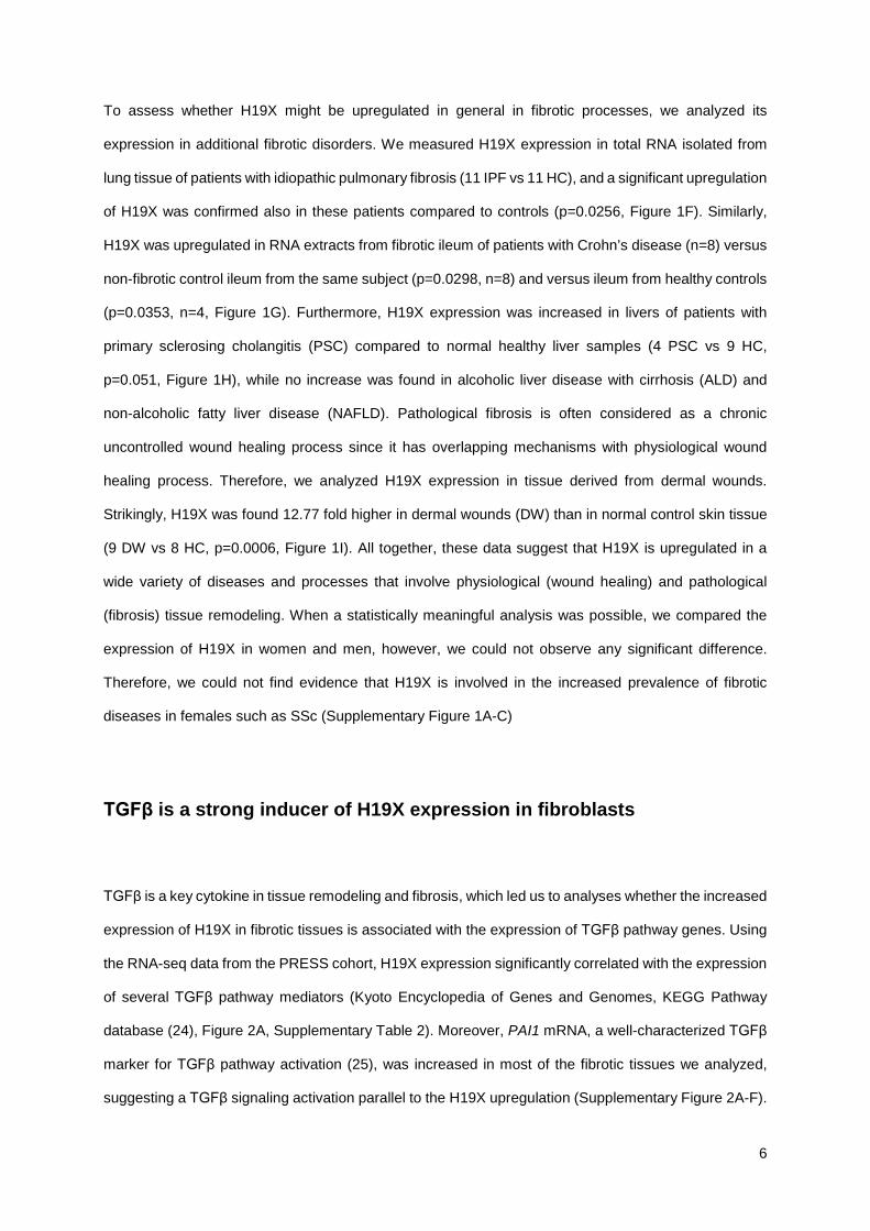
To assess whether H19X might be upregulated in general in fibrotic processes, we analyzed its
expression in additional fibrotic disorders. We measured H19X expression in total RNA isolated from
lung tissue of patients with idiopathic pulmonary fibrosis (11 IPF vs 11 HC), and a significant upregulation
of H19X was confirmed also in these patients compared to controls (p=0.0256, Figure 1F). Similarly,
H19X was upregulated in RNA extracts from fibrotic ileum of patients with Crohn’s disease (n=8) versus
non-fibrotic control ileum from the same subject (p=0.0298, n=8) and versus ileum from healthy controls
(p=0.0353, n=4, Figure 1G). Furthermore, H19X expression was increased in livers of patients with
primary sclerosing cholangitis (PSC) compared to normal healthy liver samples (4 PSC vs 9 HC,
p=0.051, Figure 1H), while no increase was found in alcoholic liver disease with cirrhosis (ALD) and
non-alcoholic fatty liver disease (NAFLD). Pathological fibrosis is often considered as a chronic
uncontrolled wound healing process since it has overlapping mechanisms with physiological wound
healing process. Therefore, we analyzed H19X expression in tissue derived from dermal wounds.
Strikingly, H19X was found 12.77 fold higher in dermal wounds (DW) than in normal control skin tissue
(9 DW vs 8 HC, p=0.0006, Figure 1I). All together, these data suggest that H19X is upregulated in a
wide variety of diseases and processes that involve physiological (wound healing) and pathological
(fibrosis) tissue remodeling. When a statistically meaningful analysis was possible, we compared the
expression of H19X in women and men, however, we could not observe any significant difference.
Therefore, we could not find evidence that H19X is involved in the increased prevalence of fibrotic
diseases in females such as SSc (Supplementary Figure 1A-C)
TGFβ is a strong inducer of H19X expression in fibroblasts
TGFβ is a key cytokine in tissue remodeling and fibrosis, which led us to analyses whether the increased
expression of H19X in fibrotic tissues is associated with the expression of TGFβ pathway genes. Using
the RNA-seq data from the PRESS cohort, H19X expression significantly correlated with the expression
of several TGFβ pathway mediators (Kyoto Encyclopedia of Genes and Genomes, KEGG Pathway
database (24), Figure 2A, Supplementary Table 2). Moreover, PAI1 mRNA, a well-characterized TGFβ
marker for TGFβ pathway activation (25), was increased in most of the fibrotic tissues we analyzed,
suggesting a TGFβ signaling activation parallel to the H19X upregulation (Supplementary Figure 2A-F).

7
Therefore, we hypothesized that TGFβ could be involved in the regulation of H19X expression. Indeed,
stimulation with TGFβ resulted in a strong upregulation of H19X in primary dermal fibroblasts from SSc
patients (p=0.0136) as well as from healthy controls (p=0.0127, Figure 2B), cultured in complete medium
with 10% FBS. The TGFβ-driven induction of H19X was even stronger when fibroblasts were cultured
in starvation medium containing 1% FBS (p=0.0413 SSc, p=0.0249 HC, Figure 2B). The TGFβ induced
upregulation of H19X was dose-dependent with a steady increase over biologically-relevant TGFβ
concentrations (26) (0.1-10 ng/ml in complete medium with 10% FBS, Figure 2C). Time course
experiments showed that the upregulation of H19X by TGFβ was strongest at 6h, reaching an 8-fold
increase in fibroblasts cultured in complete medium with 10% FBS, indicating an early response of H19X
to TGFβ. Although smaller, there was a consistent induction of H19X across all measured time points
(1h-48h, Figure 2C). Other pro-fibrotic and pro-inflammatory cytokines such as IL-1β, IL-4, IL-13 and IL-
17a did not induce H19X after 6 or 24h of stimulation. A mild absolute induction was observed after 6h
of PDGF-BB stimulation (p=0.0041), and a mild downregulation was recorded after 24h of IL-13
stimulation, but not at other time points (p=0.0030, Figure 2D and Supplementary Figure 3A).
SMAD proteins play a major role in canonical TGFβ signaling via TGFβR1 activation. To unravel the
signaling mechanisms involved in TGFβ-dependent H19X induction, we treated fibroblasts with
SB431542 and SD208, the selective inhibitors of TGFβR1 in parallel to TGFβ stimulation. The TGFβ-
driven induction of H19X significantly diminished after TGFβR1 blockage (p<0.001 SB431542 and
p=0.0017 SD208, Figure 2E). To analyze whether the canonical TGFβ pathway plays a role in the TGFβ-
induced expression of H19X, SMAD3, SMAD4 and TGFBR2 were silenced using respective siRNAs.
Indeed, the TGFβ-induced expression of H19X was reduced in silenced samples as compared to
scrambled controls (p=0.0001 SMAD3 siRNA, p=0.0006 SMAD4 siRNA, p=0.0002 TGFBR2 siRNA,
Figure 2F and Supplementary Figure 3B). These data provide further evidence that the TGFβ canonical
pathway is a key regulatory pathway of H19X expression.
Moreover, in the PRESS cohort, the expression of H19X positively correlated with the fibroblast
signature(27) (r=0.59, p<0.0001), while there was less or no correlation for other cell types (Figure 2G,
Supplementary Table 3). To assess the cell types, in which H19X can be induced by TGFβ, several cell
lines were treated with TGFβ. A strong increase in H19X expression, comparable to that observed in
dermal fibroblasts, was found in rheumatoid arthritis synovial fibroblasts (p=0.0043, Figure 2H), in the
foreskin fibroblast cell line BJ5TA (p=0.040, Figure 2H) and in fibroblast-like cells, such as pulmonary

8
artery smooth muscle cells (p=0.0723, Figure 2H). On the contrary, when we stimulated other cell types
with TGFβ for 6h, including CD14+ cells from SSc patients, human microvascular endothelial cells and
human keratinocytes (HK), a decrease or a mild induction in H19X expression was observed (p=0.0039
HK, Figure 2I). These data indicate that H19X is a TGFβ regulated lncRNA and that its induction by
TGFβ is cell type dependent. Specifically, fibroblasts of different origins and fibroblast-like cells exhibited
the strongest induction by TGFβ, further supporting the hypothesis that H19X might play a role in
fibrosis.
H19X mediates the pro-fibrotic effects of TGFβ by promoting ECM
production and myofibroblast differentiation
To study, which genes are affected by the dysregulation of H19X in fibrotic tissues, we silenced H19X
in SSc fibroblasts for 120h and stimulated with TGFβ for the last 72h (Supplementary Figure 4A). First,
we used a targeted approach by analyzing a specific set of TGFβ-responsive genes known for their
important roles in fibrosis, including genes encoding ECM components (collagen1α1, COL1A1 and
fibronectin, FN1) and myofibroblast marker molecules (α-smooth muscle actin, ACTA2). We observed
a strong and consistent reduction in the expression of these genes after H19X silencing using two
different sets of H19X anti-sense oligonucleotides (ASOs, Supplementary Figure 4C). In order to have
a broader overview on H19X target genes, microarray analysis was performed (Supplementary Figure
4D). At a false discovery rate (FDR) of <5% and absolute Log2 fold change (FC) >0.5, 162 genes were
significantly downregulated, and 155 genes were significantly upregulated in TGFβ-stimulated cells after
H19X silencing compared to TGFβ-stimulated scrambled controls. As analyzed by gene set enrichment
analysis (GSEA) of 162 downregulated genes, H19X silencing had a strong effect on genes involved in
ECM production. Gene ontology gene sets such as extracellular matrix structural constituents, collagen
containing extracellular matrix, extracellular matrix and extracellular matrix components were among
the top 10 gene sets (Figure 3A). Moreover, different collagens (COL8A2, COL15A1 and COL3A1),
other ECM genes and ECM-related genes (elastin, ELN; EGF-containing fibulin-like extracellular matrix
protein, EFEMP1; podocan, PODN; dermatopontin, DPT; metalloproteinase inhibitor 3, TIMP3;
microfibril associated protein 4, MFAP4; secreted frizzled related protein 2, SFRP2; nicastrin, NCSTN;

9
tenascin C, TNC; fibulin-5, FBLN5; glypican 6, GPC6; insulin-like growth factor-binding protein 3,
IGFBP3; insulin-like growth factor-binding protein 7, IGFBP7 and fibulin-2, FBLN2) were among the top
downregulated genes (Log2FC<-0.5, Figure 3B). These data suggest that the upregulation of H19X
observed in fibrotic tissue might promote ECM remodeling.
Given these results, we further characterized the role of H19X in the production of ECM components
and in the differentiation of fibroblasts into the ECM-producing myofibroblasts (Supplementary Figure
4A). Basal fibronectin expression was significantly reduced at the protein level following H19X silencing
in SSc fibroblasts (p=0.0051, Figure 3C). Moreover, protein secretion of total collagens (p=0.0188,
Figure 3D) and pro-collagen1α1 (p=0.0007, Figure 3D) into the supernatant of SSc fibroblasts was
impaired as revealed by Sircol assay and ELISA, respectively. Western blot analysis showed that H19X
silencing in SSc fibroblasts reduced the production of αSMA at the protein level (Figure 3E).
Immunofluorescence staining of αSMA fibers and phalloidin staining of stress fibers following H19X
silencing in SSc fibroblasts was also strongly reduced, further confirming that H19X is a crucial factor in
TGFβ mediated actin fiber formation and myofibroblast differentiation (Figure 3F). Furthermore, SSc
fibroblasts displayed a reduced capacity to contract collagen gel matrix after H19X silencing, (p=0.0026
basal condition and p=0.0007 after TGFβ stimulation, Figure 3G). These data show that H19X
expression influences ECM gene regulation and myofibroblast differentiation. Because mitochondrial
deacetylases Sirtuin-3 has recently been shown to be downregulated by TGFβ mediating effects on
collagen synthesis (28), we investigated whether H19X could be involved in the dysregulated expression
of SIRT3 in fibrosis. However, while we could confirm that protein levels of SIRT3 were reduced following
TGFβ treatment, H19X silencing did not affect SIRT3 protein expression, indicating that in this case the
TGFβ effects on SIRT3 are independent of H19X (Supplementary Figure 4E).
H19X regulates cell cycle and proliferation
We continued with the analysis of the 155 upregulated genes after H19X silencing (FDR<0.05 and
Log2FC>0.5, Supplementary Figure 4D). Gene ontology analysis revealed a strong involvement of
H19X in cell cycle regulation (top ten pathway shown in Figure 4A). Among the genes with the strongest
upregulation (Figure 4B), there were CCND1, CCNA2, CCNB2 encoding for cyclins (cell cycle

10
regulators), CDC20 (regulator of the anaphase promoting complex), PTTG1 (regulatory protein of cell
cycle progression) and MCM4 (involved in DNA replication). In order to confirm the role of H19X in
regulating the cell cycle, we performed Hoechst 33342 staining and cell cycle analysis by flow cytometry,
following H19X downregulation and TGFβ treatment. Consistent with our microarray results, H19X
silencing led to a higher percentage of living fibroblasts in S/G2 phase of the cell cycle, both, at the basal
level and after TGFβ stimulation (Figure 4C, Supplementary Figure 5A).
Indeed, H19X downregulation (Supplementary Figure 4B) led to changes in the proliferation of SSc
fibroblast as measured by X-CELLigence real-time proliferation experiments. In the exponential phase,
H19X silenced fibroblasts showed a significant increase in cell proliferation compared to controls
(p<0.0001 basal condition and p=0.0029 after TGFβ stimulation, Figure 4D, E). In the late phase of the
X-CELLigence curve, starting on average at around 104h after transfection, a strong reduction in cell
index was observed for H19X silenced cells with and without TGFβ stimulation (p=0.0002 basal
condition and p=0.0078 in the presence of TGFβ, Figure 4D, E). These data suggest that H19X silencing
might lead to late induction of cell death. Therefore, we hypothesized that H19X could exhibit anti-
apoptotic effects on myofibroblasts promoting their survival which then leads to uncontrolled ECM
deposition.
H19X promotes pro-fibrotic effects of TGFβ by favoring myofibroblast
survival
Myofibroblasts are apoptosis-resistant cells, but they are in close proximity to the apoptotic threshold
(10). As recently demonstrated, targeting apoptosis resistant myofibroblasts might be an effective
antifibrotic treatment strategy (10). Based on our results of the X-CELLigence real-time proliferation
experiments, we performed annexin V live measurements after H19X silencing with and without TGFβ
stimulation using the same experimental design that we used for the X-CELLigence analysis
(Supplementary Figure 4B). Luminescence (for annexin V) and fluorescence (DNA dye) measurements
were recorded between 24h and 126h after transfection of SSc fibroblasts. Already at 42h, we could
record a significant difference in annexin V signals in H19X silenced fibroblasts in comparison with

11
scrambled transfected cells for both untreated and TGFβ stimulated cells. This effect steadily increased
until 72h (Figure 4F). Till about 36h after cell transfection H19X silenced cells were protected from
necrosis. A delay in increased fluorescent signal was recorded starting at 54h after cell transfection for
untreated cells and 66h for TGFβ stimulated cells, marking the start of the necrotic process
(Supplementary Figure 5B). Moreover, we also tested caspase 3/7 activity as a measure of activation
of the apoptotic cascade after H19X silencing and parallel TGFβ stimulation. Indeed, 88h and 120h after
cell transfection, a strong and consistent induction of caspases 3/7 activity was recorded following H19X
silencing and TGFβ stimulation (p<0.001, Figure 4G, Supplementary Figure 5C). In summary, these
results suggest that H19X is a direct potent mediator of prolonged myofibroblast survival, potentiating
the TGFβ driven ECM production and progressive tissue fibrosis. Inducing apoptosis of myofibroblasts
by H19X silencing could be a therapeutic strategy to selectively remove ECM-over-producing cells from
fibrotic tissues (10).
H19X has a human specific function in fibrosis
Next, we wanted to see, whether H19X is conserved in mice and whether mouse models can be used
to further characterize its function. A putative murine H19X transcript is annotated in NCBI
(Supplementary Figure 6A) and it was previously reported that the microRNAs associated with H19X
(miR-503 and miR-424) have orthologous microRNAs in opossum, platypus, chicken and xenopus (29).
The genomic region spanning human and mouse transcripts has 60% homology. However, the
homologous parts consist mainly of introns, and the annotated human and mouse transcripts profoundly
differ. In addition, we analyzed the expression of the putative murine H19X (mH19X) in different well-
established mouse models of fibrosis. These analyses showed that the putative mH19X was
downregulated rather than upregulated in the lung tissues of mice with bleomycin-induced lung fibrosis,
and its expression in skin did not differ between control mice and different fibrotic mouse models
(bleomycin-induced skin fibrosis, tight skin mouse-1, injections of adenovirus overexpressing a
constitutively active TGFβR1, Supplementary Figure 6B). Moreover, the putative mH19X was not
induced when murine dermal fibroblasts were stimulated with TGFβ for different time periods
(Supplementary Figure 6C). These data suggest that the putative mH19X is differentially regulated and

12
expressed in mouse fibrotic conditions and that mouse models cannot be used for functional analysis
of the orthologue human gene.
The pro-fibrotic effects of H19X are largely independent from miR-424
and miR-503
H19X gene is localized on chromosome X and the H19X gene locus encodes two microRNAs. The
MIR424 sequence partially overlaps with the first exon of H19X, and MIR503 is embedded in the first
intron (Supplementary Figure 7A). Therefore, we tested the hypothesis that H19X could exert its function
acting as a microRNA reservoir allowing a rapid release of the two microRNAs (30). Indeed, the
expression of miR-424 and miR-503 was induced by TGFβ at 6h, 24h and 72h (p<0.05, Figure 5A). In
contrast to H19X, the expression of these microRNAs was highest at 72h and 24h, respectively, and the
early peak at 6h was not observed in SSc dermal fibroblasts. Moreover, the expression of miR-424 and
miR-503 was reduced after H19X silencing (Figure 5B). These data argue that H19X could potentially
function as a reservoir for these two microRNAs.
To test this hypothesis, we first silenced miR-424 (Supplementary Figure 7B) and miR-503
(Supplementary Figure 7C) in SSc fibroblasts. In contrast to the strong downregulation of COL1A1, FN1
and ACTA2 seen with H19X silencing (Supplementary Figure 4C), we could detect only a minor
downregulation of FN1, following the silencing of miR-424, but not miR-503 (Supplementary Figure 7B,
C). The expression of COL1A1 and ACTA2 was not affected by the silencing of these two microRNAs.
In the next step, we simultaneously downregulated H19X and overexpressed the two microRNAs to
explore whether the co-upregulation of miR-424 and miR-503 could rescue the H19X-induced
phenotype in H19X-silenced fibroblasts. However, the simultaneous overexpression of miR-503 and
miR-424 could not restore the reduced expression of COL1A1, FN1, and ACTA2 induced by H19X
silencing (Figure 5C). These results were also confirmed by the simultaneous overexpression of miR-
424 and miR-503 without H19X silencing (Supplementary Figure 8). These data suggest that minor pro-
fibrotic effects of H19X might be partially due to the microRNA reservoir function of H19X as observed
for FN1 expression, however the microRNA reservoir function of H19X cannot explain the strong and

13
widespread anti-fibrotic effects observed after H19X silencing. This suggests that H19X exerts most of
its functions through a microRNA reservoir-independent mechanism.
H19X is a nuclear trans-acting lncRNA that binds to noncoding DNA
regulatory elements across distinct chromosomes
The cellular localization can give a first indication about lncRNA mechanism of action. Therefore, we
conducted H19X FISH staining of unstimulated and stimulated (TGFβ for 6h and 48h) SSc fibroblasts.
We detected increased numbers of tightly localized, bright nuclear foci in TGFβ treated cells (p=0.0093
at 6h of TGFβ and p=0.0049 at 48h of TGFβ, Figure 6A). This pattern resembles the type of FISH
staining reported for X-inactive specific transcript (XIST), a well-characterized lncRNA, which is a part
of the X-chromosome inactivation center (31). Specificity of the results were confirmed by H19X
silencing and by using GAPDH FISH staining as validation of the method (Supplementary Figure. 9A,
B). The nuclear localization of H19X was further confirmed by cell fractionation, where nuclear H19X
expression was highest after 6h of treatment with TGFβ (p<0.0001, Figure 6B). XIST expression was
used to confirm successful cell fractionation (Supplementary Figure 9C).
Given the nuclear expression of H19X, we hypothesized that H19X regulates the pro-fibrotic responses
of fibroblasts by controlling gene expression at the transcriptional level. The pattern of H19X nuclear
foci (Figure 6A) might indicate that H19X acts at distant genomic sites as previously described for
another X-linked lncRNA (FIRRE) (32) or that it regulates the genes in its immediate genomic
neighborhood, spreading over the locus from the site of its production, as observed for XIST (31). As
revealed by microarray analysis of gene expression, none of the neighboring genes spanning 1Mb from
the H19X locus were affected by H19X silencing (Supplementary Figure 10A, Supplementary Table 4).
This suggests that H19X might exert its pro-fibrotic function at distant genomic regions, involving sites
across different chromosomes.
To identify genomic regions that directly interact with the H19X transcript, we performed Chromatin
Isolation by RNA purification and sequencing (ChIRP-seq) in the fibroblast cell line BJ5TA following 6h
of TGFβ stimulation. ChIRP-seq is an affinity capture assay that allows the identification of genomic

14
binding sites for a specific lncRNA. Telomerase RNA component (TERC) probes were used to validate
the method. LacZ probes were used as negative control as previously described (33). After probe
hybridization and pull down, RNA retrieval was assessed. On average 15.89% and 16.77% of H19X
RNA was retrieved with even probe set and odd probe set respectively (Supplementary Figure 10B).
Additionally, a specific peak at the H19X transcription start site (TSS) was identified supporting the
validity of the method (Supplementary Figure 10C). Based on ChIRP-seq, we identified 71 peaks that
were common for H19X even and odd corresponding to genomic regions that physically interacted with
the H19X transcript (Supplementary Table 5). H19X ChIRP peaks were spread across several
chromosomes, suggesting that H19X acts as a trans acting lncRNA and exerts its gene regulatory
function by modulating distant DNA regulatory elements.
H19X regulates DDIT4L expression via 3D genomic interactions and
chromatin rearrangements.
If genes interacting with the 71 identified H19X-bound genomic regions are regulated by H19X, their
transcription should be changed after H19X silencing (Supplementary Figure 4D). To address this, we
first identified genes that were potentially regulated via H19X –DNA interaction by annotating the 71
regions with the closest downstream TSS. Then, we looked for gene expression changes of the
aforementioned genes in our microarray dataset where we identified 28 genes with TSS potentially
interacting with one of the H19X-bound genomic regions (Figure 6C). Among these genes, DNA
damage-inducible transcript 4-like protein (DDIT4L, Figure 7A) had a TSS about 190kb downstream of
the H19X interaction site and, at the same time, displayed increased expression following H19X
silencing in all of the five samples paired analyzed. Given that the physical interaction of H19X could
also influence other genes in the same genomic region as DDIT4L, we checked the expression of
DDIT4L-neighbouring genes (1 Mb up and downstream) in the microarray data from the H19X silencing
experiment (Supplementary Figure 4D). However, none of the neighboring genes of DDIT4L were
changed by H19X silencing, suggesting a specific regulation of H19X with DDIT4L (Supplementary
Figure 10D). Taken together, these results identified DDIT4L as a candidate factor mediating the pro-
fibrotic effects of TGFβ-induced H19X.

15
In order to define how the interaction of H19X with the genome is influencing DDIT4L expression, we
searched publicly available datasets in the WashU Epigenome Browser (34). We aimed to identify DNA
regulatory elements that are in proximity with the H19X site of interaction and that might regulate DDIT4L
expression. Specifically, we looked for active enhancers in primary dermal fibroblasts distal to the
DDIT4L gene as defined by the presence of histone marks: histone 3 lysine 27 acetylation (H3K27ac)
and histone 3 lysine 4 mono-methylation (H3K4me1). With this strategy, we identified an active
enhancer 95kb upstream of the DDIT4L TSS and 56kb downstream of the H19X binding site (Figure
7A). The same enhancer is also annotated in Ensembl (35) for the fibroblasts cell line IMR90 and adult
normal human dermal fibroblasts (NHDF-AD, Supplementary Figure 11). GeneHancer (36) is another
database that predicts interactions between enhancers and genes, integrating data derived from
different techniques such as capture Hi-C, promoter-specific genome conformation assay and
expression quantitative trait loci (eQTLs). Data for the DDIT4L promoter derived from GeneHancer
predicted a likelihood interaction with the enhancer we had identified on the bases of eQTLs (37) (p=4.6
× 10⁻⁷).This analysis is supporting the hypothesis that this enhancer directly regulates the expression of
DDIT4L (Supplementary Figure 11).
Then, we searched for additional evidence for a role of H19X in determining the chromatin conformation
of this particular enhancer by performing Assay for Transposase-Accessible Chromatin using
sequencing (ATAC-seq) following H19X knockdown and TGF-β stimulation in SSc fibroblasts. With this
technology, we observed changes in chromatin rearrangements within the DDIT4L enhancer region
(chr4:100294478-100294699) indicating that the open chromatin of the enhancer closes upon H19X
silencing (Figure 7A).
Therefore, we hypothesized that H19X association with the genome could influence the chromatin
conformation of the enhancer which in turn regulates the expression of DDIT4L. For this purpose, we
searched publicly available datasets to identify 3D interactions using 3D Genome Browser (38) in the
fibroblast cell line IMR90 with a resolution of 10kb. Hi-C data visualized in 3D Genome Browser revealed
interactions between the H19X binding site, the active enhancer and DDIT4L promoter. When using the
DDIT4L promoter region as a query, a peak signal in the proposed DDIT4L enhancer region indicated
a chromatin interaction event, further supporting the role of the enhancer in regulating DDIT4L
expression. Notably, when using the enhancer region (chr4:100294478) as a query, a peak signal both
in the DDIT4L promoter and at the site of H19X binding, indicating a chromatin interaction event.

16
Consistently, when using the H19X binding site (chr4:100375842) as query, a peak signal in the
enhancer region indicated a chromatin interaction event (Figure 7B). All together these data indicate
that H19X might be crucial to sustain the chromatin conformation necessary for the regulation of DDIT4L
expression, promoting 3D genomic interactions between the active enhancer and DDIT4L promoter
(Figure 7C).
DDIT4L mediates the collagen-inducing effects of H19X
To further prove a functional role of DDIT4L, we next investigated whether its expression is regulated
by TGFβ. DDIT4L expression was consistently repressed by TGFβ stimulation at the mRNA and protein
level as confirmed by qPCR (p<0.0001) and Western Blot (Figure 8A), respectively. Moreover, H19X
silencing resulted in DDIT4L induction as measured by qPCR (p=0.0159 basal condition and p=0.0085
in the presence of TGFβ, Figure 8A and Supplementary Figure 12A) and Western Blot (Figure 8A),
confirming the microarray results. Furthermore, DDIT4L silencing (Supplementary Figure 12B)
increased the expression of COL1A1 that was reduced by H19X downregulation (Figure 8B,
Supplementary Figure 12C), while it did not change the expression of the cell cycle genes Cyclin A2
(CCNA2) and Cyclin D1(CCND1) (Supplementary Figure 12D). These data show that TGFβ-induced
H19X mediates its pro-fibrotic effects by disabling the transcription of DDIT4L which acts as an inhibitor
of the TGFβ pathway.

17
Discussion
In this study, we have identified lncRNA H19X as a key mediator of the TGFβ induced effects on matrix
remodeling in fibroblasts and related cell types. Matrix remodeling promoted by TGFβ is of paramount
importance in physiology (e.g. wound healing) (39,40) and fibrotic diseases (11,41,42). Targeting TGFβ
has thus been tested as a therapeutic strategy in fibrotic diseases, but this approach was limited by its
overall toxicity and the lack of appropriate drugs (43,44). Thus, the detection of key components of
TGFβ-induced fibrosis that could be therapeutically targeted has a high translational potential for the
large group of fibrotic diseases that display a high unmet clinical need.
LncRNAs represent a class of molecules that can provide novel insights into the mechanistic aspects of
regulation of gene expression (45). In comparison to other well-characterized lncRNAs, very little is
known about the function and mechanism of H19X. H19X has been found to be downregulated in
cancer. In vitro studies demonstrated that in tumor cells, H19X overexpression inhibited cell proliferation,
cell invasion and migration (46,47,48). H19X was also reported to be induced by hypoxia to promote
angiogenesis (49). No other reports about H19X are published to date.
We propose a pro-fibrotic mechanism according to which the presence of H19X is required for the
remodeling effects of TGFβ including ECM production and contraction, myofibroblast differentiation and
survival. Targeting of H19X strongly inhibited these TGFβ effects, putting H19X as a central mediator of
tissue remodeling. Moreover, we demonstrated that H19X is strongly induced by TGFβ in fibroblasts
and related cell types. In our experimental settings, we used SSc as a paradigm disease in order to
elucidate its role in TGFβ induced ECM remodeling and myofibroblast activation. Nevertheless, we show
that H19X was deregulated in a wide variety of tissues and fibrotic diseases, such as skin fibrosis, lung
fibrosis, fibrotic ileum of Crohn's disease patients, liver of patients with primary sclerosing cholangitis
and tissue undergoing physiological wound healing. This suggests that targeting H19X pathway might
be beneficial in preventing fibrosis across number of fibrotic diseases and states.
Additionally, we have identified DDIT4L as an effector of the H19X-driven effects on collagen production
following a robust and stringent strategy. First, we explored the possibility that H19X exerts its function
working as a reservoir for miR-503 and miR-424. However, we could observe only minor effects after

18
microRNA gain and loss of function studies that could not explain the strong effects of H19X on ECM
remodeling. LncRNA transcription can directly affect the expression of neighboring genes. In our
experimental settings, the loss of pro-fibrotic features observed after H19X silencing was not mediated
by H19X neighboring genes. However, when combining publicly available histone marks datasets,
ATAC-seq experiments and publicly available datasets for 3D interactions, we determined that H19X is
necessary to maintain the chromatin of a DDIT4L enhancer open most likely via 3D interactions.
Publications on DDIT4L are limited. Some studies pointed to a role in cell growth by regulating the mTOR
signaling pathway (50,51). DDIT4L was also identified as potential predictor of radiation-induced fibrosis
(52). Here, we demonstrated that under physiological, unstimulated conditions, the presence of DDIT4L
inhibits ECM production. With activation of the TGFβ pathway, the transcription of H19X expression is
induced leading to a direct physical association of H19X RNA with a DNA regulatory element upstream
to the DDIT4L gene that interacts with the DDIT4L locus, thereby suppressing DDIT4L transcription. As
a result, the inhibition of the TGFβ pathway by DDIT4L is blocked, leading to TGFβ pathway activation
and increased collagen production (Supplementary Figure 13). These results are not excluding that
mechanism other than via DDIT4L are contributing to pro-fibrotic effects of H19X.
A limitation of our study is the inapplicability of our in vitro and ex vivo human findings to an in vivo
mouse model. H19X is poorly conserved in mouse. We also showed that the putative mH19X regulation
differ from the human orthologue and that murine fibrotic models cannot be use for translational
purposes. Considering the broad biological effects of TGFβ on tissue remodeling and the key role of
H19X in this process as shown in this paper, it is also very likely that factors in addition to DDIT4L are
involved in mediating its effects. For example, in our experiments, we could not find evidence that
DDIT4L is responsible for the effects of H19x on cell cycle. In this regard, appropriate annotation of the
other 70 genomic regions might help to identify such additional factors. Genetic deletion of this regions
followed by gene expression studies could address this issue and provide a broader overview of the
molecular mechanisms of H19X.
In summary, we have identified a lncRNA H19X as being consistently upregulated across different types
of TGFβ driven fibrotic diseases. Moreover, H19X was found highly upregulated in skin tissue
undergoing wound healing providing additional evidence that H19X expression might be related to a
physiological and pathological mutual mechanism. TGFβ is the master regulator of both wound healing

19
and fibrosis. H19X downregulation followed by microarray analysis and functional assays revealed that
the induction of H19X is a condition sine qua non for TGFβ induced pro-fibrotic effects. At last, we have
identified DDIT4L as an effector of the H19X response and as a repressor of TGFβ-induced collagen
synthesis. We also described a molecular mechanism, where H19X modifies the accessibility of an
enhancer distal to DDIT4L gene altering DDIT4L expression, shedding light on players for TGFβ induced
pro-fibrotic effects.

20
Methods
Patients
Skin biopsies were obtained from the forearm of SSc patients and healthy control donors. All patients
fulfilled the American College of Rheumatology (ACR)/European League against Rheumatism (EULAR)
2013 classification criteria (53). Lung samples were obtained from patients with SSc-interstitial lung
disease (ILD) and idiopathic pulmonary fibrosis (IPF) undergoing lung transplantation. Patients were
fulfilling the respective classification and diagnostic criteria. Normal lung tissues were obtained from
organ donors whose lungs were not used for lung transplantation as previously described (54). Small
intestine samples were taken from fibrotic (stenosis) and non-fibrotic tissue (resection margin) of the
terminal ileum of Crohn’s disease patients fulfilling the Montreal classification criteria (55). Site-matched
healthy colon was obtained from adenocarcinoma patients undergoing right-sided hemicolectomy. Liver
samples were taken from healthy donors and from explanted livers from patients undergoing liver
transplantation. Tissue from normal skin wounds was obtained from volunteers by excisional biopsies,
and healthy control skin was taken from surgeries of patients with non-inflammatory, non-oncology
diseases.
RNA sequencing and RT-qPCR from tissue
Skin tissue was homogenized, and samples processed for RNA extraction with Qiagen RNeasy Fibrous
Tissue kit according to the manufacturer’s protocol. Skin biopsies of three different cohorts were used
for RNA sequencing analysis (Cohort 1, PRESS and cohort 3). Poly (A) enrichment for cohort 1 or
ribosomal depletion for PRESS and cohort 3 were carried out respectively. After retrotranscription, cDNA
was used to generate Illumina sequencing libraries according to the manufacturer’s protocol. Libraries
were then sequenced on a HiSeq 2500. Alternatively, RNA extracted form tissue was retrotranscribed
with the Transcriptor First Strand cDNA Synthesis kit (Roche) and qPCRs were performed with 2x SYBR
Green master mix (Promega) on an Agilent Technologies Stratagene Mx3005P qPCR system.
Primary dermal fibroblasts culture
Primary dermal fibroblasts from HC and SSc patients were derived by outgrowth from 3mm punch
biopsy or skin resected from donors undergoing surgery. Fibroblasts were cultured in Dulbecco’s
modified Eagle’s medium (DMEM, Sigma-Aldrich) containing 50U/ml penicillin and 50μg/ml
streptomycin (Gibco) and 10% fetal bovine serum (10% FBS, Gibco) and 100µM 2-mercaptoethanol

21
(Gibco). Fibroblasts from passages 4–10 in monolayer culture were used for the experiments. Cells
were maintained in 5% CO2 humid 37°C incubator.
RNA interference (RNAi)
Dermal SSc fibroblasts were transfected with 25nM ASO (Antisense LNA GapmeRs, Qiagen), 50nM
FlexiTube siRNA (Qiagen), 100nM Ambion Pre-miR miRNA Precursor or 100nM Ambion Anti-miR
(Thermo Fisher Scientific) using Lipofectamine 2000 (Thermo Fisher Scientific) at a final concentration
of 1.67µl/ml. Cells were incubated with the transfection mix for 6h, then washed with phosphate buffered
saline (PBS). Eventually, fresh complete medium was added to the culture. Custom designed ASO
sequences are provided in the Supplementary Table 6.
Microarray and qPCR from cells
Total RNA from cells was isolated with Zymo Quick-RNA MicroPrep RNA isolation kit. RNA was
amplified and purified using a TotalPrep RNA Amplification Kit (Applied Biosystems/Ambion) and
reverse transcription was performed using a T7 Oligo(dT) Primer to synthesize cDNA containing a T7
promoter sequence. In vitro transcription was used to amplify and label multiple copies of biotinylated
cRNA. The amplified cRNA was hybridized on Illumina HT-12 arrays. Alternatively, RNA and random
hexamers were used to carry out reverse transcription using the Transcriptor First Strand cDNA
Synthesis kit (Roche). Subsequent qPCRs were performed with 2x SYBR Green master mix (Promega)
on an Agilent Technologies Stratagene Mx3005P qPCR system.
Western blot
Cells were lysed with RIPA buffer (Sigma-Aldrich) supplemented with phosphatase inhibitors
(PhosphoStop, Roche) and the protease inhibitor cocktail (cOmplete ULTRA Tablets, Roche). Protein
concentration was measured by colorimetric BCA method according to the manufacturer’s protocol
(Thermo Fisher Scientific). Proteins were separated by SDS-PAGE and transferred to a nitrocellulose
membrane overnight at 4°C. Membranes were blocked for 1h at room temperature in Tris buffered saline
and Tween 20 (Thermo Fisher Scientific) (TBST) containing 5% milk and probed overnight with the
following antibodies: Fibronectin (1:1000, ab2413, Abcam), αSMA (1:1000, A2547, Sigma-Aldrich),
SMAD3 (1:5000, ab40854, Abcam), SMAD4 (1:5000, ab40759, Abcam), TGFβR2 (1:250, 701683,
Thermo Fisher Scientific), DDIT4L (1:1000, AM26767PU-N, Origene) and GAPDH as loading control
(1:10000, #2118, Cell signaling). HRP-conjugated secondary antibodies were used for detection with

22
ECL substrate (SuperSignal West Pico Plus, Thermo Fisher Scientific). ImageJ software was used to
semi-quantify the signal.
Secreted collagen analysis
SSc dermal fibroblasts were transfected with ASO as previously described, starved for 24h (1% FBS
medium) and treated with TGFβ in starvation medium for 72h. Pan-collagens or pro-Collagen Iα1 were
measured in the supernatants with Sircol Assay (Biocolor Life Science Assays) or ELISA (R&D systems)
respectively as per manufacturer’s instruction. Absorbance was recorded at 555nm for Sircol and 450nm
ELISA in Synergy HT (Biotek) plate reader. For both assays, samples were measured in triplicates.
Mean absorbance was calculated, and concentrations/total amount were determined using the
respective standard curves.
Cell contraction assay
ASO transfected cells were harvested and counted. After 24h in starvation medium, 1x10⁵ cells per
each condition were seeded in a collagen gel matrix in a 48-well plate at the time of TGFβ stimulation
(Cell Contraction Assay, Cell Biolabs). 72h after TGFβ stimulation pictures of the contracted collagen
gels were acquired. ImageJ software was used to measure the gel areas. Data are reported as % of gel
contraction.
Immunofluorescence and phalloidin staining
Cells were seeded in 8-chamber glass slides (Lab-Tec). 24h after seeding, cells were transfected with
25nM ASO. After 24h of starvation and additional 72h of TGFβ stimulation, cells were fixed in ice-cold
methanol-acetone (7:3, both Sigma-Aldrich) for 10 min at –20°C, washed 3 times with PBS, blocked
with PBS /10% FBS for 20 min at room temperature and then incubated with primary anti-αSMA antibody
(1:100, A2547, Sigma-Aldrich) for 1h. After 3 washes in PBS, cells were stained with the secondary
antibody for 45min at room temperature. For staining of stress fibers, cells were fixed in 4%
paraformaldehyde (PFA, Sigma-Aldrich) for 5min, washed 3 times in PBS, permeabilized in PBS 0.1%
TritonX-100 (Sigma-Aldrich) and washed again 3 times in PBS. Finally, cells were stained with 50 µg/ml
of fluorescent labelled phalloidin (Sigma-Aldrich) for 40min at room temperature. Nuclei were
counterstained with DAPI solution (1µg/ml, Roche). Images were acquired with an Olympus BX53
microscope equipped with a DP80 camera.
23
Hoechst 33342 staining and flow cytometry
Fibroblasts were transfected with 25nM of ASO as previously described for 72h and stimulated with
TGFβ for the last 48h. Next, cells were harvested, washed with PBS and stained with Hoechst 33342 at
a final concentration of 5µM for 30 minutes at 37°C. Single cell suspensions were analysed by flow
cytometry using BD FACS Aria III. Data were analysed with FlowJo software version 8. The gating
strategy is presented in Supplementary Figure 4A. Propidium iodide (PI) was used to exclude dead cells.
Hoechst 33342 was used to assess DNA content and the percentage of cells in the different cell cycle
phases G1 and S/G2.
Real-time proliferation assay
Cellular impedance was used to assess proliferation with the X-CELLigence RTCA DP instrument
(ACEA Biosciences). Briefly, cells were seeded in E-Plate VIEW 16 PET (ACEA Biosciences) and
transfected after 24h with H19X ASO or control. Fibroblasts were simultaneously treated with 10ng/ml
TGFβ. Measurements were recorded every half an hour up to 180h after cell transfection. RTCA
software 2.0 (ACEA Biosciences) was used to analyze the data. Data are normalized at the time of
transfection. The curve slope, as quantification of cell proliferation, was calculated separately for the
exponential growth phase and the late phase specifically for each patient. Every condition was tested in
quadruplicates.
Apoptosis assays
Cell were seeded in a solid white 96-well plate (Nunclon Delta Surface, Thermo Fisher Scientific)
transfected and stimulated using the same experimental set up described for real-time proliferation
experiments. 24h after transfection, RealTime-Glo Annexin V Apoptosis and Necrosis Assay (Promega)
was used to analyze apoptosis and necrosis as per manufacturer’s instructions. Luminescence was
used to assess the exposure of phosphatidylserine on the external surface of the cell membrane during
the apoptotic process. Necrosis was detected with a fluorescent DNA dye. Both signals were measured
in a Synergy HT (Biotek) plate reader up to 120h after cell transfection. All conditions were tested in
quadruplicates. The Caspase-Glo 3/7 Assay (Promega) was used as orthogonal method to assess
caspase 3 and 7 enzymatic activity at 88h and 120h after transfection as described by the
manufacturer’s protocol.

24
Fluorescent in situ hybridization (FISH)
Fifty-six Stellaris RNA FISH probes against H19X transcript were designed using Stellaris Probe
Designer tool version 4.2 (Biosearch technologies, Supplementary Table 8). Every probe was labeled
with CAL Fluor Red 610 reporter dye. Cells were grown on 18mm round coverglasses in 12-well plates,
fixed in 4% PFA, washed twice with PBS and permeabilized in 70% ethanol for 1h at 4°C. Hybridization
was carried out in 150 mm tissue culture plates; bottom lined with a water-saturated paper towel and a
single foil of Parafilm (Sigma-Aldrich) placed on top of the paper towel. Hybridization buffer containing
1µl of probe stock solution was dispensed on the Parafilm and the cover glass was flipped, cell side
down, on the hybridization buffer. Cells were incubated in the dark overnight at 37°C. After washing, cell
nuclei were counterstained with DAPI solution (1µg/ml). Cover glasses were then washed one more
time and mounted on slides. Images were acquired with an Olympus BX53 microscope equipped with
a DP80 camera. DesignReady Stellaris RNA FISH Probes, direct against human GAPDH labeled with
CAL Fluor Red 610 dye (VSMF-2149-5, Biosearch technologies) were use as positive control. Cells
incubated with hybridization buffer without probes were used to assess background signal negative
control (NC).
Cell fractionation
Cells were detached, washed with PBS and centrifuged at 500xg at 4°C for 5 min. Cell pellets were
resuspended in hypotonic lysis buffer (10mM Tris-HCl pH 7.5, 10mM NaCl, 3mM MgCl2, 0.3% NP-40
and 10% glycerol) supplemented with RNase inhibitor (SUPERase-In, Thermo Fisher Scientific) and
incubated in ice for 20 min. Next, cells were centrifuged at 1000xg at 4°C for 3 min. Cytoplasmic
supernatant was taken and RNA precipitated in ethanol and 150 mM sodium acetate at -20°C for at
least 1h. Nuclear pellets were washed with the hypotonic lysis buffer and centrifuged at 200xg at 4°C
for 2 min. Precipitated cytoplasmic RNA was pelleted, washed in ice-cold 70% ethanol and centrifuged
again at 17000xg at 4°C for 5 min. Finally, Trizol was added to all the fractions and RNA was extracted.
Chromatin Isolation by RNA Purification (ChIRP)
ChIRP experiments were carried out as previously described (56). BJ5TA cells were starved for 24h
and stimulated with TGFβ for 6h. Next, 20 million cells per ChIRP reaction were harvested, washed with
PBS and cross-linked at room temperature with 1% glutaraldehyde (glutaraldehyde solution, Sigma-
Aldrich) for 10 min on a shaker. Fixation was quenched with 1/10th volume of 1.25M glycine (Carl Roth)
at room temperature for 5 min. Cells were washed once with ice-cold PBS and resuspended in 10X the

25
mass of the pellet of lysis buffer (50mM Tris-HCl pH 7, 10mM EDTA, 1% SDS) supplemented with
protease inhibitor cocktail (cOmplete ULTRA Tablets, Roche) and RNase inhibitor SUPERase-In,
Thermo Fisher Scientific). Lysates were sonicated in Bioruptor Pico (Diagenode) with the following
settings: 30 sec ON, 30 sec OFF for a total 1 h and 40 min. DNA shearing between 100 and 500 pb was
checked on 1% agarose gel after reverse crosslinking with 100µg protease K (Thermo Fisher Scientific)
of 10µl of lysate for 45 min at 50°C and DNA fragment purification using DNA Clean & Concentrator
(Zymo Research). Then, samples were centrifuged at 16100xg at 4°C for 10 min. Ten µl for INPUT were
saved for later use. Two ml of hybridization buffer (750mM NaCl, 1% SDS, 50mM Tris-HCl pH 7, 1mM
EDTA, 15% formamide) supplemented with protease inhibitor cocktail and RNase inhibitor and 100pmol
of biotinylated probes were added to 1mL of chromatin per ChIRP reaction. Hybridization was carried
out overnight at 37°C with shaking. One hundred µl of pre-washed streptavidin magnetic beads
(Dynabeads Streptavidin C1, Thermo Fisher Scientific) were added to the hybridization reaction and
incubated at 37°C for 45 min with shaking. Beads were then washed 5 times with washing buffer (2x
SSC, 0.5% SDS) using a magnetic stand. RNA extraction was performed on a small fraction in order to
assess RNA retrieval. DNA was isolated by resuspending beads in 150µl of elution buffer (50mM
NaHCO3, 1% SDS) supplemented with 15µg of RNase A and 15U of RNase H and incubated at 37°C
for 30 min with shaking. Supernatant was separated using a magnetic stand. Similarly, DNA was also
extracted from the INPUT sample. DNA was treated with protease K (Thermo Fisher Scientific) for 45
min at 50°C. Three hundred µl of phenol: chloroform: isoamyl alcohol were then added and the samples
spun down at 16100xg for 5 min at 4°C. The aqueous phase was collected. DNA was precipitated
overnight at -20°C with 30µl NaOAc and 900µl 100% EtOH, spun down at 16100xg for 30 min at 4°C
and then washed with 1ml of 70% EtOH. Eventually, the DNA pellets were air dried, and resuspended
in 20µl of 10 mM Tris-Cl, pH 8.5. DNA samples were than processed for library preparation following
the Illumina protocol. Biotinylated probes used for ChIRP targeting H19X are listed in Supplementary
Table 8. Commercially available biotinylated probes against LacZ were used as negative control (Magna
ChIRP Negative Control Probe Set, Merk).
Assay for Transposase-Accessible Chromatin using sequencing (ATAC-seq)
ATAC-seq libraries were prepared from 50’000 transfected and TGF-β stimulated fibroblasts. Cells were
harvested, washed in PBS and re-suspended in lysis buffer (10mM Tris-HCl pH 7.4, 10mM NaCl, 3mM
MgCl2, 0.1% NP-40). Next, cells were centrifuged at 500xg at 4°C for 10 min. Transposition reaction

26
was performed on the nuclei preparation with the NexteraTn5 Transposase (Nextera kit, Illumina) and
incubated at 37°C for 30 min. Transposed DNA was purified with The MinElute PCR Purification Kit
(Qiagen) and amplified by PCR. The sequencing was performed on a NextSeq 500 platform.
Statistics
Median and non-parametric tests were used for assessing statistical significance when the population
could not be assumed to be normally distributed after Shapiro-Wilk and Kolmogorov–Smirnov normality
test. Normally distributed data were presented as mean and single values and paired two-tailed
parametric t-test was used. When more than 2 groups were compared 1-way or 2-way ANOVA test was
performed depending on the number of analyzed variables. Correlation coefficient between H19X
expression and cell type gene expression signature and TGFβ pathway effectors was calculated using
Pearson’s correlation. Probability values of less than 0.05 were considered significant; n refers to the
number of biological replicates.
Study approval
The approval for using human biosamples was obtained from the respective local ethics committees.
All patients and controls signed informed consents. All mice were bred under specific-pathogen-free
conditions, and all studies were approved by the local animal ethical committee.

27
Author contributions
O.D. directed the project and obtained funding. O.D. and E.P. designed, analyzed and interpreted
experiments. O.D., E.P. and M.F.B. wrote the manuscript. E.P. performed the experiments with the help
of M.S., F.R., A.W. and P.B.. S.A. and G.S. performed, analyzed and helped with the interpretation of
the RNA-seq and microarray data. R.L., F.K., J.d.V.B., T.M., C.F.B., G.R., W.T.v.H., G.D., F.O.
participated in acquisition and analysis of the data. J.S., B.M. and J.D. performed animal experiments.
G.K. and M.F.B participated in the design, analysis and interpretation of the experiments.
Acknowledgement
We thank PRESS PIs that collected the skin samples and gave us access to their data.
Funding
This study was funded by SNF grant 310030_166259 to O. D. and supported by the Scleroderma
Foundation PRESS (Assassi, Frech, Hinchcliff, Khanna).
Competing interest
O.D. had consultancy relationship and/or has received research funding from Actelion, Acceleron
Pharma, AnaMar, Bayer, Baecon Discovery, Blade Therapeutics, Boehringer, CSL Behring,
ChemomAb, Curzion Pharmaceuticals, Ergonex, Galapagos NV, GSK, Glenmark Pharmaceuticals,
Inventiva, Italfarmaco, iQvia, medac, Medscape, Mitsubishi Tanabe Pharma, MSD, Roche, Sanofi, UCB
in the area of potential treatments of scleroderma and its complications. In addition, Prof. Distler has a
patent mir-29 for the treatment of systemic sclerosis issued (US8247389, EP2331143).
Data availability
PRESS RNA-seq data accession code: GSE130955.
Go to https://www.ncbi.nlm.nih.gov/geo/query/acc.cgi?acc=GSE130955
Enter token cfmxwywifxgrvot into the box.
RNA sequencing files for Cohort 3 are deposited at the European Genome-phenome Archive database
under: EGAO00000000316 https://www.ebi.ac.uk/ega/organisations/EGAO00000000316
RNA microarray data accession code: GSE139334.
Go to https://www.ncbi.nlm.nih.gov/geo/query/acc.cgi?acc=GSE139334

28
Enter token wrmlkwgstfgrvgv into the box.
ChIRP-seq and ATAC-seq accession code: PRJEB34589. To review ENA accession PRJEB34589 go
to https://www.ebi.ac.uk/ena/data/view/PRJEB34589.

29
References
1. Palumbo-Zerr K, Zerr P, Distler A, et al. Orphan nuclear receptor NR4A1 regulates transforming growth factor-β signaling and fibrosis. Nat Med. 2015;21(2):150-158.
2. Varga J, Abraham D. Systemic sclerosis: A prototypic multisystem fibrotic disorder. J Clin Invest. 2007;117(3):557-567.
3. Nikpour M, Baron M. Mortality in systemic sclerosis: lessons learned from population-based and observational cohort studies. Curr Opin Rheumatol. 2014;26(2):131-137.
4. Allanore Y, Simms R, Distler O, et al. Systemic sclerosis. Nat Rev Dis Prim. 2015;1:15002.
5. Rosenbloom J, SV C, SA J. Narrative review: Fibrotic diseases: cellular and molecular mechanisms and novel therapies. Ann Intern Med. 2010;152(3):159-166.
6. Santiago B, Galindo M, Rivero M, Pablos JL. Decreased susceptibility to Fas-induced apoptosis of systemic sclerosis dermal fibroblasts. Arthritis Rheum. 2001;44(7):1667-1676.
7. Hinz B, Dugina V, Ballestrem C, Wehrle-Haller B, Chaponnier C. α-Smooth Muscle Actin Is Crucial for Focal Adhesion Maturation in Myofibroblasts. T. Matsudaira P, ed. Mol Biol Cell. 2003;14(6):2508-2519.
8. Jelaska A, Arakawa M, Broketa G, Korn JH. Heterogeneity of collagen synthesis in normal and systemic sclerosis skin fibroblasts. Increased proportion of high collagen-producing cells in systemic sclerosis fibroblasts. Arthritis Rheum. 1996;39(8):1338-1346.
9. Moulin V, Larochelle S, Langlois C, Thibault I, Lopez-Vallé CA, Roy M. Normal skin wound and hypertrophic scar myofibroblasts have differential responses to apoptotic inductors. J Cell Physiol. 2003;198(3):350-358.
10. Lagares D, Santos A, Grasberger PE, et al. Targeted apoptosis of myofibroblasts with the BH3 mimetic ABT-263 reverses established fibrosis. Sci Transl Med. 2017;9(420):eaal3765.
11. Meng X, Nikolic-Paterson DJ, Lan HY. TGF-β: the master regulator of fibrosis. Nat Rev Nephrol. 2016;12:325.
12. Lafyatis R. Transforming growth factor β-at the centre of systemic sclerosis. Nat Rev Rheumatol. 2014:1-14.
13. Goumans MJ, Mummery C. Functional analysis of the TGFbeta receptor/Smad pathway through gene ablation in mice. Int J Dev Biol. 2000;44(3):253-265.
14. Piek E, Ju WJ, Heyer J, et al. Functional Characterization of Transforming Growth Factor β Signaling in Smad2- and Smad3-deficient Fibroblasts. J Biol Chem . 2001;276(23):19945-19953.
15. Consortium IHGS, Lander ES, Linton LM, et al. Initial sequencing and analysis of the human genome. Nature. 2001;409:860.
16. Cech TR, Steitz JA. The Noncoding RNA Revolution—Trashing Old Rules to Forge New Ones. Cell. 2014;157(1):77-94.
17. Sauvageau M, Goff LA, Lodato S, et al. Multiple knockout mouse models reveal lincRNAs are required for life and brain development. Reinberg D, ed. Elife. 2013;2:e01749.
18. Mondal T, Subhash S, Vaid R, et al. MEG3 long noncoding RNA regulates the TGF-β pathway genes through formation of RNA–DNA triplex structures. Nat Commun. 2015;6:7743.
19. He Y, Wu YT, Huang C, et al. Inhibitory effects of long noncoding RNA MEG3 on hepatic stellate cells activation and liver fibrogenesis. Biochim Biophys Acta. 2014;1842(11):2204-2215.
20. Maria-Teresa P, Kumar GS, Janika V, et al. Inhibition of the Cardiac Fibroblast–Enriched lncRNA Meg3 Prevents Cardiac Fibrosis and Diastolic Dysfunction. Circ Res. 2017;121(5):575-

30
583.
21. Hu W, Alvarez-Dominguez JR, Lodish HF. Regulation of mammalian cell differentiation by long non-coding RNAs. EMBO Rep. 2012;13(11):971-983.
22. Qu X, Du Y, Shu Y, et al. MIAT Is a Pro-fibrotic Long Non-coding RNA Governing Cardiac Fibrosis in Post-infarct Myocardium. Sci Rep. 2017;7:42657.
23. Xie H, Xue J-D, Chao F, Jin Y-F, Fu Q. Long non-coding RNA-H19 antagonism protects against renal fibrosis. Oncotarget. 2016;7(32):51473-51481.
24. Kanehisa M, Goto S. KEGG: Kyoto Encyclopedia of Genes and Genomes. Nucleic Acids Res. 2000;28(1):27-30.
25. Christmann RB, Sampaio-Barros P, Stifano G, et al. Association of Interferon- and Transforming Growth Factor β–Regulated Genes and Macrophage Activation With Systemic Sclerosis–Related Progressive Lung Fibrosis. Arthritis Rheumatol (Hoboken, NJ). 2014;66(3):714-725.
26. Snowden N, Coupes B, Herrick A, Illingworth K, Jayson MI, Brenchley PE. Plasma TGF beta in systemic sclerosis: a cross-sectional study. Ann Rheum Dis. 1994;53(11):763 LP - 767.
27. Assassi S, Swindell WR, Wu M, et al. Dissecting the Heterogeneity of Skin Gene Expression Patterns in Systemic Sclerosis. Arthritis Rheumatol (Hoboken, NJ). 2015;67(11):3016-3026.
28. Akamata K, Wei J, Bhattacharyya M, et al. SIRT3 is attenuated in systemic sclerosis skin and lungs, and its pharmacologic activation mitigates organ fibrosis. Oncotarget; Vol 7, No 43. 2016.
29. Necsulea A, Soumillon M, Warnefors M, et al. The evolution of lncRNA repertoires and expression patterns in tetrapods. Nature. 2014;505:635.
30. Keniry A, Oxley D, Monnier P, et al. The H19 lincRNA is a developmental reservoir of miR-675 that suppresses growth and Igf1r. Nat Cell Biol. 2012;14(7):659-665.
31. da Rocha ST, Heard E. Novel players in X inactivation: insights into Xist-mediated gene silencing and chromosome conformation. Nat Struct &Amp; Mol Biol. 2017;24:197.
32. Hacisuleyman E, Goff LA, Trapnell C, et al. Topological organization of multichromosomal regions by the long intergenic noncoding RNA Firre. Nat Struct &Amp; Mol Biol. 2014;21:198.
33. Chu C, Qu K, Zhong FL, Artandi SE, Chang HY. Genomic Maps of Long Noncoding RNA Occupancy Reveal Principles of RNA-Chromatin Interactions. Mol Cell. 2011;44(4):667-678.
34. Li D, Hsu S, Purushotham D, Sears RL, Wang T. WashU Epigenome Browser update 2019. Nucleic Acids Res. 2019;47(W1):W158-W165.
35. Flicek P, Amode MR, Barrell D, et al. Ensembl 2014. Nucleic Acids Res. 2013;42(D1):D749-D755.
36. Fishilevich S, Nudel R, Rappaport N, et al. GeneHancer: genome-wide integration of enhancers and target genes in GeneCards. Database. 2017;2017.
37. Wang D, Rendon A, Wernisch L. Transcription factor and chromatin features predict genes associated with eQTLs. Nucleic Acids Res. 2012;41(3):1450-1463.
38. Wang Y, Song F, Zhang B, et al. The 3D Genome Browser: a web-based browser for visualizing 3D genome organization and long-range chromatin interactions. Genome Biol. 2018;19(1):151.
39. Lichtman MK, Otero-Vinas M, Falanga V. Transforming growth factor beta (TGF-β) isoforms in wound healing and fibrosis. Wound Repair Regen. 2016;24(2):215-222.
40. Penn JW, Grobbelaar AO, Rolfe KJ. The role of the TGF-β family in wound healing, burns and scarring: a review. Int J Burns Trauma. 2012;2(1):18-28.
41. Saito A, Horie M, Nagase T. TGF-β Signaling in Lung Health and Disease. Int J Mol Sci.

31
2018;19(8):2460.
42. Fabregat I, Moreno-Càceres J, Sánchez A, et al. TGF-β signalling and liver disease. FEBS J. 2016;283(12):2219-2232.
43. Varga J, Pasche B. Transforming growth factor beta as a therapeutic target in systemic sclerosis. Nat Rev Rheumatol. 2009;5(4):200-206.
44. Wei J, Melichian D, Komura K, et al. Canonical Wnt signaling induces skin fibrosis and subcutaneous lipoatrophy: A novel mouse model for scleroderma? Arthritis Rheum. 2011;63(6):1707-1717.
45. Kopp F, Mendell JT. Functional Classification and Experimental Dissection of Long Noncoding RNAs. Cell. 2018;172(3):393-407.
46. Fu J, Dong G, Shi H, et al. LncRNA MIR503HG inhibits cell migration and invasion via miR-103/OLFM4 axis in triple negative breast cancer. J Cell Mol Med. 2019;23(7):4738-4745.
47. Qiu F, Zhang M, Zhou Z, Pu J, Zhao X. lncRNA MIR503HG functioned as a tumor suppressor and inhibited cell proliferation, metastasis and epithelial-mesenchymal transition in bladder cancer. J Cell Biochem. 2019;120(6):10821-10829.
48. Wang H, Liang L, Dong Q, et al. Long noncoding RNA miR503HG, a prognostic indicator, inhibits tumor metastasis by regulating the HNRNPA2B1/NF-κB pathway in hepatocellular carcinoma. Theranostics. 2018;8(10):2814-2829.
49. Fiedler J, Breckwoldt K, Remmele CW, et al. Development of Long Noncoding RNA-Based Strategies to Modulate Tissue Vascularization. J Am Coll Cardiol. 2015;66(18):2005-2015.
50. Simonson B, Subramanya V, Chan MC, et al. DDiT4L promotes autophagy and inhibits pathological cardiac hypertrophy in response to stress. Sci Signal. 2017;10(468):eaaf5967.
51. Morquette B, Morquette P, Agostinone J, et al. REDD2-mediated inhibition of mTOR promotes dendrite retraction induced by axonal injury. Cell Death Differ. 2014;22:612.
52. Forrester HB, Li J, Leong T, McKay MJ, Sprung CN. Identification of a radiation sensitivity gene expression profile in primary fibroblasts derived from patients who developed radiotherapy-induced fibrosis. Radiother Oncol. 2014;111(2):186-193.
53. van den Hoogen F, Khanna D, Fransen J, et al. 2013 classification criteria for systemic sclerosis: an American college of rheumatology/European league against rheumatism collaborative initiative. Ann Rheum Dis. 2013;72(11):1747 LP - 1755.
54. Hsu E, Shi H, Jordan RM, Lyons-Weiler J, Pilewski JM, Feghali-Bostwick CA. Lung tissues in patients with systemic sclerosis have gene expression patterns unique to pulmonary fibrosis and pulmonary hypertension. Arthritis Rheum. 2011;63(3):783-794.
55. Silverberg MS, Satsangi J, Ahmad T, et al. Toward an integrated clinical, molecular and serological classification of inflammatory bowel disease: report of a Working Party of the 2005 Montreal World Congress of Gastroenterology. Can J Gastroenterol. 2005;19 Suppl A:5A-36A.
56. Chu C, Quinn J, Chang HY. Chromatin Isolation by RNA Purification (ChIRP). JoVE. 2012;(61):e3912.
57. Dunham I, Kundaje A, Aldred SF, et al. An integrated encyclopedia of DNA elements in the human genome. Nature. 2012;489(7414):57-74.
58. Rao SSP, Huntley MH, Durand NC, et al. A 3D Map of the Human Genome at Kilobase Resolution Reveals Principles of Chromatin Looping. Cell. 2014;159(7):1665-1680.

32
Figures and Figure legends
Figure 1. H19X is upregulated in skin from SSc patients from different cohorts, in a variety of fibrotic disorders and in physiological dermal wound healing. Heatmap showing 20 most significantly deregulated lncRNA in skin derived from SSc patients as revealed by RNA–seq analysis, H19X is indicated by the arrow (cohort 1) (A). H19X differential expression in SSc vs HC skin in the PRESS cohort (B), and cohort 3 (C), as measured by RNA-seq. H19X differential expression as measured by qPCR in cohort 4, normalized to GAPDH and RPLP0 (D). H19X differential expression in ILD-SSc vs HC lung as measured by qPCR with normalization to GAPDH and RPLP0 (E). H19X differential expression in IPF (F), Crohn's disease (G), PBC (H), and dermal wound measured by qPCR with normalization to GAPDH and RPLP0 (I). Data are presented as single values and median. Differential expression analysis was carried out on variance stabilized counts using DEseq2 package 44 (A-C). Mann–Whitney test (D-F and I). Kruskal–Wallis test (G and H). * p<0.05, ** p<0.01, *** p<0.001.

33
Figure 2. H19X is induced by TGFβ in a time- and dose-dependent manner in fibroblast and related cell types. Correlation of H19X expression with TGFβ pathway genes (KEGG Pathway database). Significant correlations are highlighted in pink (Pearson p<0.05). Genes with significant correlations and Pearson correlation coefficients r>│0.5│ are marked in red (A). H19X expression after 6h of 10ng/ml TGFβ stimulation in SSc and HC dermal fibroblasts either in complete medium (10% FBS) or starvation medium (1% FBS) (B). Dose response curve of TGFβ induced expression of H19X. SSc fibroblasts were stimulated with different concentrations of TGFβ (0.01-10ng/ml) for 6h in 10% FBS. Time-dependent induction of H19X by TGFβ. SSc dermal fibroblasts were stimulated for 1-48h with 10ng/ml TGFβ in 10% FBS medium (C). SSc fibroblasts were stimulated with PDGF-BB (20ng/ml), IL-1β (10ng/ml), IL-4 (10ng/ml), IL-13 (10ng/ml) or IL-17a (10ng/ml) for 6h in 1% FBS medium (D). H19X expression

34
in SSc fibroblasts after 6h of 10ng/ml TGFβ stimulation and concomitant chemical inhibition of TGFβR1 with SD208 (100nM) or SB431542 (10µM) in 10% FBS medium (E). Silencing of TGFBR2 and SMADs in SSc fibroblasts using 50nM siRNA for a total of 54h and stimulated with TGFβ for the last 6h in 1% FBS medium (F). Correlation of H19X expression with different cell type specific gene signatures. Significant correlations are highlighted in pink (Pearson p<0.05). Fibroblast gene signature is marked in red (G). Stimulation of other types of fibroblasts or fibroblasts-like cells with 10ng/ml of TGFβ for 6h in 1% FBS medium (H). Stimulation of other cell types with 10ng/ml of TGFβ for 6h in 10% FBS medium (I). H19X expression was measured by qPCR and normalized to GAPDH and RPLP0. Fold change was calculated respective to untreated control set as 1 (dashed line). Data are presented as single values and mean (n=3-7) biological replicates. T-test (B, C, H and I) and One-way ANOVA (DE - F). * p<0.05, ** p<0.01, *** p<0.001.

35
Figure 3. H19X silencing reduces ECM production and myofibroblast activation. H19X was silenced in dermal SSc fibroblasts with 25nM ASO for a total of 120h and stimulated with TGFβ for the last 72h in 1% FBS medium. Gene ontology enrichment analysis (Gene Set Enrichment Analysis –GSEA -software) performed on significantly downregulated genes (FDR<0.05 and absolute Log2FC<-0.5) from microarray analysis (A). Heatmap showing significant ECM-related downregulated genes as measured by microarray analysis (FDR<0.05 and Log2FC>0.5) (B). The protein levels of fibronectin and αSMA were analyzed by Western blot. Pictures are representative of n=6-7 biological replicates. The protein level was semi-quantified by densitometric analysis. For Western blot, semi-quantification fold change was calculated compared to controls set as 1 (dashed line) (C and E). The secretion of pro-collagen1α1 and pan-collagen into supernatants of SSc fibroblasts was quantified with ELISA and Sircol,

36
respectively (D). αSMA and stress fiber formation was assessed by IF staining. Scale: 50µm. Pictures are representative of n=5 biological replicates (F). Cell contraction capacity was evaluated by gel contractility assay. The picture is representative of n=6 biological replicates (G). Data are presented as single values and mean (n=5-7). One-way ANOVA (C-E and G). * p<0.05, ** p<0.01, *** p<0.001.
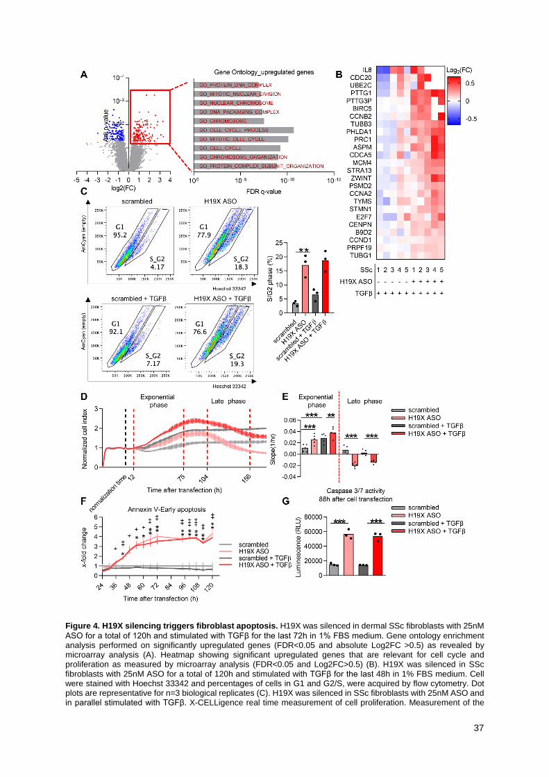
37
Figure 4. H19X silencing triggers fibroblast apoptosis. H19X was silenced in dermal SSc fibroblasts with 25nM ASO for a total of 120h and stimulated with TGFβ for the last 72h in 1% FBS medium. Gene ontology enrichment analysis performed on significantly upregulated genes (FDR<0.05 and absolute Log2FC >0.5) as revealed by microarray analysis (A). Heatmap showing significant upregulated genes that are relevant for cell cycle and proliferation as measured by microarray analysis (FDR<0.05 and Log2FC>0.5) (B). H19X was silenced in SSc fibroblasts with 25nM ASO for a total of 120h and stimulated with TGFβ for the last 48h in 1% FBS medium. Cell were stained with Hoechst 33342 and percentages of cells in G1 and G2/S, were acquired by flow cytometry. Dot plots are representative for n=3 biological replicates (C). H19X was silenced in SSc fibroblasts with 25nM ASO and in parallel stimulated with TGFβ. X-CELLigence real time measurement of cell proliferation. Measurement of the

38
cell index normalized to the cell index value at the time of transfection (24h). Experiment was recorded for 180h after cell transfection. Representative normalized cell index curve of n=5 biological replicates, variation is presented as SD of 4 technical replicates of the representative biological replicate (D). The slope of the cell index curve was calculated separately for the exponential growth phase and the late phase of the curve (E). Luminescence (for annexin V) as a measure of apoptosis was recorded every 6h starting 24h until 120h after cell transfection. Fold change was calculated respective to scrambled control. * indicate significance for untreated cells, + indicate significance for TGFβ stimulated cells (F). Caspases 3/7 activity measured at 88h after cell transfection (f). Data are presented as single values and mean (n=3-5). One-way ANOVA (C, E and G). Two-way ANOVA (F). * p<0.05, ** p<0.01, *** p<0.001.

39
Figure 5. miR-424 and miR-503 are also induced by TGFβ, but do not mediate the pro-fibrotic function of H19X. MiR-424 and miR-503 time-dependent response to TGFβ stimulation (A) and expression after H19X silencing with 25nM of ASO (B). Expression of miR-424 and miR-503 was analyzed by qPCR and normalized to RNU48. SSc fibroblasts were transfected with 25nM of ASO and 100nM of pre-miR for a total of 120h and stimulated with TGFβ for the last 72h in 1% FBS medium (C). Expression of H19X, COL1A, FN1 and ACTA2 was analyzed by qPCR and normalized to GAPDH and RPLP0. Fold change was calculated respective to untreated control set as 1 (dashed line). Data are presented as single values and mean (n=3-6). One-way ANOVA (A-C). * p<0.05, *** p<0.001; n.s., non-significant.

40
Figure 6. H19X is a nuclear lncRNA that interacts with the DNA. SSc fibroblasts were stimulated for 6 or 48h with TGFβ in 1%FBS medium and FISH staining was performed targeting H19X. Scale: 10µm. Pictures are representative of n=4 biological replicates. Percentage of nuclei positive for H19X staining (A). Cell fractions were obtained from SSc fibroblasts stimulated with TGFβ for 6 or 48h. H19X expression was analyzed by qPCR. Fold change was calculated respective to untreated control (B). Data are presented as single values and mean (n=4). T-test (A). One-way ANOVA (B). ** p<0.01, *** p<0.001. Heatmap showing expression of the gene with the closest transcription starts site to the putative sites of H91X interaction with the genome as measured by microarray analysis following H19X silencing, DDIT4L is highlighted in red (C).

41
Figure 7. H19X targets DDIT4L via 3D genomic interaction and chromatin rearrangements. BJ5TA cells were stimulated with TGFβ for 6h in 1% FBS medium. ChIRP peaks for H19X capture are shown as fold enrichment relative to input for two sets of probes (even, odd, red tracks). ChIP tracks for H3K27ac (light blue) and H3K4me1 (dark blue) were obtained from publicly available data sets (57). ATAC-seq tracks showing variations in chromatin accessibility following H19X silencing. H19X was silenced in dermal SSc fibroblasts with 25nM ASO (light green) and compared to SSc fibroblasts transfected with scrambled control (dark green) (A). Hi-C data (58) obtained from publicly available datasets revealed interactions between H19X binding site, the active enhancer and DDIT4L promoter. Query regions: DDIT4L promoter, DDIT4L enhancer (chr4:100294478), H19X binding site (chr4:100375842). Peak signals indicate interaction events and are highlighted by red circles (B). Schematic

42
representation of DDIT4L gene (blue) and its regulatory elements: DDIT4L enhancer (green, in light green it is highlighted where H19X silencing cause variation in chromatin accessibility) and H19X binding site (red) (C).

43
Figure 8. DDIT4L is a repressor of TGFβ-induced collagen production. H19X was silenced in dermal SSc fibroblasts with 25nM ASO for a total of 120h and stimulated with TGFβ for the last 72h in 1% FBS medium. The mRNA and protein levels of DDIT4L were analyzed by qPCR and Western blot respectively. Pictures are representative of n=5 biological replicates (A). SSc fibroblasts were transfected with 25nM of ASO and 50nM of siRNA for 120h and stimulated with TGFβ for the last 72h in 1% FBS medium. Expression of DDIT4L, H19X and COL1A1 was analyzed by qPCR (untreated samples) (B). qPCR data were normalized to GAPDH and RPLP0. Data are presented as single values and mean (n=5-6). Fold change was calculated respective to untreated control set as 1 (dashed line). One-way ANOVA (A, B). * p<0.05, ** p<0.01, *** p<0.001.