![[Manual] Lombardini](https://static.fdocuments.net/doc/165x107/544de6e9af7959f7178b4fa3/manual-lombardini.jpg)
Lombardini - Pathology of Laboratory Fish.ppt - … sheath tumors ... Clinical findings: Ocular...
56
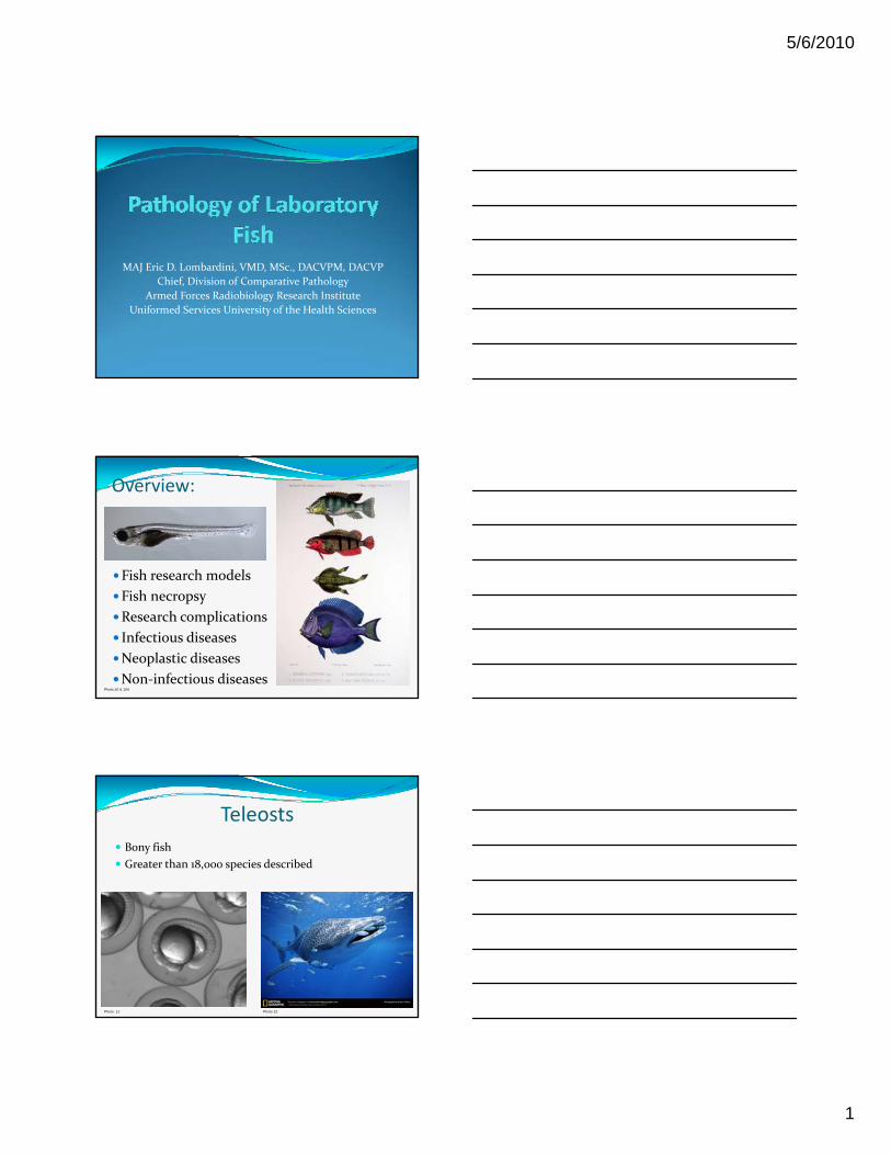
5/6/2010 1 MAJ Eric D. Lombardini, VMD, MSc., DACVPM, DACVP Chief, Division of Comparative Pathology Armed Forces Radiobiology Research Institute Uniformed Services University of the Health Sciences Overview: Fish research models Fish research models Fish necropsy Research complications Infectious diseases Neoplastic diseases Non‐infectious diseases Photo:10 & 104 Teleosts Bony fish Greater than 18,000 species described Photo: 11 Photo 12
Transcript of Lombardini - Pathology of Laboratory Fish.ppt - … sheath tumors ... Clinical findings: Ocular...
5/6/2010
1
MAJ Eric D. Lombardini, VMD, MSc., DACVPM, DACVP
Chief, Division of Comparative Pathology
Armed Forces Radiobiology Research Institute
Uniformed Services University of the Health Sciences
Overview:
Fish research modelsFish research models
Fish necropsy
Research complications
Infectious diseases
Neoplastic diseases
Non‐infectious diseases Photo:10 & 104
Teleosts
Bony fish
Greater than 18,000 species described
Photo: 11 Photo 12

5/6/2010
2
Extraordinary diversity
Photos: 1-4
Photo: 5

5/6/2010
3
Photo: 6
More Toadfish in Space: NASA will Study Balance in Two Wood’s Hole Toadfish, a Senator and Six Astronauts in Upcoming Shuttle Mission (1996 mission)
Photo: 7
1908 Nobel Prize in Physiology
Phagocytosis after experimenting on the larvae of starfish
Photo: 40
Photo: 41

5/6/2010
4
Historical use of fish in research
Genetics
Embryology
Renal physiology
Endocrinology
Nerve physiology
Toxicology
Morphology
Nutrition
Pharmacology
Parasitology
Specific neoplastic models
Photo: 8
More than just Zebrafish… Swordtail‐ Genetics of melanomas in Xiphoporus fishes
Trout‐coding sequences of the MHCII beta chain of homozygous rainbow trout
White perch‐Using white perch to study abnormal hepatic copper metabolism
Japanese medaka‐Freshwater fish in research on radiation biology
Goldfish‐The goldfish visual pathway: Intermediate filament proteins in nerve growth and development
Elasmobranchs‐In vitro metabolism of the pre‐carcinogen aflatoxin B1 by liver Elasmobranchs‐In vitro metabolism of the pre‐carcinogen aflatoxin B1 by liver preparations of the calf, nurse shark and clearnose skate
Anglerfish‐Simultaneous assessment of prohormone transport and processing in four separate islet cell types
Salmon‐Advantages of using aquatic animals for biomedical research on reproductive toxicology
Eel‐Effects of anaesthesia and surgery on levels of adrenaline and noradrenaline in blood plasma of the eel
Toadfish‐Ichthyotoxins from the oyster toadfish
Etc…
Fish Models
Examples include: Melanoma Diabetes Mellitus Neurofibromatosis Hepatic copper storage disease (Wilson’s disease)p pp g ( ) Oro‐facial‐digital syndrome Hepatocellular carcinoma Dehydration studies Na/K‐ATPase activity in muscle Renal excretion of xenobiotics Genotoxic and epigenetic carcinogenesis Gene regulation and developmental genetics Mutagenesis
5/6/2010
5
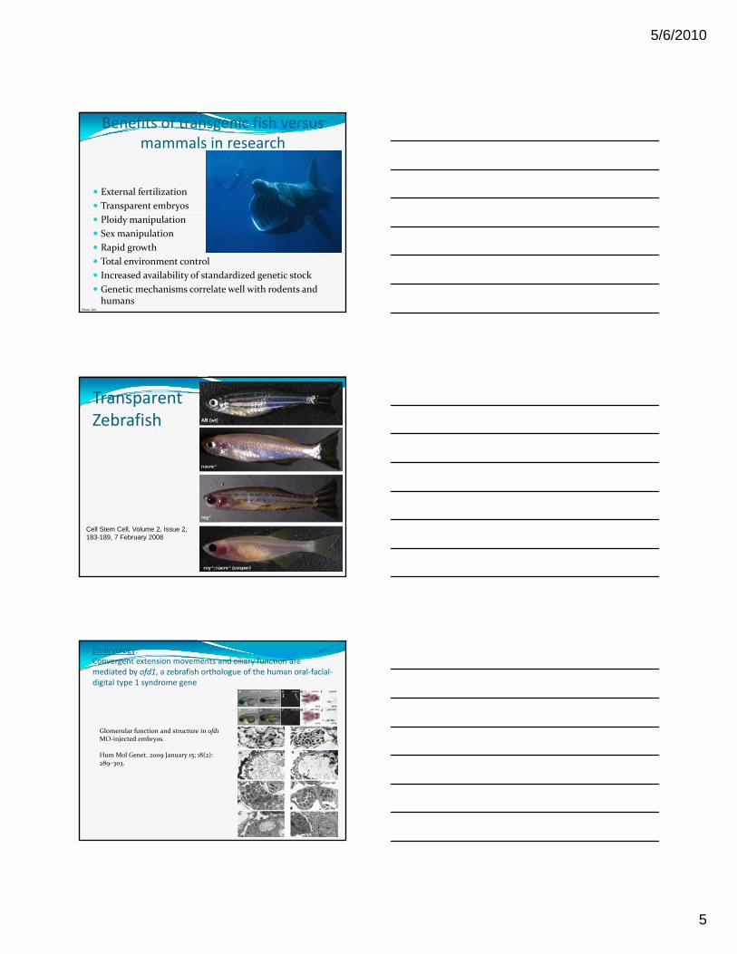
Benefits of transgenic fish versus mammals in research
External fertilization
Transparent embryos
Ploidy manipulation
Sex manipulation
Rapid growth
Total environment control
Increased availability of standardized genetic stock
Genetic mechanisms correlate well with rodents and humans
Photo: 105
TransparentZebrafish
Cell Stem Cell, Volume 2, Issue 2,183-189, 7 February 2008
Embryology:Convergent extension movements and ciliary function are mediated by ofd1, a zebrafish orthologue of the human oral‐facial‐digital type 1 syndrome gene
Glomerular function and structure in ofd1MO‐injected embryos.MO injected embryos.
Hum Mol Genet. 2009 January 15; 18(2): 289–303.

5/6/2010
6
Neuroanatomy:
Figure 1 ‐ 4 day old zebrafish embryo labelled with SV2 and acetylated tubulin antibodies showing axon tracts(green) and neuropil(red)viewed from lateral(top) and dorsal(bottom) orientations.
www.ucl.ac.uk/zebrafish‐group/research/neuroanatomy.php
Carcinogenesis:tp53mutant zebrafish develop malignant peripheral nerve sheath tumors
Fig. 5. Tumorigenesis features of tp53 M214K mutant zebrafish. (A‐D) When compared with wild‐type zebrafish (A and C), zMPNST development in tp53 mutant zebrafish is identifiable upon external observation because of ocular (B) or abdominal tumor localizations (D). (E‐H) When compared with wild‐type zebrafish (E and G),histopathology staining with hematoxylin/eosin reveals zMPNST in the eye (F) and abdominal cavity (H),as indicated by the stars (×4). (I‐K) Histopathological features of tumors (I) composed predominantly of spindle cells (J) andto a varying degree of epitheloid cells (K) are consistent with the diagnosis of zMPNST. [Bar, 200 μm (E, F, I‐K).]
PNAS January 11, 2005 vol. 102 no. 2 407‐412
Melanoma:
Review Article: Genetic and environmental melanoma models in fishPigment cell and melanoma research; 2010‐Gordon‐Kosswigmodel (Sd‐ Spotted Disease)‐Xmrk‐2 Oncogene overexpression‐loss of the Diff tumor suppressor

5/6/2010
7
Toxicology:Identification of a Primary Target of Thalidomide Teratogenicity Takumi Ito, Hideki Ando, Takayuki Suzuki, Toshihiko Ogura, Kentaro Hotta, Yoshimasa Imamura, Yuki Yamaguchi, and Hiroshi HandaScience 12 March 2010: 1345‐1350.
MUTATION In embryos, thalidomide‐induced malformations, bottom, of a fin of a zebrafish, left, and a wing of a chick, right, using mutated cereblon,a protein. NYTimes 17 March 2010
Necropsy:
Fish Necropsy (Generic)
Swim Bladder
Gills
LiverHeart
(Atrium;Ventricle; Bulbus arteriosus)
Gills

5/6/2010
8
Fish Necropsy (Generic)
Stomach Intestine/rectum
ovaryPyloric ceca
Fish Necropsy (Generic)
Kidney

5/6/2010
9
Pathology General Concepts Necrotizing versus Granulomatous disease
Variation in leukocytes between species
74 bacterial pathogens of fish, dozens of fungi, h d d f k i h d f i hundreds of known viruses, thousands of parasites, innumerable toxins and miscellaneous or physiological pathologies.
Emerging diseases
Viral Infections DNA:
Herpesvirus
Iridovirus
Adenovirus
RNA: RNA:
Picornavirus
Paramyxovirus
Reovirus
Togavirus
Birnavirus
Rhabdovirus
Retrovirus
Piscine iridovirus Gross findings:
Range in appearance from miliary pale nodules to a cutaneous mass effect.
Histopathological findings: Histopathological findings:
Markedly hypertrophic dermal fibroblasts (lymphocystis cells). Cells may measure > 300 µm with vacuolated to granular pale basophilic cytoplasm surrounded by a 10‐30µm amphophilic hyaline wall. Occasionally there is the presence of basophilic fibrillar inclusion material.

5/6/2010
10
Lymphocystis
Photo: 20
Lymphocystis
Photo: 46
Lymphocysitis
Photo: 45

5/6/2010
11
Lymphocystis
Lymphocystis
Lymphocystis
Photo: 21

5/6/2010
12
Journal of Fish Diseases, Volume 33 Issue 2 , Pages 93 ‐ 186 (February 2010)
Iridovirus infections in finfish – critical review with emphasis on ranaviruses (p 95‐122)
Liver of redfin perch, found dead 10 days post‐bath inoculation with epizootic haematopoietic necrosis virus. Hepatocytes that surround a focus of hepatic necrosis frequently contain basophilic intracytoplasmic inclusion bodies (arrows) (H & E, bar = 50 µm).
Photo: 114
Carp Pox (CyHV‐1)
Photo: 34 & 35
Carp Pox
Photo: 44

5/6/2010
13
KHV Koi herpesvirus (KHV) is a highly contagious viral disease that may cause significant morbidity and mortality in common carp (Cyprinus carpio) (Hedrick et al 2000; OATA 2001)et al. 2000; OATA 2001)
The white patches are due to necrosis (death) of the gill tissue. Gill lesions caused by KHV disease are the most common clinical signs in affected koi. Other external signs of KHV may include bleeding gills, sunken eyes, pale patches or blisters on the skin.
Koi herpesvirus (KHV) disease (CyHV‐3)
Photo: 14
Koi Herpesvirus
Photos: 70 & 71

5/6/2010
14
KHV
Photo: 80
IcHV‐1
Photo: 99 & 100
VHS (Viral hemorrhagic septicemia)
Affects >50 species of fish (Fresh water and marine)
Multiple strains
Family Rhabdoviridae
en.wikipedia.org/wiki/File:VHS.png
y
Gross: Visceral, cutaneous and muscular petechial hemorrhage
Exopthalmus, ascites, eccymosesaround eyes, skin, gills and fins
Histo: Variable necrosis of kidneys (hematopoetic 1st), spleen, liver, and skeletal muscle

5/6/2010
15
VHS
Photo: 29
VHS
Photo: 30
Spring Viremia of Carp
Photo: 83

5/6/2010
16
Ulcerative Dermatitis in Winter Flounder
Photo: 31
Bacterial Infectious Diseases In zebrafish, the pneumatic duct acts as the primary point of invasion for systemic fungal and bacterial infections, spreading to the gas bladder
Primarily opportunistic infections of which the 1° Primarily opportunistic infections of which the 1°agents: Aeromonas sp. and Pseudomonas sp.
Clinical signs of Gram negative septicemia are non‐specific and include: ascites, exopthalmia, cutaneous hemorrhages (body and fins), cutaneous ulceration
Non‐specific signs of septicemia Ascites or “Dropsy”
Photo: 43 &45

5/6/2010
17
Non‐specific signs of septicemia Exopthalmia
Photo: 28
Non‐specific signs of septicemia Cutaneous hemorrhages
Photo: 19
Non‐specific signs of septicemia Ulceration
Photo: 32

5/6/2010
18
Aeromonas salmonicida Furunculosis of salmonids
Goldfish Ulcer Disease
Carp Erythrodermatitis
Trout Ulcer Disease.
Several other species of Aeromonas, including: A. hydrophila, A. formicans, A. liquefaciens, and A. hydrophila complex are capable of causing a disease known as “Motile Aeromonas Septicemia” or “Bacterial Hemorrhagic Septicemia”.
Photo: 36
Furunculosis Aeromonas salmonicida
(4 subspecies: salmonicida, achromogenes, masoucida and smithia
Opportunist resulting in septicemia
Predominantly salmonid fishes, but also affects goldfish and other cyprinids.
“Furunculosis” is derived from the presence of “blisters” or furuncules on the surface of chronically infected fish
Significant resistance to both Terramycin and sulfamerazine
Furunculosis
Photo: 78

5/6/2010
19
Photos: 37 & 91
Koi Ulcer Disease
Photo: 33
Yersinia ruckeri
Photos: 48 & 49

5/6/2010
20
Enteric septicemia of catfish (ESC)
Edwardsiella ictaluri
Photo: 53
Edwardsiella tarda (Emphysematous Putrefactive Disease EPD)
Photos: 38 & 39
Renibacterium salmonarium Gross findings: Muscular cavitation; Vesicular dermatitis (Spawning rash); necrotizing nephritis & splenitis.
Histopathological findings: Granulomatous and necrotizing nephritis, splenitis, hepatitis, endocarditis and pancreatitis; occasionally with giant cell formation. Histiocytes are expanded by intracytoplasmic gram‐positive bacteria

5/6/2010
21
Renibacterium salmonarium
Renibacterium salmonrium
Streptococcus
Photo: 17

5/6/2010
22
Streptococcus Clinical findings: Ocular hemorrhage, cutaneous hemorrhage, exopthalmia, corneal opacity, dropsy and ulceration.
Gross Findings: Abdominal serosanguinous fluid, splenomegaly, hepatic pallor, endocarditis and nephritis Many Streps infect the brain and nervous nephritis. Many Streps infect the brain and nervous system of fish, explaining the erratic swimming frequently observed in infected fish.
Isolated Streptococcal pathogens in fish: S. iniae
S. difficilis
S. parauberis
S. milleri
S. shiloi
Mycobacteriosis
Mycobacterium marinum
Mycobacterium chelonae
Mycobacterium fortuitum
Photo: 89
Mycobacteriosis
Abdominal cavity: Peritonitis, hepatitis, splenitis, granulomatous, multifocal, severePhoto: 13

5/6/2010
23
Mycobacteriummarinum
Mycobacteriosis Mycobacteria as Environmental Portent in Chesapeake Bay Fish Species, Emerging Infectious Diseases: Volume 13, Number 2–February 2007
Occurrence of Mycobacterium spp. in ornamental fish in Italy, Journal of Fish Diseases, 2008 Jun;31(6):433‐41.
Husbandry stress exacerbates mycobacterial infections in adult zebrafish, Danio rerio (Hamilton), Journal of Fish Diseases, 2009, 32, 931–941
Gliding bacteria
Photos 18 & 90

5/6/2010
24
Flavobacterium columnare
Photo: 42 & 98
Photo: 50
Histopathology and Ultrastructure of Segmented Filamentous Bacteria–Associated Rainbow Trout Gastroenteritis; Veterinary Pathology 47(2) 220‐230
Photo: 93

5/6/2010
25
Piscirickettsia salmonis
Photo: 81
Protozoae
EpistylisIchthyobodo necator
Epistylis
Trichodina Glossatella
Microsporidiae Glugea, Ichthyosporidium, Loma, Pseudoloma, Microgemma, Microsporidium Mrazekia, Nosema, Pleistophora, Spraguea, Tetramicra, and Theragra.
Photo: 54

5/6/2010
26
Microsporidiae
Microsporidiae
Microsporidiae

5/6/2010
27
Pleistophora hyphessobryconis
Pseudoloma neurophilia
Photo: 75
Glugea stephani
Photo: 67

5/6/2010
28
Glugea sp.
Photos: 68 & 69
Myxosporidiae
Photo: 51
Henneguya salminicola
Photos: 63 & 94

5/6/2010
29
Henneguya sp. xenomas
Photos: 65 & 66
Myxoma and trematode
Hamburger gill disease

5/6/2010
30
Myxobolus cerebralis Gross Findings: Severe scoliosis, frequently with black discoloration of the tail, and with chronicity, misshapen head and jaws
Histopathological Findings: Cartilage degeneration and necrosis, granulomatous chondritis with intralesional ovoid to ellipsoidal spores (5 15µm) with intralesional ovoid to ellipsoidal spores (5‐15µm) with 2 piriform‐shaped polar capsules at the anterior end
Photos: 119
Myxobolus cerebralis
Photos: 76 & 77
Myxobolus cerebralis

5/6/2010
31
Myxobolus sp.
Figure 2—Photomicrograph of an impression smear prepared from a biopsy specimen of the oral cavity mass in the goldfish in Figure 1. Notice a large cluster of epithelial cells (blue) and numerous mature Myxobolus sp spores (red). Modified acid‐fast stain; bar = 25 μm.
JAVMA, Vol 236, No. 6, March 15, 2010
Myxobolus sp.
Figure 3—Photomicrograph of a paraffin‐embedded section of a biopsy specimen of the oral cavity mass in the goldfish in Figure 1. The mass tissue contains a single large, late‐stage, polysporous, histozoic plasmodial pseudocyst (borders of this structure are demarcated by arrows) and evidence of inflammation. H&E stain; bar = 100 μm.
JAVMA, Vol 236, No. 6, March 15, 2010
Myxozoa
Renal myxozoanosis in weedy sea dragons, Phyllopteryxtaeniolatus (Lacepe` de), caused by Sinuolinea phyllopteryxan. sp.Journal of Fish Diseases 2008, 31, 27–35

5/6/2010
32
Tetrahymena sp. infection in guppies, Poecilia reticulata Peters: parasite characterization and pathology of infected fish, Journal of Fish Diseases 2009, 32, 845–855
Severe Scuticociliate (Philasterides dicentrarchi) Infection in a Population of Sea Dragons, Vet Pathol 45:546–550 (2008)
Photos: 115 & 116
Tetrahymena corlissi
Tetrahymena corlissi

5/6/2010
33
Tetrahymenacorlissi
Ichthyophthirius multifiliis Gross findings:
Up to 1 mm elevated nodules in a miliary pattern along body, gills and fins
May coalesce or progress to erosion and ulceration due to irritationto irritation
Histopathological findings:
Oval holotrich ciliated parasite which forms intraepithelial cysts composed of a 1‐2 µm hyaline wall.
Small circular oral opening, dark granules and a horseshoe/crescent shaped basophilic macronucleus
+/‐ Hyperplasia of neighboring epithelium
Photos: 95 & 96

5/6/2010
34
Ichthyophthirius multifiliis
Ichthyophthirius multifiliis
Piscinoodinium sp.

5/6/2010
35
Piscinoodinium sp.
Ichthyobodonecator
Photos: 118
Coccidiae
Photos: 101 & 103

5/6/2010
36
Hole‐in‐the‐Head DiseaseHead and Lateral Line Erosion
Hexamita sp.?
Photo: 52
Fungal
Photos: 79 & 92
Saprolegnia
Photo: 22

5/6/2010
37
Saprolegnia
Photo: 23
Parasitic Nematodes
Cestodes
Trematodes
Pentastomes
Arthropods
Nematodes
Photo: 47

5/6/2010
38
Eustrongylides sp.Eustrongylides ignotus infecting commercial bass, and other fish in the southeastern USA, Journal of Fish Diseases 2009, 32, 795–799
Photos: 15 & 26
Acanthocephalids
Photo: 107
Cestodes

5/6/2010
39
Asian Fish Tapeworm: Bothriocephalus acheilognathi
Photo: 97
Ligula intestinalis
Photo: 24
Digenetic trematodes
Photomicrographs: MAJ Jeremy Bearss

5/6/2010
40
Black spot disease
Photo: 86
Trematode metacercariae
Photo: 27

5/6/2010
41
Monogenean trematodes
Monogeneantrematodes
Photomicrographs: MAJ Jeremy Bearss

5/6/2010
42
Diplostomum sp.
Photo: 109
Pentastomes
Photo: 16 & 108
Arthropods
Photo: 25 & 117

5/6/2010
43
Neoplastic
Photo: Sarah Johnson & 120
Melanoma
Melanoma

5/6/2010
44
Fibrosarcoma
Photo: 61
Bi‐colored Damselfish Schwannoma
Photo: 73
Neurofibroma

5/6/2010
45
PNST
Peripheral Nerve Sheath tumor
Dermatofibroma of Angelfish
Photo: 113

5/6/2010
46
Walleye Dermal sarcoma
Photo: 87
Walleye Dermal Sarcoma
Photo: 88
Lymphoma
Photo: 85 & 110

5/6/2010
47
Lymphoma
Lymphoma
Basal cell carcinoma
5/6/2010
48
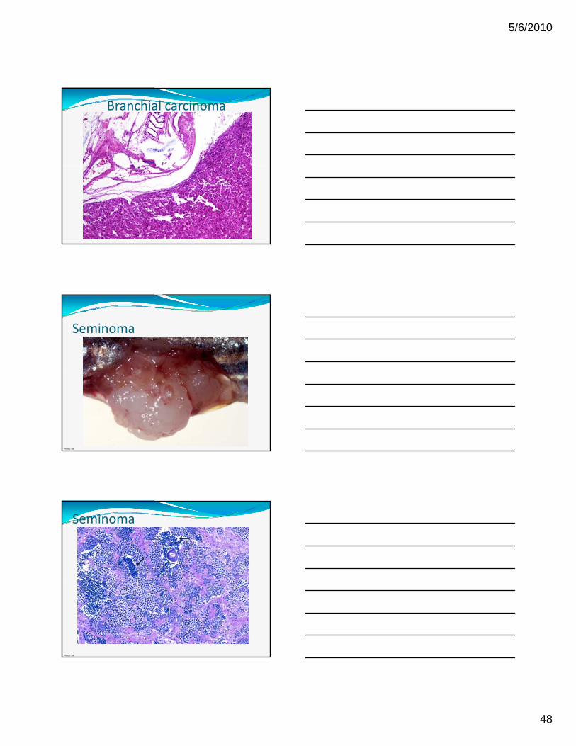
Branchial carcinoma
Seminoma
Photo: 58
Seminoma
Photo: 59

5/6/2010
49
Nephroblastoma
Ependymoma

5/6/2010
50
Neuronal Embryonal Tumors in Fish; Vet Pathol published online 31 December 2009
Photo: 92
Hepatocellular carcinoma
Photo: 102
Cholangiocarcinoma
Photo: 60 112

5/6/2010
51
Papilloma (Cyrinid herpesvirus 1)
Photo: 72
Viral papillomas of Brown bullhead catfish
Photo: 111
Reported Zebrafish tumors Seminoma (Most common spontaneous
tumor)
Ovarian papillary adenocarcinoma
Adenocarcinoma
Small cell carcinoma of intestine at Ampulla of Vater
Hepatocellular adenoma/carcinoma
Biliary carcinoma/Cholangiocarcinoma
Myxoma/myxosarcoma
Fibrosarcoma
Chordoma
Hemangioma (Retrobulbar hemangioma)
Rhabomyoma/rhabdomyosarcoma
Chondrosarcoma
Osteoma/osteosarcoma
PNST Hepatoblastoma
Nephroblastoma
Renal cell carcinoma
Thyroid adenocarcinoma
Ultimobranchial tumor
Gas bladder adenoma/adenocarcinoma
Pancreatic acinar cell adenoma/carcinoma
Pancreatic ductal adenoma
Islet cell adenoma
Lymphoma
Acute lymphoblastic leukemia
PNST
Melanoma
Complex‐compound odotoma of the pharyngeal teeth
Neuroblastoma/neuroectodermal tumor
Medulloepithelioma
Ganglioglioma
Branchioblastoma
Epidermal papilloma
SCC
Teratoma

5/6/2010
52
Miscellaneous
Figure 2. Adult female sand tiger shark withstomach prolapse through the right third gillslit.
Figure 1. Internal view of stomach prolapsein a sand tiger shark. Note also the spleen(arrow) is pulled up into the ventral throatregion.
Journal of Fish Diseases 2008, 31, 311–315; Gastric prolapse in sand tiger sharks
Pericardial effusion and dilated cardiomyopathy
Photo: 55
Nephrocalcinosis

5/6/2010
53
Hepatic megalocytosis
Photo: 56
Gas Bubble Disease
Photo: 57
Gas Bubble Disease: Supersaturation
Photo: 84
5/6/2010
54
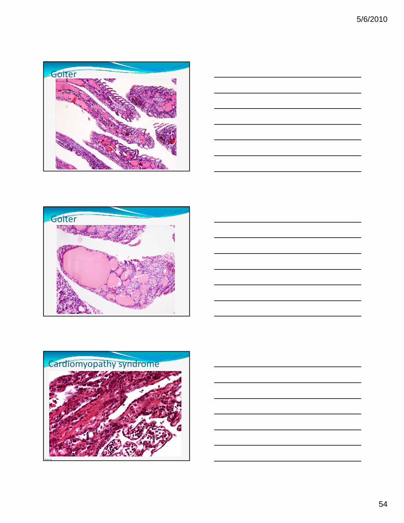
Goiter
Goiter
Cardiomyopathy syndrome
Photo: 82

5/6/2010
55
Knockdown of Bicaudal C in Zebrafish (Danio rerio) Causes Cystic Kidneys: A Nonmammalian Model of Polycystic Kidney Disease
Bouvrette et al.
Comparative Medicine, Volume 60, Number 2, April 2010 , pp. 96‐106(11)
Dilatation of the pronephric ducts (arrows) and cysts which form in the single glomerulus (arrowheads)
Photo: 106
Cardiac dissecting hemorrhage
Figure 1 Dissecting haemorrhage of the ventricle in rainbow trout. (a) Cut surfaceof the formalin‐fixed ventricle showing haemorrhage along the lateral wall. (b) Sickle‐shaped haemorrhagebetween compact and spongious ventricular myocardium (H&E, bar = 1 cm). (c) Detailshowing haemorrhage and compact myocardium with focus of inflammation to the left (H&E, bar = 50 lm).Journal of Fish Diseases 2009, 32, 1041–1043; Dissecting haemorrhage in rainbow trout
Photography credits: 1.en.wikipedia.org/wiki/File:Leafydragon.jpg
2.en.wikipedia.org/wiki/File:Pterois_volitans_Manado‐e_edit.jpg
3. en.wikipedia.org/wiki/File:Georgia_Aquarium_‐_Giant_Grouper_edit.jpg
4. en.wikipedia.org/wiki/File:White_shark.jpg
5. en.wikipedia.org/wiki/File:Lamprey_mouth.jpg
6. en.wikipedia.org/wiki/File:GambianMudskippers.jpg
7. en.wikipedia.org/wiki/Oyster_toadfish
8. blogs.nature.com/nm/spoonful/zebrafish.jpg
9. dx.doi.org/10.1371/journal.pbio.0020148
10. www.genome.gov/pressDisplay.cfm?photoID=95
11. www.realscience.us/blog/wp‐content/uploads/2007/06/zebrafish‐embryo.jpg
12.images.google.com/imgres?imgurl=http://dreamsforpeace.files.wordpress.com
13.http://edis.ifas.ufl.edu/LyraEDISServlet?command=getImageDetail&image_soid=FIGURE%201&document_soid=VM055&document_version=42850
14. http://edis.ifas.ufl.edu/vm113
15.http://edis.ifas.ufl.edu/LyraEDISServlet?command=getImageDetail&image_soid=FIGURE%2012&document_soid=FA091&document_version=1943678729
16.http://edis.ifas.ufl.edu/LyraEDISServlet?command=getImageDetail&image_soid=FIGURE%204&document_soid=FA090&document_version=37638
17.http://edis.ifas.ufl.edu/LyraEDISServlet?command=getImageDetail&image_soid=FIGURE%201&document_soid=FA057&document_version=48516
18. http://zebrafish.org/zirc/health/Disease_images/TailRot4.jpg
19. http://zebrafish.org/zirc/health/Disease_images/Erythema.jpg
20. http://vp4.afip.org/wsco/do_slideshow2.php?pic_series=082303&cYear=2008
21. http://vp4.afip.org/wsco/do_slideshow2.php?pic_series=082303&cYear=2008
22. http://www.frw.ca/albums/Correcting‐Stormwater‐Mistakes
23.http://media.photobucket.com/image/fish%20fungus/aquaticquotient/disease/fungus.jpg
24. http://apscience.org.au/projects/APSF_04_2/apsf_04_2.htm
25. http://apscience.org.au/projects/APSF_04_2/apsf_04_2.htm
26. http://apscience.org.au/projects/APSF_04_2/apsf_04_2.htm
27. http://apscience.org.au/projects/APSF_04_2/apsf_04_2.htm
65. http://www.maine.gov/ifw/fishing/health/vol4issue5.htm 66. http://ag.ansc.purdue.edu/courses/aq448/images/henneguya1.jpg
67. http://upload.wikimedia.org/wikipedia/commons/3/3e/Glugea_stephani.jpg
68.http://www.wildaboutbritain.co.uk/archive/data/517/glugea_anomala_stickleback.jpg
69.http://tolweb.org/onlinecontributors/app?page=ContributorImagesPage&service=external&sp=3682
70.http://www.koihealth.org/images/Interocean_KHV/images/Allen_Hsu_KHV%20272_jpg
71.http://www.koihealth.org/images/Interocean_KHV/images/Allen_Hsu_KHV%20080_jpg
72.http://www.koihealth.org/images/Interocean_KHV/pages/Allen_Hsu_KHV%20180_jpg.htm
73. http://ehp.niehs.nih.gov/docs/1995/103‐3/niehsnewsfigfish2.GIF
74. http://zebrafish.org/zirc/health/Disease_images/ultimobranchialTumors4.jpg
75. http://zebrafish.org/zirc/health/diseaseManual.php
76. http://en.wikipedia.org/wiki/File:Deformed_Brook_Trout.jpg
77. http://en.wikipedia.org/wiki/File:Whirling_disease_pathology.jpg
78. http://www.fws.gov/midwest/lacrossefishhealthcenter/Index.htm
79. http://biology.unm.edu/ccouncil/Biology_203/Summaries/Protists.htm
80. http://www.fao.org/docrep/009/a0192e/A0192E07.htm
81. http://www.cdc.gov/ncidod/eid/vol3no2/fryer.htm
82. http://www.marlab.ac.uk/Delivery/standalone.aspx?contentid=2015
83.http://www.lethbridgecollege.ab.ca/grasscarp/images/stories/projects/Spring%20Viremia%20in%20Carp/DSC01878.JPG
84. http://www.advancedaquarist.com/2010/3/fish
85. http://www.ramp‐alberta.org/ramp/gallery.aspx?galleryimage=493
86. http://www.ramp‐alberta.org/ramp/gallery.aspx?galleryimage=503
87. http://www.ohio.edu/news/pix/FISH.JPG
88. http://i244.photobucket.com/albums/gg22/way2phat/walleyefungus2.jpg
89. http://ag.ansc.purdue.edu/courses/aq448/images/tangmycobact.jpg
90. http://www.fishdoc.co.uk/photos/Fin_rot_02.JPG
91 http://www koi fish co uk/acatalog/ulcer jpg 28. http://www.focusonfishhealth.org/signs‐of‐disease.php
29. http://www.focusonfishhealth.org/signs‐of‐disease.php
30. http://www.focusonfishhealth.org/signs‐of‐disease.php
31. www.mwra.state.ma.us/harbor/omsap/graphic/omsap_050811_flounder.ppt
32. http://www.blueridgekoi.com/images/categories/C76.jpg
33. http://www.ponddoc.com/koihospital/Diagnostic/Bacterial/AdvancedUlcer.jpg
34. http://www.backyardpuddle.ca/images/healthpix/smapril02‐4.jpg
35.http://www.spfdwatergardensoc.bizland.com/sitebuildercontent/sitebuilderpictures/pox.jp36.http://www.cnr.vt.edu/fisheries/afs/AFS_education_fisheries_techniques_visuals_chap_14_add.htm
37.http://www.aquacultuur.wur.nl/NL/Visteelt/Productie+en+teelt/Ziekten/Bacteri%C3%ABn
38.http://www.aquacultuur.wur.nl/NL/Visteelt/Productie+en+teelt/Ziekten/Bactari%C3%ABn
39. http://www.time4fish.net/FDB.html
40. http://phlogthat.wordpress.com/2007/07/16/two‐star‐fish‐making‐love‐in‐the‐sand‐at‐morro‐bay‐ca/
41. http://www.odec.ca/projects/2007/sank7b2/figure1.jpg
42. http://www.blueridgekoi.com/Disease‐Fish‐Health‐sc‐17.html
43. http://www.flippersandfins.net/Dropsy.htm
44. http://www.blueridgekoi.com/Disease‐Fish‐Health‐sc‐17.html
45. http://www.drpez.net/panel/showthread.php?t=167187
46. http://www.drpez.net/panel/showthread.php?t=167187
47. http://www.fishbase.org/diseases/DisPic/NemtodD5.jpg
48. http://web.abo.fi/instut/fisk/Fig/yr1.jpg
49. http://www.daff.gov.au/_media/images/animal‐plant/aquatic/aquatic‐cd/enteric‐redmouth1.jpg
50. http://en.wikipedia.org/wiki/Columnaris
51. http://en.wikipedia.org/wiki/File:Henneguya_zschokkei.jpg
52. http://en.wikipedia.org/wiki/File:Oscar2.jpg
53.http://coursewares.mju.ac.th/section2/fb406/0002lecture/htm/003allchapter3/image/image003_5/edwardsiella.gif
54. http://zebrafish.org/zirc/health/diseaseManual.php
55. http://zebrafish.org/zirc/health/Disease_images/BigHeartGross4.jpg
56. http://zebrafish.org/zirc/health/diseaseManual.php
57. http://zebrafish.org/zirc/health/Disease_images/GasBubbleDis4.jpg
58. http://zebrafish.org/zirc/health/Disease_images/GrossSeminoma4.jpg
59. http://zebrafish.org/zirc/health/Disease_images/seminoma24.jpg
60. http://zebrafish.org/zirc/health/Disease_images/LiverTumorsDMBA4.jpg
61. http://zebrafish.org/zirc/health/Disease_images/SpincleCellGrossB4.jpg
62. http://zebrafish.org/zirc/health/Disease_images/SpindleCellEye4.jpg
63.http://en.wikipedia.org/wiki/File:Henneguya_salminicola_in_flesh_of_coho_salmon,_BC,_Canada.JPG
64. http://www.virtualslides.psu.edu/zebrafish.jsp?compare1=08B98770‐2FC7‐5057‐42CF‐8695A7F9C030&
91. http://www.koi‐fish.co.uk/acatalog/ulcer.jpg
92. http://kentsimmons.uwinnipeg.ca/16cm05/16labman05/lb2pg3_files/saprolegnia4.jpg
93. http://vet.sagepub.com/content/47/2/220.full.pdf+html
94. http://www.scielo.br/scielo.php?pid=S0074‐02762005000200011&script=sci_arttext
95. http://nippyfish.net/Ich4.jpg
96. http://www.vet.cornell.edu/PUBLIC/FishDisease/AquaticProg/highlights/Ich/ICH1.JPG
97. http://www.cnr.vt.edu/fisheries/afs/AFS_education_fisheries_techniques_visuals_chap_14_add.htm
98. http://www.cnr.vt.edu/fisheries/afs/fisheries_techniques
99. http://www.cnr.vt.edu/fisheries/afs/fisheries_techniques/Chapter14/Channel%20catfish%20virus%202.jpg
100. http://www.cnr.vt.edu/fisheries/afs/fisheries_techniques/Chapter14/Channel%20catfish%20virus%201.jpg
101. http://www.alaineknipes.com/parasite_photos/index.html
102. http://www.nature.com/nprot/journal/v5/n3/thumbs/nprot.2010.8‐F5.jpg
103. http://www.alaineknipes.com/parasite_photos/index.html
104. http://upload.wikimedia.org/wikipedia/commons/7/75/F_de_Castelnau‐poissonsPl12.jpg
105. http://www.sciencedaily.com/images/2009/05/090507121953‐large.jpg
106. http://renalfellow.blogspot.com/2009/05/zebrafish‐model‐of‐cystic‐kidney.html
107. http://www.mybay.umd.edu/parasites.html
108. http://edis.ifas.ufl.edu/pdffiles/FA/FA11400.pdf
109. http://www.thefishsite.com/articles/102/invasion‐of‐the‐eye‐flukes\
110. http://www.seagrant.umn.edu/fisheries/img/parasites/lymphosarcoma_b.jpg
111. http://www.glfc.org/tumor/f2b.gif
112. http://www.glfc.org/tumor/f5a.gif
113. http://www.ultimatebettas.com/index.php?showtopic=8863
114. http://www3.interscience.wiley.com/cgi‐bin/fulltext/123227302/PDFSTART
115. http://vet.sagepub.com/content/45/4/546.full.pdf+html
116. http://vet.sagepub.com/content/45/4/546.full.pdf+html
117. http://www.state.me.us/ifw/fishing/health/vol2issue2.htm
118. http://www.drpez.net/panel/showthread.php?t=330799
119. http://www.forumaquario.com.br/portal/tx_doencas.html
120. http://www.epa.gov/glnpo/aoc/presque/catfish‐tumors.jpg

5/6/2010
56
References Histology:
1. http://zfatlas.psu.edu/reference.php
2. http://training.fws.gov/CSP/fish/histo1.html
3. http://aquaticpath.umd.edu/fhm/
4. http://www.vspo.us/vspo
Neoplasia:
5 D J S itb POLA t 2008 5. Dr. Jan Spitbergen POLA notes 2008
6. Registry for Tumors in Lower Animals
General:
7. Edward Noga, Fish Diseases, Diagnosis and treatment, 2000
8. Ronald Roberts, Fish Pathology, 2003
9. Greg Lewbart, Ornamental fish (Self assessment colour review), 1998
10. Gary Ostrander, The laboratory fish, 2000
11. Erwin Amlacher, Textbook of fish diseases, 1970
12. Fish diseases vol 1 and vol 2, (Ed.) J Eiras et al., 2008