Localization of the polyol pathway in the human kidney of the... · enzyme sorbitol dehydrogenase...
9
Summary. Sorbitol plays an important role in the osmotic regulation of the mammalian kidney. Sorbitol synthesis is regulated by the enzyme aldose reductase (AR) and its degradation to fructose is catalyzed by the enzyme sorbitol dehydrogenase (SDH). Various data exist on the polyol pathway on the rat kidney, but little is known about the distribution of the polyol pathway enzymes in the human kidney. Determination of enzyme activities and a semiquantitative determination of mRNA expression, immunohistochemistry and in-situ hybridisation in healthy human kidney tissue was carried out. The enzyme activity of AR showed a fourfold increase from cortex to papilla, while SDH-activity dropped from cortex to papilla by a factor of four. Corresponding data was obtained at the mRNA level from the semiquantitative polymerase chain reaction (PCR). Additional differentiation at the cellular level reveals both enzymes in cells of the proximal and distal tubules, thick ascending loop, thin loop and collecting duct. Studies of enzyme activity and expression by immunohistochemistry, PCR and in-situ hybridization presented corresponding results with respect to the localization of the enzymes, which match the experimental data obtained from rats very well. Thus, the established rat model might well represent the situation in the human kidney, too. Key words: Polyol pathway, Aldose reductase, Sorbitol dehydrogenase, Human kidney Introduction The polyol (or sorbitol) pathway consists of two enzymes, aldose reductase (AR) and sorbitol dehydrogenase (SDH). AR (EC 1.1.1.21) catalyzes the reduction of glucose to sorbitol by NADPH and SDH (EC 1.1.1.21) converts sorbitol to fructose dependent on NAD + (Jeffery and Joernvall, 1983; Kador et al., 1985; Yabe-Nishimura, 1998). The physiological role of aldose reductase in the kidney, the rate limiting enzyme of the polyol pathway, is the intracellular accumulation of the osmolyte sorbitol as an important mechanism in the long-term adaptation of the cell to a rise in the extracellular osmolarity (Grunewald and Kinne, 1989). High extracellular osmolarities induce renal sorbitol synthesis by increasing AR activity and vice versa (Garcia-Perez et al., 1989; Grunewald and Kinne, 1989). However, the polyol pathway plays an important role in the pathogenesis of diabetic nephropathy (Dunlop, 2000; Forbes et al., 2007). Under normoglycaemic conditions, glucose utilization by the polyol pathway is limited in tissues taking up glucose independent of insulin, such as in the kidney (Tomlinson, 1993). This is due to the relatively low intracellular glucose concentration and the low affinity of AR for glucose (Swidan and Montgomery, 1998). Only 3% of the glucose is utilized by AR (Morrison et al., 1970). Here, most of the cellular glucose is phosphorylated into glucose 6-phosphate by hexokinase and glucokinase (Philips and Steadman, 2003). In the case of hyperglycaemia, however, when hexokinase and glucokinase appear to be saturated, the proportion of glucose utilized by the polyol pathway is about 30% of the total, but a corresponding rise of SDH does not occur, causing an intracellular rise of sorbitol concentration (Gonzales et al., 1984; Kador et al., 1985). Diabetic nephropathy is the main reason for terminal renal insufficiency and for 30-40% of all kidney transplantations (Philips and Steadman, 2003). An important co-factor is the glucose-stimulated activation of the polyol pathway, which influences the sorbitol and myo-inositol pathway (Greene et al., 1987; Meyer et al., 2005). This activation of the polyol pathway produces several effects. The osmotic balance of the tissue is Localization of the polyol pathway in the human kidney Steffen Zopf 1 , Jakob Flämig 2 , Heide Schmid 3 , Nicolai Miosge 4 , Sabine Blaschke 2 , Eckhart G. Hahn 1 , Gerhard A. Müller 2 and Rolf W. Grunewald 2,5 1 Department of Internal Medicine 1, University of Erlangen-Nuremberg, Erlangen, Germany, 2 Department of Nephrology and Rheumatology, University of Goettingen, Goettingen, Germany, 3 Department of Pathology, University of Tuebingen, Tuebingen, Germany, 4 Department of Anatomy, University of Goettingen, Goettingen, Germany and 5 Department of Internal Medicine I, St. Antonius Hospital Kleve, Kleve, Germany Histol Histopathol (2009) 24: 447-455 Offprint requests to: Steffen Zopf, MD, Department of Medicine 1, University of Erlangen-Nuremberg, Ulmenweg 18, D-91054 Erlangen, Germany. e-mail: [email protected] http://www.hh.um.es Histology and Histopathology Cellular and Molecular Biology
Transcript of Localization of the polyol pathway in the human kidney of the... · enzyme sorbitol dehydrogenase...
Summary. Sorbitol plays an important role in theosmotic regulation of the mammalian kidney. Sorbitolsynthesis is regulated by the enzyme aldose reductase(AR) and its degradation to fructose is catalyzed by theenzyme sorbitol dehydrogenase (SDH). Various dataexist on the polyol pathway on the rat kidney, but little isknown about the distribution of the polyol pathwayenzymes in the human kidney. Determination of enzymeactivities and a semiquantitative determination of mRNAexpression, immunohistochemistry and in-situhybridisation in healthy human kidney tissue was carriedout. The enzyme activity of AR showed a fourfoldincrease from cortex to papilla, while SDH-activitydropped from cortex to papilla by a factor of four.Corresponding data was obtained at the mRNA levelfrom the semiquantitative polymerase chain reaction(PCR). Additional differentiation at the cellular levelreveals both enzymes in cells of the proximal and distaltubules, thick ascending loop, thin loop and collectingduct. Studies of enzyme activity and expression byimmunohistochemistry, PCR and in-situ hybridizationpresented corresponding results with respect to thelocalization of the enzymes, which match theexperimental data obtained from rats very well. Thus,the established rat model might well represent thesituation in the human kidney, too.
Key words: Polyol pathway, Aldose reductase, Sorbitoldehydrogenase, Human kidney
Introduction
The polyol (or sorbitol) pathway consists of twoenzymes, aldose reductase (AR) and sorbitoldehydrogenase (SDH). AR (EC 1.1.1.21) catalyzes the
reduction of glucose to sorbitol by NADPH and SDH(EC 1.1.1.21) converts sorbitol to fructose dependent onNAD+ (Jeffery and Joernvall, 1983; Kador et al., 1985;Yabe-Nishimura, 1998).
The physiological role of aldose reductase in thekidney, the rate limiting enzyme of the polyol pathway,is the intracellular accumulation of the osmolyte sorbitolas an important mechanism in the long-term adaptationof the cell to a rise in the extracellular osmolarity(Grunewald and Kinne, 1989). High extracellularosmolarities induce renal sorbitol synthesis by increasingAR activity and vice versa (Garcia-Perez et al., 1989;Grunewald and Kinne, 1989).
However, the polyol pathway plays an importantrole in the pathogenesis of diabetic nephropathy(Dunlop, 2000; Forbes et al., 2007). Undernormoglycaemic conditions, glucose utilization by thepolyol pathway is limited in tissues taking up glucoseindependent of insulin, such as in the kidney(Tomlinson, 1993). This is due to the relatively lowintracellular glucose concentration and the low affinityof AR for glucose (Swidan and Montgomery, 1998).Only 3% of the glucose is utilized by AR (Morrison etal., 1970). Here, most of the cellular glucose isphosphorylated into glucose 6-phosphate by hexokinaseand glucokinase (Philips and Steadman, 2003).
In the case of hyperglycaemia, however, whenhexokinase and glucokinase appear to be saturated, theproportion of glucose utilized by the polyol pathway isabout 30% of the total, but a corresponding rise of SDHdoes not occur, causing an intracellular rise of sorbitolconcentration (Gonzales et al., 1984; Kador et al., 1985).
Diabetic nephropathy is the main reason for terminalrenal insufficiency and for 30-40% of all kidneytransplantations (Philips and Steadman, 2003). Animportant co-factor is the glucose-stimulated activationof the polyol pathway, which influences the sorbitol andmyo-inositol pathway (Greene et al., 1987; Meyer et al.,2005). This activation of the polyol pathway producesseveral effects. The osmotic balance of the tissue is
Localization of the polyol pathway in the human kidneySteffen Zopf1, Jakob Flämig2, Heide Schmid3, Nicolai Miosge4, Sabine Blaschke2, Eckhart G. Hahn1, Gerhard A. Müller2 and Rolf W. Grunewald2,5
1Department of Internal Medicine 1, University of Erlangen-Nuremberg, Erlangen, Germany, 2Department of Nephrology and
Rheumatology, University of Goettingen, Goettingen, Germany, 3Department of Pathology, University of Tuebingen, Tuebingen,
Germany, 4Department of Anatomy, University of Goettingen, Goettingen, Germany and 5Department of Internal Medicine I, St.
Antonius Hospital Kleve, Kleve, Germany
Histol Histopathol (2009) 24: 447-455
Offprint requests to: Steffen Zopf, MD, Department of Medicine 1,University of Erlangen-Nuremberg, Ulmenweg 18, D-91054 Erlangen,Germany. e-mail: [email protected]
http://www.hh.um.es
Histology andHistopathology
Cellular and Molecular Biology
disturbed by an accumulation of sorbitol, whileconsumption of NAD(P)H in these processes may alterthe redox potential of the cells, making them lessresistant to oxidative stress. Finally, sorbitol may beconverted to fructose, which promotes nonenzymaticglycation of proteins (Lane, 2002).
To understand the effect of the polyol pathway, it isimportant to know something about the localization ofthe enzymes. Several investigations on the enzymelocalization of AR and SDH and their expression in therat have been carried out, but few data exist on thehuman kidney. Corder et al. (1977) were the first tolocalize AR and SDH in rat kidneys byimmunohistochemistry and showed that the mainlocalization of AR was in the inner medulla, whereasSDH was detected with an opposite distribution patternalong the cortico-papillary gradient. The aim of ourinvestigations was to localize protein and mRNA of bothenzymes in individual structures of the normal humankidney along the cortico-papillary gradient in order tocompare this data with the results of the animal model.For the first time, we describe the localization of polyolpathway enzymes in the human kidney as a whole at themRNA and protein levels.
Materials and methods
Tissue
The human kidney tissue was taken from tumornephrectomies (Dept. of Urology, University HospitalGoettingen). From the tumor-free region, a tissue piece,macroscopically containing all parts from cortex topapilla, was removed within 2 hours after kidneyresection. Immediately after preparation, the tissue waseither frozen or formalin-fixed or used for determinationof enzyme activity. Microscopically, the tissue did notshow any tumorous areas. The various nephron segmentswere identified by their difference in length, cellularmorphology and distribution in the renal sections(Pfaller, 1982; Kriz and Bankir, 1988).
All patients had normal creatinine and blood glucoselevels before kidney resection. 4 of 20 patients showedhigh blood-pressure.
On the basis of the limited amount of tissue, it wasnot possible to perform all investigations on everykidney.
Determination of aldose reductase activity (AR)
The determination of AR activity is based on thereduction of DL-glyceraldehyde to glycerol by NADPHin the presence of AR. The assay contained 50 mmol/lphosphate buffer (pH 6.2), 400 mmol Li2SO4, 10 mmol/lDL-glyceraldehyde and 0.1 mmol/l NADPH (Grunewaldet al., 2001a; Sato and Kador, 1993). The decrease inabsorbance was monitored at 340 nm at 37°C for 5minutes in the presence or absence of DL-glyceraldehyde to correct for unspecific NADPH
dehydrogenase activity. Li2SO4 stimulates AR and notaldehyde reductase (Das and Srivastava, 1985).
Determination of sorbitol dehydrogenase activity (SDH)
This assay is based on the reduction of fructose tosorbitol by NADH in the presence of SDH. It wasperformed in 106 mmol/l triethanolamine buffer pH 7.4,containing 1.2 mmol/l NADH and 1.19 mmol/l D-fructose. The decrease in absorbance at 340 nm wasmeasured at 37°C for 5 minutes. Unspecific oxidation ofNADH was corrected by parallel monitoring of thesample in the absence of fructose (Gerlach, 1983).
PAP immunodetection
Here, we used 5 µm thick, paraffin-embedded tissuesections for the peroxidase-antiperoxidase (PAP)technique (Sternberger et al., 1970). First, the tissuesections were deparaffinized in xylol and rehydratedwith ethanol/H2O. All tissue sections were consecutivelyincubated with polyclonal antibodies against AR(provided by Prof. Dr. WG Guder, Munich) (Moeckel etal., 1995; Grunewald et al., 2001a) or SDH (Dako,Hamburg, Germany, antibodies were verified by westernblots on rat and human kidney cell proteins) in a dilutionof 1:400 (one hour, 22°C), swine anti-rabbit bridgeantibody (Dako, Hamburg, Germany) (1:50, 30 minutes,22°C) and a peroxidase anti-peroxidase complex (Dako,Hamburg, Germany) (1:150). The presence of AR orSDH was indicated by the precipitation of DAB (3,3’-diaminobenzidine tetrahydrochloride). To minimize theunspecific background, the endogenous peroxidase wasblocked with a methanol/H2O2-mixture before antigen-antibody binding. In tests, the primary antiserum wasomitted.
RNA-purification
Total RNA from the tissue was isolated by using acommercial silica-membrane based system following themanufacturer’s protocol (RNeasy, Quiagen, Hilden,Germany). Tissue was homogenized (Ultra-Turrax,Janke and Kunkel, Staufen, Germany) after separatingthe different kidney sections macroscopically. RNAyield and purity were estimated spectrophotometricallyby determining absorbance at 260 and 280 nm. Theintegrity of the RNA was checked on a non-denaturatingagarose gel.
Reverse transcription polymerase chain reaction (RT-PCR)
1 µg of total-RNA from the tissue was reverselytranscribed by the Superscript™ II reverse transcriptaseat 42°C for 50 minutes. A 5 µl aliquot of the RT wasused for the PCR reaction. The following specificprimers were added were added to the assay: AR:forward, 5’-AAG CGT GAG GAG CTC TTC ATC G-
448
Polyol pathway in human kidney
3’; reverse, 5’-TTA TTG TGC TTG GCT GCG ATC G-3’, product length 523 bp; SDH: forward, 5’-CAC TACTGG GAG TAT GGT CG-3’; reverse 5’-GAC CTT CTACTT TCC TGG CG-3’; product length 567 bp; thehousekeeping gene beta actin: forward, 5’-AGC CATGTA CGT TGC TAT-3’; reverse, 5’TGC CAA TGGTGA TGA CCT-3’; product length 364 bp. Using Taqpolymerase purchased from Qiagen (Hilden, Germany),the PCR reaction was performed (1 minute, 94°Cdenaturating; 2 minutes, 55°C for AR or 51°C for SDHannealing; 3 minutes, 72°C elongation). The adequatenumber of PCR cycles to reach the log-phaseamplification was determined in experiments with 25, 30and 35 PCR cycles. The signal generated by the 30-cyclePCR was significantly lower than that generated by the35-cycle PCR. This result demonstrated that at 30 cycle-PCR, the amplification of AR, SDH and beta actin wasat log-phase and was adopted in this investigation.Following PCR, an aliquot was separated on a 1.8%agarose gel.
For further specification, PCR products were cut byspecific restriction enzymes (PST1 (AR, SDH), EcoRI(SDH)). Furthermore, a sequencing of the PCR-productwith the automatic sequencer ABI PRISM 310 (PerkinElmer, Ueberlingen, Germany) was performed.
Additionally, a semiquantitative analysis of mRNAexpression by using beta actin as external standard wascarried out by densitometric analysis of the bands withthe computer-based Molecular Analyst (Bio-Rad,Munich, Germany).
Synthesis of ribonucleotide probes
The synthesis of the ribonucleotide probes (AR,SDH) was carried out by in-vitro transcription of PCRproducts. The PCR was performed as described withspecific primers, containing an additional bindingsequence for a T7- (forward) and T3-polymerase(reverse).
The probes for the ribonuclease protection assay andthe in-situ hybridisation were psoralen-biotin-labeledfollowing the manufacturer’s protocol (NonisotopicLabeling Kit, Ambion, Austin, USA).
In-situ hybridization
The paraffin sections (5 µm) were deparaffinized inxylol and rehydrated in decreasing concentrations ofethanol followed by a pretreatment with proteinase K in100 mM Tris-HCl, pH 8.0, and 50 mM EDTA at 37°Cfor 30 minutes. The sections were postfixed for 5minutes at 4°C in 4% paraformaldehyde, 0.1 Mphosphate buffer, pH 7.4, followed by an acetylationprocedure for 2x5 minutes in 100 mM triethanolamineand 0.25% acetic anhydride. The tissue sections (2 min.,85°C) and probes (5 min, 55°C) were denaturated andhybridized overnight at 48°C in a hybridization buffer(Amersham, Buckinghamshire, England) containing thebiotin-labeled probes at a final concentration of 4 ng/µl.
After hybridization, the sections were immersed in5% 20x standard saline citrate (SSC) and 1% SDS (10%)for 2x10 minutes. Finally, 20 µg/ml RNase A in NTEbuffer were added and incubated for 30 minutes,followed by another immersion, as previously describedfor 2x10 minutes at 50°C. After being washed in Tris-HCl buffer, the sections were incubated in a blockingbuffer (Roche, Mannheim, Germany) for 30 minutes.The detection was identically performed for all tissuesections with 4-nitroblue tetrazolium chloride (NBT)plus 5-bromo-4-chloro-3-indolyl-phosphate (BCIP).Nuclei were stained briefly with Meyer’s haemalaun(Merck, Darmstadt, Germany). Negative controls wereperformed with sense probes.
Statistical analysis
All results are expressed as mean +/- standarddeviation (SD). Statistical significance was evaluatedusing analysis of variance (ANOVA). The differencebetween the kidney sections was tested with an analysisaccording to Tukey. Difference was consideredstatistically significant at p<0.05.
Results
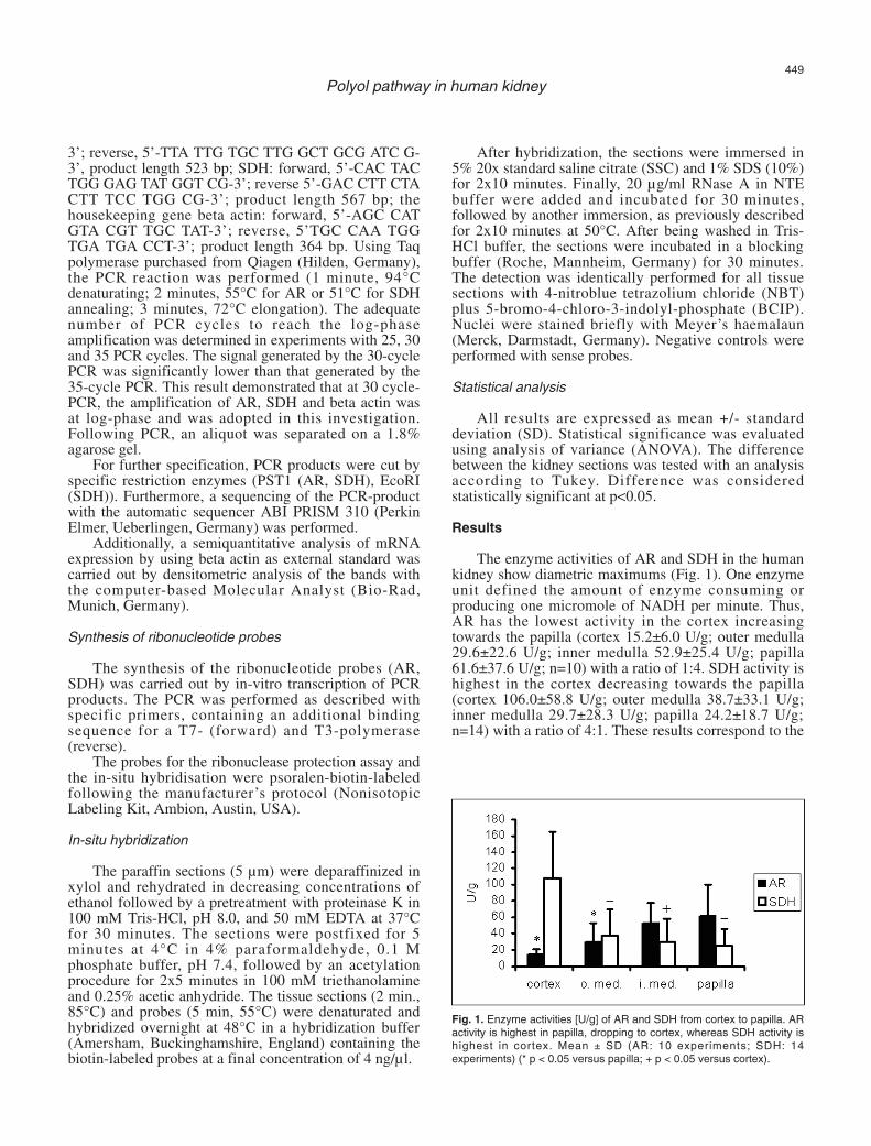
The enzyme activities of AR and SDH in the humankidney show diametric maximums (Fig. 1). One enzymeunit defined the amount of enzyme consuming orproducing one micromole of NADH per minute. Thus,AR has the lowest activity in the cortex increasingtowards the papilla (cortex 15.2±6.0 U/g; outer medulla29.6±22.6 U/g; inner medulla 52.9±25.4 U/g; papilla61.6±37.6 U/g; n=10) with a ratio of 1:4. SDH activity ishighest in the cortex decreasing towards the papilla(cortex 106.0±58.8 U/g; outer medulla 38.7±33.1 U/g;inner medulla 29.7±28.3 U/g; papilla 24.2±18.7 U/g;n=14) with a ratio of 4:1. These results correspond to the
449
Polyol pathway in human kidney
Fig. 1. Enzyme activities [U/g] of AR and SDH from cortex to papilla. ARactivity is highest in papilla, dropping to cortex, whereas SDH activity ishighest in cortex. Mean ± SD (AR: 10 experiments; SDH: 14experiments) (* p < 0.05 versus papilla; + p < 0.05 versus cortex).
450
Polyol pathway in human kidney
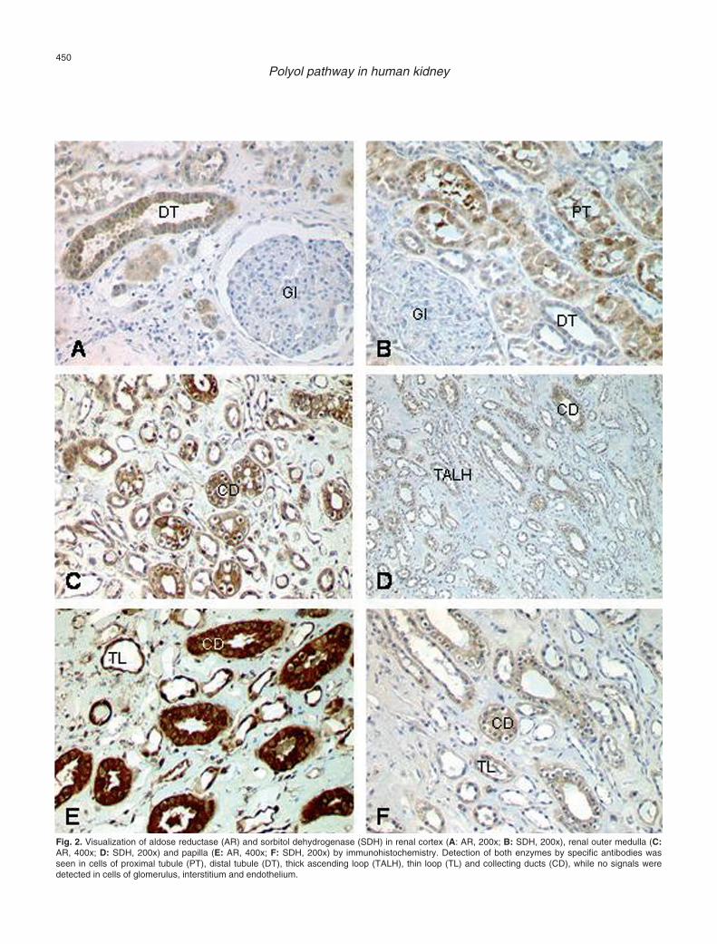
Fig. 2. Visualization of aldose reductase (AR) and sorbitol dehydrogenase (SDH) in renal cortex (A: AR, 200x; B: SDH, 200x), renal outer medulla (C:AR, 400x; D: SDH, 200x) and papilla (E: AR, 400x; F: SDH, 200x) by immunohistochemistry. Detection of both enzymes by specific antibodies wasseen in cells of proximal tubule (PT), distal tubule (DT), thick ascending loop (TALH), thin loop (TL) and collecting ducts (CD), while no signals weredetected in cells of glomerulus, interstitium and endothelium.
451
Polyol pathway in human kidney
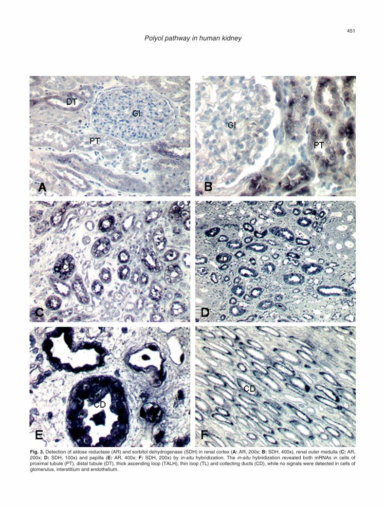
Fig. 3. Detection of aldose reductase (AR) and sorbitol dehydrogenase (SDH) in renal cortex (A: AR, 200x; B: SDH, 400x), renal outer medulla (C: AR,200x; D: SDH, 100x) and papilla (E: AR, 400x; F: SDH, 200x) by in-situ hybridization. The in-situ hybridization revealed both mRNAs in cells ofproximal tubule (PT), distal tubule (DT), thick ascending loop (TALH), thin loop (TL) and collecting ducts (CD), while no signals were detected in cells ofglomerulus, interstitium and endothelium.
enzyme localization visualized byimmunohistochemistry. Detection of the enzymes byspecific antibodies was seen for AR (n=10) and SDH(n=11) in cells of the proximal tubule (PT), distal tubule(DT), thick ascending loop (TALH), thin loop (TL) andcollecting ducts (CD), while no signals were detected incells of the glomerulus, interstitium and endothelium.Here, AR showed an increasing amount of staining fromcortex to papilla, with the highest intensity in collectingducts and thick ascending loop. This rise in signalintensity was not seen for SDH, where the collectingducts and proximal tubules showed the highest intensity(Fig. 2).
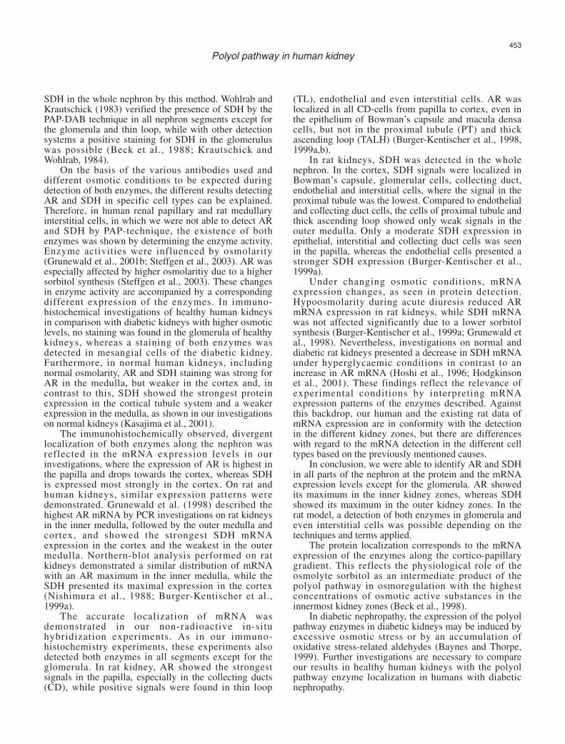
Furthermore, the in-situ hybridization experimentsrevealed the mRNA of AR (n=7) and SDH (n=7) in thesame structures as the proteins shown byimmunohistochemistry, again with an opposite gradient,as previously described (Fig. 3). The AR signal intensitywas strongest in collecting ducts and thin loop, while thehighest signal of SDH was detected in collecting ducts,proximal tubule and thick ascending loop. Analysis byRT-PCR presented a single band for AR and SDH eachat the expected size (AR, 523 bp; SDH 567) (Fig. 4). Forspecification of the PCR products, the restrictiondigestion generated two fragments of the expected size(AR, PST1; SDH, PST1, EcoRI), while the sequencingof the PCR-products showed an identity correspondingto the sequences published.
AR- and SDH-mRNA-expression, each compared tobeta actin, presented opposite gradients in thedistribution, as previously described by enzymeactivities. AR displayed the lowest mRNA expression inthe cortex increasing towards the papilla, while SDHmRNA expression declined from cortex to papilla.
A correspondence between enzyme activity, proteinlocalization and mRNA expression of both enzymesalong the cortico-papillary gradient was observed.
Discussion
For the first time, we presented the localization ofthe polyol pathway enzymes (AR, SDH) throughout the
entire nephron of the normal human kidney using acombination of methods. Various investigators describedthe localization of these two key enzymes, AR and SDH,in human or rat kidneys as the most frequently usedanimal model. Several investigations were performeddoing in vitro cell experimental techniques in singlekidney cell types under special osmotic conditions. Thisfact is important to consider when comparing differentresults concerning single nephron segments or interstitialcells.
Regarding enzyme activities, we demonstrated thehighest AR activity in the papilla, which is the innermostkidney zone, with a decrease towards the cortex. Theseresults were comparable with the detection of ARactivity and protein localization in the rat kidney (Corderet al., 1977; Nishimura et al., 1993; Robinson et al.,1993; Chauncey et al., 1998). In contrast, SDH activitydecreased from the cortex towards the papilla in ourinvestigations, as was also shown for the rat model(Heinz et al., 1975; Cirder et al., 1977; Chauncey et al.,1988). These specific enzyme activities imply thatsorbitol in the polyol pathway is more stronglysynthesized in the papilla and less widely in the cortex.In conclusion, the distribution of the enzyme activitiesmeasured is similar in human and rat kidneys. Theseresults correlate with the physiologic role of sorbitol inosmoregulation, the intermediate product of the polyolpathway, and reflect the osmotic gradient of the kidney(Beck et al., 1988).
The localization of both proteins along the nephronin specific cell types was performed using specificpolyclonal antibodies (Moeckel et al., 1995; Grunewaldet al., 2001a) and PAP-DAB detection. We were able todetect both enzymes in the whole nephron, with theexception of the glomerula. In the rat model, conflictingresults of immunohistochemical detection of AR,depending on the antibodies used, have been reported.
For example, the use of antibodies against rat lensAR and rat placenta AR localized the enzyme in ratkidneys in cells of the nephron of inner and outermedulla, whereas immunohistochemical detection failedin the cortex (Terubayashi et al., 1989). However,detection of AR with an antibody against seminal vesicleAR succeeded only in glomerulus, distal tubule, thinloop and epithelium of the kidney pelvis. (Ludvigsonand Sorenson, 1980; Terubayashi et al., 1989). On theother hand, AR was detected immunohistochemically inall parts of the rat kidney nephron (Corder et al., 1977;Wirth and Wermuth, 1985). These different results pointout the high dependence of the PAP-technique on theantibody used. Aida et al. (2000) localized AR in themouse model from an inner stripe of outer medulla toinner medulla (including papilla), where they detected astrong staining with the avidin-biotin-peroxidasecomplex. They used the same antibody in an ARknockout mouse as well, where no staining was visible.
With regard to SDH, hardly any immuno-histochemical investigations have been undertaken forthe rat kidney, although Corder et al. (1977) detected
452
Polyol pathway in human kidney
Fig. 4. Semiquantitative RT-PCR of aldose reductase (AR) and sorbitoldehydrogenase (SDH) mRNA with beta actin as standard (30 cycles).Highest SDH mRNA was seen in cortex dropping towards papilla, whereAR mRNA expression is the weakest, rising towards papilla.
SDH in the whole nephron by this method. Wohlrab andKrautschick (1983) verified the presence of SDH by thePAP-DAB technique in all nephron segments except forthe glomerula and thin loop, while with other detectionsystems a positive staining for SDH in the glomeruluswas possible (Beck et al., 1988; Krautschick andWohlrab, 1984).
On the basis of the various antibodies used anddifferent osmotic conditions to be expected duringdetection of both enzymes, the different results detectingAR and SDH in specific cell types can be explained.Therefore, in human renal papillary and rat medullaryinterstitial cells, in which we were not able to detect ARand SDH by PAP-technique, the existence of bothenzymes was shown by determining the enzyme activity.Enzyme activities were influenced by osmolarity(Grunewald et al., 2001b; Steffgen et al., 2003). AR wasespecially affected by higher osmolaritiy due to a highersorbitol synthesis (Steffgen et al., 2003). These changesin enzyme activity are accompanied by a correspondingdifferent expression of the enzymes. In immuno-histochemical investigations of healthy human kidneysin comparison with diabetic kidneys with higher osmoticlevels, no staining was found in the glomerula of healthykidneys, whereas a staining of both enzymes wasdetected in mesangial cells of the diabetic kidney.Furthermore, in normal human kidneys, includingnormal osmolarity, AR and SDH staining was strong forAR in the medulla, but weaker in the cortex and, incontrast to this, SDH showed the strongest proteinexpression in the cortical tubule system and a weakerexpression in the medulla, as shown in our investigationson normal kidneys (Kasajima et al., 2001).
The immunohistochemically observed, divergentlocalization of both enzymes along the nephron wasreflected in the mRNA expression levels in ourinvestigations, where the expression of AR is highest inthe papilla and drops towards the cortex, whereas SDHis expressed most strongly in the cortex. On rat andhuman kidneys, similar expression patterns weredemonstrated. Grunewald et al. (1998) described thehighest AR mRNA by PCR investigations on rat kidneysin the inner medulla, followed by the outer medulla andcortex, and showed the strongest SDH mRNAexpression in the cortex and the weakest in the outermedulla. Northern-blot analysis performed on ratkidneys demonstrated a similar distribution of mRNAwith an AR maximum in the inner medulla, while theSDH presented its maximal expression in the cortex(Nishimura et al., 1988; Burger-Kentischer et al.,1999a).
The accurate localization of mRNA wasdemonstrated in our non-radioactive in-situhybridization experiments. As in our immuno-histochemistry experiments, these experiments alsodetected both enzymes in all segments except for theglomerula. In rat kidney, AR showed the strongestsignals in the papilla, especially in the collecting ducts(CD), while positive signals were found in thin loop
(TL), endothelial and even interstitial cells. AR waslocalized in all CD-cells from papilla to cortex, even inthe epithelium of Bowman’s capsule and macula densacells, but not in the proximal tubule (PT) and thickascending loop (TALH) (Burger-Kentischer et al., 1998,1999a,b).
In rat kidneys, SDH was detected in the wholenephron. In the cortex, SDH signals were localized inBowman’s capsule, glomerular cells, collecting duct,endothelial and interstitial cells, where the signal in theproximal tubule was the lowest. Compared to endothelialand collecting duct cells, the cells of proximal tubule andthick ascending loop showed only weak signals in theouter medulla. Only a moderate SDH expression inepithelial, interstitial and collecting duct cells was seenin the papilla, whereas the endothelial cells presented astronger SDH expression (Burger-Kentischer et al.,1999a).
Under changing osmotic conditions, mRNAexpression changes, as seen in protein detection.Hypoosmolarity during acute diuresis reduced ARmRNA expression in rat kidneys, while SDH mRNAwas not affected significantly due to a lower sorbitolsynthesis (Burger-Kentischer et al., 1999a; Grunewald etal., 1998). Nevertheless, investigations on normal anddiabetic rat kidneys presented a decrease in SDH mRNAunder hyperglycaemic conditions in contrast to anincrease in AR mRNA (Hoshi et al., 1996; Hodgkinsonet al., 2001). These findings reflect the relevance ofexperimental conditions by interpreting mRNAexpression patterns of the enzymes described. Againstthis backdrop, our human and the existing rat data ofmRNA expression are in conformity with the detectionin the different kidney zones, but there are differenceswith regard to the mRNA detection in the different celltypes based on the previously mentioned causes.
In conclusion, we were able to identify AR and SDHin all parts of the nephron at the protein and the mRNAexpression levels except for the glomerula. AR showedits maximum in the inner kidney zones, whereas SDHshowed its maximum in the outer kidney zones. In therat model, a detection of both enzymes in glomerula andeven interstitial cells was possible depending on thetechniques and terms applied.
The protein localization corresponds to the mRNAexpression of the enzymes along the cortico-papillarygradient. This reflects the physiological role of theosmolyte sorbitol as an intermediate product of thepolyol pathway in osmoregulation with the highestconcentrations of osmotic active substances in theinnermost kidney zones (Beck et al., 1998).
In diabetic nephropathy, the expression of the polyolpathway enzymes in diabetic kidneys may be induced byexcessive osmotic stress or by an accumulation ofoxidative stress-related aldehydes (Baynes and Thorpe,1999). Further investigations are necessary to compareour results in healthy human kidneys with the polyolpathway enzyme localization in humans with diabeticnephropathy.
453
Polyol pathway in human kidney
Recapitulating, our results in human healthy kidneysare consistent with the rat kidney, with speciesdifferences, so that many research results from the ratmodel might be valid for human tissues as well.
References
Aida K., Ikegishi Y., Chen J., Tawata M., Ito S., Maeda S. and Onaya T.(2000). Disruption of aldose reductase gene (Akr1b1) causes defectin urinary concentrating ability and divalent cation homeostasis.Biochem. Biophys. Res. Commun. 277, 281-286.
Baynes J.W. and Thorpe S.R. (1999). Role of oxidative stress indiabetic complications. A new perspective on an old paradigm.Diabetes 48, 1-9.
Beck F.X., Droege A. and Thurau K. (1988). Cellular osmoregulation inthe renal papilla. Klin. Wochenschr. 66, 843-848.
Burger-Kentischer A., Muller E., Neuhofer W., Marz J., Thurau K. andBeck F.X. (1998). Expression of Na+/Cl-/betaine and Na+/myo-inositol transporters, aldose reductase and sorbitol dehydrogenasein macula densa cells of the kidney. Pflugers Arch. 436, 807-809.
Burger-Kentischer A., Muller E., Neuhofer W., Marz J., Thurau K. andBeck F.X. (1999a). Expression of aldose reductase, sorbitoldehydrogenase and Na+/myo-inositol and Na+/Cl-/betainetransporter mRNAs in individual cells of the kidney during changesin the diuretic state. Pflugers Arch. 437, 248-254.
Burger-Kentischer A., Muller E., Marz J., Fraek M.L., Thurau K. andBeck F.X. (1999b). Hypertonicity-induced accumulation of organicosmolytes in papillary interstitial cells. Kidney Int. 55, 1417-1425.
Chauncey B., Leite M.V. and Goldstein L. (1988). Renal sorbitolaccumulation and associated enzyme activities in diabetes. Enzyme39, 231-234.
Corder C.N., Collins J.G., Brannan T.S. and Sharma J. (1977). Aldosereductase and sorbitol dehydrogenase distribution in rat kidney. J.Histochem. Cytochem. 25, 1-8.
Das B. and Srivastava S.K. (1985). Purification and properties of aldosereductase and aldehyde reductase II from human erythrocyte. Arch.Biochem. Biophys. 238, 670-679.
Dunlop M. (2000). Aldose reductase and the role of the polyol pathwayin diabetic nephropathy. Kidney. Int. 77, 3-12.
Forbes J.M., Fukami K. and Cooper M.E. (2007). Diabetic Nephropathy:Where hemodynamics meets metabolism. Exp. Clin. Endocrinol.Diabetes 115, 69-84.
Garcia-Perez A., Martin B., Murphy H.R., Uchida S., Murer H., CowleyB.D. Jr., Handler J.S. and Burg M.B. (1989). Molecular cloning ofcDNA coding for kidney aldose reductase. Regulation of specificmRNA accumulation by NaCl-mediated osmotic stress. J. Biol.Chem. 264, 16815-16821.
Gerlach U. (1983). Sorbitol dehydrogenase. In: Methods of enzymaticanalysis. Bergmeyer H.U. (ed). Verlag Chemie. Deersfield Beach. pp112-117.
Gonzalez R.G., Barnett P., Aguayo J., Cheng H.M. and Chylack L.T. Jr(1984). Direct measurement of polyol pathway activity in the ocularlens. Diabetes 33, 196-199.
Greene D.A., Latt imer S.A. and Sima A.A. (1987). Sorbitol,phosphoinosit ides, and sodium-potassium-ATPase in thepathogenesis of diabetic complications. N. Engl. J. Med. 316, 599-606.
Grunewald R.W. and Kinne R.K. (1989). Intracellular sorbitol content inisolated rat inner medullary collecting duct cells. Regulation by
extracellular osmolarity. Pflugers Arch. 414, 178-184.Grunewald R.W., Wagner M., Schubert I., Franz H.E., Mueller G.A. and
Steffgen J. (1998). Rat renal expression of mRNA coding for aldosereductase and sorbitol dehydrogenase and its osmotic regulation ininner medullary collecting duct cells. Cell. Physiol. Biochem. 8, 293-303.
Grunewald R.W., Eckstein A., Reisse C.H. and Mueller G.A. (2001a).Characterization of aldose reductase from the thick ascending limbof Henle's loop of rabbit kidney. Nephron 89, 73-81.
Grunewald R.W., Ehrhard M., Fiedler G.M., Schuettert J.B., OppermannM. and Mueller G.A. (2001b). Evidence for a sorbitol transportsystem in immottalized human renal interstitial cells. Exp. Nephrol.9, 405-411.
Heinz F., Schlegel F. and Krause P.H. (1975). Enzymes of fructosemetabolism in human kidney. Enzyme 19, 85-92.
Hodgkinson A.D., Sondergaard K.L., Yang B., Cross D.F., Millward B.A.and Demaine A.G. (2001). Aldose reductase expression is inducedby hyperglycemia in diabetic nephropathy. Kidney Int. 60, 211-218.
Hoshi A., Takahashi M., Fujii J., Myint T., Kaneto H., Suzuki K.,Yamasaki Y., Kamada T. and Taniquchi N. (1996). Glycyation andinactivation of sorbitol dehydrogenase in normal and diabetic rats.Biochem. J. 318, 119-123.
Jeffery J. and Joernvall H. (1983). Enzyme relationships in a sorbitolpathway that bypasses glycolysis and pentose phosphates inglucose metabolism. Proc. Natl. Acad. Sci. 80, 901-907.
Kador P.F., Robison W.G. Jr and Kinoshita J.H. (1985). Thepharmacology of aldose reductase inhibitors. Annu. Rev.Pharmacol. Toxicol. 25, 691-714.
Kasajima H., Yamagishi S., Sugai S., Yagihashi N. and Yagihashi S.(2001). Enhanced in situ expression of aldose reductase inperipheral nerve and renal glomeruli in diabetic patients. VirchowsArch. 439, 46-54.
Krautschick I. and Wohlrab F. (1984). Histochemical determination ofsorbitol dehydrogenase in the rat kidney. Indicator histochemical,immunohistochemical and microelectrophoretic studies. ActaHistochem. Suppl. 30, 365-368.
Kriz W. and Bankir L. (1988). A standard nomenclature for structures ofthe kidney. Kidney Int. 33, 1-7.
Lane P.H. (2002). Diabetic kidney disease: impact of puberty. Am. J.Physiol. Renal Physiol. 283, 589-600.
Ludvigson M.A. and Sorenson R.L. (1980). Immunohistochemicallocalization of aldose reductase. II. Rat eye and kidney. Diabetes 29,450-459.
Meyer C., Tolias A., Platanisios D., Stumvoll M., Vlachos L. andMitrakou A. (2005). Increased renal glucose metabolism in type 1diabetes mellitus. Diabetic Medicine 22, 453-459.
Moeckel G., Hallbach J. and Guder W.G. (1995). Purification of humanand rat kidney aldose reductase. Enzyme Protein 48, 45-50.
Morrison A.D., Clements R.S. Jr, Travis S.B., Oski F. and Winegrad A.I.(1970). Glucose utilization by the polyol pathway in humanerythrocytes. Biochem. Biophys. Res. Commun. 40, 199-205.
Nishimura C., Graham C., Hohman T.C., Nagata M., Robison W.G. Jrand Carper D. (1988). Characterization of mRNA and genes foraldose reductase in rat. Biochem. Biophys. Res. Commun. 153,1051-1059.
Nishimura C., Furue M., Ito T., Omori Y. and Tanimoto T. (1993).Quantitative determination of human aldose reductase by enzyme-linked immunosorbent assay. Immunoassay of human aldosereductase. Biochem. Pharmacol. 46, 21-28.
454
Polyol pathway in human kidney
Pfaller W. (1982). Structure function correlation on rat kidney.Quantitative correlation of structure and function in the normal andinjured rat kidney. Adv. Anat. Embryol. Cell Biol. 70, 1-106.
Philips A.O. and Steadman R. (2003). Diabetic nephropathy: the centralrole of renal proximal tubular cells in tubulointerstitial injury. Histol.Histopathol. 17, 247-252.
Robinson B., Hunsaker L.A., Stangebye L.A. and Vander Jagt D.L.(1993). Aldose and aldehyde reductases from human kidney cortexand medulla. Biochim. Biophys. Acta 1203, 260-266.
Sato S. and Kador P.F. (1993). Human kidney aldose and aldehydereductases. J. Diabetes Complications 7, 179-187.
Steffgen J., Kampfer K., Grupp C., Langenberg C., Mueller G.A. andGrunewald R.W. (2003). Osmoregulation of aldose reductase andsorbitol dehydrogenase in cultivated interstitial cells of rat renal innermedulla. Nephrol. Dial. Transplant. 18, 2255-2261.
Sternberger L.A., Hardy P.H. Jr, Cuculis J.J. and Meyer H.G. (1970).The unlabeled antibody enzyme method of immunohistochemistry:preparation and properties of soluble antigen-antibody complex(horseradish peroxidase-antihorseradish peroxidase) and its use inidentification of spirochetes. J. Histochem. Cytochem. 18, 315-333.
Swidan S.Z. and Montgomery P.A. (1998). Effect of blood glucoseconcentrations on the development of chronic complications ofdiabetes mellitus. Pharmacotherapy 18, 961-972.
Terubayashi H., Sato S., Nishimura C., Kador P.F. and Kinoshita J.H.(1989). Localization of aldose and aldehyde reductase in the kidney.Kidney Int. 36, 843-851.
Tomlinson D.R. (1993). Aldose reductase inhibitors and thecomplications of diabetes mellitus. Diabet. Med. 10, 214-230.
Yabe-Nishimura C. (1998). Aldose reductase in glucose toxicity: apotential target for the prevention of diabetic complications.Pharmacol. Rev. 50, 21-33.
Wirth H.P. and Wermuth B. (1985). Immunochemical characterization ofaldo-keto reductases from human tissues. FEBS Lett. 187, 280-282.
Wohlrab F. and Krautschick I. (1983). Specificity of histochemicaldemonstration of sorbitol dehydrogenase - Comparativeinvestigations by indicator- and immunohistochemical techniques inthe rat kidney. Acta Histochem. 72, 133-151.
Accepted November 3, 2008
455
Polyol pathway in human kidney