Localization of Arabinogalactan Proteins in the Cell Walls ... · stage with the functional...
3
Central Bringing Excellence in Open Access International Journal of Plant Biology & Research Cite this article: Leszczuk A, Szczuka E (2017) Localization of Arabinogalactan Proteins in the Cell Walls of Developing Ovule in Strawberry (Fragaria X Ananassaduch.). Int J Plant Biol Res 5(2): 1066. *Corresponding author Ewa Szczuka, Institute of Agrophysics, Polish Academy of Science, Poland, Email: Submitted: 30 May 2017 Accepted: 10 June 2017 Published: 12 June 2017 ISSN: 2333-6668 Copyright © 2017 Szczuka et al. OPEN ACCESS Keywords • Arabinogalactan proteins • Development • Fragaria x ananassa • Ovule Review Article Localization of Arabinogalactan Proteins in the Cell Walls of Developing Ovule in Strawberry (Fragaria X Ananassaduch.) Agata Leszczuk 1 and Ewa Szczuka 2 * 1 Institute of Agrophysics, Polish Academy of Science, Poland 2 Department of Plant Anatomy and Cytology, Maria Curie Skłodowska University, Poland Abstract The aim of the study was to investigate changes in the localization of arabinogalactan proteins (AGPs) during ovule development in Fragaria x ananassa Duch. cv. ‘Mount Everest’. For the first time, the localization of AGPs was studied in a hybrid Fragaria x ananassa, which is a facultative apomict. The AGP epitopes were revealed with the use of JIM13 mAb after immune cyto chemical reaction and immune gold labeling. The fluorescence after immune cyto chemical reaction and gold particles indicated the presence of AGPs, which changed during the consecutive stages of ovule development. AGPs were localized in the walls of nucellus cells lying on the possible route of pollen tube growth, functional megaspore, and embryo sac. Such a distribution of AGPs resembles the localization of these proteins in plants that reproduce in the amphimiticmode. Moreover, the AGPs were observed in the cell walls of the embryo. In addition to the wall of the embryo sac, they were also present in the cytoplasm of the egg cell, near the secondary nuclei in the central cell, and in the endosperm. In the nucellus cells, AGPs were observed in the area of starch grains. The possible functions in Fragaria x ananassa of AGPs are discussed. INTRODUCTION Arabinogalactan proteins (AGPs) as one of the most abundant and highly diverse classes of cell surface proteins occur commonly in the plant kingdom. These macromolecules are integral components of the extracellular matrix and their occurrence is developmentally regulated [1]. Available papers usually provide a description of the localization of AGPs and their implication in many biological processes like cell division, programmed cell death, organ abscission, or interactions with growth regulators [2]. The research presented by us focused on the developing ovule, i.e. a key organ in plant reproduction. So far, no analysis of AGP distribution in the ovules of a facultative apomictic plant has been reported. Therefore, in this study, we undertook description of the localization of AGPs in the ovule cells of a widely cultivated facultative apomict – Fragaria x ananassa Duch. We chose the cv. ‘Mount Everest’, i.e. a new strawberry cultivar that, in favorable conditions, produces fruits three times in one growing season. PLANT MATERIAL One cultivar of Fragaria x ananssa -’Mount Everest’ was used in this study. Flower buds were collected in different stages of development during the last vegetation period in 2016.The plants were cultivated in the Botanical Garden of Maria Curie- Skłodowska University in Lublin (Poland). METHODS In these studies, we used two methods to examine the AGP localization. The first method was the immunocyto chemical reaction with the use a primary monoclonal antibody against AGPs epitope according to [3]. The plant material was fixed in a mixture of 4% paraformal dehyde and 0,5% glutaraldehyde in 0,2M phosphate-buffered saline (PBS, pH 7). After fixation, ovules were rinsed in PBS and dehydrated in a graded ethanol series. Then, the material was embedded in LR White (Sigma-Aldrich) and was sectioned using a Reichert Ultracut S ultramicrotome (Leica, Wetzlar, Germany). For immune fluorescence labeling, the sections (1 µm) were dried on poly-L-lysine slides (Sigma- Aldrich), rehydrated with PBS, preincubatedin a solution consisting of 1% BSA in PBS for 1 h, and washed with PBS. Primary antibodies (diluted 1:50 in 0,1% BSA) were added to each sample and incubated at 4 °C overnight. The following mAbagainst AGP was used: JIM13 recognizing arabinogalactan protein epitopes containing trisaccharide β-D-GlcA-(1,3)-α-DGalA(1,2)-α-L-Rha [4]. The samples were incubated with the secondary fluorescein isothiocynate (FITC)-conjugated antibody (Sigma-Aldrich) diluted 1:200 in the blocking solution at 4 °C overnight. This facilitated visualization of the AGP epitope localization under confocal laser scanning microscopy (Zeiss Axiovert 200M equipped with an LSM 5 Pascal laser scanning head, Germany). The second method was the immune gold labeling at the transmission electron microscopy (TEM) level. Reactions were carried out as previously described by [5]. Ultrathin sections (60 nm) were collected on form var-coated nickel grids (100 MESH).
Transcript of Localization of Arabinogalactan Proteins in the Cell Walls ... · stage with the functional...
CentralBringing Excellence in Open Access
International Journal of Plant Biology & Research
Cite this article: Leszczuk A, Szczuka E (2017) Localization of Arabinogalactan Proteins in the Cell Walls of Developing Ovule in Strawberry (Fragaria X Ananassaduch.). Int J Plant Biol Res 5(2): 1066.
*Corresponding authorEwa Szczuka, Institute of Agrophysics, Polish Academy of Science, Poland, Email:
Submitted: 30 May 2017
Accepted: 10 June 2017
Published: 12 June 2017
ISSN: 2333-6668
Copyright© 2017 Szczuka et al.
OPEN ACCESS
Keywords•Arabinogalactan proteins•Development•Fragariax ananassa•Ovule
Review Article
Localization of Arabinogalactan Proteins in the Cell Walls of Developing Ovule in Strawberry (Fragaria X Ananassaduch.)Agata Leszczuk1 and Ewa Szczuka2*1Institute of Agrophysics, Polish Academy of Science, Poland2Department of Plant Anatomy and Cytology, Maria Curie Skłodowska University, Poland
Abstract
The aim of the study was to investigate changes in the localization of arabinogalactan proteins (AGPs) during ovule development in Fragaria x ananassa Duch. cv. ‘Mount Everest’. For the first time, the localization of AGPs was studied in a hybrid Fragaria x ananassa, which is a facultative apomict. The AGP epitopes were revealed with the use of JIM13 mAb after immune cyto chemical reaction and immune gold labeling. The fluorescence after immune cyto chemical reaction and gold particles indicated the presence of AGPs, which changed during the consecutive stages of ovule development. AGPs were localized in the walls of nucellus cells lying on the possible route of pollen tube growth, functional megaspore, and embryo sac. Such a distribution of AGPs resembles the localization of these proteins in plants that reproduce in the amphimiticmode. Moreover, the AGPs were observed in the cell walls of the embryo. In addition to the wall of the embryo sac, they were also present in the cytoplasm of the egg cell, near the secondary nuclei in the central cell, and in the endosperm. In the nucellus cells, AGPs were observed in the area of starch grains. The possible functions in Fragaria x ananassa of AGPs are discussed.
INTRODUCTIONArabinogalactan proteins (AGPs) as one of the most abundant
and highly diverse classes of cell surface proteins occur commonly in the plant kingdom. These macromolecules are integral components of the extracellular matrix and their occurrence is developmentally regulated [1]. Available papers usually provide a description of the localization of AGPs and their implication in many biological processes like cell division, programmed cell death, organ abscission, or interactions with growth regulators [2]. The research presented by us focused on the developing ovule, i.e. a key organ in plant reproduction. So far, no analysis of AGP distribution in the ovules of a facultative apomictic plant has been reported. Therefore, in this study, we undertook description of the localization of AGPs in the ovule cells of a widely cultivated facultative apomict – Fragaria x ananassa Duch. We chose the cv. ‘Mount Everest’, i.e. a new strawberry cultivar that, in favorable conditions, produces fruits three times in one growing season.
PLANT MATERIALOne cultivar of Fragaria x ananssa -’Mount Everest’ was used
in this study. Flower buds were collected in different stages of development during the last vegetation period in 2016.The plants were cultivated in the Botanical Garden of Maria Curie-Skłodowska University in Lublin (Poland).
METHODSIn these studies, we used two methods to examine the AGP
localization. The first method was the immunocyto chemical reaction with the use a primary monoclonal antibody against AGPs epitope according to [3]. The plant material was fixed in a mixture of 4% paraformal dehyde and 0,5% glutaraldehyde in 0,2M phosphate-buffered saline (PBS, pH 7). After fixation, ovules were rinsed in PBS and dehydrated in a graded ethanol series. Then, the material was embedded in LR White (Sigma-Aldrich) and was sectioned using a Reichert Ultracut S ultramicrotome (Leica, Wetzlar, Germany). For immune fluorescence labeling, the sections (1 µm) were dried on poly-L-lysine slides (Sigma-Aldrich), rehydrated with PBS, preincubatedin a solution consisting of 1% BSA in PBS for 1 h, and washed with PBS. Primary antibodies (diluted 1:50 in 0,1% BSA) were added to each sample and incubated at 4 °C overnight. The following mAbagainst AGP was used: JIM13 recognizing arabinogalactan protein epitopes containing trisaccharide β-D-GlcA-(1,3)-α-DGalA(1,2)-α-L-Rha [4]. The samples were incubated with the secondary fluorescein isothiocynate (FITC)-conjugated antibody (Sigma-Aldrich) diluted 1:200 in the blocking solution at 4 °C overnight. This facilitated visualization of the AGP epitope localization under confocal laser scanning microscopy (Zeiss Axiovert 200M equipped with an LSM 5 Pascal laser scanning head, Germany).
The second method was the immune gold labeling at the transmission electron microscopy (TEM) level. Reactions were carried out as previously described by [5]. Ultrathin sections (60 nm) were collected on form var-coated nickel grids (100 MESH).
CentralBringing Excellence in Open Access
Szczuka et al. (2017)Email:
Int J Plant Biol Res 5(1): 1066 (2017) 2/3
The grids were washed in deionized water and incubated in the blocking solution containing 1% BSA in PBS for 1 h at room temperature. Then, the samples were incubated with the JIM13 antibody (diluted 1:10 in 0,1% BSA) for 3 h at 37 °C and then with the secondary antibody conjugated with 10 nm gold particles (Sigma-Aldrich) diluted 1:50 in 0,1 BSA for 1 h at 37 °C. The ultrathin sections were double-stained with 1% aqueous uranyl acetate and Reynold’s lead citrate. After the reaction, the samples were examined with TEM (ZEISS LEO 912 AB, Germany).
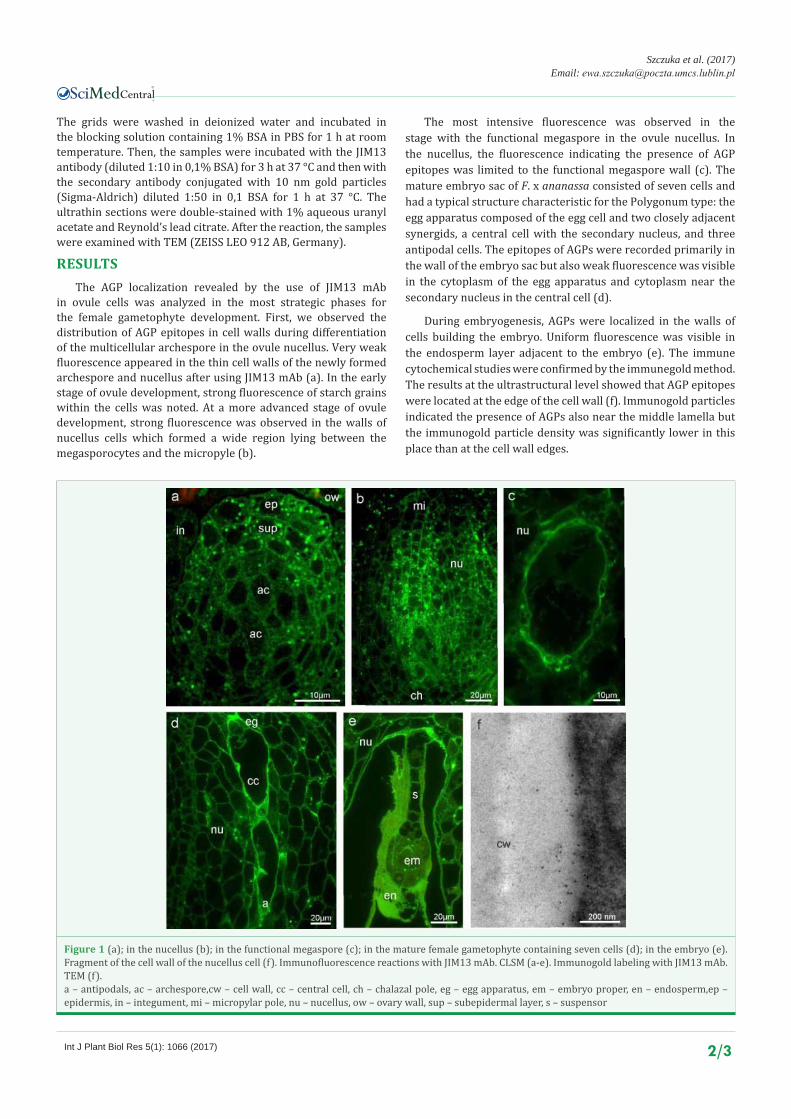
RESULTSThe AGP localization revealed by the use of JIM13 mAb
in ovule cells was analyzed in the most strategic phases for the female gametophyte development. First, we observed the distribution of AGP epitopes in cell walls during differentiation of the multicellular archespore in the ovule nucellus. Very weak fluorescence appeared in the thin cell walls of the newly formed archespore and nucellus after using JIM13 mAb (a). In the early stage of ovule development, strong fluorescence of starch grains within the cells was noted. At a more advanced stage of ovule development, strong fluorescence was observed in the walls of nucellus cells which formed a wide region lying between the megasporocytes and the micropyle (b).
The most intensive fluorescence was observed in the stage with the functional megaspore in the ovule nucellus. In the nucellus, the fluorescence indicating the presence of AGP epitopes was limited to the functional megaspore wall (c). The mature embryo sac of F. x ananassa consisted of seven cells and had a typical structure characteristic for the Polygonum type: the egg apparatus composed of the egg cell and two closely adjacent synergids, a central cell with the secondary nucleus, and three antipodal cells. The epitopes of AGPs were recorded primarily in the wall of the embryo sac but also weak fluorescence was visible in the cytoplasm of the egg apparatus and cytoplasm near the secondary nucleus in the central cell (d).
During embryogenesis, AGPs were localized in the walls of cells building the embryo. Uniform fluorescence was visible in the endosperm layer adjacent to the embryo (e). The immune cytochemical studies were confirmed by the immunegold method. The results at the ultrastructural level showed that AGP epitopes were located at the edge of the cell wall (f). Immunogold particles indicated the presence of AGPs also near the middle lamella but the immunogold particle density was significantly lower in this place than at the cell wall edges.
Figure 1 (a); in the nucellus (b); in the functional megaspore (c); in the mature female gametophyte containing seven cells (d); in the embryo (e). Fragment of the cell wall of the nucellus cell (f). Immunofluorescence reactions with JIM13 mAb. CLSM (a-e). Immunogold labeling with JIM13 mAb. TEM (f).a – antipodals, ac – archespore,cw – cell wall, cc – central cell, ch – chalazal pole, eg – egg apparatus, em – embryo proper, en – endosperm,ep – epidermis, in – integument, mi – micropylar pole, nu – nucellus, ow – ovary wall, sup – subepidermal layer, s – suspensor
CentralBringing Excellence in Open Access
Szczuka et al. (2017)Email:
Int J Plant Biol Res 5(1): 1066 (2017) 3/3
Leszczuk A, Szczuka E (2017) Localization of Arabinogalactan Proteins in the Cell Walls of Developing Ovule in Strawberry (Fragaria X Ananassaduch.). Int J Plant Biol Res 5(2): 1066.
Cite this article
DISCUSSIONNumerous publications on arabinogalactan proteins
reported results proving their involvement in diverse processes of growth and development in plants [6,7]. The presence and variable localization of arabinogalactan proteins in cells during plant development indicated that AGPs act as proteogly cans necessary to proceed into consecutive developmental stages. The possibility of migration through the extracellular matrix of the cell walls in ovules is associated with their role as signal molecules. Their localization in the specific area of the ovule and temporary distribution allowed to determination of their supposed functions.
In our studies performed on developing ovules of F. x ananassa cv. ‘Mount Everest’, AGPs epitopes were localized in ovules in early stages of development. The presence of AGPs at the beginning of this process may indicate and confirm the involvement of these macromolecules in cell differentiation. The assumption that AGPs, as structural elements constituting the extracellular matrix of the cell wall, are a source of nutrients for the developing ovule cells has been proposed by [8].
Observations of AGPs in the wall of the functional megaspore allowed a conclusion that the examined proteins, as signal molecules, can mark the beginning of the female gametophyte development. Similarly, genetic studies of AGP18 expression showed that the proteins were involved in determination of functional megaspores and their selection [9]. The observation performed by us confirms the results obtained during the investigation of AGP distribution in amphimictic plants.
Distribution of arabinogalactan proteins in the cell wall of the embryo sac may be associated with their function as an attractant for the growing pollen tube. Similar findings were described in different species e.g. Arabidopsis thaliana [10] or Olea europaea [5]. In our studies, we observed a fluorescence signal in the egg apparatus and in the cytoplasm near the secondary nucleus, which can be evidence of their involvement in the double fertilization process. Other authors describe AGPs as the first source of calcium, a key element to start the fusion of gametes [3].
During early embryogenesis, AGPs were distributed in suspensor cells and newly formed cell walls of the embryo proper as well as the endosperm layer. This result refers to their function of supporting the development process as structural proteins, which may provide favorable conditions for the newly formed embryo. Other authors discussed the functions of AGPs in the interaction between the embryo proper and the suspensor, in signaling transduction and transportation of material [11].
The presented results provide the first information on specific epitopes of arabinogalactan proteins in the cells of Fragaria x ananassa (Duch) ovules. The immune cytochemical and immune gold analyses showed spatial and temporal patterns in the distribution of AGPs during female gametophyte development in the facultative apomict, which allowed formulation of a hypothesis of their functions in this plant.
REFERENCES1. Serpe MD, Nothnagel EA. 1999. Arabinogalactan-proteins in the
multiple domains of the plant cell surface. Advances in Botanical Research. 199; 30: 207-289.
2. Seifert GJ, Roberts K. The Biology of Arabinogalactan Proteins. Annu Rev Plant Biol. 2007; 58: 137-161.
3. Lopez RA, Renzaglia KS. 2016. Arabinogalactan proteins and arabinanpectins abound in the specialized matrices surrounding female gametes of the fern Ceratopterisrichardii. Planta. 2016; 243: 1-11.
4. Knox JP, Linstead PJ, Peart J, Cooper C, Roberts K. 1991. Developmentally regulated epitopes of cell surface arabinogalactan proteins and their relation to root tissue pattern formation. Plant Journal. 1991; 1: 317-326.
5. Suárez C, Zienkiewicz A, Castro AJ, Zienkiewicz K, Majewska-Sawka A, Rodríguez-García MI. 2013. Cellular localization and levels of pectins and arabinogalactan proteins in olive (Olea europaeaL.) pistil tissues during development: implications for pollen-pistil interaction. Planta. 2013; 237: 305-319.
6. Showalter AM. Arabinogalactan-proteins: structure, expression and function. Cell Mol Life Sci. 2013; 58: 1399-1417.
7. Pereira AM, Pereira LG, Coimbra S. 2015. Arabinogalactan proteins: rising attention from plant biologists. Plant Reprod. 2015; 28: 1-15.
8. Rafińska K, Bednarska E. Localization pattern of homogalacturonan and arabinogalactan proteins in developing ovules of the gymnosperm plant Larix decidua Mill. Sexual Plant Reprod. 2011; 24: 75-87.
9. Demesa-ArévaloE, Vielle-Calzada JP. The Classical Arabinogalactan Protein AGP18 Mediates Megaspore Selection in Arabidopsis. Plant Cell. 2013; 25: 1274-1287.
10. Coimbra S, Almeida J, Junqueira V, Costa ML, Pereira LG. Arabinogalactan proteins as molecular markers in Arabidopsis thaliana sexual reproduction. J Exp Bot. 2007; 58: 4027-4035.
11. Qin Y, Zhao J. Localization of arabinogalactan proteins in different stages of embryos and their role in cotyledon formation Nicotianatabacum L. Sexual Plant Reproduction. 2007; 20: 213-224.
![i5 Literature [081610DP] · Chicory), Beta Glucans, Oat Fiber) Sunflower oil, Proprietary Immune Blend, Natural Flavors, Medium Chain Triglycerides, Arabinogalactan, Broccoli Seed](https://static.fdocuments.net/doc/165x107/6013a3b270ad005e46206382/i5-literature-081610dp-chicory-beta-glucans-oat-fiber-sunflower-oil-proprietary.jpg)