Llama-Derived Single-Chain Antibody Fragments Directed...
12
JOURNAL OF VIROLOGY, Oct. 2008, p. 9753–9764 Vol. 82, No. 19 0022-538X/08/$08.000 doi:10.1128/JVI.00436-08 Copyright © 2008, American Society for Microbiology. All Rights Reserved. Llama-Derived Single-Chain Antibody Fragments Directed to Rotavirus VP6 Protein Possess Broad Neutralizing Activity In Vitro and Confer Protection against Diarrhea in Mice Lorena Garaicoechea, 1 *† Aurelien Olichon, 2 † Gisela Marcoppido, 1 Andre ´s Wigdorovitz, 1 Marina Mozgovoj, 1 Linda Saif, 3 Thomas Surrey, 2 and Viviana Parren ˜o 1 Instituto de Virologı ´a, CICV y A–INTA, Buenos Aires, Argentina 1 ; European Molecular Biology Laboratory, Cell Biology and Biophysics Unit, Meyerhofstr. 1, 69117 Heidelberg, Germany 2 ; and Food Animal Health Research Program, Department of Veterinary Preventive Medicine, Ohio Agricultural Research and Development Center, The Ohio State University, Wooster, Ohio 44691-40961 3 Received 27 February 2008/Accepted 7 July 2008 Group A rotavirus is one of the most common causes of severe diarrhea in human infants and newborn animals. Rotavirus virions are triple-layered particles. The outer capsid proteins VP4 and VP7 are highly variable and represent the major neutralizing antigens. The inner capsid protein VP6 is conserved among group A rotaviruses, is highly immunogenic, and is the target antigen of most immunodiagnosis tests. Llama-derived single-chain antibody fragments (VHH) are the smallest molecules with antigen-binding ca- pacity and can therefore be expected to have properties different from conventional antibodies. In this study a library containing the VHH genes of a llama immunized with recombinant inner capsid protein VP6 was generated. Binders directed to VP6, in its native conformation within the viral particle, were selected and characterized. Four selected VHH directed to conformational epitopes of VP6 recognized all human and animal rotavirus strains tested and could be engineered for their use in immunodiagnostic tests for group A rotavirus detection. Three of the four VHH neutralized rotavirus in vivo independently of the strain serotype. Further- more, this result was confirmed by in vivo partial protection against rotavirus challenge in a neonatal mouse model. The present study demonstrates for the first time a broad neutralization activity of VP6 specific VHH in vitro and in vivo. Neutralizing VHH directed to VP6 promise to become an essential tool for the prevention and treatment of rotavirus diarrhea. Group A rotavirus (RV) is the leading cause of acute gas- troenteritis in human infants less than 5 years old, causing 611,000 deaths per year (41). It is also the main cause of severe diarrhea in the neonates of many animal species of economic interest (43, 47). RV virions are triple-layered particles com- posed by a core (protein VP2), an inner capsid (protein VP6), and an outer capsid (proteins VP7 and VP4) (16, 29). The inner capsid protein, VP6, is a trimer representing 51% of the virion mass. According to the antigenic variation of VP6, RVs are classified into seven groups (A to G) (16). Depending on the presence or absence of two different epitopes in the VP6 protein, group A RV strains are further divided into subgroups (Sb) I, II, III, and no I no II. Despite the different subgroups mentioned, VP6 is a strongly conserved protein among all group A RVs (90% amino acid homology). It is highly im- munogenic and constitutes the target antigen of most immu- nodiagnosis tests for group A RV detection. In contrast, the outer capsid proteins VP7 (glycoprotein) and VP4 (protease sensitive) are highly variable and constitute the major neutral- izing antigens. Based on the variation of VP7 and VP4, group A RVs are further classified into G and P types, respectively. RVs with different G- and P-type combinations induce low or no cross neutralization in vitro. The neutralizing antibodies directed to VP7 and VP4 correlate with protection in vivo against subsequent homologous RV infection (16, 29). Since VP6 is a highly conserved protein, several attempts to investigate its use as a broadly protective antigen were carried out. Contradictory results were obtained. Some studies showed that anti-VP6 maternal antibodies did not induce passive pro- tection against RV-induced diarrhea in neonatal mice (7, 8), and active vaccination with VP2/6 virus-like particles failed to protect against RV infection and diarrhea in gnotobiotic pigs (27), while other studies of vaccination with VP6 protein or DNA induced protection in vivo in a mouse model (10–13). Anti-VP6 secretory immunoglobulin A (IgA) binds to RV and mediates protection by intracellular neutralization during transcytosis in mice (3, 6, 52). VP6 could, therefore, be con- sidered as a potential broadly reactive vaccine. Regarding in vitro neutralization, most studies showed that antibodies to VP6 lack neutralizing activity (20, 21, 44, 56). However, it has been reported that a monospecific polyclonal antiserum to VP6 of C486 RV has low neutralizing activity in vitro (46). It is well known that the continuous presence of high titers of passive RV antibodies in the gut lumen (naturally produced or artificially added to the milk) fully protects against diarrhea and significantly reduces virus shedding (20, 48, 49). Passive immunity strategies such as oral administration of specific an- tibodies from different sources (bovine colostrum or chicken egg yolk) have been explored and were shown to be effective * Corresponding author. Mailing address: Instituto de Virologı ´a, CICV y A–INTA, CC 25 (1712) Castelar, Buenos Aires, Argentina. Phone: 54 11 4621 9050. Fax: 54 11 4621 9050. E-mail: lgaraicoechea @cnia.inta.gov.ar. † L.G. and A.O. contributed equally to this study. Published ahead of print on 16 July 2008. 9753 on May 1, 2018 by guest http://jvi.asm.org/ Downloaded from
Transcript of Llama-Derived Single-Chain Antibody Fragments Directed...
JOURNAL OF VIROLOGY, Oct. 2008, p. 9753–9764 Vol. 82, No. 190022-538X/08/$08.00�0 doi:10.1128/JVI.00436-08Copyright © 2008, American Society for Microbiology. All Rights Reserved.
Llama-Derived Single-Chain Antibody Fragments Directed to RotavirusVP6 Protein Possess Broad Neutralizing Activity In Vitro and Confer
Protection against Diarrhea in Mice�
Lorena Garaicoechea,1*† Aurelien Olichon,2† Gisela Marcoppido,1 Andres Wigdorovitz,1Marina Mozgovoj,1 Linda Saif,3 Thomas Surrey,2 and Viviana Parreno1
Instituto de Virologıa, CICV y A–INTA, Buenos Aires, Argentina1; European Molecular Biology Laboratory, Cell Biology andBiophysics Unit, Meyerhofstr. 1, 69117 Heidelberg, Germany2; and Food Animal Health Research Program, Department of
Veterinary Preventive Medicine, Ohio Agricultural Research and Development Center, The Ohio State University,Wooster, Ohio 44691-409613
Received 27 February 2008/Accepted 7 July 2008
Group A rotavirus is one of the most common causes of severe diarrhea in human infants and newbornanimals. Rotavirus virions are triple-layered particles. The outer capsid proteins VP4 and VP7 are highlyvariable and represent the major neutralizing antigens. The inner capsid protein VP6 is conserved amonggroup A rotaviruses, is highly immunogenic, and is the target antigen of most immunodiagnosis tests.Llama-derived single-chain antibody fragments (VHH) are the smallest molecules with antigen-binding ca-pacity and can therefore be expected to have properties different from conventional antibodies. In this studya library containing the VHH genes of a llama immunized with recombinant inner capsid protein VP6 wasgenerated. Binders directed to VP6, in its native conformation within the viral particle, were selected andcharacterized. Four selected VHH directed to conformational epitopes of VP6 recognized all human and animalrotavirus strains tested and could be engineered for their use in immunodiagnostic tests for group A rotavirusdetection. Three of the four VHH neutralized rotavirus in vivo independently of the strain serotype. Further-more, this result was confirmed by in vivo partial protection against rotavirus challenge in a neonatal mousemodel. The present study demonstrates for the first time a broad neutralization activity of VP6 specific VHHin vitro and in vivo. Neutralizing VHH directed to VP6 promise to become an essential tool for the preventionand treatment of rotavirus diarrhea.
Group A rotavirus (RV) is the leading cause of acute gas-troenteritis in human infants less than 5 years old, causing611,000 deaths per year (41). It is also the main cause of severediarrhea in the neonates of many animal species of economicinterest (43, 47). RV virions are triple-layered particles com-posed by a core (protein VP2), an inner capsid (protein VP6),and an outer capsid (proteins VP7 and VP4) (16, 29). Theinner capsid protein, VP6, is a trimer representing 51% of thevirion mass. According to the antigenic variation of VP6, RVsare classified into seven groups (A to G) (16). Depending onthe presence or absence of two different epitopes in the VP6protein, group A RV strains are further divided into subgroups(Sb) I, II, I�II, and no I no II. Despite the different subgroupsmentioned, VP6 is a strongly conserved protein among allgroup A RVs (�90% amino acid homology). It is highly im-munogenic and constitutes the target antigen of most immu-nodiagnosis tests for group A RV detection. In contrast, theouter capsid proteins VP7 (glycoprotein) and VP4 (proteasesensitive) are highly variable and constitute the major neutral-izing antigens. Based on the variation of VP7 and VP4, groupA RVs are further classified into G and P types, respectively.
RVs with different G- and P-type combinations induce low orno cross neutralization in vitro. The neutralizing antibodiesdirected to VP7 and VP4 correlate with protection in vivoagainst subsequent homologous RV infection (16, 29).
Since VP6 is a highly conserved protein, several attempts toinvestigate its use as a broadly protective antigen were carriedout. Contradictory results were obtained. Some studies showedthat anti-VP6 maternal antibodies did not induce passive pro-tection against RV-induced diarrhea in neonatal mice (7, 8),and active vaccination with VP2/6 virus-like particles failed toprotect against RV infection and diarrhea in gnotobiotic pigs(27), while other studies of vaccination with VP6 protein orDNA induced protection in vivo in a mouse model (10–13).Anti-VP6 secretory immunoglobulin A (IgA) binds to RV andmediates protection by intracellular neutralization duringtranscytosis in mice (3, 6, 52). VP6 could, therefore, be con-sidered as a potential broadly reactive vaccine. Regarding invitro neutralization, most studies showed that antibodies toVP6 lack neutralizing activity (20, 21, 44, 56). However, it hasbeen reported that a monospecific polyclonal antiserum toVP6 of C486 RV has low neutralizing activity in vitro (46).
It is well known that the continuous presence of high titersof passive RV antibodies in the gut lumen (naturally producedor artificially added to the milk) fully protects against diarrheaand significantly reduces virus shedding (20, 48, 49). Passiveimmunity strategies such as oral administration of specific an-tibodies from different sources (bovine colostrum or chickenegg yolk) have been explored and were shown to be effective
* Corresponding author. Mailing address: Instituto de Virologıa,CICV y A–INTA, CC 25 (1712) Castelar, Buenos Aires, Argentina.Phone: 54 11 4621 9050. Fax: 54 11 4621 9050. E-mail: [email protected].
† L.G. and A.O. contributed equally to this study.� Published ahead of print on 16 July 2008.
9753
on May 1, 2018 by guest
http://jvi.asm.org/
Dow
nloaded from
immunotherapies to prevent RV infections in both humansand animals (15, 23, 31–33, 51). However, there are somepractical limitations related to the high cost of scaling up im-munoglobulin purification and sterilization and the fact thatthey are animal-derived products. To develop a pharmaceuti-cal product complying with the rigorous requirements for hu-man application (i.e., high purity, high batch reproducibility,absence of adventitious viruses derived from an animal host ormammalian cell cultures, etc.), a more reliable technologybased on recombinant antibodies might be an ideal choice.Especially, fragments of heavy-chain antibodies (VHH) ofCamelidae come into the focus of research and are expected tohave superior properties compared to conventional recombi-nant antibodies (scFv) (28). The VHH domain (15 kDa) is thesmallest known natural domain with full antigen-binding ca-pacity. It is ideal for the generation of libraries of single-chainantibodies (24, 25, 38). Due to their small size, VHH moleculeshave been shown to act as strong enzyme inhibitors, reachingenzyme pockets not accessible to common antibodies (4). Re-combinant VHH antibody fragments are emerging as new ver-satile reagents for the diagnosis and also for the therapy ofinfectious diseases, including RV-induced diarrhea (40, 57).We describe here the selection and characterization of VHHantibodies directed to the inner capsid protein VP6 of group ARV. We show that these VHH are broadly reactive reagentsthat can be engineered for their use in immunodiagnostic testsfor group A RV detection. Furthermore, we demonstrate abroad neutralization activity of VP6 specific VHH in vitro and
confirmed this result by in vivo protection against RV chal-lenge in a neonatal mouse model.
MATERIALS AND METHODS
RVs. The bovine RV (BRV) reference strain IND (SbI, P[5]G6) was used asan antigen in enzyme-linked immunosorbent assay (ELISA) and virus neutral-ization (VN) tests to monitor the immune response of the llama and the bio-panning process for binder selection.
In order to have a panel of RVs representing different subgroup reactivitiesand G- and P-type combinations from different species, the following strainswere included in the different assays (ELISA and VN) for binder characteriza-tion: BRV strain C486 (SbI, P[1]G6), homologous to the VP6 used as immuniz-ing antigen; BRV B223 (SbI, P[11]G10); human RV Wa (SbII, P1A[8]G1); andequine H2 (Sb no I no II, P[12]G3).
Reference RVs were propagated in monkey kidney cells (MA-104) for use inWestern blot, enzyme-linked immunospot (ELISPOT), ELISA, and VN assays.The tissue culture-adapted BRV C486 (Sb1, P[1]G6) was used for mouse chal-lenge. Fecal samples from newborn colostrum-deprived calves experimentallyinfected with BRV IND, taken at preinoculation and at the peak of virus shed-ding, were also included (42).
Wild-type murine RV strain ECw (Sb no I no II, P[17]G3) was kindly suppliedby A. Castello, Quilmes University. Stock EC strain was prepared as 10% intes-tinal homogenate derived from infected suckling mice as previously described(17).
Llama immunization. A 1-year-old male llama received five doses of crude Sf9cell extract containing 500 �g of recombinant VP6 protein derived from the BRVC486 strain (SbI, P[1]G6; kindly supplied by L. Babiuk, VIDO, Canada), emul-sified in INTA oil adjuvant (42.5% Marcol 52, 6.5% Arlacel C, 1% Tween 80),at days 0, 21, 28, 35, and 246. Serum and blood samples were taken at days 0, 4,and 7 after each inoculation (Fig. 1). The antibody response was monitored byELISA and VN assay (see below). To evaluate the effector B-cell response, anELISPOT assay determining the number of RV-specific antibody-secreting cells
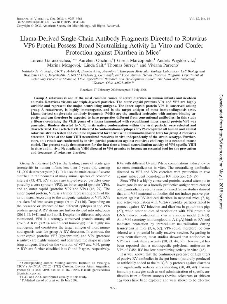
FIG. 1. Llama immunization. The schedule for immunization, sample collection, and final bleeding is shown. The evaluation of RV antibodyresponse in serum during the time course of immunization is depicted. Antibody titers were measured by (i) ELISA using recombinant VP6, (ii)ELISA using RV (IND, Sb I, P[5]G6), (iii) VN assay, and (iv) ELISPOT assay using the same RV (IND, Sb I, P[5]G6). Vaccination time pointsare represented by arrows.
9754 GARAICOECHEA ET AL. J. VIROL.
on May 1, 2018 by guest
http://jvi.asm.org/
Dow
nloaded from
(ASC) in the peripheral blood of the inoculated llama was adapted from previousELISPOT assays conducted in pigs and calves (42, 58). Briefly, IND BRV-infected MA-104 cells (with �80% infection as detected by immunofluores-cence) grown in 96-well plates were fixed with 70% acetone, air dried, and storedat �20°C until used. Suspensions of mononuclear cells derived from peripheralblood of the inoculated llama were added to quadruplicate wells (1 � 106, 5 �105, 2.5 � 105, and 1.25 � 105 cells/well). After centrifugation at 500 � g for 5min, plates were incubated for 12 to 14 h at 37°C in 5% CO2. The plates werewashed with phosphate-buffered saline (PBS)–0.05% Tween 20 to remove ad-herent cells, and the spots were developed by adding a peroxidase-labeled goatanti-llama immunoglobulin G (IgG; H�L; Bethyl Labs, Inc., Montgomery, CA)at a 1/1,500 dilution for 2 h at 37°C, followed by 50 �l of a tetramethylbenzidinemembrane peroxidase substrate system (KLP, Maryland).
Llama management, inoculation, and sample collection were conducted bytrained personnel under the supervision of a veterinarian and in accordance withprotocols approved by the INTA’s ethical committee of animal welfare.
VHH library production and enrichment of VP6 binders. From a total of 900ml of blood collected 4 days after the last injection, 6 � 108 mononuclear cellswere extracted by Ficoll-Paque gradient centrifugation, pelleted, frozen in liquidnitrogen, and then kept at �80°C. The total RNA was extracted by using an RNAextraction kit (Nucleospin RNA II; Macherey Nagel), yielding 256 �g of RNA.Subsequently, first-strand cDNA was synthesized from 210 �g of RNA by usingSuperscript III reverse transcriptase (Invitrogen), with oligo(dT)12-18 primers(Invitrogen) or random primers (Invitrogen). In a 20-�l reaction mixture 0.2, 1,or 5 �g of total RNA was used. The cDNA encoding VHH and VH wasspecifically amplified by PCR using the primers CALL01 and CALL02 (GTCCTGGCTGCTCTTCTACAAGG and GGTACGTGCTGTTGAACTGTTCC,respectively) annealing at the leader and at the CH2 sequences. The 600-bpfragment (VHH-CH2 without the CH1 exon) was eluted from an 1.6% agarosegel after separation from the 900-bp fragment (VH-CH1-CH2 exons). VHHfragments were then amplified with one additional nested PCR with primersannealing at the framework 1 and framework 4 regions (AACATGCCATGACTCGCGGCTCAACCGGCCATGGCTGAKGTBCAGCTGCAGGCGTCTGGRGGAGG and ATTATTATTCAGATTATTAGTGCGG CCGCGTGAGGAGACGGTGACCWGGGTCC, respectively), followed by the use of primerscontaining the restriction sites for further cloning steps: VHHfor2 (GGCTGAKGTBCAGCTGCAGGCGTCTGGRGGAGG) contained the NcoI and PstIrestriction sites, and VHHrev2 (GTTATTATTATTCAGATTATTAGTGCGGCCGCTGGAGACGGTGACCWGGGTCC) contained the NotI restriction site.The final PCR fragments were ligated by using the upstream restriction sitesNcoI or PstI and the downstream NotI site into the phagemid vector pAO-Lib,a modified version of pHEN4 (1) carrying a long irrelevant sequence that isremoved upon VHH insertion in order to slow down the potential propagationof vector without a VHH insert. Ligated material was transformed into Esche-richia coli TG1 cells. The colonies from the plated cells were collected, washed,and stored at �80°C in LB medium supplemented with glycerol (50% finalconcentration).
Specific VHH were selected from the library using the phage display technol-ogy. The VHH library was infected with M13 helper phages (Invitrogen), andphage particles expressing the VHH repertoire were rescued and precipitatedwith polyethylene glycol as described previously (37). Enrichment in specificbinders was performed by three rounds of in vitro selection, i.e., by the so-called“biopanning.” Immunotubes were coated overnight at 4°C with an anti-RVantiserum from guinea pig (1/5,000 dilution in carbonate buffer [pH 9.6]), and2 � 105 focus-forming units (FFU) of BRV IND (SbI; P[5]G6) were capturedafter a blocking step. Phages were incubated with the captured BRV IND andwashed, and bound phage particles were eluted with 100 mM triethylamine (pH10.0) and immediately neutralized with Tris (pH 7.4). The eluted phages wereused to infect exponentially growing TG1 cells. After the second or third roundof biopanning, individual colonies were grown, and the corresponding VHHclones were analyzed by phage ELISA.
Screening for RV and VP6 specific VHH fragments (phage ELISA). Phagesdisplaying the selected VHH were produced by the individual TG1 E. coli clonesas previously described (38). Produced phages were tested by ELISA using twodifferent conditions: a capture ELISA using RV and a direct ELISA usingrecombinant VP6. MA-104 mock-infected cells and an unrelated protein ex-pressed in baculovirus (BVDV E2) were used as negative antigens. PBS was usedas blank, and a normal serum from a nonimmunized guinea pig was used as anegative capture. After the coating step, all plates were blocked with 4% skimmilk in 0.5% Tween 20–PBS. Phages were added, followed by incubation at roomtemperature for 60 min. The assays were developed using a 1/5,000 dilution of ahorseradish peroxidase–anti-M13p8 conjugate (Amersham/Pharmacia Biotech)
for 40 min at room temperature, followed by H2O2/ABTS [2,2�azinobis(3-ethyl-benzthiazolinesulfonic acid)] as a substrate chromogen reagent.
Expression and purification of recombinant VHH. VHH cDNA of the clonesthat scored positive in phage ELISA for VP6 and for RV were recloned using therestriction enzymes NcoI and NotI into the expression vector pHEN6 (4), whichprovides a pelB targeting sequence for the periplasm and a C-terminal His6 tag.Bivalent VHH were constructed by PCR amplification of the VHH sequenceusing the primers Bivfor2 (CTCGCGGCCCAGCCGGCCATGGCGGATGTGCAGCTTCAGGCGTCTGGG) and Bivrev2 (GCATTGGTTCTGCAGTTGCACATCTGACGGCGGGGTGGACGGAGACGCCGCGGCGGGGTAGACGGGCCCGATGAGGAGACGGTGACCTG) encoding a linker related to thehuman IgA hinge. The PCR product and the pHEN6 vector containing the templateVHH were digested by NcoI and PstI and ligated to produce the pAO-biv vectorcontaining the bivalent VHH. Production of recombinant monovalent or bivalentVHH was performed in shaker flasks by growing cells in Terrific Broth supple-mented with 0.1% glucose and ampicillin (50). E. coli XL1-Blue cells were freshlytransformed with the different plasmid constructs. VHH expression was theninduced with 1 mM IPTG (isopropyl-�-D-thiogalactopyranoside) for 16 h at27°C. After the cells were pelleted, the periplasmic proteins were extracted byosmotic shock (53). The VHH were purified from this periplasmic extract byusing a High-Trap HP Ni-chelating column (Amersham Biosciences).
ELISAs. The assays were performed in 96-well plates (Nunc Maxisorp), eitherby direct coating of BRV IND, by capture of BRV IND, or by use of a recom-binant VP6 with a polyclonal antiserum to RV made in a germfree pig (20).MA-104 mock-infected cells and an unrelated protein expressed in the baculo-virus system (bovine diarrhea virus E2 protein) were used as negative controlantigens. PBS was used as blank, and a serum from a nonimmunized guinea pigwas used as a negative capture.
The llama serum was tested for anti-RV antibodies as previously described(42) and for anti-VP6 antibodies with a protocol adapted from Fernandez et al.(20). Llama IgG were detected by using a peroxidase-labeled goat anti-llama IgG(H�L; Bethyl Labs, Inc.) at a 1:2,000 dilution.
Purified monovalent VHH molecules with the C-terminal His6 tag were firsttested in an ELISA as a RV capture reagent either by direct coating of 10 �g ofVHH/ml or by first coating with 10 �g of anti-pentahistidine monoclonal anti-body (1/500; Qiagen)/ml, followed by an incubation with 20 �g of VHH/ml. Theassays were developed by using an RV polyclonal antiserum made in a co-lostrum-deprived calf hyperimmunized with BRV IND (1/2,000 dilution) and aperoxidase-labeled anti-bovine IgG (H�L; KPL, Gaithersburg, MD) in a 1/5,000dilution). Monovalent VHH molecules were also tested as secondary antibodies,and the ELISA was then revealed by the monoclonal anti-pentahistidine anti-body and horseradish peroxidase-conjugated goat anti-mouse (1:1,000 dilution;Amersham/Pharmacia Biotech). Bivalent VHH were tested in ELISA as a RVcapture reagent at 10 �g/ml.
Western blotting for VHH characterization. VP6 expressed in Sf9 cells usingthe baculovirus system and concentrated BRV IND were either resuspended inLaemmli sample buffer and boiled for 10 min or resuspended in nonreducingsample buffer and heated at 65°C for 15 min. Negative controls of concentratedMA-104 cells or wild-type baculovirus-infected Sf9 cells were treated the sameway. Samples were subjected to sodium dodecyl sulfate-polyacrylamide gel elec-trophoresis in a 12% gel and blotted onto a nitrocellulose membrane (Bio-Rad).The membrane was blocked for 1 h with PBS-Tween (0.05%) containing 3%skim milk. To test the VHH, the membranes were incubated with each VHH (4�g/ml) in PBS-Tween 20 (0.05%)–skim milk (1%) overnight at 4°C, and then themembranes were washed with PBS-Tween 20 (0.05%) and incubated 1 h at 37°Cwith the anti-pentahistidine monoclonal antibody at a 1/500 dilution in PBS-Tween 20 (0.05%)–skim milk (1%). A polyclonal serum from a colostrum-deprived calf experimentally infected with RV (42) and monoclonal antibody toVP6 (RG25A10) (9) were used as positive controls. Nonrelated VHH was usedas a negative control. Finally, the membranes were incubated with alkalinephosphatase-conjugated anti-mouse or anti-bovine IgG (1/1,000 dilution; KPL)for 1 h at room temperature. The assay was developed by the nitroblue tetrazo-lium/BCIP (5-bromo-4-chloro-3-indolylphosphate) colorimetric method.
VN test. Neutralization of RV strains IND, C486, B223, Wa, and H2 wasdetermined in llama serum samples and for purified VHH molecules by afluorescent focus neutralization test as described previously (55). Briefly, 100 �lof serial dilutions of llama serum or selected purified VHH molecules was mixedwith an equal volume of virus in order to have 100 FFU/100 �l of mixture,followed by incubation for 1 h at 37°C. Then, 100 �l of the antibody-virus mixturewas plated onto a MA-104 monolayer (four replicates), followed by incubationfor 48 h at 37°C. The plates were fixed with 70% acetone, and the assay wasdeveloped using a fluorescein isothiocyanate-labeled anti-RV polyclonal anti-serum derived from a colostrum-deprived calf by hyperimmunization. VHH VN
VOL. 82, 2008 ANTI-VP6 VHH NEUTRALIZE ROTAVIRUSES 9755
on May 1, 2018 by guest
http://jvi.asm.org/
Dow
nloaded from
capacity was expressed as the minimum concentration resulting in a �80%reduction in the number of fluorescent foci. The monovalent or bivalent VHHwere assayed from 6.25 to 0.02 �g/well.
Furthermore, to evaluate whether the VHH neutralization activity was asso-ciated with VP6 binding, an assay to block VHH neutralization was performed bypreincubation of recombinant VP6. Briefly, serial fourfold dilutions of VHH2KD1, 3A6, and 3B2 (6.25 to 0.02 �g/well) were preincubated with 5 �g ofrecombinant VP6 produced in Sf9 cells or Sf9 cells infected with wild-typebaculovirus for 1 h at 37°C. The residual neutralizing activity of VHH was thentested by VN assay against 100 FFU of BRV C486. An affinity-purified IgGderived from a polyclonal antiserum made in guinea pig with a VN titer of 2,048to the homologous RV and a monoclonal antibody directed to a neutralizingepitope of VP7 serotype G6 (MA-49 IC3) (35) were used as controls.
RV protection assay in neonatal mice. Four-day-old BALB/c mouse pupsreceived 100 �l of a solution containing 100 �g of monovalent anti-VP6 VHH,using an intragastric gauge, once a day, starting on day 0 for 5 days. The pupswere challenged either with BRV C486 or murine RV ECw. Pup challengeconditions for both strains were standardized in order to produce diarrhea in100% of the nontreated control pups similarly to a previous report (26, 36). Onday 1, at 2 h after the VHH administration pups received 20 �l of 5% bicarbon-ate solution, they were also challenged by the intragastric route with 100 �l ofBRV C486 containing a total of 30 times the dose that caused diarrhea in 50%of suckling mice (DD50) (6 � 105 FFU/mouse) (experiment A) or with 100 �l ofmurine RV ECw containing 316 DD50 (experiment B). Experiment A was con-ducted in three independent assays of five mice each, while experiment B wasconducted in two independent assays of five mice each. Control groups used inthe experiments included (i) RV-inoculated mice and not treated with antibodiesand (ii) mice treated with the same amount of a nonrelated VHH (directedagainst a nonrelated protein). In addition, for experiment A, a control group ofmice treated with 450 �g of the affinity-purified IgG derived from a polyclonalantiserum made in guinea pig with a VN titer of 2,048 to the homologous RV wasalso included. Pups were clinically evaluated for RV-induced diarrhea by directpalpation of the abdomen during the 5 days of the study by a well-trained personworking blindly regarding the treatment groups. As murine RV ECw spread inlittermates, cases of diarrhea that developed 24 h later or more after the first casein a litter could not be due to the initial inoculum and therefore were notconsidered (18). For the murine RV challenge, the pups were euthanatized 4days postinoculation, and the entire small intestines were removed, frozen-thawed, and homogenized in 10% (wt/vol) Hanks balanced salt solution. Thehomogenates were assayed for detection of RV by ELISA (5). The Fisher exacttest was used to compare the proportions of pups with diarrhea among thevarious groups. The Kruskal-Wallis rank sum (nonparametric) test was used tocompare days to onset, mean duration of diarrhea, and mean cumulative diar-rhea scores among the treatment groups of experiment A (Statistix 8.0; Analyt-ical Software, Tallahassee, FL). One-way analysis of variance and Tukey’s testwere used to compare the average values for virus shedding among the groups.
RESULTS
Llama immunization. To evaluate the llama immune re-sponse, antibody titers to RV and VP6 were monitored byELISA and a VN assay. The number of specific ASC circulat-ing in peripheral blood was followed by ELISPOT assay (Fig.1). As expected, before the first vaccination dose, at 0 dayspostinoculation, the llama was already positive for antibodiesto RV as determined by ELISA (RV and VP6) and VN assay,indicating a previous contact with the antigen. However, nospecific ASC were circulating in blood. At 7 days postinocula-tion, ELISA antibody titers to IND RV or to VP6 were signif-icantly increased, and a peak of RV-specific ASC was detectedin peripheral blood (16 anti-RV IgG ASC/5 � 105 mononu-clear cells). After the following boosters the antibody responsereached a plateau at 14 days postinoculation, with high ELISAantibody titers for both the whole virus and the VP6 protein. Incontrast, the VN titers remained very low for RV (Fig. 1), asexpected for an antibody response directed against VP6. Al-though very high antibody titers were obtained in serum, thenumber of ASC to RV detected in blood decreased after each
booster (Fig. 1). For this reason and in order to give plenty oftime to favor antibody affinity maturation, the llama receivedthe final dose of VP6 (Fig. 1) after about 7 months. Finally, thellama was bled 4 days after the last booster, and a very highnumber of RV-specific plasmocytes was obtained (26.8 anti-RVIgG ASC/5 � 105 mononuclear cells).
From the total 900 ml of blood, 6 � 108 mononuclear cellswere extracted (expected to contain at least 32,160 IgG ASCspecific for RV according to the ELISPOT result). From theprocessed RNA (210 �g), a VHH phage display library con-taining 6 � 107 clones was generated.
Phage display selection of VHH specific for VP6 and RV. Toselect phages displaying VHH specific for RV, three rounds ofin vitro selection (biopanning) were performed using immobi-lized BRV IND as an antigen. The VP6 binders in the VHHlibrary showed a large diversity, as judged from a restrictionanalysis of 192 clones that were randomly chosen after thesecond and third rounds of biopanning (data not shown). Thecapacity of the 192 clones to bind RV and VP6 was tested in aphage ELISA. The 10 clones showing the strongest specificbinding were subcloned into an expression vector providing acarboxy-terminal His6 tag for purification. From these 10clones, four VHH, named 2KA4, 2KD1, 3A6, and 3B2, thatwere produced with the highest yields, between 17 and 52mg/liter of culture, and that strongly recognized RV strainswith different subgroup specificities as determined by ELISA,were selected for further analysis.
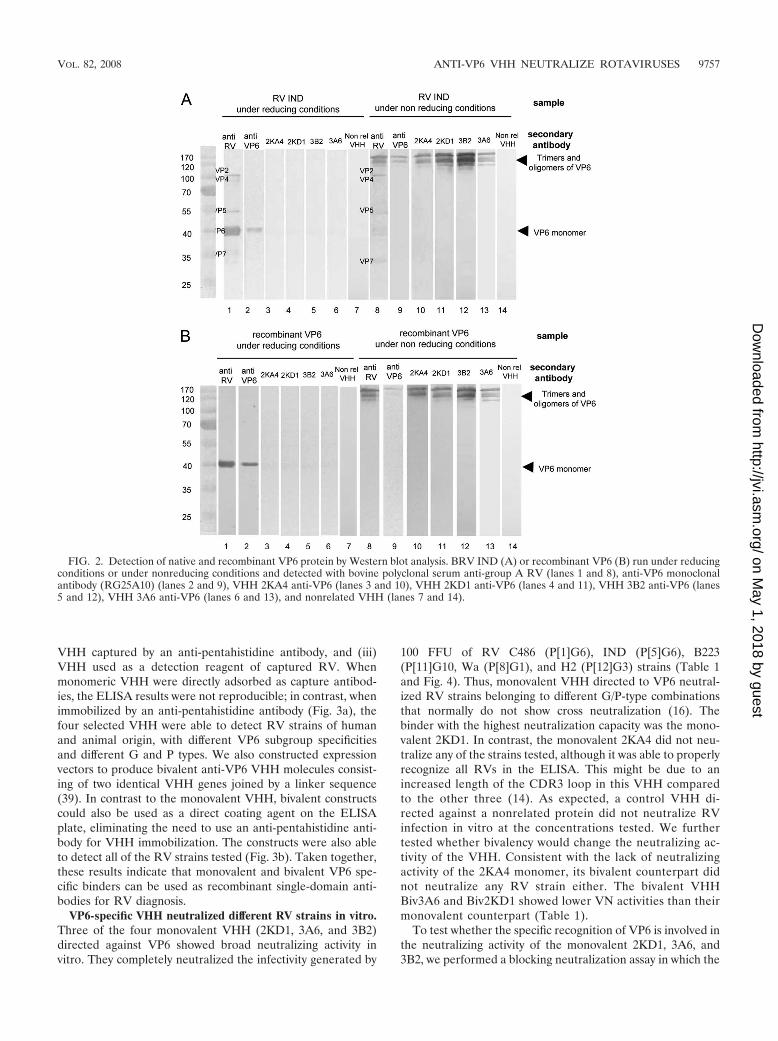
Characterization of the specificity of the isolated VHH byWestern blotting. Using Western blot analysis we found thatthe four VHH selectively recognized bands correspondingto VP6 in samples consisting either of recombinant VP6 ornative RV VP6 (Fig. 2). A monoclonal anti-VP6 antibody(RG25A10) was used as a positive control for VP6 detection.Under nonreducing conditions the four VHH recognized thehigher-molecular-weight bands that correspond to trimers andoligomers of VP6 (56). The band corresponding to monomericVP6 obtained under reducing conditions could be detectedonly using a more sensitive chemoluminescent method (datanot shown). This indicates that the four selected VHH recog-nize conformational epitopes of VP6, in contrast to the mono-clonal antibody that recognizes both unfolded and oligomericVP6. The four tested VHH did not recognize other bands ofthe typical protein pattern of RV samples, as detected by apolyclonal serum to RV (Fig. 2) and by monoclonal antibodiesto VP7 (common 60) and VP5 (BR16D3 F7.G11) (data notshown). In control experiments, no bands were observed inlanes with wild-type baculovirus-infected Sf9 cells and MA-104cells in both reducing and nonreducing conditions, demon-strating that the anti-VP6 VHH did not detect any cellularproteins (data not shown). As expected, the nonrelated VHHdid not recognize any RV protein, demonstrating that recog-nition of VP6 is not a general property of a VHH.
These experiments demonstrate that the four selected VHHspecifically recognize VP6.
VP6-specific VHH as broadly reactive reagents for group ARV immunodiagnosis. Because VP6 is a conserved proteinamong group A RVs, we assessed the usefulness of the se-lected VHH as broadly reactive reagents for RV diagnosis. Tothis end, we tested the use of VHH monomers in three types ofELISAs: (i) VHH directly adsorbed as a capture antibody, (ii)
9756 GARAICOECHEA ET AL. J. VIROL.
on May 1, 2018 by guest
http://jvi.asm.org/
Dow
nloaded from
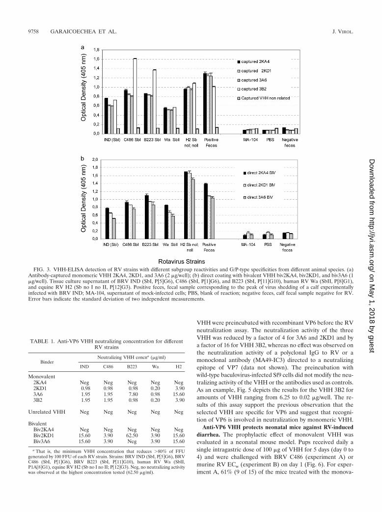
VHH captured by an anti-pentahistidine antibody, and (iii)VHH used as a detection reagent of captured RV. Whenmonomeric VHH were directly adsorbed as capture antibod-ies, the ELISA results were not reproducible; in contrast, whenimmobilized by an anti-pentahistidine antibody (Fig. 3a), thefour selected VHH were able to detect RV strains of humanand animal origin, with different VP6 subgroup specificitiesand different G and P types. We also constructed expressionvectors to produce bivalent anti-VP6 VHH molecules consist-ing of two identical VHH genes joined by a linker sequence(39). In contrast to the monovalent VHH, bivalent constructscould also be used as a direct coating agent on the ELISAplate, eliminating the need to use an anti-pentahistidine anti-body for VHH immobilization. The constructs were also ableto detect all of the RV strains tested (Fig. 3b). Taken together,these results indicate that monovalent and bivalent VP6 spe-cific binders can be used as recombinant single-domain anti-bodies for RV diagnosis.
VP6-specific VHH neutralized different RV strains in vitro.Three of the four monovalent VHH (2KD1, 3A6, and 3B2)directed against VP6 showed broad neutralizing activity invitro. They completely neutralized the infectivity generated by
100 FFU of RV C486 (P[1]G6), IND (P[5]G6), B223(P[11]G10, Wa (P[8]G1), and H2 (P[12]G3) strains (Table 1and Fig. 4). Thus, monovalent VHH directed to VP6 neutral-ized RV strains belonging to different G/P-type combinationsthat normally do not show cross neutralization (16). Thebinder with the highest neutralization capacity was the mono-valent 2KD1. In contrast, the monovalent 2KA4 did not neu-tralize any of the strains tested, although it was able to properlyrecognize all RVs in the ELISA. This might be due to anincreased length of the CDR3 loop in this VHH comparedto the other three (14). As expected, a control VHH di-rected against a nonrelated protein did not neutralize RVinfection in vitro at the concentrations tested. We furthertested whether bivalency would change the neutralizing ac-tivity of the VHH. Consistent with the lack of neutralizingactivity of the 2KA4 monomer, its bivalent counterpart didnot neutralize any RV strain either. The bivalent VHHBiv3A6 and Biv2KD1 showed lower VN activities than theirmonovalent counterpart (Table 1).
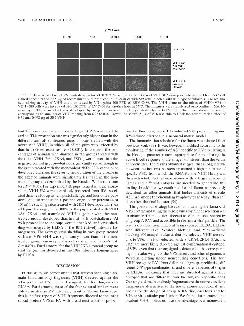
To test whether the specific recognition of VP6 is involved inthe neutralizing activity of the monovalent 2KD1, 3A6, and3B2, we performed a blocking neutralization assay in which the
FIG. 2. Detection of native and recombinant VP6 protein by Western blot analysis. BRV IND (A) or recombinant VP6 (B) run under reducingconditions or under nonreducing conditions and detected with bovine polyclonal serum anti-group A RV (lanes 1 and 8), anti-VP6 monoclonalantibody (RG25A10) (lanes 2 and 9), VHH 2KA4 anti-VP6 (lanes 3 and 10), VHH 2KD1 anti-VP6 (lanes 4 and 11), VHH 3B2 anti-VP6 (lanes5 and 12), VHH 3A6 anti-VP6 (lanes 6 and 13), and nonrelated VHH (lanes 7 and 14).
VOL. 82, 2008 ANTI-VP6 VHH NEUTRALIZE ROTAVIRUSES 9757
on May 1, 2018 by guest
http://jvi.asm.org/
Dow
nloaded from
VHH were preincubated with recombinant VP6 before the RVneutralization assay. The neutralization activity of the threeVHH was reduced by a factor of 4 for 3A6 and 2KD1 and bya factor of 16 for VHH 3B2, whereas no effect was observed onthe neutralization activity of a polyclonal IgG to RV or amonoclonal antibody (MA49-IC3) directed to a neutralizingepitope of VP7 (data not shown). The preincubation withwild-type baculovirus-infected Sf9 cells did not modify the neu-tralizing activity of the VHH or the antibodies used as controls.As an example, Fig. 5 depicts the results for the VHH 3B2 foramounts of VHH ranging from 6.25 to 0.02 �g/well. The re-sults of this assay support the previous observation that theselected VHH are specific for VP6 and suggest that recogni-tion of VP6 is involved in neutralization by monomeric VHH.
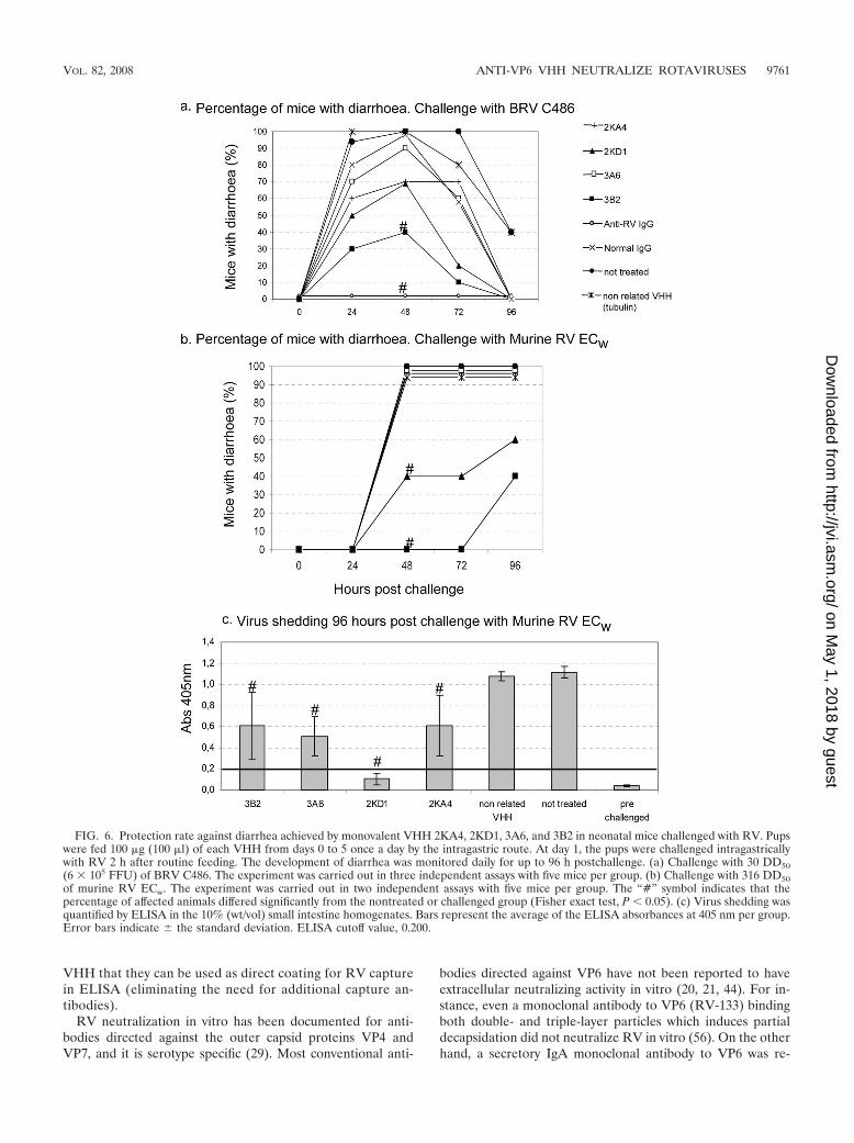
Anti-VP6 VHH protects neonatal mice against RV-induceddiarrhea. The prophylactic effect of monovalent VHH wasevaluated in a neonatal mouse model. Pups received daily asingle intragastric dose of 100 �g of VHH for 5 days (day 0 to4) and were challenged with BRV C486 (experiment A) ormurine RV ECw (experiment B) on day 1 (Fig. 6). For exper-iment A, 61% (9 of 15) of the mice treated with the monova-
FIG. 3. VHH-ELISA detection of RV strains with different subgroup reactivities and G/P-type specificities from different animal species. (a)Antibody-captured monomeric VHH 2KA4, 2KD1, and 3A6 (2 �g/well); (b) direct coating with bivalent VHH biv2KA4, biv2KD1, and biv3A6 (1�g/well). Tissue culture supernatant of BRV IND (SbI, P[5]G6), C486 (SbI, P[1]G6), and B223 (SbI, P[11]G10), human RV Wa (SbII, P[8]G1),and equine RV H2 (Sb no I no II, P[12]G3). Positive feces, fecal sample corresponding to the peak of virus shedding of a calf experimentallyinfected with BRV IND; MA-104, supernatant of mock-infected cells; PBS, blank of reaction; negative feces, calf fecal sample negative for RV.Error bars indicate the standard deviation of two independent measurements.
TABLE 1. Anti-VP6 VHH neutralizing concentration for differentRV strains
BinderNeutralizing VHH concna (�g/ml)
IND C486 B223 Wa H2
Monovalent2KA4 Neg Neg Neg Neg Neg2KD1 0.98 0.98 0.98 0.20 3.903A6 1.95 1.95 7.80 0.98 15.603B2 1.95 1.95 0.98 0.20 3.90
Unrelated VHH Neg Neg Neg Neg Neg
BivalentBiv2KA4 Neg Neg Neg Neg NegBiv2KD1 15.60 3.90 62.50 3.90 15.60Biv3A6 15.60 3.90 Neg 3.90 15.60
a That is, the minimum VHH concentration that reduces �80% of FFUgenerated by 100 FFU of each RV strain. Strains: BRV IND (SbI, P�5G6), BRVC486 (SbI, P�1G6), BRV B223 (SbI, P�11G10), human RV Wa (SbII,P1A�8G1), equine RV H2 (Sb no I no II; P�12G3). Neg, no neutralizing activitywas observed at the highest concentration tested (62.50 �g/ml).
9758 GARAICOECHEA ET AL. J. VIROL.
on May 1, 2018 by guest
http://jvi.asm.org/
Dow
nloaded from
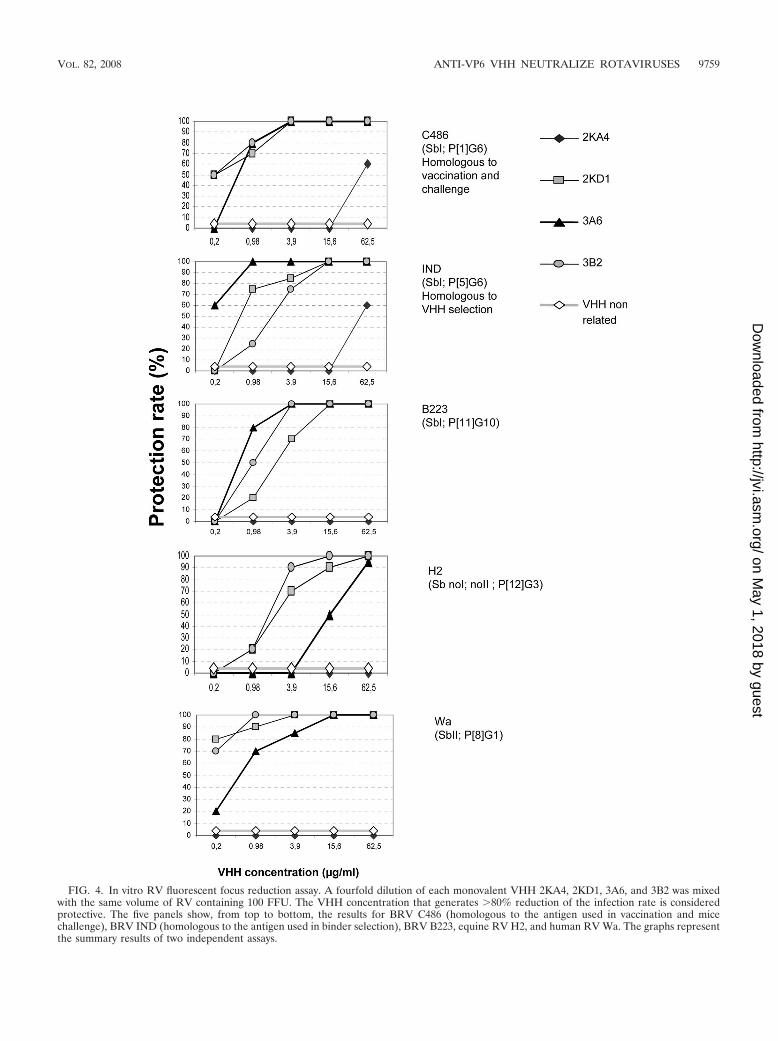
FIG. 4. In vitro RV fluorescent focus reduction assay. A fourfold dilution of each monovalent VHH 2KA4, 2KD1, 3A6, and 3B2 was mixedwith the same volume of RV containing 100 FFU. The VHH concentration that generates �80% reduction of the infection rate is consideredprotective. The five panels show, from top to bottom, the results for BRV C486 (homologous to the antigen used in vaccination and micechallenge), BRV IND (homologous to the antigen used in binder selection), BRV B223, equine RV H2, and human RV Wa. The graphs representthe summary results of two independent assays.
VOL. 82, 2008 ANTI-VP6 VHH NEUTRALIZE ROTAVIRUSES 9759
on May 1, 2018 by guest
http://jvi.asm.org/
Dow
nloaded from
lent 3B2 were completely protected against RV-associated di-arrhea. This protection rate was significantly higher than in thedifferent controls (untreated pups or pups treated with thenonrelated VHH), in which all of the pups were affected bydiarrhea (Fisher exact test; P 0.001). In contrast, the per-centages of animals with diarrhea in the groups treated withthe other VHH (3A6, 2KA4, and 2KD1) were lower than thenegative control groups—but not significantly so. Although inthe group treated with the monovalent 2KD1 73% of the pupsdeveloped diarrhea, the severity and duration of the disease inthe affected animals were significantly less than in the non-treated group (as determined by the Kruskal-Wallis rank sumtest; P 0.05). For experiment B, pups treated with the mono-valent VHH 3B2 were completely protected from RV-associ-ated diarrhea for up to 72 h postchallenge, and four of ten pupsdeveloped diarrhea at 96 h postchallenge. Forty percent (4 of10) of the suckling mice treated with 2KD1 developed diarrhea48 h postchallenge, while 100% of the pups treated with VHH3A6, 2KA4, and nonrelated VHH, together with the non-treated group, developed diarrhea at 48 h postchallenge. At96 h postchallenge the pups were euthanized, and virus shed-ding was assayed by ELISA in the 10% (wt/vol) intestine ho-mogenates. The average virus shedding in each group treatedwith anti-VP6 VHH was significantly lower than in the non-treated group (one-way analysis of variance and Tukey’s test,P � 0.001). Furthermore, for the VHH 2KD1-treated group noviral antigen was detected in the 10% intestine homogenatesby ELISA.
DISCUSSION
In this study we demonstrated that recombinant single-do-main llama antibody fragments (VHH) directed against theVP6 protein of RV are ideal reagents for RV diagnosis byELISA. Furthermore, three of the four selected binders wereable to neutralize RV infectivity in vitro. To our knowledge,this is the first report of VHH fragments directed to the innercapsid protein VP6 of RV with broad neutralization proper-
ties. Furthermore, two VHH conferred 60% protection againstRV-induced diarrhea in a neonatal mouse model.
The immunization schedule for the llama was adapted fromprevious work (39). It was, however, modified according to themonitoring of the number of ASC specific to RV circulating inthe blood, a parameter more appropriate for monitoring theactive B-cell response to the antigen of interest than the serumantibody titer. The results obtained suggest that a long intervalbetween the last two boosters promoted a higher number ofspecific ASC, from which the RNA for the VHH library wasthen extracted. Further experiments with a larger number ofllamas and different antigens are needed to generalize thisfinding. In addition, we confirmed for this llama, as previouslydescribed for other animals, that higher amounts of specificASC are among the circulating lymphocytes at 4 days than at 7days after the final booster (54).
The goal of our strategy based on immunizing the llama withVP6 protein and using the whole virus for binder selection wasto obtain VHH molecules directed to VP6 epitopes shared byall group A RVs and accessible in the intact viral particle. Theresults obtained from different assays (phage ELISA, ELISAswith different RVs, Western blotting, and VP6-mediatedblocking VN assays) indicates that the selected VHH are spe-cific to VP6. The four selected binders (2KA4, 2KD1, 3A6, and3B2) are most likely directed against conformational epitopesof VP6, given that a strong signal is detected at the correspond-ing molecular weight of the VP6 trimers and other oligomers inWestern blotting under nonreducing conditions. The fourVHH recognize RVs from different subgroup specificities, dif-ferent G/P-type combinations, and different species of origin,by ELISA, indicating that they are directed against sharedepitopes that are different from the subgroup-specific ones.Our single-domain antibody fragments are therefore excellent,inexpensive alternatives to the use of mouse monoclonal anti-bodies for the design of group A RV diagnosis tests and forVP6 or virus affinity purification. We found, furthermore, thatbivalent VHH molecules have the advantage over monovalent
FIG. 5. In vitro blocking of RV neutralization for VHH 3B2. Serial fourfold dilutions of VHH 3B2 were preincubated for 1 h at 37°C witha fixed concentration of 5 �g of recombinant VP6 produced in Sf9 cells or with Sf9 cells infected with wild-type baculovirus. The residualneutralizing activity of VHH was then tested by VN against 100 FFU of BRV C486. The VHH alone or the mixes of VHH�VP6 orVHH�Sf9 cells were incubated with 100 FFU of RV C486 for another hour at 37°C. The mixtures were transferred onto confluent MA-104monolayer. The virus effect was developed by using a fluorescein isothiocyanate-labeled anti-RV IgG. The figure shows the resultscorresponding to amounts of VHH ranging from 6.25 to 0.02 �g/well. As shown, 5 �g of VP6 was able to block the neutralization effect of0.39 and 0.098 �g of 3B2 VHH.
9760 GARAICOECHEA ET AL. J. VIROL.
on May 1, 2018 by guest
http://jvi.asm.org/
Dow
nloaded from
VHH that they can be used as direct coating for RV capturein ELISA (eliminating the need for additional capture an-tibodies).
RV neutralization in vitro has been documented for anti-bodies directed against the outer capsid proteins VP4 andVP7, and it is serotype specific (29). Most conventional anti-
bodies directed against VP6 have not been reported to haveextracellular neutralizing activity in vitro (20, 21, 44). For in-stance, even a monoclonal antibody to VP6 (RV-133) bindingboth double- and triple-layer particles which induces partialdecapsidation did not neutralize RV in vitro (56). On the otherhand, a secretory IgA monoclonal antibody to VP6 was re-
FIG. 6. Protection rate against diarrhea achieved by monovalent VHH 2KA4, 2KD1, 3A6, and 3B2 in neonatal mice challenged with RV. Pupswere fed 100 �g (100 �l) of each VHH from days 0 to 5 once a day by the intragastric route. At day 1, the pups were challenged intragastricallywith RV 2 h after routine feeding. The development of diarrhea was monitored daily for up to 96 h postchallenge. (a) Challenge with 30 DD50(6 � 105 FFU) of BRV C486. The experiment was carried out in three independent assays with five mice per group. (b) Challenge with 316 DD50of murine RV ECw. The experiment was carried out in two independent assays with five mice per group. The “#” symbol indicates that thepercentage of affected animals differed significantly from the nontreated or challenged group (Fisher exact test, P 0.05). (c) Virus shedding wasquantified by ELISA in the 10% (wt/vol) small intestine homogenates. Bars represent the average of the ELISA absorbances at 405 nm per group.Error bars indicate � the standard deviation. ELISA cutoff value, 0.200.
VOL. 82, 2008 ANTI-VP6 VHH NEUTRALIZE ROTAVIRUSES 9761
on May 1, 2018 by guest
http://jvi.asm.org/
Dow
nloaded from
ported to mediate protection by intracellular neutralizationduring transcytosis in mice and shown to inhibit RV replicationand transcription (3, 6, 19, 21, 30, 34, 52, 56).
In agreement with the general statement that antibodies toVP6 do not have neutralizing activity in vitro, the baculovirusrecombinant VP6 derived from BRV C486, used in our study,was reported to induce partial protection against RV challengein a neonatal mouse model but did not induce the productionof neutralizing antibodies in the vaccinated dams (44). In con-trast, monospecific rabbit antiserum to this protein, as well asmonoclonal antibodies to a VP6 peptide corresponding to theregion from amino acids 40 to 60 of SA11 simian RV, had beenreported to have very low neutralization activity in vitro (45,46). Our discovery of anti-VP6 VHH antibodies with neutral-izing activity is in concordance with the latter observation. Therecent finding that VP6 is involved in RV cell entry via itsbinding to the cellular protein hsp70 (22) might be related tothe presence of neutralizing epitopes in VP6. Due to theirsmall size, VHH directed to VP6 can, therefore, be expected toaccess these neutralizing epitopes more easily than the con-ventional anti-VP6 antibodies, and this highlights the potentialuse of VHH as excellent tools for studying the role of VP6 invirus-cell interactions.
Interestingly, three of the four monovalent anti-VP6 VHHwere able to neutralize all of the RV strains tested indepen-dently of their G and P types, demonstrating broad neutraliza-tion activity in vitro. In addition, the preincubation of VHHwith recombinant VP6 strongly reduced this neutralization ac-tivity. This suggests that binding of the VHH to VP6 of theinfectious TLP causes neutralization of virus infectivity in apolyreactive way. The capacity of the monomeric VHH to haveneutralizing activity could be related to the small size of theVHH molecule. This hypothesis is supported by our observa-tion that the bivalent VHH Biv3A6 and Biv2KD1 showedlower VN activity than their monovalent counterpart.
Several mechanisms could be responsible for the neutraliz-ing activity of the obtained VHH. Their binding could blockVP6 interaction with a cellular receptor or induce a conforma-tional change in the viral particle, interfering with virus attach-ment to the cell.
This hypothesis is supported by previous data demonstratingthat VP4 and VP6 bind the hsp70 cellular protein (22). On theother hand, if the VHH-virus complex can attach and enter thecell, virus decapsidation or transcriptional activity and mRNAexit might be blocked. Further studies are needed to elucidatethe mechanism behind this neutralizing behavior.
Recently, the generation of a VHH library from lymphocytesof a llama immunized with the RV strain RRV was reported.The binders selected from that library showed high serotype-specific neutralizing activity in vitro (57). The four VP6 specificbinders characterized in the present study show similar neu-tralizing capacity with the advantage of neutralizing group ARV strains of different subgroups and serotypes because theyare directed against a highly conserved protein. Interestingly,the highest neutralizing titer was found against the human RVstrain (Wa, SbII, P[8]G1), which is the most common strainassociated with gastroenteritis in human infants worldwide(29).
The intragastric administration of the VHH 3B2 inducedpartial protection against RV-induced diarrhea caused by both
BRV C486 and by a highly infectious murine RV EC in aneonatal mouse model. Furthermore, for mice treated withVHH 3B2 and challenged with murine EC RV, there was adelay of 2 days in the onset of diarrhea compared to thenontreated group.
In the group treated with the monovalent VHH 2KD1, only27% of the pups were protected against diarrhea caused byBRV C486. However, after challenge with the RV murinestrain, 60% of the pups were protected, and no virus wasdetected in the intestine homogenates at 96 h postchallenge.This observation is hard to explain but is in agreement with thehigh neutralization capacity of this VHH. Pups were eutha-nized at 96 h postinoculation, given that the peak of intestinalvirus shedding in suckling mice was reported to be at that point(2). Considering that the virus shedding pattern trough time ishighly variable, the absence of virus detection in the 2KD1VHH-treated group could also be due to a modification in thepeak of virus shedding (2). In the present study, challenge ofRV was performed by the intragastric route without the needfor premixing the virus with the VHH, in contrast to a previousreport (57).
The results obtained indicate that the 2KD1 and 3B2 bindersare polyreactive tools that could be applied for the preventiveor therapeutic treatment of RV-associated diarrhea, avoidingthe need to prepare a cocktail of different antibodies directedto the common VP7 and VP4 types. Following these prelimi-nary results obtained in mice, it will be interesting to evaluatethe protective capacity of the selected VHH against the viru-lent HRV Wa in a gnotobiotic pig model for RV infection anddisease.
Finally, we described here for the first time a broad neutral-ization activity of VP6-specific VHH in vitro. The engineeredVHH could be applied in group A RV diagnosis and representexcellent tools for the study of RV-cell interaction. Further-more, if future protection studies are successful, heterologouspassive protection treatments based on recombinant llamaVP6 VHH might offer promising alternative strategies to pre-vent RV-induced diarrhea in premature infants and provideother means to complement RV vaccination to reduce diar-rhea severity and associated deaths.
ACKNOWLEDGMENTS
We are grateful to Diego Franco and Daniela Rodriguez for theircontribution to the llama management. We also thank Alejandro Cas-tello, Quilmes University, for providing murine RV strain ECw withthe permissions of Ninguo Feng and Harry Greenberg, and LicDemian Bellido for help with the mouse experiments.
V.P. is member of CONICET Scientist carrier, Argentina.V.P., L.G., and T.S. received support from the BMBF (grant ARG
05/Z07) and SecyT AL/PA/05/BI6. L.G. was also supported by a fel-lowship from the German Academic Exchange Service. A.O. was sup-ported by the European Commission (MRTN-CT-2004-512348) andby the German Research Foundation (DFG, Su 175/5).
REFERENCES
1. Arbabi-Ghahroudi, M., A. Desmyter, L. Wyns, R. Hamers, and S. Muylder-mans. 1997. Selection and identification of single domain antibody fragmentsfrom camel heavy-chain antibodies. FEBS Lett. 414:521–526.
2. Burns, J. W., A. A. Krishnaney, P. T. Vo, R. V. Rouse, L. J. Anderson, andH. B. Greenberg. 1995. Analyses of homologous rotavirus infection in themouse model. Virology 207:143–153.
3. Burns, J. W., M. Siadat-Pajouh, A. A. Krishnaney, and H. B. Greenberg.1996. Protective effect of rotavirus VP6-specific IgA monoclonal antibodiesthat lack neutralizing activity. Science 272:104–107.
9762 GARAICOECHEA ET AL. J. VIROL.
on May 1, 2018 by guest
http://jvi.asm.org/
Dow
nloaded from
4. Conrath, K. E., M. Lauwereys, M. Galleni, A. Matagne, J. M. Frere, J.Kinne, L. Wyns, and S. Muyldermans. 2001. Beta-lactamase inhibitors de-rived from single-domain antibody fragments elicited in the Camelidae. An-timicrob. Agents Chemother. 45:2807–2812.
5. Cornaglia, E. M., M. Barrandeguy, M. Fitjman, and A. A. Schudel. 1989.Enzyme linked immunosorbent assay, immunofluorescent test and electro-phoresis analysis of rotaviral RNA in the diagnosis and characterization ofthe bovine rotavirus. Rev. Latinoam. Microbiol. 1989:59–62.
6. Corthesy, B., Y. Benureau, C. Perrier, C. Fourgeux, N. Parez, H. Greenberg,and I. Schwartz-Cornil. 2006. Rotavirus anti-VP6 secretory immunoglobulinA contributes to protection via intracellular neutralization but not via im-mune exclusion. J. Virol. 80:10692–10699.
7. Coste, A., J. Cohen, M. Reinhardt, J. P. Kraehenbuhl, and J. C. Sirard. 2001.Nasal immunization with Salmonella typhimurium producing rotavirus VP2and VP6 antigens stimulates specific antibody response in serum and milkbut fails to protect offspring. Vaccine 19:4167–4174.
8. Coste, A., J. C. Sirard, K. Johansen, J. Cohen, and J. P. Kraehenbuhl. 2000.Nasal immunization of mice with virus-like particles protects offspringagainst rotavirus diarrhea. J. Virol. 74:8966–8971.
9. Chang, K. O., O. H. Vandal, L. Yuan, D. C. Hodgins, and L. J. Saif. 2001.Antibody-secreting cell responses to rotavirus proteins in gnotobiotic pigsinoculated with attenuated or virulent human rotavirus. J. Clin. Microbiol.39:2807–2813.
10. Chen, S. C., E. F. Fynan, H. L. Robinson, S. Lu, H. B. Greenberg, J. C.Santoro, and J. E. Herrmann. 1997. Protective immunity induced by rota-virus DNA vaccines. Vaccine 15:899–902.
11. Chen, S. C., D. H. Jones, E. F. Fynan, G. H. Farrar, J. C. Clegg, H. B.Greenberg, and J. E. Herrmann. 1998. Protective immunity induced by oralimmunization with a rotavirus DNA vaccine encapsulated in microparticles.J. Virol. 72:5757–5761.
12. Choi, A. H., M. Basu, M. M. McNeal, J. D. Clements, and R. L. Ward. 1999.Antibody-independent protection against rotavirus infection of mice stimu-lated by intranasal immunization with chimeric VP4 or VP6 protein. J. Virol.73:7574–7581.
13. Choi, A. H., K. Smiley, M. Basu, M. M. McNeal, M. Shao, J. A. Bean, J. D.Clements, R. R. Stout, and R. L. Ward. 2007. Protection of mice againstrotavirus challenge following intradermal DNA immunization by Biojectorneedle-free injection. Vaccine 25:3215–3218.
14. De Genst, E., K. Silence, K. Decanniere, K. Conrath, R. Loris, J. Kinne, S.Muyldermans, and L. Wyns. 2006. Molecular basis for the preferential cleftrecognition by dromedary heavy-chain antibodies. Proc. Natl. Acad. Sci.USA 103:4586–4591.
15. Erhard, M. H., E. Gobel, B. Lewan, U. Losch, and M. Stangassinger. 1997.Systemic availability of bovine immunoglobulin G and chicken immunoglob-ulin Y after feeding colostrum and whole egg powder to newborn calves.Arch. Tierernahr. 50:369–380. (In German.)
16. Estes, M. K. 2007. Rotaviruses and their replication, p. 1917–1974. In B. N.Fields, D. M. Knipe, P. M. Howley, D. E. Griffin, R. A. Lamb, M. A. Martin,B. Roizman, and S. E. Straus (ed.), Fields virology, 5th ed. LippincottWilliams & Wilkins, Philadelphia, PA.
17. Feng, N., J. W. Burns, L. Bracy, and H. B. Greenberg. 1994. Comparison ofmucosal and systemic humoral immune responses and subsequent protectionin mice orally inoculated with a homologous or a heterologous rotavirus.J. Virol. 68:7766–7773.
18. Feng, N., M. A. Franco, and H. B. Greenberg. 1997. Murine model ofrotavirus infection. Adv. Exp. Med. Biol. 412:233–240.
19. Feng, N., J. A. Lawton, J. Gilbert, N. Kuklin, P. Vo, B. V. Prasad, and H. B.Greenberg. 2002. Inhibition of rotavirus replication by a non-neutralizing,rotavirus VP6-specific IgA Mab. J. Clin. Investig. 109:1203–1213.
20. Fernandez, F. M., M. E. Conner, D. C. Hodgins, A. V. Parwani, P. R. Nielsen,S. E. Crawford, M. K. Estes, and L. J. Saif. 1998. Passive immunity to bovinerotavirus in newborn calves fed colostrum supplements from cows immu-nized with recombinant SA11 rotavirus core-like particle (CLP) or virus-likeparticle (VLP) vaccines. Vaccine 16:507–516.
21. Fleming, F. E., K. L. Graham, K. Taniguchi, Y. Takada, and B. S. Coulson.2007. Rotavirus-neutralizing antibodies inhibit virus binding to integrins 2�1 and 4�1. Arch. Virol. 152:1087–1101.
22. Gualtero, D. F., F. Guzman, O. Acosta, and C. A. Guerrero. 2007. Aminoacid domains 280–297 of VP6 and 531–554 of VP4 are implicated in heatshock cognate protein hsc70-mediated rotavirus infection. Arch. Virol. 152:2183–2196.
23. Hammarstrom, L. 1999. Passive immunity against rotavirus in infants. ActaPaediatr. Suppl. 88:127–132.
24. Harmsen, M. M., C. B. van Solt, H. P. Fijten, L. van Keulen, R. A. Rosalia,K. Weerdmeester, A. H. Cornelissen, M. G. De Bruin, P. L. Eble, and A.Dekker. 2007. Passive immunization of guinea pigs with llama single-domainantibody fragments against foot-and-mouth disease. Vet. Microbiol. 120:193–206.
25. Harmsen, M. M., C. B. van Solt, A. Hoogendoorn, F. G. van Zijderveld, T. A.Niewold, and J. van der Meulen. 2005. Escherichia coli F4 fimbriae specificllama single-domain antibody fragments effectively inhibit bacterial adhesionin vitro but poorly protect against diarrhea. Vet. Microbiol. 111:89–98.
26. Ijaz, M. K., D. Dent., D. Haines, and L. A. Babiuk. 1989. Development of amurine model to study the pathogenesis of rotavirus infection. Exp. Mol.Pathol. 51:186–204.
27. Iosef, C., T. Van Nguyen, K. Jeong, K. Bengtsson, B. Morein, Y. Kim, K. O.Chang, M. S. Azevedo, L. Yuan, P. Nielsen, and L. J. Saif. 2002. Systemic andintestinal antibody secreting cell responses and protection in gnotobiotic pigsimmunized orally with attenuated Wa human rotavirus and Wa 2/6-rotavi-rus-like-particles associated with immunostimulating complexes. Vaccine 20:1741–1753.
28. Joosten, V., C. Lokman, C. van den Hondel, and P. J. Punt. 2003. Theproduction of antibody fragments and antibody fusion proteins by yeasts andfilamentous fungi. Microb. Cell Factories 2:15.
29. Kapikian, A. Z. 2001. Rotaviruses, p. 1787–1834. In D. M. Knipe, P. M.Howley, D. E. Griffin, R. A. Lamb, M. A. Martin, B. Roizman, and S. E.Straus (ed.), Fields virology, 4th ed. Lippincott Williams & Wilkins, Phila-delphia, PA.
30. Klasse, P. J., and Q. J. Sattentau. 2002. Occupancy and mechanism inantibody-mediated neutralization of animal viruses. J. Gen. Virol. 83:2091–2108.
31. Korhonen, H., P. Marnila, and H. S. Gill. 2000. Bovine milk antibodies forhealth. Br. J. Nutr. 84(Suppl. 1):S135–S146.
32. Kuroki, M., M. Ohta, Y. Ikemori, F. C. Icatlo, Jr., C. Kobayashi, H.Yokoyama, and Y. Kodama. 1997. Field evaluation of chicken egg yolkimmunoglobulins specific for bovine rotavirus in neonatal calves. Arch. Vi-rol. 142:843–851.
33. Kuroki, M., M. Ohta, Y. Ikemori, R. C. Peralta, H. Yokoyama, and Y.Kodama. 1994. Passive protection against bovine rotavirus in calves by spe-cific immunoglobulins from chicken egg yolk. Arch. Virol. 138:143–148.
34. Lawton, J. A., M. K. Estes, and B. V. Prasad. 1999. Comparative structuralanalysis of transcriptionally competent and incompetent rotavirus-antibodycomplexes. Proc. Natl. Acad. Sci. USA 96:5428–5433.
35. Lucchelli, A., S. Y. Kang, M. K. Jayasekera, A. V. Parwani, D. H. Zeman, andL. J. Saif. 1994. A survey of G6 and G10 serotypes of group A bovinerotaviruses from diarrheic beef and dairy calves using monoclonal antibodiesin ELISA. J. Vet. Diagn. Investig. 6:175–181.
36. Mackow, E. R., P. T. Vo, R. Broome, D. Bass, and H. B. Greenberg. 1990.Immunization with baculovirus-expressed VP4 protein passively protectsagainst simian and murine rotavirus challenge. J. Virol. 64:1698–1703.
37. Marks, J. D., H. R. Hoogenboom, T. P. Bonnert, J. McCafferty, A. D. Grif-fiths, and G. Winter. 1991. By-passing immunization. Human antibodiesfrom V-gene libraries displayed on phage. J. Mol. Biol. 222:581–597.
38. Muyldermans, S. 2001. Single domain camel antibodies: current status.J. Biotechnol. 74:277–302.
39. Olichon, A., and T. Surrey. 2007. Selection of genetically encoded fluores-cent single domain antibodies engineered for efficient expression in Esche-richia coli. J. Biol. Chem. 282:36314–36320.
40. Pant, N., A. Hultberg, Y. Zhao, L. Svensson, Q. Pan-Hammarstrom, K.Johansen, P. H. Pouwels, F. M. Ruggeri, P. Hermans, L. Frenken, T.Boren, H. Marcotte, and L. Hammarstrom. 2006. Lactobacilli expressingvariable domain of llama heavy-chain antibody fragments (lactobodies)confer protection against rotavirus-induced diarrhea. J. Infect. Dis. 194:1580–1588.
41. Parashar, U. D., C. J. Gibson, J. S. Bresse, and R. I. Glass. 2006. Rotavirusand severe childhood diarrhea. Emerg. Infect. Dis. 12:304–306.
42. Parreno, V., C. Bejar, A. Vagnozzi, M. Barrandeguy, V. Costantini, M. I.Craig, L. Yuan, D. Hodgins, L. Saif, and F. Fernandez. 2004. Modulation bycolostrum-acquired maternal antibodies of systemic and mucosal antibodyresponses to rotavirus in calves experimentally challenged with bovine rota-virus. Vet. Immunol. Immunopathol. 100:7–24.
43. Parreno, V., V. Constantini, S. Cheetham, J. Blanco Viera, L. J. Saif, F.Fernandez, L. Leoni, and A. Schudel. 2001. First isolation of rotavirus asso-ciated with neonatal diarrhea in guanacos (Lama guanicoe) in the Argentin-ean Patagonia region. J. Vet. Med. B Infect. Dis. Vet. Public Health 48:713–720.
44. Redmond, M. J., M. K. Ijaz, M. D. Parker, M. I. Sabara, D. Dent., E.Gibbons, and L. A. Babiuk. 1993. Assembly of recombinant rotavirus pro-teins into virus-like particles and assessment of vaccine potential. Vaccine11:273–281.
45. Sabara, M., P. Frenchick, A. Potter, M. K. Ijaz, and J. E. Gilchrist. 1987.Peptides corresponding to antigenic and immunogenic determinants of ma-jor neutralizing proteins of rotaviruses. Canada.
46. Sabara, M., J. E. Gilchrist, G. R. Hudson, and L. A. Babiuk. 1985. Prelim-inary characterization of an epitope involved in neutralization and cell at-tachment that is located on the major bovine rotavirus glycoprotein. J. Virol.53:58–66.
47. Saif, L. J., and A. Parwani. 1994. Animal rotaviruses, p. 279–367. In D. A. J.Tyrrell and A. Z. Kapikian (ed.), Virus infections of the gastrointestinaltract, 2nd ed. Marcel-Dekker, New York, NY.
48. Saif, L. J., D. R. Redman, K. L. Smith, and K. W. Theil. 1983. Passiveimmunity to bovine rotavirus in newborn calves fed colostrum supple-ments from immunized or nonimmunized cows. Infect. Immun. 41:1118–1131.
VOL. 82, 2008 ANTI-VP6 VHH NEUTRALIZE ROTAVIRUSES 9763
on May 1, 2018 by guest
http://jvi.asm.org/
Dow
nloaded from
49. Saif, L. J., P. Weilnau, K. Miller, and L. Stitzlein. 1987. Isotypes of intestinaland systemic antibodies in colostrum-fed and colostrum-deprived calveschallenged with rotavirus. Adv. Exp. Med. Biol. 216B:1815–1823.
50. Sambrook, J., and D. W. Russell. 2001. Molecular cloning: a laboratorymanual, 3rd ed. Cold Spring Harbor Laboratory Press, Cold Spring Har-bor, NY.
51. Sarker, S. A., T. H. Casswall, L. R. Juneja, E. Hoq, I. Hossain, G. J. Fuchs,and L. Hammarstrom. 2001. Randomized, placebo-controlled, clinical trialof hyperimmunized chicken egg yolk immunoglobulin in children with rota-virus diarrhea. J. Pediatr. Gastroenterol. Nutr. 32:19–25.
52. Schwartz-Cornil, I., Y. Benureau, H. Greenberg, B. A. Hendrickson, and J.Cohen. 2002. Heterologous protection induced by the inner capsid proteinsof rotavirus requires transcytosis of mucosal immunoglobulins. J. Virol.76:8110–8117.
53. Skerra, A., and A. Pluckthun. 1988. Assembly of a functional immunoglob-ulin Fv fragment in Escherichia coli. Science 240:1038–1041.
54. Somroop, S., P. Tongtawe, U. Chaisri, P. Tapchaisri, M. Chongsa-nguan, P.Srimanote, and W. Chaicumpa. 2006. Traffic of antibody-secreting cells after
immunization with a liposome-associated, CpG-ODN-adjuvanted oral chol-era vaccine. Asian Pac. J. Allergy Immunol. 24:229–238.
55. To, T. L., L. A. Ward, L. Yuan, and L. J. Saif. 1998. Serum and intestinalisotype antibody responses and correlates of protective immunity to humanrotavirus in a gnotobiotic pig model of disease. J. Gen. Virol. 79(Pt. 11):2661–2672.
56. Tosser, G., T. Delaunay, E. Kohli, J. Grosclaude, P. Pothier, and J. Cohen.1994. Topology of bovine rotavirus (RF strain) VP6 epitopes by real-timebiospecific interaction analysis. Virology 204:8–16.
57. van der Vaart, J. M., D. Wolvers, S. Bezemer, P. W. Hermans, K. Bellamy,S. A. Sarker, C. P. van der Logt, L. Svensson, C. T. Verrips, L. Ham-marstrom, and B. J. van Klinken. 2006. Reduction in morbidity of rotavirusinduced diarrhea in mice by yeast produced monovalent llama-derived an-tibody fragments. Vaccine 24:4130–4137.
58. Yuan, L., L. A. Ward, B. I. Rosen, T. L. To, and L. J. Saif. 1996. Systematicand intestinal antibody-secreting cell responses and correlates of protectiveimmunity to human rotavirus in a gnotobiotic pig model of disease. J. Virol.70:3075–3083.
9764 GARAICOECHEA ET AL. J. VIROL.
on May 1, 2018 by guest
http://jvi.asm.org/
Dow
nloaded from