LL-37 Immunomodulatory Activity during Mycobacterium ...iai.asm.org/content/83/12/4495.full.pdf ·...
9
LL-37 Immunomodulatory Activity during Mycobacterium tuberculosis Infection in Macrophages Flor Torres-Juarez, a Albertina Cardenas-Vargas, a Alejandra Montoya-Rosales, a Irma González-Curiel, a Mariana H. Garcia-Hernandez, a Jose A. Enciso-Moreno, a Robert E. W. Hancock, b Bruno Rivas-Santiago a Medical Research Unit-Zacatecas, Mexican Institute for Social Security-IMSS, Zacatecas, Mexico a ; Centre for Microbial Diseases and Immunity Research, University of British Columbia, Vancouver, BC, Canada b Tuberculosis is one of the most important infectious diseases worldwide. The susceptibility to this disease depends to a great extent on the innate immune response against mycobacteria. Host defense peptides (HDP) are one of the first barriers to coun- teract infection. Cathelicidin (LL-37) is an HDP that has many immunomodulatory effects besides its weak antimicrobial activ- ity. Despite advances in the study of the innate immune response in tuberculosis, the immunological role of LL-37 during M. tuberculosis infection has not been clarified. Monocyte-derived macrophages were infected with M. tuberculosis strain H37Rv and then treated with 1, 5, or 15 g/ml of exogenous LL-37 for 4, 8, and 24 h. Exogenous LL-37 decreased tumor necrosis factor alpha (TNF-) and interleukin-17 (IL-17) while inducing anti-inflammatory IL-10 and transforming growth factor (TGF-) production. Interestingly, the decreased production of anti-inflammatory cytokines did not reduce antimycobacterial activity. These results are consistent with the concept that LL-37 can modulate the expression of cytokines during mycobacterial infec- tion and this activity was independent of the P2X7 receptor. Thus, LL-37 modulates the response of macrophages during infec- tion, controlling the expression of proinflammatory and anti-inflammatory cytokines. T uberculosis (TB), caused by Mycobacterium tuberculosis, is the single deadliest communicable disease. In 2013, an estimated 9.0 million people developed TB and 1.5 million died from the disease (1). The main cells involved in the control of tuberculosis are mac- rophages, and together with epithelial cells, they are the first anti- infective immunological barriers encountered, with a primary task to initiate pathogen clearance. In the progress of cellular im- munity against M. tuberculosis, macrophages can also function as antigen-presenting cells, in which the antigens of M. tuberculosis are degraded into immunogenic polypeptides and presented on T lymphocytes by the major histocompatibility complex to trigger adaptive immunity. However, M. tuberculosis has developed a wide assortment of strategies to counteract the bactericidal activ- ities of these cells, enabling it to successfully establish a niche for long-term survival within macrophages. This M. tuberculosis rep- lication causes mild inflammation, which promotes cell-mediated immunity that often leads to M. tuberculosis retention through granuloma (tubercle) formation (2). When infection becomes re- activated at a low rate, the granuloma suffers caseous necrosis, and this results in lung cavitation and pulmonary disease, inducing a prominent inflammation (3). Several molecules of the immune system are involved throughout this process, including host de- fense peptides (HDP) such as cathelicidin and defensins (4–6). LL-37 is the unique member of cathelicidin family in humans; this multifunctional immunomodulatory HDP is produced mainly by phagocytic leukocytes and epithelial cells as well as be- ing normally found at concentrations varying from 2 to 5 g/ml in several fluids and tissues; however, this concentration increases during inflammatory processes, including those associated with infections (7). Besides its weak antimicrobial activity, LL-37 has both proinflammatory and anti-inflammatory immunomodula- tory effects (8). Thus, it seems that LL-37 can selectively modulate host immune responses to enable the resolution of pathogen-in- duced inflammation while maintaining or enhancing anti-infec- tive immunity. The mechanisms used by LL-37 to achieve this modulation are complex; however, it has been demonstrated that LL-37 targets inflammatory pathways such as Toll-like receptor to NF-kB in the presence of either pathogenic or immune-mediated inflammatory stimuli, resulting in selective suppression of proin- flammatory responses, while maintaining or enhancing critical immune responses such as cell recruitment (9). The suppression of specific-pathogen-induced proinflammatory responses, such as the induction of tumor necrosis factor alpha (TNF-), interleu- kin-1 (IL-1), TNF--induced protein-2, matrix metalloprotei- nase 3 (MMP-3), and nitric oxide, occurs in part through the enhancement of the production of TNF--induced protein-3, NF-B inhibitor NFKBIA, and expression of IL-10 and the IL-1 antagonist (IL-1RA) (9–12). Moreover, this human cathelicidin modulates gamma interferon (IFN-) responses during both in- nate and adaptive phases of the immune response, promoting an anti-inflammatory milieu (7). Studies have suggested that in in- tracellular uptake it is important for the immunomodulatory ac- tivity of LL-37 (8, 13), and this peptide also interacts with multiple cell surface receptors such as FPRL-1, CCR6, and P2X7, mediating different events depending on the cell type and perhaps the exog- Received 17 July 2015 Returned for modification 18 August 2015 Accepted 1 September 2015 Accepted manuscript posted online 8 September 2015 Citation Torres-Juarez F, Cardenas-Vargas A, Montoya-Rosales A, González-Curiel I, Garcia-Hernandez MH, Enciso-Moreno JA, Hancock REW, Rivas-Santiago B. 2015. LL-37 immunomodulatory activity during Mycobacterium tuberculosis infection in macrophages. Infect Immun 83:4495–4503. doi:10.1128/IAI.00936-15. Editor: S. Ehrt Address correspondence to Bruno Rivas-Santiago, [email protected]. F.T.-J. and A.C.-V. contributed equally to this article. Copyright © 2015, American Society for Microbiology. All Rights Reserved. December 2015 Volume 83 Number 12 iai.asm.org 4495 Infection and Immunity on May 4, 2018 by guest http://iai.asm.org/ Downloaded from
-
Upload
truongdang -
Category
Documents
-
view
214 -
download
0
Transcript of LL-37 Immunomodulatory Activity during Mycobacterium ...iai.asm.org/content/83/12/4495.full.pdf ·...

LL-37 Immunomodulatory Activity during Mycobacterium tuberculosisInfection in Macrophages
Flor Torres-Juarez,a Albertina Cardenas-Vargas,a Alejandra Montoya-Rosales,a Irma González-Curiel,a Mariana H. Garcia-Hernandez,a
Jose A. Enciso-Moreno,a Robert E. W. Hancock,b Bruno Rivas-Santiagoa
Medical Research Unit-Zacatecas, Mexican Institute for Social Security-IMSS, Zacatecas, Mexicoa; Centre for Microbial Diseases and Immunity Research, University of BritishColumbia, Vancouver, BC, Canadab
Tuberculosis is one of the most important infectious diseases worldwide. The susceptibility to this disease depends to a greatextent on the innate immune response against mycobacteria. Host defense peptides (HDP) are one of the first barriers to coun-teract infection. Cathelicidin (LL-37) is an HDP that has many immunomodulatory effects besides its weak antimicrobial activ-ity. Despite advances in the study of the innate immune response in tuberculosis, the immunological role of LL-37 during M.tuberculosis infection has not been clarified. Monocyte-derived macrophages were infected with M. tuberculosis strain H37Rvand then treated with 1, 5, or 15 �g/ml of exogenous LL-37 for 4, 8, and 24 h. Exogenous LL-37 decreased tumor necrosis factoralpha (TNF-�) and interleukin-17 (IL-17) while inducing anti-inflammatory IL-10 and transforming growth factor � (TGF-�)production. Interestingly, the decreased production of anti-inflammatory cytokines did not reduce antimycobacterial activity.These results are consistent with the concept that LL-37 can modulate the expression of cytokines during mycobacterial infec-tion and this activity was independent of the P2X7 receptor. Thus, LL-37 modulates the response of macrophages during infec-tion, controlling the expression of proinflammatory and anti-inflammatory cytokines.
Tuberculosis (TB), caused by Mycobacterium tuberculosis, is thesingle deadliest communicable disease. In 2013, an estimated
9.0 million people developed TB and 1.5 million died from thedisease (1).
The main cells involved in the control of tuberculosis are mac-rophages, and together with epithelial cells, they are the first anti-infective immunological barriers encountered, with a primarytask to initiate pathogen clearance. In the progress of cellular im-munity against M. tuberculosis, macrophages can also function asantigen-presenting cells, in which the antigens of M. tuberculosisare degraded into immunogenic polypeptides and presented on Tlymphocytes by the major histocompatibility complex to triggeradaptive immunity. However, M. tuberculosis has developed awide assortment of strategies to counteract the bactericidal activ-ities of these cells, enabling it to successfully establish a niche forlong-term survival within macrophages. This M. tuberculosis rep-lication causes mild inflammation, which promotes cell-mediatedimmunity that often leads to M. tuberculosis retention throughgranuloma (tubercle) formation (2). When infection becomes re-activated at a low rate, the granuloma suffers caseous necrosis, andthis results in lung cavitation and pulmonary disease, inducing aprominent inflammation (3). Several molecules of the immunesystem are involved throughout this process, including host de-fense peptides (HDP) such as cathelicidin and defensins (4–6).
LL-37 is the unique member of cathelicidin family in humans;this multifunctional immunomodulatory HDP is producedmainly by phagocytic leukocytes and epithelial cells as well as be-ing normally found at concentrations varying from 2 to 5 �g/ml inseveral fluids and tissues; however, this concentration increasesduring inflammatory processes, including those associated withinfections (7). Besides its weak antimicrobial activity, LL-37 hasboth proinflammatory and anti-inflammatory immunomodula-tory effects (8). Thus, it seems that LL-37 can selectively modulatehost immune responses to enable the resolution of pathogen-in-duced inflammation while maintaining or enhancing anti-infec-
tive immunity. The mechanisms used by LL-37 to achieve thismodulation are complex; however, it has been demonstrated thatLL-37 targets inflammatory pathways such as Toll-like receptor toNF-kB in the presence of either pathogenic or immune-mediatedinflammatory stimuli, resulting in selective suppression of proin-flammatory responses, while maintaining or enhancing criticalimmune responses such as cell recruitment (9). The suppressionof specific-pathogen-induced proinflammatory responses, suchas the induction of tumor necrosis factor alpha (TNF-�), interleu-kin-1� (IL-1�), TNF-�-induced protein-2, matrix metalloprotei-nase 3 (MMP-3), and nitric oxide, occurs in part through theenhancement of the production of TNF-�-induced protein-3,NF-�B inhibitor NFKBIA, and expression of IL-10 and the IL-1antagonist (IL-1RA) (9–12). Moreover, this human cathelicidinmodulates gamma interferon (IFN-�) responses during both in-nate and adaptive phases of the immune response, promoting ananti-inflammatory milieu (7). Studies have suggested that in in-tracellular uptake it is important for the immunomodulatory ac-tivity of LL-37 (8, 13), and this peptide also interacts with multiplecell surface receptors such as FPRL-1, CCR6, and P2X7, mediatingdifferent events depending on the cell type and perhaps the exog-
Received 17 July 2015 Returned for modification 18 August 2015Accepted 1 September 2015
Accepted manuscript posted online 8 September 2015
Citation Torres-Juarez F, Cardenas-Vargas A, Montoya-Rosales A, González-Curiel I,Garcia-Hernandez MH, Enciso-Moreno JA, Hancock REW, Rivas-Santiago B. 2015.LL-37 immunomodulatory activity during Mycobacterium tuberculosis infection inmacrophages. Infect Immun 83:4495–4503. doi:10.1128/IAI.00936-15.
Editor: S. Ehrt
Address correspondence to Bruno Rivas-Santiago, [email protected].
F.T.-J. and A.C.-V. contributed equally to this article.
Copyright © 2015, American Society for Microbiology. All Rights Reserved.
December 2015 Volume 83 Number 12 iai.asm.org 4495Infection and Immunity
on May 4, 2018 by guest
http://iai.asm.org/
Dow
nloaded from
enous stimuli. Nevertheless, it has not been determined whetherthe inflammasome-linked receptor P2X7 contributes to cathelici-din anti-inflammatory activity.
Several studies have shown the importance of LL-37 in con-taining M. tuberculosis growth during the early stages of the infec-tion (4, 14, 15). Furthermore, our group determined the kineticsof cathelicidin expression during M. tuberculosis infection in amouse experimental model, showing a high cathelicidin expres-sion in three peaks: the first peak observed very early after 1 day ofinfection, a second one at day 21, when the peak of protectiveimmunity in this model was achieved, and a third peak (associatedwith strong cathelicidin immunostaining in vacuolated macro-phages filled with bacilli) observed at day 60 postinfection, whenadvanced progressive disease was well established and character-ized by high bacillary loads and extensive tissue damage (16). Thisstrong expression in vacuolated macrophages suggests that cathe-licidin might have greater immunomodulatory effects than anti-microbial activity during advanced disease, consistent with theknown suppression of cathelicidin activity under physiologicalconditions. Thus, cathelicidin may have a dual function; duringearly infection, it might be an important factor, expressed by lungepithelial cells and alveolar macrophages, that contributes to thecontrol of mycobacterial growth, and during advanced progres-sive disease it could be a significant immunomodulatory factor,perhaps in suppressing excessive inflammation.
This report provides evidence that LL-37 promotes anti-in-flammatory cytokines in M. tuberculosis-infected macrophages.
MATERIALS AND METHODSM. tuberculosis culture. Drug-sensitive M. tuberculosis strain H37Rv(ATCC 27294, Manassas, VA, USA) was cultured in 25-cm2 plastic cultureflasks with 10 ml of Middlebrook 7H9 broth (Difco, Detroit, MI, USA)supplemented with 0.2% (vol/vol) glycerol, 10% oleic acid, albumin, dex-trose, and catalase (OADC enrichment medium; BBL, Becton Dickinson,Franklin Lakes, NJ) and incubated at 37°C with 5% CO2 atmosphere untilthe bacteria reached the logarithmic phase of growth, which was deter-mined by daily measurements of the optical density at 600 nm (OD600).Once determined to be in the log phase, the culture was divided intoworking aliquots of 2 � 108 cells/ml and frozen at �80°C until use. Forexperiments involving heat-killed mycobacteria, a 1,000-�l vial contain-ing 1 � 109 bacilli was submerged (using a lead weight) in a water bathpreheated and maintained at 80°C for 2 h, after which the mycobacteriawere washed three times with phosphate-buffered saline (PBS) and platedonto a 7H10 agar plate to confirm inactivation.
Cell preparation and infection. The study was approved by the Na-tional Committee of Ethics and National Commission of Scientific Re-search of the Mexican Institute of Social Security (IMSS). The study wasperformed according to the Declaration of Helsinki. After written in-formed consent was obtained, subjects underwent venipuncture, andheparinized blood was obtained from the 6 purified protein derivative(PPD)-negative healthy donors, none of them having a history of priorexposure to TB patients. Peripheral blood mononuclear cells (PBMC)were isolated by Ficoll-Hypaque (Nycomed Pharma AS, Oslo, Norway).PBMC were cultured in RPMI medium using 24-well dishes (Costar On-tario, Canada) or chamber slides (Costar, Corning, NY). After 2 h, non-adherent cells were removed. The remaining adherent cells were washed atleast three times with Hanks’ balanced salt solution (BioWhittaker, Walk-ersville, MD). Cells were incubated with homologous serum, and after 7days, monocyte-derived macrophages (MDMs) were used for infection.Cytospin preparations were prepared from adherent uninfected cells toallow evaluation of the nuclear and cellular morphology by Wright’sstaining. The purity of MDMs was assessed by flow cytometry (90%).
The human U937 promonocytic cell line was obtained from American
Type Culture Collection (ATCC) and maintained in RPMI 1640, supple-mented with 10% fetal bovine serum (FBS; Sigma) and 5,000 units/mlpenicillin. The cells were incubated in an atmosphere of 5% CO2 at 37°C.All cells used in this study were between passages 5 and 15. Viability waschecked by the Guava Viacount Assay (Millipore, Billerica, MA, USA)showing 95% of viability. Cells were cultured in 24-well dishes or inchamber slides and incubated overnight under 5% CO2 at 37°C with 1ng/ml of phorbol 12-myristate 13-acetate (PMA; Sigma-Aldrich, St.Louis, MO, USA) to transform U937 cells into an adherent macrophage-like state (MDMs), supernatants were removed the next day, and theadherent cells were washed thoroughly with Hanks’ salt solution.
The in vitro infection was done as previously reported (4). Briefly,MDMs either from healthy donors or from U937 cells were plated sepa-rately in 24-well dishes at a concentration of 5 �105 cells per well, inaddition to plating in 4-well chamber slides. The cells were infected withM. tuberculosis in RPMI 1640 supplemented with 30% of non-heat-inac-tivated pooled human AB serum. MDMs were left untreated (mediumalone) or infected at a multiplicity of infection (MOI) of 5:1, and then cellswere incubated at 37°C in a 5% CO2 atmosphere for 2 h to allow phago-cytosis, and the nonphagocytosed mycobacteria were removed throughvigorous washing with Hanks’ solution supplemented with streptomycin.
For stimulations, infected (after 2 h of infection, when bacilli werephagocytosed) or uninfected cells were incubated at 37°C and 5% CO2
with 1, 5, or 15 �g/ml of LL-37 for 4, 8, and 24 h as described previously(17). Then, supernatant was collected and stored at �70°C in the presenceof protease inhibitor or 200 �l TRIzol (Life Technologies, Inc., Gaithers-burg, MD) until use. Before RNA isolation, cell culture viability waschecked, showing 99% viability. To abrogate P2X7 activity, we used ananti-P2X7 monoclonal antibody (Abcam, Cambridge, MA).
Phagocytosis assays. To determine whether MDMs from the U937cell line and those from healthy donors were comparable, we assessed thepercentage of phagocytosis and calculated a phagocytosis index. MDMs(5 � 105 cells/ml) were cultured and adhered into 4 well-chamber slides(Lab Tek) and infected as described above for 2 h, and then cells werevigorously washed with Hanks’ solution to eliminate nonphagocytosedmycobacteria. Cells were fixed and stained with the conventional Ziehl-Neelsen (ZN) method. To determine the percentage of phagocytosis, atleast 3,000 cells were counted and those cells with at least one mycobac-terium inside were counted as positive. To determine the phagocytosisindex, in the cells that were positive for phagocytosis, the phagocytosedbacilli per infected cell were counted. Experiments were done in triplicatein three independent experiments.
Intracellular M. tuberculosis growth with exogenous LL-37. Afterinfection with M. tuberculosis as described above, we wanted to determinewhether the exogenous LL-37 would improve the capacity of the MDMsto kill mycobacteria; several concentrations of LL-37 (1, 5, or 15 �g/ml)were separately added to the infected MDMs and incubated for 24 h at37°C, 5% CO2. Then, cells were lysed with SDS, serial dilutions wereplated in triplicate onto Middlebrook 7H10 agar (Difco), and CFU weredetermined after 21 days of incubation at 37°C, 5% CO2. In representativeexperiments, the viability of the cell cultures was assessed at 12 and 24 hpostinfection by Trypan blue exclusion.
The mean concentrations of the frozen H37Rv stock suspensions weredetermined by counting CFU on 7H10 agar plates in triplicate serial dilu-tions of unclumped stock suspensions between days 21 and 28. This un-clumping procedure was performed in each experiment to ensure that wewere using single-bacterial-cell suspensions and to establish the inputamounts of bacteria for the infections at the various MOIs.
RNA isolation and RT. Reverse transcription (RT) of mRNA was per-formed using 5 �g RNA, 2 �M oligo(dT) 15 primer (Promega, Ontario,Canada), 10 units RNase inhibitor (10 units/�l; Invitrogen, Carlsbad,CA), 1� RT buffer, 0.5 mM each deoxynucleoside triphosphate (dNTP),and 4 units Omniscript reverse transcriptase (Qiagen, Inc., Mexico). Real-time PCR was performed using a Light Cycler 2.0 (Roche, Germany),Light Cycler TaqMan Mastermix, and the specific probe for each gene that
Torres-Juarez et al.
4496 iai.asm.org December 2015 Volume 83 Number 12Infection and Immunity
on May 4, 2018 by guest
http://iai.asm.org/
Dow
nloaded from
had been designed using the Universal Probe Library software (Roche,Germany) (Table 1). The relative expression of each sample was calcu-lated using human hypoxanthine-guanine phosphoribosyltransferase(HPRT) mRNA as a reference gene and the 2�CT method (where CT isthreshold cycle) as described previously (18). This method was based onthe expression levels of a target gene compared to a reference gene(HPRT), comparing between control group and target group.
Flow cytometry assay. To determine surface markers, 1 � 106 MDMcells uninfected or infected with M. tuberculosis were stained with variousantibodies. An anti-HLA-DR allophycocyanin (APC)-labeled antibody(BD Biosciences, San Diego, CA, USA) was used to determine the matu-ration state of the MDMs. For P2X7 receptor determinations in MDMs, amonoclonal rabbit anti-human antibody (Abcam, Cambridge, UnitedKingdom) was used together with a goat anti-rabbit phycoerythrin (PE)-labeled antibody (Santacruz Biotechnology, Dallas, TX, USA) as a secondstaining step. Cells were analyzed in a FACS-CANTO II flow cytometerusing the FACS DIVA software, (where FACS is fluorescence-activatedcell sorter) v6.1.3 for analysis (Becton Dickinson, San Jose, CA, USA).
Cytokine determinations by CBAs. Supernatant cytokine concentra-tions for IL-10, TNF-�, IL-1� (mature form), IL-6, IL-8, IL-17, trans-forming growth factor � (TGF-�), and IFN-� were measured using theBD cytometric bead array (CBA) human Flex Set system (BD Biosciences,San Diego, CA, USA). For these determinations, 50 �l from each cellsupernatant 1 was used. Fluorescence intensity (FI) from the immunoas-say was measured using a BD FACS-CANTO II cytometry system and theFACS DIVA v6.1.3, and for the data analysis the BD FCAP Array Softwarev3.0.1 (BD Biosciences, San Diego, CA, USA) was used according to themanufacturer’s instructions.
Statistical analysis. Statistical analyses were performed using theGraphPad Prism software for Mac (GraphPad Software version 6.01, SanDiego, CA). Normal distribution was assessed using the Kolmogorov-Smirnov test for each data set, together with a nonparametric two-groupcomparison U-Mann-Whitney or multiple-comparison Kruskal-Wallistest to identify differences among the groups. When statistical significance(P � 0.05) was found, a Dunn’s posttest was performed. Two-sided Pvalues of �0.05 were considered statistically significant.
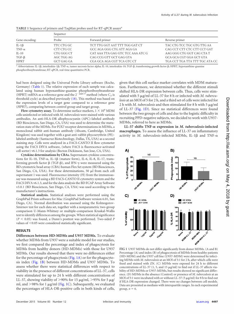
RESULTSDifferences between HD-MDMs and U937 MDMs. To evaluatewhether MDMs from U937 were a suitable model for our studies,we first compared the percentage and index of phagocytosis forMDMs from healthy donors (HD-MDMs) with those for U937MDMs. Our results showed that there were no differences eitherfor the percentage of phagocytosis (Fig. 1A) or for the phagocyto-sis index (Fig. 1B) between HD-MDMs and U937 MDMs. Toassess whether there were statistical differences with respect toviability in the presence of different concentrations of LL-37, cellswere stimulated for up to 24 h with different concentrations ofLL-37, showing viability of 90% for 15 �g/ml, 95% for 5 �g/ml, and 98% for 1 �g/ml (Fig. 1C). Subsequently, we evaluatedthe percentages of HLA-DR-positive cells in both kinds of cells,
given that this cell surface marker correlates with MDM matura-tion. Furthermore, we determined whether the different stimulishifted HLA-DR expression between cells. Thus, cells were stim-ulated with 5 �g/ml of LL-37 for 8 h or infected with M. tubercu-losis at an MOI of 5 for 2 h, and a third set of cells were infected for2 h with M. tuberculosis and then stimulated for 8 h with 5 �g/mlof LL-37 (Fig. 1D). Since no statistical differences were foundbetween the two groups of cells and due to the logistic difficulty inrecruiting PPD-negative subjects, we decided to work with U937-MDMs, referred to here as MDMs.
LL-37 shifts TNF-� expression in M. tuberculosis-infectedmacrophages. To assess the influence of LL-37 on inflammatoryactivity in M. tuberculosis-infected MDMs, IL-1� and TNF-�
TABLE 1 Sequence of primers and TaqMan probes used for RT-qPCR assaysa
Gene encoding:
Sequence
Probe Forward primer Reverse primer
IL-1� TTC CTG GC TCT TTG GGT AAT TTT TGG GAT CT TAC CTG TCC TGC GTG TTG AATNF-� CTT CTG CC GCC AGA GGG CTG ATT AGA GA CAG CCT CTT CTC CTT CCT GATIL-10 CTG GGG CT CAT AAA TTA GAG GTC TCC AAA ATC G AAG GGG CTG GGT CAG CTA TTGF-� AGC TGG AG CAG CCG GTT GCT GAG GTA GCA GCA CGT GGA GCT GTAHPRT GCT GAG GA CGA GCA AGA CGT TCA GTC CT TGA CCT TGA TTA TTT TGC ATA CCa Abbreviations: IL-1�, interleukin-1�; TNF-�, tumor necrosis factor alpha; IL-10, interleukin-10; TGF-�, transforming growth factor �; HPRT, hypoxanthine-guaninephosphoribosyltransferase; RT-qPCR, real-time quantitative PCR.
FIG 1 U937 MDMs do not differ significantly from donor MDMs. (A and B)Percentage (A) and index (B) of phagocytosis of MDMs from healthy patients(HD-MDM) and the U937 cell line (U937-MDM) were determined by infect-ing MDMs with M. tuberculosis at an MOI of 5:1 for 2 h, after which cells werefixed and stained with ZN. (C) MDMs were exposed for 24 h to differentconcentrations of LL-37 (1, 5, and 15 �g/ml) to find out if LL-37 affects via-bility of HD-MDMs or U937-MDMs, but results showed no significant differ-ence. (D) MDMs in the absence (Control) or presence of M. tuberculosis at anMOI of 5:1 were incubated with or without LL-37 (5 �g/ml) for 8 h to find outif HLA-DR expression changed. There were no changes between cell models.Data are presented as medians with interquartile ranges. In each experimentalgroup, n � 6.
Activity of LL37 during M. tuberculosis Infection
December 2015 Volume 83 Number 12 iai.asm.org 4497Infection and Immunity
on May 4, 2018 by guest
http://iai.asm.org/
Dow
nloaded from
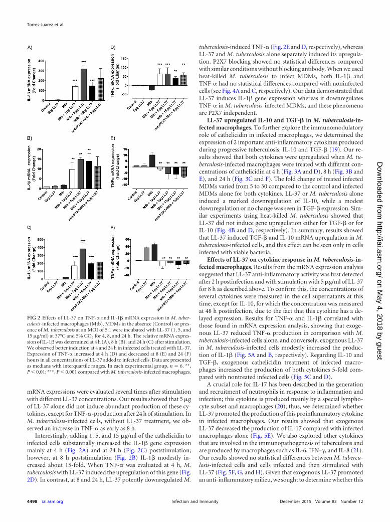
mRNA expressions were evaluated several times after stimulationwith different LL-37 concentrations. Our results showed that 5 �gof LL-37 alone did not induce abundant production of these cy-tokines, except for TNF-�-production after 24 h of stimulation. InM. tuberculosis-infected cells, without LL-37 treatment, we ob-served an increase in TNF-� as early as 8 h.
Interestingly, adding 1, 5, and 15 �g/ml of the cathelicidin toinfected cells substantially increased the IL-1� gene expressionmainly at 4 h (Fig. 2A) and at 24 h (Fig. 2C) poststimulation;however, at 8 h poststimulation (Fig. 2B) IL-1� modestly in-creased about 15-fold. When TNF-� was evaluated at 4 h, M.tuberculosis with LL-37 induced the upregulation of this gene (Fig.2D). In contrast, at 8 and 24 h, LL-37 potently downregulated M.
tuberculosis-induced TNF-� (Fig. 2E and D, respectively), whereasLL-37 and M. tuberculosis alone separately induced its upregula-tion. P2X7 blocking showed no statistical differences comparedwith similar conditions without blocking antibody. When we usedheat-killed M. tuberculosis to infect MDMs, both IL-1� andTNF-� had no statistical differences compared with noninfectedcells (see Fig. 4A and C, respectively). Our data demonstrated thatLL-37 induces IL-1� gene expression whereas it downregulatesTNF-� in M. tuberculosis-infected MDMs, and these phenomenaare P2X7 independent.
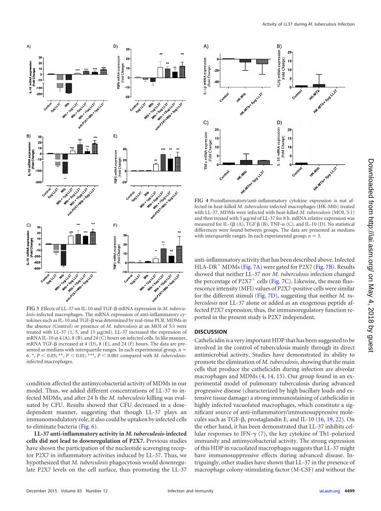
LL-37 upregulated IL-10 and TGF-� in M. tuberculosis-in-fected macrophages. To further explore the immunomodulatoryrole of cathelicidin in infected macrophages, we determined theexpression of 2 important anti-inflammatory cytokines producedduring progressive tuberculosis: IL-10 and TGF-� (19). Our re-sults showed that both cytokines were upregulated when M. tu-berculosis-infected macrophages were treated with different con-centrations of cathelicidin at 4 h (Fig. 3A and D), 8 h (Fig. 3B andE), and 24 h (Fig. 3C and F). The fold change of treated infectedMDMs varied from 5 to 30 compared to the control and infectedMDMs alone for both cytokines. LL-37 or M. tuberculosis aloneinduced a marked downregulation of IL-10, while a modestdownregulation or no change was seen in TGF-� expression. Sim-ilar experiments using heat-killed M. tuberculosis showed thatLL-37 did not induce gene upregulation either for TGF-� or forIL-10 (Fig. 4B and D, respectively). In summary, results showedthat LL-37 induced TGF-� and IL-10 mRNA upregulation in M.tuberculosis-infected cells, and this effect can be seen only in cellsinfected with viable bacteria.
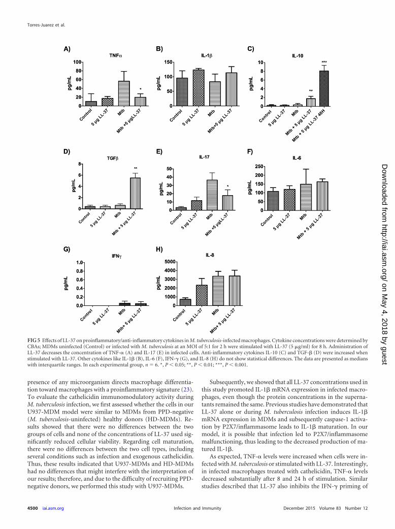
Effects of LL-37 on cytokine response in M. tuberculosis-in-fected macrophages. Results from the mRNA expression analysissuggested that LL-37 anti-inflammatory activity was first detectedafter 2 h postinfection and with stimulation with 5 �g/ml of LL-37for 8 h as described above. To confirm this, the concentrations ofseveral cytokines were measured in the cell supernatants at thistime, except for IL-10, for which the concentration was measuredat 48 h postinfection, due to the fact that this cytokine has a de-layed expression. Results for TNF-� and IL-1� correlated withthose found in mRNA expression analysis, showing that exoge-nous LL-37 reduced TNF-� production in comparison with M.tuberculosis-infected cells alone, and conversely, exogenous LL-37in M. tuberculosis-infected cells modestly increased the produc-tion of IL-1� (Fig. 5A and B, respectively). Regarding IL-10 andTGF-�, exogenous cathelicidin treatment of infected macro-phages increased the production of both cytokines 5-fold com-pared with nontreated infected cells (Fig. 5C and D).
A crucial role for IL-17 has been described in the generationand recruitment of neutrophils in response to inflammation andinfection; this cytokine is produced mainly by a special lympho-cyte subset and macrophages (20); thus, we determined whetherLL-37 promoted the production of this proinflammatory cytokinein infected macrophages. Our results showed that exogenousLL-37 decreased the production of IL-17 compared with infectedmacrophages alone (Fig. 5E). We also explored other cytokinesthat are involved in the immunopathogenesis of tuberculosis andare produced by macrophages such as IL-6, IFN-�, and IL-8 (21).Our results showed no statistical differences between M. tubercu-losis-infected cells and cells infected and then stimulated withLL-37 (Fig. 5F, G, and H). Given that exogenous LL-37 promotedan anti-inflammatory milieu, we sought to determine whether this
FIG 2 Effects of LL-37 on TNF-� and IL-1� mRNA expression in M. tuber-culosis-infected macrophages (Mtb). MDMs in the absence (Control) or pres-ence of M. tuberculosis at an MOI of 5:1 were incubated with LL-37 (1, 5, and15 �g/ml) at 37°C and 5% CO2 for 4, 8, and 24 h. The relative mRNA expres-sion of IL-1� was determined at 4 h (A), 8 h (B), and 24 h (C) after stimulation.We observed better induction at 4 and 24 h in infected cells treated with LL-37.Expression of TNF-� increased at 4 h (D) and decreased at 8 (E) and 24 (F)hours in all concentrations of LL-37 added to infected cells. Data are presentedas medians with interquartile ranges. In each experimental group, n � 6. **,P � 0.01; ***, P � 0.001 compared with M. tuberculosis-infected macrophages.
Torres-Juarez et al.
4498 iai.asm.org December 2015 Volume 83 Number 12Infection and Immunity
on May 4, 2018 by guest
http://iai.asm.org/
Dow
nloaded from
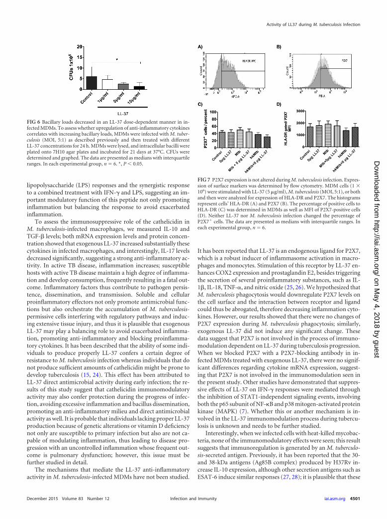
condition affected the antimycobacterial activity of MDMs in ourmodel. Thus, we added different concentrations of LL-37 to in-fected MDMs, and after 24 h the M. tuberculosis killing was eval-uated by CFU. Results showed that CFU decreased in a dose-dependent manner, suggesting that though LL-37 plays animmunomodulatory role, it also could be uptaken by infected cellsto eliminate bacteria (Fig. 6).
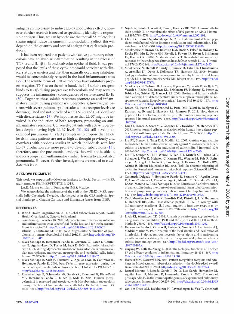
LL-37 anti-inflammatory activity in M. tuberculosis-infectedcells did not lead to downregulation of P2X7. Previous studieshave shown the participation of the nucleotide scavenging recep-tor P2X7 in inflammatory activities induced by LL-37. Thus, wehypothesized that M. tuberculosis phagocytosis would downregu-late P2X7 levels on the cell surface, thus promoting the LL-37
anti-inflammatory activity that has been described above. InfectedHLA-DR MDMs (Fig. 7A) were gated for P2X7 (Fig. 7B). Resultsshowed that neither LL-37 nor M. tuberculosis infection changedthe percentage of P2X7 cells (Fig. 7C). Likewise, the mean fluo-rescence intensity (MFI) values of P2X7-positive cells were similarfor the different stimuli (Fig. 7D), suggesting that neither M. tu-berculosis nor LL-37 alone or added as an exogenous peptide af-fected P2X7 expression; thus, the immunoregulatory function re-ported in the present study is P2X7 independent.
DISCUSSION
Cathelicidin is a very important HDP that has been suggested to beinvolved in the control of tuberculosis mainly through its directantimicrobial activity. Studies have demonstrated its ability topromote the elimination of M. tuberculosis, showing that the maincells that produce the cathelicidin during infection are alveolarmacrophages and MDMs (4, 14, 15). Our group found in an ex-perimental model of pulmonary tuberculosis during advancedprogressive disease (characterized by high bacillary loads and ex-tensive tissue damage) a strong immunostaining of cathelicidin inhighly infected vacuolated macrophages, which constitute a sig-nificant source of anti-inflammatory/immunosuppressive mole-cules such as TGF-�, prostaglandin E, and IL-10 (16, 19, 22). Onthe other hand, it has been demonstrated that LL-37 inhibits cel-lular responses to IFN-� (7), the key cytokine of Th1-polarizedimmunity and antimycobacterial activity. The strong expressionof this HDP in vacuolated macrophages suggests that LL-37 mighthave immunosuppressive effects during advanced disease. In-triguingly, other studies have shown that LL-37 in the presence ofmacrophage colony-stimulating factor (M-CSF) and without the
FIG 3 Effects of LL-37 on IL-10 and TGF-� mRNA expression in M. tubercu-losis-infected macrophages. The mRNA expression of anti-inflammatory cy-tokines such as IL-10 and TGF-� was determined by real-time PCR. MDMs inthe absence (Control) or presence of M. tuberculosis at an MOI of 5:1 weretreated with LL-37 (1, 5, and 15 �g/ml). LL-37 increased the expression ofmRNA IL-10 at 4 (A), 8 (B), and 24 (C) hours on infected cells. In like manner,mRNA TGF-� increased at 4 (D), 8 (E), and 24 (F) hours. The data are pre-sented as medians with interquartile ranges. In each experimental group, n �6. *, P � 0.05; **, P � 0.01; ***, P � 0.001 compared with M. tuberculosis-infected macrophages.
FIG 4 Proinflammatory/anti-inflammatory cytokine expression is not af-fected in heat-killed M. tuberculosis-infected macrophages (HK-Mtb) treatedwith LL-37. MDMs were infected with heat-killed M. tuberculosis (MOI, 5:1)and then treated with 5 �g/ml of LL-37 for 8 h. mRNA relative expression wasmeasured for IL-1� (A), TGF � (B), TNF-� (C), and IL-10 (D). No statisticaldifferences were found between groups. The data are presented as medianswith interquartile ranges. In each experimental group, n � 3.
Activity of LL37 during M. tuberculosis Infection
December 2015 Volume 83 Number 12 iai.asm.org 4499Infection and Immunity
on May 4, 2018 by guest
http://iai.asm.org/
Dow
nloaded from
presence of any microorganism directs macrophage differentia-tion toward macrophages with a proinflammatory signature (23).To evaluate the cathelicidin immunomodulatory activity duringM. tuberculosis infection, we first assessed whether the cells in ourU937-MDM model were similar to MDMs from PPD-negative(M. tuberculosis-uninfected) healthy donors (HD-MDMs). Re-sults showed that there were no differences between the twogroups of cells and none of the concentrations of LL-37 used sig-nificantly reduced cellular viability. Regarding cell maturation,there were no differences between the two cell types, includingseveral conditions such as infection and exogenous cathelicidin.Thus, these results indicated that U937-MDMs and HD-MDMshad no differences that might interfere with the interpretation ofour results; therefore, and due to the difficulty of recruiting PPD-negative donors, we performed this study with U937-MDMs.
Subsequently, we showed that all LL-37 concentrations used inthis study promoted IL-1� mRNA expression in infected macro-phages, even though the protein concentrations in the superna-tants remained the same. Previous studies have demonstrated thatLL-37 alone or during M. tuberculosis infection induces IL-1�mRNA expression in MDMs and subsequently caspase-1 activa-tion by P2X7/inflammasome leads to IL-1� maturation. In ourmodel, it is possible that infection led to P2X7/inflammasomemalfunctioning, thus leading to the decreased production of ma-tured IL-1�.
As expected, TNF-� levels were increased when cells were in-fected with M. tuberculosis or stimulated with LL-37. Interestingly,in infected macrophages treated with cathelicidin, TNF-� levelsdecreased substantially after 8 and 24 h of stimulation. Similarstudies described that LL-37 also inhibits the IFN-� priming of
FIG 5 Effects of LL-37 on proinflammatory/anti-inflammatory cytokines in M. tuberculosis-infected macrophages. Cytokine concentrations were determined byCBAs; MDMs uninfected (Control) or infected with M. tuberculosis at an MOI of 5:1 for 2 h were stimulated with LL-37 (5 �g/ml) for 8 h. Administration ofLL-37 decreases the concentration of TNF-� (A) and IL-17 (E) in infected cells. Anti-inflammatory cytokines IL-10 (C) and TGF-� (D) were increased whenstimulated with LL-37. Other cytokines like IL-1� (B), IL-6 (F), IFN-� (G), and IL-8 (H) do not show statistical differences. The data are presented as medianswith interquartile ranges. In each experimental group, n � 6. *, P � 0.05; **, P � 0.01; ***, P � 0.001.
Torres-Juarez et al.
4500 iai.asm.org December 2015 Volume 83 Number 12Infection and Immunity
on May 4, 2018 by guest
http://iai.asm.org/
Dow
nloaded from
lipopolysaccharide (LPS) responses and the synergistic responseto a combined treatment with IFN-� and LPS, suggesting an im-portant modulatory function of this peptide not only promotinginflammation but balancing the response to avoid exacerbatedinflammation.
To assess the immunosuppressive role of the cathelicidin inM. tuberculosis-infected macrophages, we measured IL-10 andTGF-� levels; both mRNA expression levels and protein concen-tration showed that exogenous LL-37 increased substantially thesecytokines in infected macrophages, and interestingly, IL-17 levelsdecreased significantly, suggesting a strong anti-inflammatory ac-tivity. In active TB disease, inflammation increases; susceptiblehosts with active TB disease maintain a high degree of inflamma-tion and develop consumption, frequently resulting in a fatal out-come. Inflammatory factors thus contribute to pathogen persis-tence, dissemination, and transmission. Soluble and cellularproinflammatory effectors not only promote antimicrobial func-tions but also orchestrate the accumulation of M. tuberculosis-permissive cells interfering with regulatory pathways and induc-ing extensive tissue injury, and thus it is plausible that exogenousLL-37 may play a balancing role to avoid exacerbated inflamma-tion, promoting anti-inflammatory and blocking proinflamma-tory cytokines. It has been described that the ability of some indi-viduals to produce properly LL-37 confers a certain degree ofresistance to M. tuberculosis infection whereas individuals that donot produce sufficient amounts of cathelicidin might be prone todevelop tuberculosis (15, 24). This effect has been attributed toLL-37 direct antimicrobial activity during early infection; the re-sults of this study suggest that cathelicidin immunomodulatoryactivity may also confer protection during the progress of infec-tion, avoiding excessive inflammation and bacillus dissemination,promoting an anti-inflammatory milieu and direct antimicrobialactivity as well. It is probable that individuals lacking proper LL-37production because of genetic alterations or vitamin D deficiencynot only are susceptible to primary infection but also are not ca-pable of modulating inflammation, thus leading to disease pro-gression with an uncontrolled inflammation whose frequent out-come is pulmonary dysfunction; however, this issue must befurther studied in detail.
The mechanisms that mediate the LL-37 anti-inflammatoryactivity in M. tuberculosis-infected MDMs have not been studied.
It has been reported that LL-37 is an endogenous ligand for P2X7,which is a robust inducer of inflammasome activation in macro-phages and monocytes. Stimulation of this receptor by LL-37 en-hances COX2 expression and prostaglandin E2, besides triggeringthe secretion of several proinflammatory substances, such as IL-1�, IL-18, TNF-�, and nitric oxide (25, 26). We hypothesized thatM. tuberculosis phagocytosis would downregulate P2X7 levels onthe cell surface and the interaction between receptor and ligandcould thus be abrogated, therefore decreasing inflammation cyto-kines. However, our results showed that there were no changes ofP2X7 expression during M. tuberculosis phagocytosis; similarly,exogenous LL-37 did not induce any significant change. Thesedata suggest that P2X7 is not involved in the process of immuno-modulation dependent on LL-37 during tuberculosis progression.When we blocked P2X7 with a P2X7-blocking antibody in in-fected MDMs treated with exogenous LL-37, there were no signif-icant differences regarding cytokine mRNA expression, suggest-ing that P2X7 is not involved in the immunomodulation seen inthe present study. Other studies have demonstrated that suppres-sive effects of LL-37 on IFN-� responses were mediated throughthe inhibition of STAT1-independent signaling events, involvingboth the p65 subunit of NF-�B and p38 mitogen-activated proteinkinase (MAPK) (7). Whether this or another mechanism is in-volved in the LL-37 immunomodulation process during tubercu-losis is unknown and needs to be further studied.
Interestingly, when we infected cells with heat-killed mycobac-teria, none of the immunomodulatory effects were seen; this resultsuggests that immunoregulation is generated by an M. tuberculo-sis-secreted antigen. Previously, it has been reported that the 30-and 38-kDa antigens (Ag85B complex) produced by H37Rv in-crease IL-10 expression, although other secretion antigens such asESAT-6 induce similar responses (27, 28); it is plausible that these
FIG 6 Bacillary loads decreased in an LL-37 dose-dependent manner in in-fected MDMs. To assess whether upregulation of anti-inflammatory cytokinescorrelates with increasing bacillary loads, MDMs were infected with M. tuber-culosis (MOI, 5:1) as described previously and then treated with differentLL-37 concentrations for 24 h. MDMs were lysed, and intracellular bacilli wereplated onto 7H10 agar plates and incubated for 21 days at 37°C. CFUs weredetermined and graphed. The data are presented as medians with interquartileranges. In each experimental group, n � 6. *, P � 0.05.
FIG 7 P2X7 expression is not altered during M. tuberculosis infection. Expres-sion of surface markers was determined by flow cytometry. MDM cells (1 �106) were stimulated with LL-37 (5 �g/ml), M. tuberculosis (MOI, 5:1), or bothand then were analyzed for expression of HLA-DR and P2X7. The histogramsrepresent cells’ HLA-DR (A) and P2X7 (B). The percentage of positive cells toHLA-DR (C) was determined in MDMs as well as MFI of P2X7-positive cells(D). Neither LL-37 nor M. tuberculosis infection changed the percentage ofP2X7 cells. The data are presented as medians with interquartile ranges. Ineach experimental group, n � 6.
Activity of LL37 during M. tuberculosis Infection
December 2015 Volume 83 Number 12 iai.asm.org 4501Infection and Immunity
on May 4, 2018 by guest
http://iai.asm.org/
Dow
nloaded from
antigens are necessary to induce LL-37 modulatory effects; how-ever, further research is needed to specifically identify the respon-sible antigen. Thus, we can hypothesize that not all M. tuberculosisstrains might induce the same immunomodulatory effect; this willdepend on the quantity and sort of antigen that each strain pro-duces.
It has been reported that patients with active pulmonary tuber-culosis have an alveolar inflammation resulting in the release ofTNF-� and IL-1� in bronchoalveolar epithelial fluid. It was pro-posed that the levels of these cytokines would correlate with clin-ical status parameters and that their naturally occurring inhibitorswould be concomitantly released in the local inflammatory sites(29). The soluble forms of TNF-� receptors have inhibitory prop-erties against TNF-�; on the other hand, the IL-1 soluble receptorbinds to IL-1� during progressive tuberculosis and may serve tosuppress the inflammatory consequences of early-released IL-1�(30). Together, these soluble receptors promote an anti-inflam-matory milieu during pulmonary tuberculosis; however, in pa-tients with severe pulmonary tuberculosis these receptor levels aredownregulated and not correlated with TNF-� and IL-1� levels orwith disease status (29). We hypothesize that LL-37 might be in-volved in the induction of both receptors, promoting an anti-inflammatory response. Conversely, patients with active tubercu-losis despite having high LL-37 levels (31, 32) still develop anextended pneumonia; this fact prompts us to propose that LL-37levels in these patients are not as high as thought previously andcorrelates with previous studies in which individuals with lowLL-37 production are more prone to develop tuberculosis (15);therefore, once the pneumonia develops, they will not be able toinduce a proper anti-inflammatory milieu, leading to exacerbatedpneumonia. However, further investigations are needed to eluci-date this issue.
ACKNOWLEDGMENTS
This work was supported by Mexican Institute for Social Security—IMSS,grant number FIS/IMSS/PROT/G14/1318.
J.A.E.-M. is a Scholar of Fundación IMSS, Mexico.We acknowledge the assistance of the staff at the UIMZ-IMSS, espe-
cially Julio Castañeda-Delgado, who helped us in the CBA analysis. Spe-cial thanks go to Claudia Carranza and Esmeralda Juarez from INER.
REFERENCES1. World Health Organization. 2014. Global tuberculosis report. World
Health Organization, Geneva, Switzerland.2. Sasindran SJ, Torrelles JB. 2011. Mycobacterium tuberculosis infection
and inflammation: what is beneficial for the host and for the bacterium?Front Microbiol 2:2. http://dx.doi.org/10.3389/fmicb.2011.00002.
3. Ulrichs T, Kaufmann SH. 2006. New insights into the function of gran-ulomas in human tuberculosis. J Pathol 208:261–269. http://dx.doi.org/10.1002/path.1906.
4. Rivas-Santiago B, Hernandez-Pando R, Carranza C, Juarez E, Contre-ras JL, Aguilar-Leon D, Torres M, Sada E. 2008. Expression of catheli-cidin LL-37 during Mycobacterium tuberculosis infection in human alve-olar macrophages, monocytes, neutrophils, and epithelial cells. InfectImmun 76:935–941. http://dx.doi.org/10.1128/IAI.01218-07.
5. Rivas-Santiago B, Sada E, Tsutsumi V, Aguilar-Leon D, Contreras JL,Hernandez-Pando R. 2006. beta-Defensin gene expression during thecourse of experimental tuberculosis infection. J Infect Dis 194:697–701.http://dx.doi.org/10.1086/506454.
6. Rivas-Santiago B, Schwander SK, Sarabia C, Diamond G, Klein-PatelME, Hernandez-Pando R, Ellner JJ, Sada E. 2005. Human {beta}-defensin 2 is expressed and associated with Mycobacterium tuberculosisduring infection of human alveolar epithelial cells. Infect Immun 73:4505– 4511. http://dx.doi.org/10.1128/IAI.73.8.4505-4511.2005.
7. Nijnik A, Pistolic J, Wyatt A, Tam S, Hancock RE. 2009. Human catheli-cidin peptide LL-37 modulates the effects of IFN-gamma on APCs. J Immu-nol 183:5788–5798. http://dx.doi.org/10.4049/jimmunol.0901491.
8. Choi KY, Chow LN, Mookherjee N. 2012. Cationic host defence pep-tides: multifaceted role in immune modulation and inflammation. J In-nate Immun 4:361–370. http://dx.doi.org/10.1159/000336630.
9. Mookherjee N, Brown KL, Bowdish DM, Doria S, Falsafi R, Hokamp K,Roche FM, Mu R, Doho GH, Pistolic J, Powers JP, Bryan J, BrinkmanFS, Hancock RE. 2006. Modulation of the TLR-mediated inflammatoryresponse by the endogenous human host defense peptide LL-37. J Immu-nol 176:2455–2464. http://dx.doi.org/10.4049/jimmunol.176.4.2455.
10. Mookherjee N, Hamill P, Gardy J, Blimkie D, Falsafi R, ChikatamarlaA, Arenillas DJ, Doria S, Kollmann TR, Hancock RE. 2009. Systemsbiology evaluation of immune responses induced by human host defencepeptide LL-37 in mononuclear cells. Mol Biosyst 5:483– 496. http://dx.doi.org/10.1039/b813787k.
11. Mookherjee N, Wilson HL, Doria S, Popowych Y, Falsafi R, Yu JJ, Li Y,Veatch S, Roche FM, Brown KL, Brinkman FS, Hokamp K, Potter A,Babiuk LA, Griebel PJ, Hancock RE. 2006. Bovine and human catheli-cidin cationic host defense peptides similarly suppress transcriptional re-sponses to bacterial lipopolysaccharide. J Leukoc Biol 80:1563–1574. http://dx.doi.org/10.1189/jlb.0106048.
12. Brown KL, Poon GF, Birkenhead D, Pena OM, Falsafi R, Dahlgren C,Karlsson A, Bylund J, Hancock RE, Johnson P. 2011. Host defensepeptide LL-37 selectively reduces proinflammatory macrophage re-sponses. J Immunol 186:5497–5505. http://dx.doi.org/10.4049/jimmunol.1002508.
13. Lau YE, Rozek A, Scott MG, Goosney DL, Davidson DJ, Hancock RE.2005. Interaction and cellular localization of the human host defense pep-tide LL-37 with lung epithelial cells. Infect Immun 73:583–591. http://dx.doi.org/10.1128/IAI.73.1.583-591.2005.
14. Liu PT, Stenger S, Tang DH, Modlin RL. 2007. Cutting edge: vitaminD-mediated human antimicrobial activity against Mycobacterium tuber-culosis is dependent on the induction of cathelicidin. J Immunol 179:2060 –2063. http://dx.doi.org/10.4049/jimmunol.179.4.2060.
15. Liu PT, Stenger S, Li H, Wenzel L, Tan BH, Krutzik SR, Ochoa MT,Schauber J, Wu K, Meinken C, Kamen DL, Wagner M, Bals R, Stein-meyer A, Zugel U, Gallo RL, Eisenberg D, Hewison M, Hollis BW,Adams JS, Bloom BR, Modlin RL. 2006. Toll-like receptor triggering ofa vitamin D-mediated human antimicrobial response. Science 311:1770 –1773. http://dx.doi.org/10.1126/science.1123933.
16. Castaneda-Delgado J, Hernandez-Pando R, Serrano CJ, Aguilar-LeonD, Leon-Contreras J, Rivas-Santiago C, Mendez R, Gonzalez-Curiel I,Enciso-Moreno A, Rivas-Santiago B. 2010. Kinetics and cellular sourcesof cathelicidin during the course of experimental latent tuberculous infec-tion and progressive pulmonary tuberculosis. Clin Exp Immunol 161:542–550. http://dx.doi.org/10.1111/j.1365-2249.2010.04199.x.
17. Yu J, Mookherjee N, Wee K, Bowdish DM, Pistolic J, Li Y, RehaumeL, Hancock RE. 2007. Host defense peptide LL-37, in synergy withinflammatory mediator IL-1beta, augments immune responses bymultiple pathways. J Immunol 179:7684 –7691. http://dx.doi.org/10.4049/jimmunol.179.11.7684.
18. Livak KJ, Schmittgen TD. 2001. Analysis of relative gene expression datausing real-time quantitative PCR and the 2(-delta delta C(T)) method.Methods 25:402– 408. http://dx.doi.org/10.1006/meth.2001.1262.
19. Hernandez-Pando R, Orozco H, Arriaga K, Sampieri A, Larriva-Sahd J,Madrid-Marina V. 1997. Analysis of the local kinetics and localization ofinterleukin-1 alpha, tumour necrosis factor-alpha and transforminggrowth factor-beta, during the course of experimental pulmonary tuber-culosis. Immunology 90:607– 617. http://dx.doi.org/10.1046/j.1365-2567.1997.00193.x.
20. Ouyang W, Kolls JK, Zheng Y. 2008. The biological functions of T helper17 cell effector cytokines in inflammation. Immunity 28:454 – 467. http://dx.doi.org/10.1016/j.immuni.2008.03.004.
21. Hossain MM, Norazmi MN. 2013. Pattern recognition receptors and cyto-kines in Mycobacterium tuberculosis infection—the double-edged sword?Biomed Res Int 2013:179174. http://dx.doi.org/10.1155/2013/179174.
22. Rangel Moreno J, Estrada Garcia I, De La Luz Garcia Hernandez M,Aguilar Leon D, Marquez R, Hernandez Pando R. 2002. The role ofprostaglandin E2 in the immunopathogenesis of experimental pulmonarytuberculosis. Immunology 106:257–266. http://dx.doi.org/10.1046/j.1365-2567.2002.01403.x.
23. van der Does AM, Beekhuizen H, Ravensbergen B, Vos T, Ottenhoff
Torres-Juarez et al.
4502 iai.asm.org December 2015 Volume 83 Number 12Infection and Immunity
on May 4, 2018 by guest
http://iai.asm.org/
Dow
nloaded from
TH, van Dissel JT, Drijfhout JW, Hiemstra PS, Nibbering PH. 2010.LL-37 directs macrophage differentiation toward macrophages with aproinflammatory signature. J Immunol 185:1442–1449. http://dx.doi.org/10.4049/jimmunol.1000376.
24. Mily A, Rekha RS, Kamal SM, Akhtar E, Sarker P, Rahim Z, Gud-mundsson GH, Agerberth B, Raqib R. 2013. Oral intake of phenylbu-tyrate with or without vitamin D3 upregulates the cathelicidin LL-37 inhuman macrophages: a dose finding study for treatment of tuberculosis.BMC Pulm Med 13:23. http://dx.doi.org/10.1186/1471-2466-13-23.
25. Alves LA, Bezerra RJ, Faria RX, Ferreira LG, da Silva Frutuoso V. 2013.Physiological roles and potential therapeutic applications of the P2X7 re-ceptor in inflammation and pain. Molecules 18:10953–10972. http://dx.doi.org/10.3390/molecules180910953.
26. Elssner A, Duncan M, Gavrilin M, Wewers MD. 2004. A novel P2X7receptor activator, the human cathelicidin-derived peptide LL37, inducesIL-1 beta processing and release. J Immunol 172:4987– 4994. http://dx.doi.org/10.4049/jimmunol.172.8.4987.
27. Freeman S, Post FA, Bekker LG, Harbacheuski R, Steyn LM, Ryffel B,Connell ND, Kreiswirth BN, Kaplan G. 2006. Mycobacterium tubercu-losis H37Ra and H37Rv differential growth and cytokine/chemokine in-duction in murine macrophages in vitro. J Interferon Cytokine Res 26:27–33. http://dx.doi.org/10.1089/jir.2006.26.27.
28. Heuer M, Behlich AS, Lee JS, Ribechini E, Jo EK, Lutz MB. 2013. The30-kDa and 38-kDa antigens from Mycobacterium tuberculosis inducepartial maturation of human dendritic cells shifting CD4( ) T cell re-sponses towards IL-4 production. BMC Immunol 14:48. http://dx.doi.org/10.1186/1471-2172-14-48.
29. Tsao TC, Li L, Hsieh M, Liao S, Chang KS. 1999. Soluble TNF-alphareceptor and IL-1 receptor antagonist elevation in BAL in active pulmo-nary TB. Eur Respir J 14:490 – 495. http://dx.doi.org/10.1034/j.1399-3003.1999.14c03.x.
30. Granowitz EV, Clark BD, Mancilla J, Dinarello CA. 1991. Interleukin-1receptor antagonist competitively inhibits the binding of interleukin-1 tothe type II interleukin-1 receptor. J Biol Chem 266:14147–14150.
31. Gonzalez-Curiel I, Castaneda-Delgado J, Lopez-Lopez N, Araujo Z,Hernandez-Pando R, Gandara-Jasso B, Macias-Segura N, Enciso-Moreno A, Rivas-Santiago B. 2011. Differential expression of antimicro-bial peptides in active and latent tuberculosis and its relationship withdiabetes mellitus. Hum Immunol 72:656 – 662. http://dx.doi.org/10.1016/j.humimm.2011.03.027.
32. Zhan Y, Jiang L. 2015. Status of vitamin D, antimicrobial peptide cathe-licidin and T helper-associated cytokines in patients with diabetes mellitusand pulmonary tuberculosis. Exp Ther Med 9:11–16.
Activity of LL37 during M. tuberculosis Infection
December 2015 Volume 83 Number 12 iai.asm.org 4503Infection and Immunity
on May 4, 2018 by guest
http://iai.asm.org/
Dow
nloaded from