Liver trauma: WSES 2020 guidelines · Keywords: Liver trauma, Adult, Pediatric, Minor, Moderate,...
15
REVIEW Open Access Liver trauma: WSES 2020 guidelines Federico Coccolini 1* , Raul Coimbra 2 , Carlos Ordonez 3 , Yoram Kluger 4 , Felipe Vega 5 , Ernest E. Moore 6 , Walt Biffl 7 , Andrew Peitzman 8 , Tal Horer 9,36 , Fikri M. Abu-Zidan 10 , Massimo Sartelli 11 , Gustavo P. Fraga 12 , Enrico Cicuttin 1 , Luca Ansaloni 13 , Michael W. Parra 14 , Mauricio Millán 3 , Nicola DeAngelis 15 , Kenji Inaba 16 , George Velmahos 17 , Ron Maier 18 , Vladimir Khokha 19 , Boris Sakakushev 20 , Goran Augustin 21 , Salomone di Saverio 22 , Emanuil Pikoulis 23 , Mircea Chirica 24 , Viktor Reva 25 , Ari Leppaniemi 26 , Vassil Manchev 27 , Massimo Chiarugi 1 , Dimitrios Damaskos 28 , Dieter Weber 29 , Neil Parry 30 , Zaza Demetrashvili 31 , Ian Civil 32 , Lena Napolitano 33 , Davide Corbella 34 , Fausto Catena 35 and the WSES expert panel Abstract Liver injuries represent one of the most frequent life-threatening injuries in trauma patients. In determining the optimal management strategy, the anatomic injury, the hemodynamic status, and the associated injuries should be taken into consideration. Liver trauma approach may require non-operative or operative management with the intent to restore the homeostasis and the normal physiology. The management of liver trauma should be multidisciplinary including trauma surgeons, interventional radiologists, and emergency and ICU physicians. The aim of this paper is to present the World Society of Emergency Surgery (WSES) liver trauma management guidelines. Keywords: Liver trauma, Adult, Pediatric, Minor, Moderate, Severe, Classification, Guidelines, Surgery, Hemorrhage, Operative management, Non-operative management, Interventional, Radiology, Intensive care Background Liver trauma is one of the most common abdominal le- sions in severely injured trauma patients [1]. Diagnosis and treatment of hepatic trauma has evolved with the use of modern diagnostic and therapeutic tools [2–4]. Until two to three decades ago, most cases with blunt abdominal trauma and possible injury in parenchymat- ous organs were managed by exploratory laparotomy [5]. Several innovative multimodal approaches as EVTM (endovascular trauma and bleeding management) have allowed to greatly increase the likelihood of non- operative management (NOM) for selected patients. Nowadays, even borderline patients or transient re- sponder, without other indications for laparotomy, may be considered for NOM in selected and well-developed trauma centers. This advanced strategy necessitates a multidisciplinary approach to deal with the complexity of moderate and severe liver injury. The majority of pa- tients admitted with liver injuries have minor or moder- ate injuries (WSES I, II, III) (AAST-OIS I, II, or III) and are successfully treated by NOM. In contrast, one third of severe injuries (WSES IV, V) (AAST-OIS IV, V) allow for NOM [6]. In pediatric patients, NOM should be con- sidered the optimal management approach. In determin- ing the optimal treatment strategy, the anatomical description of liver lesions is fundamental but not suffi- cient. In fact, the decision whether patients need to be managed operatively or undergo NOM is based mainly on the hemodynamic status, associated injuries, and on the anatomical liver injury grade. The aim of this manuscript is to present the updated World Society of Emergency Surgery (WSES) liver trauma management guidelines. © The Author(s). 2020 Open Access This article is licensed under a Creative Commons Attribution 4.0 International License, which permits use, sharing, adaptation, distribution and reproduction in any medium or format, as long as you give appropriate credit to the original author(s) and the source, provide a link to the Creative Commons licence, and indicate if changes were made. The images or other third party material in this article are included in the article's Creative Commons licence, unless indicated otherwise in a credit line to the material. If material is not included in the article's Creative Commons licence and your intended use is not permitted by statutory regulation or exceeds the permitted use, you will need to obtain permission directly from the copyright holder. To view a copy of this licence, visit http://creativecommons.org/licenses/by/4.0/. The Creative Commons Public Domain Dedication waiver (http://creativecommons.org/publicdomain/zero/1.0/) applies to the data made available in this article, unless otherwise stated in a credit line to the data. * Correspondence: [email protected] 1 General, Emergency and Trauma Surgery Department, Pisa University Hospital, Via Paradisia 1, 56100 Pisa, Italy Full list of author information is available at the end of the article Coccolini et al. World Journal of Emergency Surgery (2020) 15:24 https://doi.org/10.1186/s13017-020-00302-7
Transcript of Liver trauma: WSES 2020 guidelines · Keywords: Liver trauma, Adult, Pediatric, Minor, Moderate,...
REVIEW Open Access
Liver trauma: WSES 2020 guidelinesFederico Coccolini1*, Raul Coimbra2, Carlos Ordonez3, Yoram Kluger4, Felipe Vega5, Ernest E. Moore6, Walt Biffl7,Andrew Peitzman8, Tal Horer9,36, Fikri M. Abu-Zidan10, Massimo Sartelli11, Gustavo P. Fraga12, Enrico Cicuttin1,Luca Ansaloni13, Michael W. Parra14, Mauricio Millán3, Nicola DeAngelis15, Kenji Inaba16, George Velmahos17,Ron Maier18, Vladimir Khokha19, Boris Sakakushev20, Goran Augustin21, Salomone di Saverio22, Emanuil Pikoulis23,Mircea Chirica24, Viktor Reva25, Ari Leppaniemi26, Vassil Manchev27, Massimo Chiarugi1, Dimitrios Damaskos28,Dieter Weber29, Neil Parry30, Zaza Demetrashvili31, Ian Civil32, Lena Napolitano33, Davide Corbella34,Fausto Catena35 and the WSES expert panel
Abstract
Liver injuries represent one of the most frequent life-threatening injuries in trauma patients. In determining theoptimal management strategy, the anatomic injury, the hemodynamic status, and the associated injuries should betaken into consideration. Liver trauma approach may require non-operative or operative management with theintent to restore the homeostasis and the normal physiology. The management of liver trauma should bemultidisciplinary including trauma surgeons, interventional radiologists, and emergency and ICU physicians. The aimof this paper is to present the World Society of Emergency Surgery (WSES) liver trauma management guidelines.
Keywords: Liver trauma, Adult, Pediatric, Minor, Moderate, Severe, Classification, Guidelines, Surgery, Hemorrhage,Operative management, Non-operative management, Interventional, Radiology, Intensive care
BackgroundLiver trauma is one of the most common abdominal le-sions in severely injured trauma patients [1]. Diagnosisand treatment of hepatic trauma has evolved with theuse of modern diagnostic and therapeutic tools [2–4].Until two to three decades ago, most cases with bluntabdominal trauma and possible injury in parenchymat-ous organs were managed by exploratory laparotomy [5].Several innovative multimodal approaches as EVTM(endovascular trauma and bleeding management) haveallowed to greatly increase the likelihood of non-operative management (NOM) for selected patients.Nowadays, even borderline patients or transient re-sponder, without other indications for laparotomy, maybe considered for NOM in selected and well-developed
trauma centers. This advanced strategy necessitates amultidisciplinary approach to deal with the complexityof moderate and severe liver injury. The majority of pa-tients admitted with liver injuries have minor or moder-ate injuries (WSES I, II, III) (AAST-OIS I, II, or III) andare successfully treated by NOM. In contrast, one thirdof severe injuries (WSES IV, V) (AAST-OIS IV, V) allowfor NOM [6]. In pediatric patients, NOM should be con-sidered the optimal management approach. In determin-ing the optimal treatment strategy, the anatomicaldescription of liver lesions is fundamental but not suffi-cient. In fact, the decision whether patients need to bemanaged operatively or undergo NOM is based mainlyon the hemodynamic status, associated injuries, and onthe anatomical liver injury grade.The aim of this manuscript is to present the updated
World Society of Emergency Surgery (WSES) livertrauma management guidelines.
© The Author(s). 2020 Open Access This article is licensed under a Creative Commons Attribution 4.0 International License,which permits use, sharing, adaptation, distribution and reproduction in any medium or format, as long as you giveappropriate credit to the original author(s) and the source, provide a link to the Creative Commons licence, and indicate ifchanges were made. The images or other third party material in this article are included in the article's Creative Commonslicence, unless indicated otherwise in a credit line to the material. If material is not included in the article's Creative Commonslicence and your intended use is not permitted by statutory regulation or exceeds the permitted use, you will need to obtainpermission directly from the copyright holder. To view a copy of this licence, visit http://creativecommons.org/licenses/by/4.0/.The Creative Commons Public Domain Dedication waiver (http://creativecommons.org/publicdomain/zero/1.0/) applies to thedata made available in this article, unless otherwise stated in a credit line to the data.
* Correspondence: [email protected], Emergency and Trauma Surgery Department, Pisa UniversityHospital, Via Paradisia 1, 56100 Pisa, ItalyFull list of author information is available at the end of the article
Coccolini et al. World Journal of Emergency Surgery (2020) 15:24 https://doi.org/10.1186/s13017-020-00302-7
Notes on the use of the guidelinesThe guidelines are evidence-based, with the grade of rec-ommendation based on the evidence. The guidelinespresent the diagnostic and therapeutic methods for opti-mal management of liver trauma. The practice guide-lines promulgated in this work do not represent astandard of practice. These are suggested plans of care,based on best available evidence and the consensus ofexperts, but they do not exclude other approaches as be-ing within the standard of practice. For example, theyshould not be used to compel adherence to a givenmethod of medical management, which method shouldbe finally determined after taking account of the condi-tions at the relevant medical institution (staff levels, ex-perience, equipment, etc.), and the characteristics of theindividual patient. However, responsibility for the resultsof treatment rests with those who are directly engagedtherein, and not with the consensus group.
MethodsA computerized search was done by the bibliographer indifferent databanks (MEDLINE, Scopus, EMBASE).Citations were included for the period between January1990 and October 2019 using the primary search strat-egy: liver, injuries, trauma, hepatic, adult, pediatric,hemodynamic instability/stability, angioembolization,management, nonoperative, conservative, operative, sur-gery, diagnosis, and follow-up, combined with AND/OR.No search restrictions were imposed. The dates were se-lected to allow comprehensive published abstracts ofclinical trials, consensus conference, comparative studies,congresses, guidelines, government publication, multi-center studies, systematic reviews, meta-analysis, largecase series, original articles, and randomized controlledtrials. Case reports and small case series were excluded.Narrative review articles were also analyzed to determineif other cited studies should be included.The level of evidence (LE) was evaluated using the
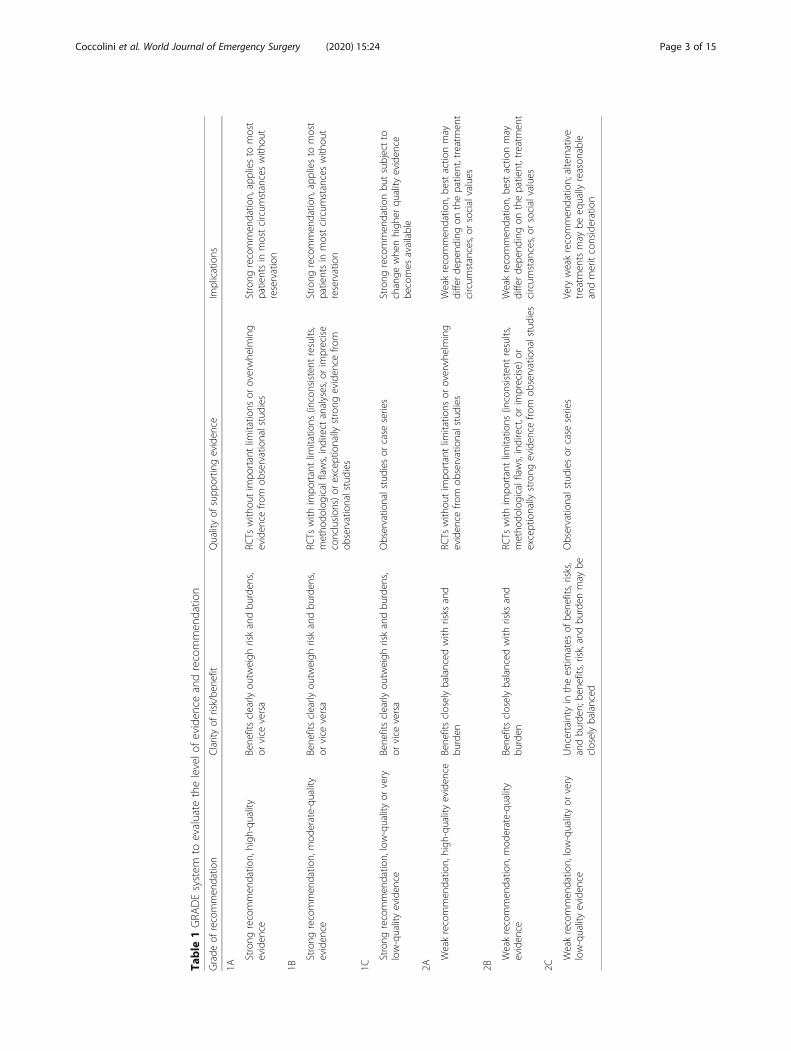
GRADE system [7] (Table 1).A group of experts in the field coordinated by a cen-
tral coordinator was contacted to express their evidence-based opinion on several issues about the pediatric (<16 years old) and adult liver trauma [8, 9]. Hepatictrauma was assessed by the anatomy of the injury, typeof injury (blunt and penetrating injury), management(conservative and operative management), and type ofpatient (adults, pediatrics). Through the Delphi process,different issues were discussed in subsequent rounds.The central coordinator assembled the different answersderived from each round. Each version was then revisedand improved. An expert group discussed the definitiveversion. The final version about on agreement wasreached resulted in the present manuscript. Statementsare summarized in Table 4.
DefinitionsIn adult patients, hemodynamic instability is consideredthe condition in which admission systolic blood pressureis < 90mmHg with clinical evidence of hemorrhagicshock with skin vasoconstriction (cool, clammy, de-creased capillary refill), altered level of consciousnessand/or shortness of breath, or > 90 mmHg but requiringbolus infusions/transfusions and/or vasopressor drugsand/or admission base excess (BE) > -5 mmol/l or trans-fusion requirement of at least > 4 units of packed redblood cells within the first 8 h. Transient responder pa-tients (adult and pediatric) are those showing an initialresponse to adequate fluid resuscitation, but then subse-quent signs of ongoing blood loss and perfusion deficits.These patients have an initial response to therapy but donot reach sufficient stabilization to undergo endovascu-lar procedures or NOM.In pediatric patients, hemodynamic stability is consid-
ered a systolic blood pressure of 70 mmHg plus twicethe child’s age in years. An acceptable hemodynamic sta-tus in children is considered a positive response to fluidresuscitation: 2 boluses of 20 mL/kg of crystalloid re-placement should be administered before blood replace-ment leading to heart rate reduction, cleared sensorium,return of peripheral pulses, normal skin color, increasein blood pressure and urinary output, and an increase inwarmth of the skin in the extremities. Clinical judgmenthowever is fundamental in evaluating pediatric patients.
WSES classificationThe WSES classification (Table 2) divides liver injuriesinto four classes considering the AAST-OIS classifica-tion (Table 3) and the hemodynamic status (Table 4):
� Minor (WSES grade I)� Moderate (WSES grade II)� Severe (WSES grade III and IV)
Minor hepatic injuries:
� WSES grade I includes AAST-OIS grade I–IIhemodynamically stable lesions.
Moderate hepatic injuries:
� WSES grade II includes AAST-OIS grade IIIhemodynamically stable lesions.
Severe hepatic injuries:
� WSES grade III includes AAST-OIS grade IV–Vhemodynamically stable lesions.
� WSES grade IV includes AAST-OIS grade I–VIhemodynamically unstable lesions.
Coccolini et al. World Journal of Emergency Surgery (2020) 15:24 Page 2 of 15
Table
1GRA
DEsystem
toevaluate
thelevelo
feviden
ceandrecommen
datio
n
Grade
ofrecommen
datio
nClarityof
risk/be
nefit
Qualityof
supp
ortin
geviden
ceIm
plications
1A
Strong
recommen
datio
n,high
-quality
eviden
ceBene
fitsclearly
outw
eigh
riskandbu
rden
s,or
vice
versa
RCTs
with
outim
portantlim
itatio
nsor
overwhe
lming
eviden
cefro
mob
servationalstudies
Strong
recommen
datio
n,appliesto
most
patientsin
mostcircum
stanceswith
out
reservation
1B
Strong
recommen
datio
n,mod
erate-qu
ality
eviden
ceBene
fitsclearly
outw
eigh
riskandbu
rden
s,or
vice
versa
RCTs
with
impo
rtantlim
itatio
ns(inconsistent
results,
metho
dologicalflaws,indirect
analyses,orim
precise
conclusion
s)or
exceptionally
strong
eviden
cefro
mob
servationalstudies
Strong
recommen
datio
n,appliesto
most
patientsin
mostcircum
stanceswith
out
reservation
1C
Strong
recommen
datio
n,low-qualityor
very
low-qualityeviden
ceBene
fitsclearly
outw
eigh
riskandbu
rden
s,or
vice
versa
Observatio
nalstudies
orcase
series
Strong
recommen
datio
nbu
tsubjectto
change
whe
nhigh
erqu
ality
eviden
cebe
comes
available
2A
Weakrecommen
datio
n,high
-qualityeviden
ceBene
fitscloselybalanced
with
risks
and
burden
RCTs
with
outim
portantlim
itatio
nsor
overwhe
lming
eviden
cefro
mob
servationalstudies
Weakrecommen
datio
n,be
stactio
nmay
differde
pend
ingon
thepatient,treatmen
tcircum
stances,or
socialvalues
2B
Weakrecommen
datio
n,mod
erate-qu
ality
eviden
ceBene
fitscloselybalanced
with
risks
and
burden
RCTs
with
impo
rtantlim
itatio
ns(inconsistent
results,
metho
dologicalflaws,indirect,orim
precise)
orexceptionally
strong
eviden
cefro
mob
servationalstudies
Weakrecommen
datio
n,be
stactio
nmay
differde
pend
ingon
thepatient,treatmen
tcircum
stances,or
socialvalues
2C
Weakrecommen
datio
n,low-qualityor
very
low-qualityeviden
ceUncertainty
intheestim
ates
ofbe
nefits,risks,
andbu
rden
;ben
efits,risk,andbu
rden
may
becloselybalanced
Observatio
nalstudies
orcase
series
Very
weakrecommen
datio
n;alternative
treatm
entsmay
beeq
ually
reason
able
andmeritconsideration
Coccolini et al. World Journal of Emergency Surgery (2020) 15:24 Page 3 of 15

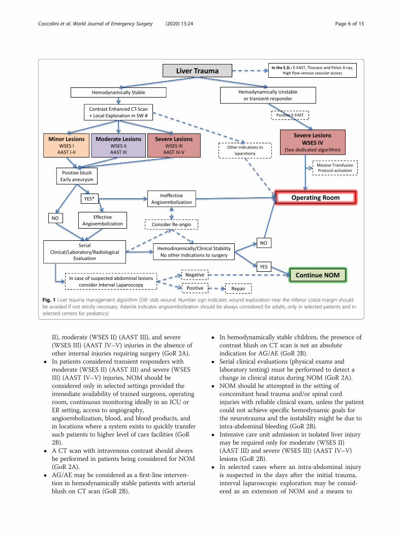
Based on the present classification, we suggest twomanagement algorithms: one general (Fig. 1) and onespecifically dedicated to hemodynamically unstable pa-tients (Fig. 2).
Diagnosis
� The diagnostic methods on admission aredetermined by the hemodynamic status (GoR 1A).
� Extended-focused abdominal sonography for trauma(E-FAST) is rapid in detecting intra-abdominal freefluid (GoR 1A).
� CT scan with intravenous contrast is the goldstandard in hemodynamically stable trauma patients(GoR 1A).
Careful physical examination is of paramount import-ance in determining the need for exploratory laparotomy[10]. E-FAST is useful and generally reliable in traumain general. However, abdominal ultrasound may be
falsely negative due to clotted blood or suboptimal qual-ity views [11–13]. In the pediatric population, reportedsensitivity and specificity ranges from 42 to 52% and 96to 98%, with a negative predicting value for intra-abdominal fluid of 93–96% [8, 9, 14–16]. The low sensi-tivity of E-FAST in hemodynamically stable pediatric pa-tients may warrant further investigation, specificallycontrast-enhanced ultrasound (US) or abdomen/pelvisCT scan or magnetic resonance, in hemodynamicallystable pediatric patients with a high degree of suspicionfor intra-abdominal injury (abnormal physical examin-ation, abnormal laboratory values, or other radiologicstudies).Computed tomography (CT) scan is considered the
gold standard in trauma imaging assessment with a sen-sitivity and specificity approaching 96–100% [17–19].CT must be immediately available and performed onlyin hemodynamically stable or stabilized patients or inthose who transiently responded to fluid resuscitation inspecial circumstances and under the supervision of thetrauma team [20, 21]. Delayed-phase CT helps in differ-entiating patients with active bleeding from those withcontained vascular injuries [22]. This data is importantto reduce the risk of discrepancy between CT scan im-ages and angiographic images (only 47% of patients havea confirmation of the CT findings at angiography) [22].Active contrast extravasation is a sign of activehemorrhage [23]. CT scan may help in subsequent
Table 3 AAST liver trauma classification
Table 2 WSES liver trauma classification
WSES grade AAST Hemodynamic
Minor WSES grade I I–II Stable
Moderate WSES grade II III Stable
Severe WSES grade III IV–V Stable
WSES grade IV I–VI Unstable
Coccolini et al. World Journal of Emergency Surgery (2020) 15:24 Page 4 of 15
surgical procedures and angiography/angioembolization(AG/AE) [24–32].Diagnostic peritoneal lavage (DPL) should be consid-
ered diagnostic modality in low-resource settings, whereCT scan or US is not promptly available [33]. It shouldbe considered in the presence of massive subcutaneousemphysema in a shocked patient in whom ultrasoundcannot be done and/or in the presence of free peritoneal
fluid without solid organ injury in a hemodynamicallystable patient. The possibility of DPL-related complica-tions (up to 2%) should be considered [33].
Non-operative management
� NOM should be the treatment of choice for allhemodynamically stable minor (WSES I) (AAST I–
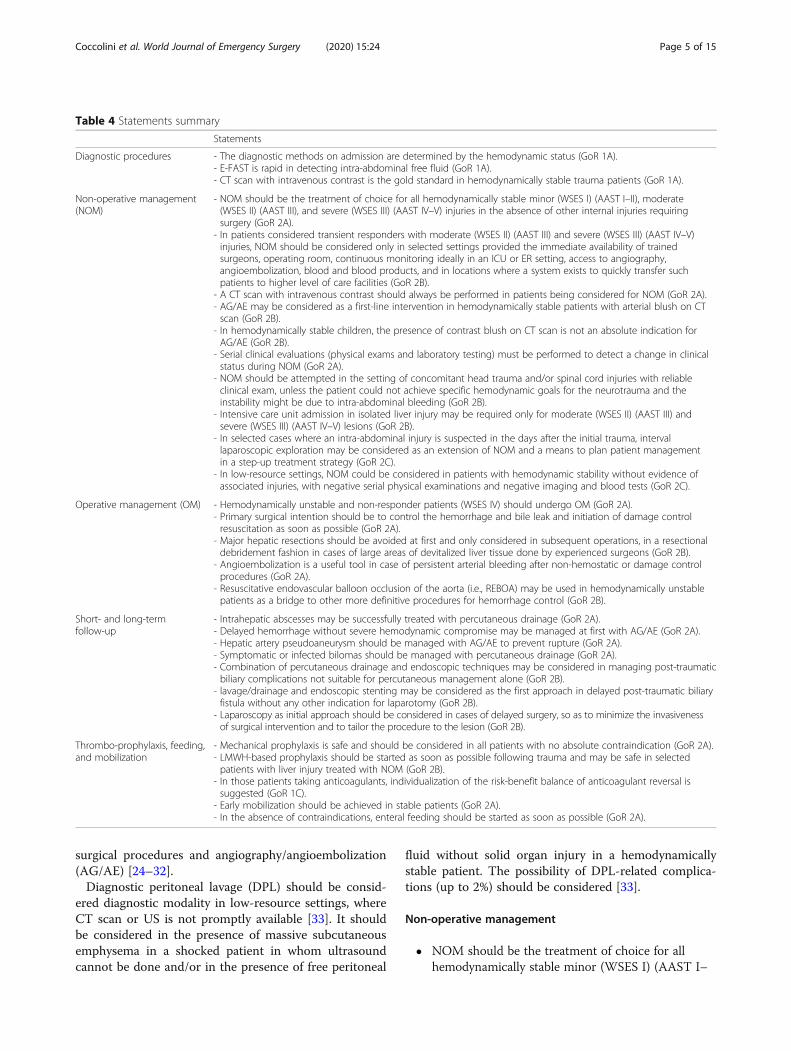
Table 4 Statements summary
Statements
Diagnostic procedures - The diagnostic methods on admission are determined by the hemodynamic status (GoR 1A).- E-FAST is rapid in detecting intra-abdominal free fluid (GoR 1A).- CT scan with intravenous contrast is the gold standard in hemodynamically stable trauma patients (GoR 1A).
Non-operative management(NOM)
- NOM should be the treatment of choice for all hemodynamically stable minor (WSES I) (AAST I–II), moderate(WSES II) (AAST III), and severe (WSES III) (AAST IV–V) injuries in the absence of other internal injuries requiringsurgery (GoR 2A).
- In patients considered transient responders with moderate (WSES II) (AAST III) and severe (WSES III) (AAST IV–V)injuries, NOM should be considered only in selected settings provided the immediate availability of trainedsurgeons, operating room, continuous monitoring ideally in an ICU or ER setting, access to angiography,angioembolization, blood and blood products, and in locations where a system exists to quickly transfer suchpatients to higher level of care facilities (GoR 2B).
- A CT scan with intravenous contrast should always be performed in patients being considered for NOM (GoR 2A).- AG/AE may be considered as a first-line intervention in hemodynamically stable patients with arterial blush on CTscan (GoR 2B).
- In hemodynamically stable children, the presence of contrast blush on CT scan is not an absolute indication forAG/AE (GoR 2B).
- Serial clinical evaluations (physical exams and laboratory testing) must be performed to detect a change in clinicalstatus during NOM (GoR 2A).
- NOM should be attempted in the setting of concomitant head trauma and/or spinal cord injuries with reliableclinical exam, unless the patient could not achieve specific hemodynamic goals for the neurotrauma and theinstability might be due to intra-abdominal bleeding (GoR 2B).
- Intensive care unit admission in isolated liver injury may be required only for moderate (WSES II) (AAST III) andsevere (WSES III) (AAST IV–V) lesions (GoR 2B).
- In selected cases where an intra-abdominal injury is suspected in the days after the initial trauma, intervallaparoscopic exploration may be considered as an extension of NOM and a means to plan patient managementin a step-up treatment strategy (GoR 2C).
- In low-resource settings, NOM could be considered in patients with hemodynamic stability without evidence ofassociated injuries, with negative serial physical examinations and negative imaging and blood tests (GoR 2C).
Operative management (OM) - Hemodynamically unstable and non-responder patients (WSES IV) should undergo OM (GoR 2A).- Primary surgical intention should be to control the hemorrhage and bile leak and initiation of damage controlresuscitation as soon as possible (GoR 2A).
- Major hepatic resections should be avoided at first and only considered in subsequent operations, in a resectionaldebridement fashion in cases of large areas of devitalized liver tissue done by experienced surgeons (GoR 2B).
- Angioembolization is a useful tool in case of persistent arterial bleeding after non-hemostatic or damage controlprocedures (GoR 2A).
- Resuscitative endovascular balloon occlusion of the aorta (i.e., REBOA) may be used in hemodynamically unstablepatients as a bridge to other more definitive procedures for hemorrhage control (GoR 2B).
Short- and long-termfollow-up
- Intrahepatic abscesses may be successfully treated with percutaneous drainage (GoR 2A).- Delayed hemorrhage without severe hemodynamic compromise may be managed at first with AG/AE (GoR 2A).- Hepatic artery pseudoaneurysm should be managed with AG/AE to prevent rupture (GoR 2A).- Symptomatic or infected bilomas should be managed with percutaneous drainage (GoR 2A).- Combination of percutaneous drainage and endoscopic techniques may be considered in managing post-traumaticbiliary complications not suitable for percutaneous management alone (GoR 2B).
- lavage/drainage and endoscopic stenting may be considered as the first approach in delayed post-traumatic biliaryfistula without any other indication for laparotomy (GoR 2B).
- Laparoscopy as initial approach should be considered in cases of delayed surgery, so as to minimize the invasivenessof surgical intervention and to tailor the procedure to the lesion (GoR 2B).
Thrombo-prophylaxis, feeding,and mobilization
- Mechanical prophylaxis is safe and should be considered in all patients with no absolute contraindication (GoR 2A).- LMWH-based prophylaxis should be started as soon as possible following trauma and may be safe in selectedpatients with liver injury treated with NOM (GoR 2B).
- In those patients taking anticoagulants, individualization of the risk-benefit balance of anticoagulant reversal issuggested (GoR 1C).
- Early mobilization should be achieved in stable patients (GoR 2A).- In the absence of contraindications, enteral feeding should be started as soon as possible (GoR 2A).
Coccolini et al. World Journal of Emergency Surgery (2020) 15:24 Page 5 of 15
II), moderate (WSES II) (AAST III), and severe(WSES III) (AAST IV–V) injuries in the absence ofother internal injuries requiring surgery (GoR 2A).
� In patients considered transient responders withmoderate (WSES II) (AAST III) and severe (WSESIII) (AAST IV–V) injuries, NOM should beconsidered only in selected settings provided theimmediate availability of trained surgeons, operatingroom, continuous monitoring ideally in an ICU orER setting, access to angiography,angioembolization, blood, and blood products, andin locations where a system exists to quickly transfersuch patients to higher level of care facilities (GoR2B).
� A CT scan with intravenous contrast should alwaysbe performed in patients being considered for NOM(GoR 2A).
� AG/AE may be considered as a first-line interven-tion in hemodynamically stable patients with arterialblush on CT scan (GoR 2B).
� In hemodynamically stable children, the presence ofcontrast blush on CT scan is not an absoluteindication for AG/AE (GoR 2B).
� Serial clinical evaluations (physical exams andlaboratory testing) must be performed to detect achange in clinical status during NOM (GoR 2A).
� NOM should be attempted in the setting ofconcomitant head trauma and/or spinal cordinjuries with reliable clinical exam, unless the patientcould not achieve specific hemodynamic goals forthe neurotrauma and the instability might be due tointra-abdominal bleeding (GoR 2B).
� Intensive care unit admission in isolated liver injurymay be required only for moderate (WSES II)(AAST III) and severe (WSES III) (AAST IV–V)lesions (GoR 2B).
� In selected cases where an intra-abdominal injuryis suspected in the days after the initial trauma,interval laparoscopic exploration may be consid-ered as an extension of NOM and a means to
Fig. 1 Liver trauma management algorithm (SW: stab wound. Number sign indicates wound exploration near the inferior costal margin shouldbe avoided if not strictly necessary. Asterisk indicates angioembolization should be always considered for adults, only in selected patients and inselected centers for pediatrics)
Coccolini et al. World Journal of Emergency Surgery (2020) 15:24 Page 6 of 15
plan patient management in a step-up treatmentstrategy (GoR 2C).
� In low-resource settings, NOM could be consideredin patients with hemodynamic stability without evi-dence of associated injuries, with negative serialphysical examinations and negative imaging andblood tests (GoR 2C).
Absolute requirements for NOM are hemodynamicstability and absence of other lesions requiring surgery[9, 15, 34–39]. In hemodynamically stable patients with-out other associated injuries requiring OM, NOM isconsidered the standard of care [8, 14, 15]. The conceptis valid for both: blunt (BT) and penetrating trauma(PT). Attempting NOM in moderate (WSES II) (AAST-OIS III) and severe (WSES III) (AAST-OIS IV–V) bluntor penetrating injuries requires the ability to diagnose allassociated injuries and to provide intensive management(continuous clinical monitoring, serial hemoglobin mon-itoring, and around-the-clock availability of trained
surgeons, CT scanning, angiography, OR, and blood andblood products) [16, 40–44].As a general consideration, great attention should be
paid in selecting PT for NOM especially in the case ofgunshot wound (GSW) and even more if thoraco-abdominal. They should be considered for NOM only incenters with experience in dealing with PT. Even in pa-tients presenting with stable conditions and with no evi-dence of other intra-abdominal/internal injuries, intervallaparoscopy should be always considered in order toconfirm the absence of other injuries requiring surgicalrepair.In PT, NOM feasibility has been reported [35–37, 45–
49] with 50% and 85% success rate of NOM for stabwounds (SW) in anterior and posterior abdomen re-spectively [34, 50]. Similar managing strategy can be ap-plied to GSWs [35, 45]. Necessary distinction betweenlow- and high-energy penetrating trauma however ismandatory when deciding for OM or NOM. Low-energyPT (SW and low-energy GSW) may be safely treated
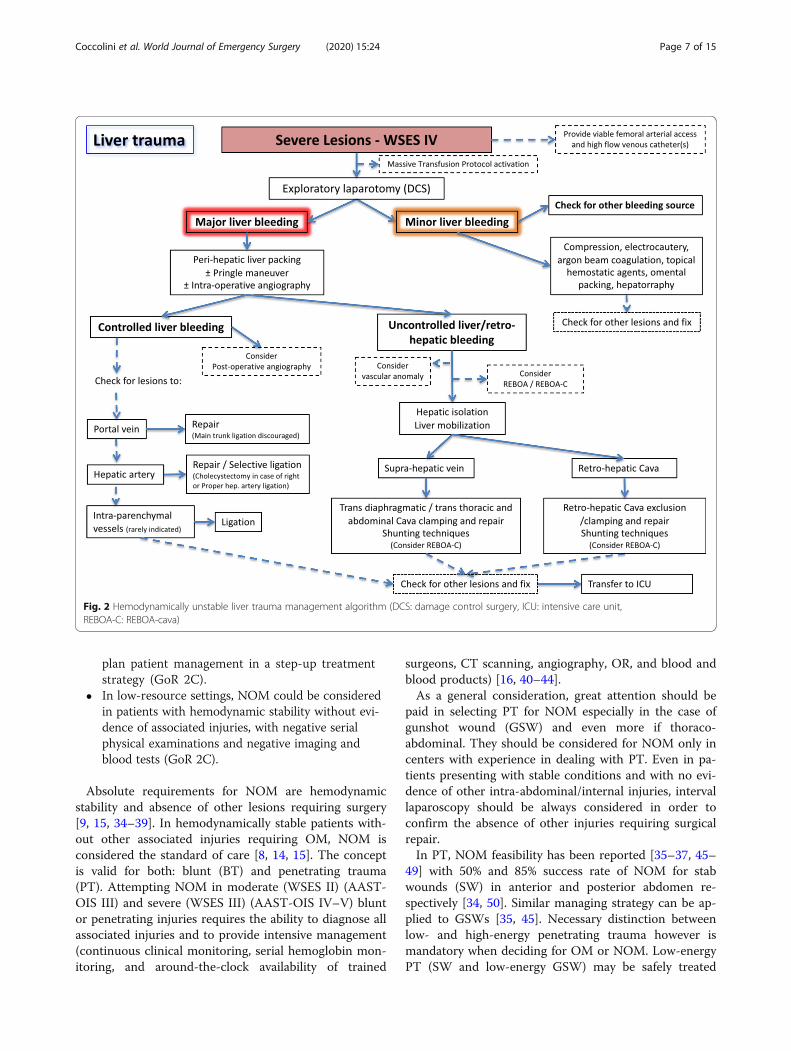
Fig. 2 Hemodynamically unstable liver trauma management algorithm (DCS: damage control surgery, ICU: intensive care unit,REBOA-C: REBOA-cava)
Coccolini et al. World Journal of Emergency Surgery (2020) 15:24 Page 7 of 15
with NOM at first, provided the patient ishemodynamically stable and no other injuries requiresurgery. In considering NOM, interval laparoscopyshould be considered to rule out missed intra-abdominalinjuries. High-energy GSW and other ballistic injuriesare less amenable to NOM, and in 90% of cases, OM isrequired [34, 36, 51]. In abdominal GSWs, up to 25% ofnon-therapeutic laparotomy has been reported [51], con-firming the need to have strict selection criteria for OMor NOM even in the GSW cohort. Associated head andspinal cord injuries (that preclude affordable clinicalexamination) and significant reduction in hemoglobinrequiring > 4 units of blood transfusion in the first 8 h[34, 45] have been suggested as predictive criteria ofNOM failure in abdominal GSWs.Patient selection is influenced by the diagnostic cap-
ability and accuracy. In fact, the accuracy of CT scanin SWs has been questioned [37, 50]. Even in thepresence of a negative CT scan, exploratory laparos-copy/laparotomy may be necessary [37]. Intervallaparoscopy is a useful tool to be considered in obesepatients or in the presence a long and tangentialwound tract or when the trajectory is difficult to de-termine on CT scan [34, 37]. In anterior abdominalSW, local wound exploration (LWE) is generally ac-curate in evaluating penetration depth; small externalwounds may be enlarged for precise LWE and deter-mination of anterior fascia violation [34, 35]. LWE,however, may be misleading, and patients should beadmitted for observation if equivocal. Wounds closeto the inferior costal margin should be evaluated byLWE with caution and only if strictly necessary.GSWs undergoing NOM may warrant a CT scan to
determine the trajectory [45, 51]. CT scan specificity andsensitivity of 96% and 90.5% respectively for GSWs re-quiring laparotomy have been reported [52]. The goldstandard to decide for OM or NOM remains the clinicalexamination [34, 51] associated with laboratory andradiological evaluation. Strict clinical and hemoglobinevaluation should be done (every 6 h for at least 24 h);after index CT scan allowing for NOM, serial ecoghra-phical evaluation may be utilized to help in defining pa-tient clinical evolution. Once stabilized, patients areusually transferred from ICU to the ward [35, 45, 50].NOM is contraindicated if free intra- or retro-
peritoneal air, free intra-peritoneal fluid in the absenceof solid organ injury, localized bowel wall thickening,bullet tract close to hollow viscus with surroundinghematoma [46], and in high-energy penetrating traumaare detected at CT scan.In selected centers, AE is considered as an “extension”
of NOM in patients with liver injuries presenting withongoing resuscitative needs [9, 53, 54]. If required, AEcan be safely repeated.
In children, the use of primary hepatic AE has been re-ported rarely and is debated even in the presence of ar-terial blush where it seems to increase NOM failurerates [55], or according to some studies, it does not cor-relate with decrease odds of laparotomy [30]. In thepediatric population, AE use is associated with older ageand is not completely defined in terms of efficacy andcost-effectiveness, especially in low-resource settings [30,55–61]. Some authors, however, identify the presence ofactive contrast extravasation as an independent predictorfor pseudoaneurysm (PSA) formation in children, re-gardless of injury grade. This suggests a thoroughfollow-up during NOM of these patients, so to obtain anearly identification and angiographic treatment of PSA[62].The biggest risk of NOM in penetrating trauma is a
missed abdominal injury, especially hollow viscus perfor-ation [34, 46]. However, no increase in mortality rateswith missed hollow viscus perforation has been reportedin patients without peritonitis on admission [63]. As acounterpart, non-therapeutic laparotomy leads to an in-crease in morbidity [63]. Moreover, OM in penetratingliver injuries has a higher liver-related complication rate(50–52%) compared to blunt injuries [34, 46].During NOM for liver injuries, no standard early
follow-up and monitoring protocols exist in adult or inchildren [34]. Serial clinical evaluation and hemoglobinmeasurement represent the cornerstone in evaluatingNOM patients [14]. Bedsides, US may represent an af-fordable tool during early follow-up. Presence of largesubcapsular hematomas is not a strict indication forOM, but a higher risk of NOM failure exists. In any case,these patients should undergo serial blood test: increas-ing levels of transaminases could indicate the presenceof intrahepatic parenchymal ischemia or rare cases oftorsion of suprahepatic veins [64]. ICU admission maybe indicated for moderate (WSES II) (AAST III) and se-vere (WSES III–IV) (AAST IV–V) liver trauma in orderto reduce the mortality risk [26].If available, interval laparoscopy during NOM provides
important information about the evolution of the injury.Laparoscopy should be considered an important tool inthe NOM of liver injuries, and it could be used as abridge strategy to plan an immediate or subsequent lap-aroscopic/laparotomy intervention [65].Particular attention should be paid in managing
hemodynamically stable patients with liver trauma as-sociated with spinal trauma (ST) and severe traumaticbrain injury (STBI). In blunt trauma, NOM shouldapply to all patients with no other indication to lapar-otomy. However, the optimal management of con-comitant STBI and/or ST and penetrating liverinjuries is debated and OM in general could be sug-gested as safer [45, 48, 66].
Coccolini et al. World Journal of Emergency Surgery (2020) 15:24 Page 8 of 15
Patients affected by neurotrauma (i.e., spinal cordor moderate-severe traumatic brain injury) in fact, forseveral instances, differ from the others because theyneed a higher perfusion pressure to adequately supplyoxygen to the brain and to the spinal cord to reducethe subsequent burden of disability and mortality. Adisruption of the normal blood flow regulation in thecentral nervous system (CNS) characterizes thetrauma and eventually leads to a blood flowdependent on perfusion pressure in ischemic tissue[67]. Specific hemodynamic goals for ST and STBIare defined as SBP > 110 mmHg and/or a CPP be-tween 60 and 70 mmHg in the case of moderate/se-vere TBI and an MBP > 80 mmHg in case of ST [68,69]. To date, no study specifically addressed theNOM of abdominal solid organ injuries in the neuro-trauma patient, and several authors have considered itan exclusion criterion from NOM [45, 48, 70]. How-ever, since the first goal is to have a stable patientwith adequate perfusion pressure, there is no rationalein denying NOM to these patients, as long as thespecific hemodynamic goals are met.
Operative management
� Hemodynamically unstable and non-responder pa-tients (WSES IV) should undergo OM (GoR 2A).
� Primary surgical intention should be to control thehemorrhage and bile leak and initiation of damagecontrol resuscitation as soon as possible (GoR 2A).
� Major hepatic resections should be avoided at firstand only considered in subsequent operations, in aresectional debridement fashion in cases of largeareas of devitalized liver tissue done by experiencedsurgeons (GoR 2B).
� Angioembolization is a useful tool in case ofpersistent arterial bleeding after non-hemostatic ordamage control procedures (GoR 2A).
� Resuscitative endovascular balloon occlusion of theaorta (i.e., REBOA) may be used inhemodynamically unstable patients as a bridge toother more definitive procedures for hemorrhagecontrol (GoR 2B).
At laparotomy, if no major bleeding is present, com-pression alone or electrocautery, bipolar devices, argonbeam coagulation, topical hemostatic agents, simple su-ture of the hepatic parenchyma, or omental patchingmay be sufficient to stop the bleeding [34, 66, 71–73].In case of major hemorrhage, more aggressive proce-
dures including manual compression and hepatic pack-ing, ligation of vessels in the wound, hepaticdebridement and finger fracture, balloon tamponade,shunting procedures, or hepatic vascular isolation and
exclusion may be used [64, 74]. Of paramount import-ance is to provide simultaneous intraoperative intensiveresuscitation with early institution of a massive transfu-sion protocol (MTP) aiming to maintain organ perfusionand ultimately reverse all trauma-induced physiologicalderangements [34, 71, 73, 75].In case of evident injury to the proper hepatic artery,
an attempt to control and repair it should be made. Ifnot effective or not possible, selective hepatic arteryligation should be considered as a viable option. If theinjury is on the right or left branches of the proper hep-atic artery, selective ligation is advisable. If the right orcommon hepatic artery must be ligated, cholecystectomyshould be performed to avoid gallbladder necrosis [2,76]. If the patient’s condition allows for it, post-operativeAE represents a viable alternative allowing hemorrhagecontrol while reducing complications [34, 66, 71, 77].Hepatic artery ligation increases the risk of hepatic ne-crosis, abscesses, and biloma formation [34].Portal vein injuries should be repaired primarily. Portal
vein main branch ligation should not be considered andshould be avoided because of the high risk of liver ne-crosis or massive bowel edema. If no other option exists,ligation can be used, but only in patients with an intacthepatic artery. Liver packing or liver resection should bepreferred to ligation in case of lobar or segmental/sub-segmental portal venous branch injuries [34, 76].Whenever Pringle maneuver or arterial control fails
and bleeding persists, the presence of an aberrant hep-atic artery should be considered. If the bleeding comesfrom behind the liver, retro-hepatic caval or hepatic veininjury should be highly suspected [34, 77]. Three viableoptions exist for the management of retrohepatic caval/suprahepatic venous injuries: (1) tamponade with hepaticpacking, (2) direct repair (with or without vascular isola-tion), and (3) lobar resection [38, 78–80]. Liver packingis the least risky method to temporarily deal with severevenous injuries [34, 66, 81–83]. Direct venous repair isdifficult especially in non-experienced hands, with highmortality rates [34, 66].Different techniques of hepatic vascular exclusion with
shunting procedures have been described, most of themanecdotally. The veno-veno bypass (femoral vein and in-ferior mesenteric vein to axillary or jugular vein by pass)and the use of fenestrated stent grafts are the most fre-quently used [66, 71, 76, 84]. The atrio-caval shunt by-passes the retro-hepatic cava blood through the rightatrium using a chest tube put into the inferior vena cava.Mortality rates in such a complicated situations are veryhigh and usually related to the fact that the decision toperform the shunt is made late in the case [71].Complete vascular exclusion of the liver is generallypoorly tolerated in the unstable patient with major bloodloss [34].
Coccolini et al. World Journal of Emergency Surgery (2020) 15:24 Page 9 of 15
Resuscitative endovascular balloon occlusion of theaorta (REBOA) catheter in zone I should be consideredif despite all damage control procedures, there is still ac-tive surgical bleeding. Simultaneously, the large highflow femoral venous catheter should be exchanged overa guide wire to an introducer with the aim of floating upand inflating a resuscitative endovascular balloon occlu-sion of the vena cava (REBOVC) at the level of theretro-hepatic vena cava. The goal is to achieve proximaland distal vascular control of a possible retro-hepatic/supra-hepatic vessel injury with the REBOVC and ultim-ately obtaining complete combined endovascular/openliver isolation with the Pringle maneuver. A supra-diaphragmatic central venous access must be obtainedprior to inflating the REBOA/REBOVC [85–91].In cases of liver avulsion or total crush injury, when a
total hepatic resection is indicated, hepatic transplant-ation has been described [76]. A retrospective studybased on the European Liver Transplant Registry identi-fies an ISS score less than 33 for recipient selection, soto avoid futile procedures [92].Anatomic hepatic resection may seldom be considered
as a surgical option [6, 93, 94]. In unstable patients andduring damage control surgery, it should be avoided, butin case of need, a non-anatomic resection is safer andeasier [34, 66, 71, 76]. For staged liver procedures, eitheranatomic or non-anatomic resections may be safely per-formed by experienced surgeons [76].Temporary abdominal closure may be indicated if the
risk of abdominal compartment syndrome is high or inthose situation where a “second look” operation isneeded [71–73].Two principal indications for post-operative
angiography-embolization (AG-AE) have been proposed:(1) after initial operative hemostasis, in stable or stabi-lized patients with contrast blush at completion CTscan; and (2) as adjunctive hemostatic tool in patientswith uncontrolled suspected arterial bleeding despiteemergency laparotomy and hemostasis attempt [34, 54,95–99]. Recent evidence suggests that routine use of im-mediate post-damage control hepatic angiography re-duces mortality in grade IV/V hepatic injuries [100].
Complications
� Intrahepatic abscesses may be successfully treatedwith percutaneous drainage (GoR 2A).
� Delayed hemorrhage without severe hemodynamiccompromise may be managed at first with AG/AE(GoR 2A).
� Hepatic artery pseudoaneurysm should be managedwith AG/AE to prevent rupture (GoR 2A).
� Symptomatic or infected bilomas should bemanaged with percutaneous drainage (GoR 2A).
� Combination of percutaneous drainage andendoscopic techniques may be considered inmanaging post-traumatic biliary complications notsuitable for percutaneous management alone (GoR2B).
� Laparoscopic lavage/drainage and endoscopicstenting may be considered as the first approach indelayed post-traumatic biliary fistula without anyother indication for laparotomy (GoR 2B).
� Laparoscopy as initial approach should beconsidered in cases of delayed surgery, so as tominimize the invasiveness of surgical interventionand to tailor the procedure to the lesion (GoR 2B).
In blunt hepatic trauma, particularly after high-gradeinjury, complications occur in 12–14% of patients [9,66]. Diagnostic tools for complications after NOM in-clude clinical examination, blood tests, ultrasound, andCT scan. Routine follow-up with CT scan is not neces-sary unless there is clinical suspicion of a complication[6, 9, 66]. In the presence of abnormal inflammatory re-sponse, abdominal pain, fever, jaundice, or drop ofhemoglobin level, repeated CT scan is recommended[9]. Bleeding, abdominal compartment syndrome, infec-tions (abscesses and other infections), biliary complica-tions (bile leak, hemobilia, biloma, biliary peritonitis,biliary fistula), and liver necrosis are the most frequentcomplications associated with NOM [16, 66]. Ultrasoundis useful in the assessment of bile leak/biloma in gradeIV–V injuries, especially with a central laceration.Re-bleeding or secondary hemorrhage is the most fre-
quently reported complications after NOM as in subcap-sular hematoma or pseudo-aneurysm (PSA) rupture(range 1.7–5.9%) with a mortality rate up to 18% [9, 66,101, 102]. In the majority of cases (69%), “late” bleedingcan be treated non-operatively [9, 66].Hepatic artery PSA is a rare complication with a
prevalence of 1% [103]. Asymptomatic PSA should betreated as early as possible with AE because of the highrisk of rupture and the associated high morbidity [34,104, 105]. In patients with melena or hematemesis fol-lowing liver trauma, bleeding from the ampulla of Vater(hemobilia) is highly suggestive of ruptured intrahepaticPSA [106, 107]. AE is the treatment of choice [6, 34, 66].In the presence of intrahepatic bilio-venous fistula (fre-quently associated with bilemia), endoscopic retrogradecholangiopancreatography (ERCP) represents an effect-ive tool [108].Biliary complications include biloma, biliary fistula, bil-
hemia, and bile peritonitis (incidence 2.8–30%) [8, 40].Most traumatic bilomas regress spontaneously. Enlar-ging, symptomatic or infected bilomas can be success-fully managed with percutaneous drainage. Percutaneousdrainage may be combined with therapeutic ERCP with
Coccolini et al. World Journal of Emergency Surgery (2020) 15:24 Page 10 of 15
eventual endobiliary stent placement [9, 101, 109–111].Bile peritonitis has been usually treated with laparotomy.Combination of laparoscopic irrigation/drainage andendoscopic bile duct stent placement may represent avalid alternative [101, 102, 112, 113].Abscesses are rare after NOM and usually happen in
severe lesions (prevalence 0.6–7%) [9, 66, 114–117]. CTscan or ultrasound-guided percutaneous drainage is thetreatment of choice with high success rate and no re-ported mortality [106]. In the presence of necrosis anddevascularization of hepatic segments, surgical manage-ment may be indicated whenever affecting patient condi-tion [34, 66].Generally, once stabilization of traumatized patient is
obtained, late complications should be managed prefer-entially by minimally invasive procedures. Laparoscopyand endoscopy are part of this approach, which becamepossible in a delayed surgery setting [64, 65, 118, 119].
Thromboprophylaxis, feeding, and mobilization
� Mechanical prophylaxis is safe and should beconsidered in all patients with no absolutecontraindication (GoR 2A).
� LMWH-based prophylaxis should be started as soonas possible following trauma and may be safe inselected patients with liver injury treated with NOM(GoR 2B).
� In those patients taking anticoagulants,individualization of the risk-benefit balance of anti-coagulant reversal is suggested (GoR 1C).
� Early mobilization should be achieved in stablepatients (GoR 2A).
� In the absence of contraindications, enteral feedingshould be started as soon as possible (GoR 2A).
Venous thromboembolism (VTE) is one of the greatrisks of trauma victims, because patients enter a hyper-coagulation state within 48 h from injury [120–122].More than 50% of patients without thrombo-prophylaxismay develop deep vein thrombosis (DVT) and subse-quent pulmonary embolism (PE) which carries a moral-ity rate up to 50% [120, 121]. PE is the third leadingcause of death in trauma patients.No differences in complication, mortality, and NOM
failure rate were demonstrated when thrombo-prophylaxis was administered within and after 48 and72 h from the initial injury in patients without STBI andBST [123–125]. Early mobilization is not related toNOM failure and secondary bleeding [126]. However,VTE rates seem to be over fourfold when LMWH is ad-ministered > 72 h from admission [120].In patients taking anticoagulants, it is important to
evaluate the eventual need for reversal therapy in order
to balance the risk of bleeding against the benefit of pre-venting thrombotic complications. Poor outcomes derivefrom the failure to restore the anticoagulation as soon aspossible [127].Early enteral feeding is associated with improved clin-
ical outcomes when administered within the first 72 hfrom admission in ICU [128], and it should be delayedonly in cases of uncontrolled shock, use of vasopressortherapy, uncontrolled hypoxaemia and acidosis, uncon-trolled upper GI bleeding, gastric aspirate > 500 ml/6 h,bowel ischemia, bowel obstruction, abdominal compart-ment syndrome, and high-output fistula without distalfeeding access [129]. Oral intake, when possible, shouldbe initiated after 24–48 h from the traumatic event.
Follow-upMandatory late follow-up imaging is not indicated, andit should be used only if the patient’s clinical conditionand/or symptoms indicating a complication require itfor diagnosis. The majority of liver lesions heal in about4 months [14, 66]. After moderate and severe liver injur-ies, patients may usually resume normal physical activ-ities after 3–4 months.During the recovery phase, patients should be encour-
aged to not remain alone for long periods and to returnimmediately to the hospital in case of increasing abdom-inal pain, lightheadedness, nausea, or vomiting [14, 34].
ConclusionsManagement of liver trauma is multidisciplinary. Whenfeasible, non-operative management should always beconsidered as the first option in adult and in thepediatric populations. For this reason, clinical condition,anatomical injury grade, and associated injuries shouldbe considered together in deciding the best treatmentoption.
AbbreviationsNOM: Non-operative management; OM: Operative management;AAST: American Association for Surgery for Trauma; WSES: World Society ofEmergency Surgery; PTS: Panamerican Trauma Society; ATLS: Advancedtrauma life support; ERCP: Endoscopic retrograde cholangiopancreatography;BLT: Blunt liver trauma; SW: Stab wounds; GSW: Gunshot wound;DCS: Damage control surgery; OR: Operating room; AG: Angiography;AE: Angioembolization; EVTM: Endovascular bleeding and traumamanagement; STBI: Severe traumatic brain injury; ST: Spine trauma;CNS: Central nervous system; PSA: Pseudoaneurysm; REBOA: Resuscitativeendovascular balloon occlusion of the aorta; MTP: Massive transfusionprotocol
AcknowledgementsNone(1) General and Trauma Surgery, Rambam Medical Centre, Tel Aviv, Israel(2) General Surgery Dept., Mehilati Hospital, Helsinki, Finland(3) General Emergency and Trauma Surgery, Bufalini Hospital, Cesena, Italy(4) Fundación Valle del Lili, Division of Trauma and Acute Care Surgery, Cali,Colombia(5) Division of Trauma and Acute Care Surgery, Universidad del Valle –Hospital Universitario del Valle, Cali-Colombia(6) Fundacion Valle del Lili, Clinical Research Center, Cali, Colombia
Coccolini et al. World Journal of Emergency Surgery (2020) 15:24 Page 11 of 15
(7) Trauma/Acute Care Surgery & Surgical Critical Care, University ofCampinas, Campinas, Brazil(8) General, Acute Care, Abdominal Wall Reconstruction, and Trauma SurgeryFoothills Medical Centre, Calgary, Alberta, Canada(9) Department of Surgery and Obstetrics and Gynecology, University ofBuea, Buea, Cameroon(10) Emergency Surgery Dept., Parma University Hospital, Parma, Italy(11) Emergency and Trauma Surgery Dept., Niguarda Hospital, Milano, Italy(12) Hospital Universitário Terezinha De Jesus, Faculdade De CiênciasMédicas E Da Saúde De Juiz De Fora (Suprema) Brazil(13) Trauma and Acute Care Surgery and Surgical Critical Care Trauma,Department of Surgery University of California, Davis, USA(14) General Surgery Dept., Hadassah Medical Centre, Jerusalem, Israel(15) Emergency Medicine Dept., Verona Hospital, Verona, Italy(20) General and Emergency Surgery Dept., Montevideo Hospital,Montevideo, Paraguay(21) Trauma and Surgical Critical Care, University of Michigan Health System,East Medical Center Drive, Ann Arbor, MI, USA(22) Papa Giovanni XXIII Hospital, Bergamo, Italy(23) Emergency medicine dept., Pisa University Hospital, Pisa, Italy(24) Intensive Care Dept., Pisa University Hospital, Pisa, Italy(25) General, Emergency and Trauma Surgery Dept., Pisa University Hospital,Pisa, Italy(26) General, Emergency and Trauma Surgery Dept., Monza UniversityHospital, Monza, Italy(27) Chirurgie D’Urgence et Digestive, CHUGA-CHU Grenoble Alpes, Gre-noble, France(28) Interventional Radiology Dept., Mozir City Hospital, Mozir, Belarus(29) Department of Surgery, Stanford University, Stanford, CA, USA(30) Erzincan University Faculty Of Medicine Mengucek Gazi TrainingResearch Hospital Erzincan, TurkeyMembers of WSES Expert Panel: Hany Bahouth (1), Matti Tolonen (2), PaolaFugazzola (3), Jose Julian Serna (4), Fernando Rodriguez (4), Alberto F. García(4), Adolfo Gonzalez (5), Luis Fernando Pino (5), Mónica Guzmán-Rodríguez(6), Bruno M Pereira (7), Andrew Kirkpatrick (8), Alain Chichom Mefire (9),Antonio Tarasconi (10), Osvaldo Chiara (11), Carlos Augusto Gomes (12),Joseph Galante (13), Miklosh Bala (14), Paola Perfetti (19), Fernando Machado(20), Oreste Romeo (21), Francesco Salvetti (22), Lorenzo Ghiadoni (23),Francesco Forfori (24), Paolo Malacarne (24), Silvia Pini (24), Marsia Pucciarelli(25), Marco Ceresoli (26), Catherine Arvieux (27), Denis Khokha (28), David A.Spain (29) and Arda Isik (30).
Authors’ contributionsFC, RC, CA, YK, FV, EM, WB, AP, TH, FAZ, MS, GPF, EC, LA, MWP, MM, NDA, KI,GV, RM, VK, BS, GA, SDS, MP, MC, VR, AL, VM, MC, DD, DW, NP, ZD, IC, LN, DC,and FCa contributed to the manuscript conception and draft, criticallyrevised the manuscript, contributed important scientific knowledge, andapproved the final manuscript.
FundingNone
Availability of data and materialsNot applicable
Ethics approval and consent to participateNot applicable
Consent for publicationNot applicable
Competing interestsThe authors declare that they have no competing interests.
Author details1General, Emergency and Trauma Surgery Department, Pisa UniversityHospital, Via Paradisia 1, 56100 Pisa, Italy. 2Riverside University Health System,CECORC Research Center, Loma Linda University, Loma Linda, USA. 3Divisionof Trauma and Acute Care Surgery, Fundación Valle del Lili, Cali, Colombia.4Division of General Surgery, Rambam Health Care Campus Haifa, Haifa,Israel. 5Department of Surgery, Hospital Angeles Lomas, Huixquilucan,
Mexico. 6Trauma Surgery, Denver Health, Denver, CO, USA. 7Trauma SurgeryDepartment, Scripps Memorial Hospital La Jolla, San Diego, CA, USA.8Surgery Department, University of Pittsburgh, Pittsburgh, PA, USA.9Department of Cardiothoracic and Vascular Surgery, Örebro UniversityHospital, Örebro University, Örebro, Sweden. 10Department of Surgery,College of Medicine and Health Sciences, UAE University, Al-Ain, United ArabEmirates. 11General and Emergency Surgery, Macerata Hospital, Macerata,Italy. 12Trauma/Acute Care Surgery & Surgical Critical Care, University ofCampinas, Campinas, Brazil. 13General, Emergency and Trauma SurgeryDepartment, Bufalini Hospital, Cesena, Italy. 14Department of Trauma CriticalCare, Broward General Level I Trauma Center, Fort Lauderdale, FL, USA. 15Unitof Digestive Surgery, HPB Surgery and Liver Transplant, Henri MondorHospital, Créteil, France. 16General and Trauma Surgery, LAC+USC MedicalCenter, Los Angeles, CA, USA. 17General and Emergency Surgery,Massachusetts General Hospital, Boston, MA, USA. 18Department of Surgery,Harborview Medical Centre, Seattle, USA. 19General Surgery Department,Mozir City Hospital, Mozir, Belarus. 20General Surgery Department, MedicalUniversity, University Hospital St George, Plovdiv, Bulgaria. 21Department ofSurgery, Zagreb University Hospital Centre and School of Medicine,University of Zagreb, Zagreb, Croatia. 22General and Trauma SurgeryAddenbrooke’s Hospital, Cambridge University Hospitals NHS FoundationTrust, Cambridge, UK. 233rd Department of Surgery, Attiko Hospital, National& Kapodistrian University of Athens, Athens, Greece. 24Chirurgie Digestive,CHUGA-CHU Grenoble Alpes, Grenoble, France. 25General and EmergencySurgery, Sergei Kirov Military Academy, Saint Petersburg, Russia. 26GeneralSurgery Department, Mehilati Hospital, Helsinki, Finland. 27General andTrauma Surgery Department, Pietermaritzburg Hospital, Pietermaritzburg,South Africa. 28General and Emergency Surgery, NHS Lothian, Edinburgh, UK.29Department of General Surgery, Royal Perth Hospital, Perth, Australia.30General and Trauma Surgery Department, London Health Sciences Centre,Victoria Hospital, London, ON, Canada. 31General Surgery, Tbilisi StateMedical University, Tbilisi, Georgia. 32Trauma Surgery, Auckland UniversityHospital, Auckland, New Zealand. 33Division of Acute Care Surgery, Universityof Michigan Health System, Ann Arbor, MI, USA. 34ICU Department, PapaGiovanni XXII Hospital, Bergamo, Italy. 35Emergency and Trauma Surgery,Maggiore Hospital, Parma, Italy. 36Department of Surgery, Örebro UniversityHospital, Örebro University, Örebro, Sweden.
Received: 17 January 2020 Accepted: 6 March 2020
References1. Brillantino A, Iacobellis F, Festa P, Mottola A, Acampora C, Corvino F, Del
Giudice S, Lanza M, Armellino M, Niola R, Romano L, Castriconi M, De PalmaM, Noschese G. Non-operative management of blunt liver trauma: safety,efficacy and complications of a standardized treatment protocol. Bull Emergtrauma. 2019;7(1):49–54.
2. David Richardson J, Franklin GA, Lukan JK, Carrillo EH, Spain DA, Miller FB,Wilson MA, Polk HC, Flint LM. Evolution in the management of hepatictrauma: a 25-year perspective. Ann Surg. 2000;232(3):324–30.
3. Badger SA, Barclay R, Campbell P, Mole DJ, Diamond T. Management ofliver trauma. World J Surg. 2009;33(12):2522–37.
4. Peitzman AB, Richardson JD. Surgical treatment of injuries to the solidabdominal organs: a 50-year perspective from the Journal of Trauma. JTrauma. 2010;69(5):1011–21.
5. Morrison JJ, Bramley KE, Rizzo AG. Liver trauma--operative management. J RArmy Med Corps. 2011;157(2):136–44.
6. Piper GL, Peitzman AB. Current management of hepatic trauma. Surg ClinNorth Am. 2010;90(4):775–85.
7. Oxford Centre for Evidence-based Medicine - Levels of Evidence (March2009) - CEBM [Internet]. Available from: http://www.cebm.net/oxford-centre-evidence-based-medicine-levels-evidence-march-2009/.
8. Boese CK, Hackl M, Müller LP, Ruchholtz S, Frink M, Lechler P. Nonoperativemanagement of blunt hepatic trauma: a systematic review. J Trauma AcuteCare Surg. 2015;79(4):654–60.
9. Kozar RA, Moore FA, Moore EE, West M, Cocanour CS, Davis J, Biffl WL,McIntyre RC. Western Trauma Association critical decisions in trauma:nonoperative management of adult blunt hepatic trauma. J Trauma. 2009;67(6):1144–8 discussion 1148-9.
10. Fodor M, Primavesi F, Morell-Hofert D, Haselbacher M, Braunwarth E, CardiniB, Gassner E, Öfner D, Stättner S. Non-operative management of blunt
Coccolini et al. World Journal of Emergency Surgery (2020) 15:24 Page 12 of 15

hepatic and splenic injuries-practical aspects and value of radiologicalscoring systems. Eur Surg. 2018;50(6):285–98.
11. Becker A, Lin G, McKenney MG, Marttos A, Schulman CI. Is the FAST examreliable in severely injured patients? Injury. 2010 May;41(5).
12. Kirkpatrick AW, Sirois M, Laupland KB, Liu D, Rowan K, Ball CG, Hameed SM,Brown R, Simons R, Dulchavsky SA, Hamiilton DR, Nicolaou S. Hand-heldthoracic sonography for detecting post-traumatic pneumothoraces: theExtended Focused Assessment with Sonography for Trauma (EFAST). JTrauma. 2004;57(2):288–95.
13. Kirkpatrick AW, Sirois M, Ball CG, Laupland KB, Goldstein L, Hameed M,Brown DR, Simons RK, Kortbeek J, Dulchavsky S, Boulanger BB. The hand-held ultrasound examination for penetrating abdominal trauma. Am J Surg.2004;187(5):660–5.
14. Parks NA, Davis JW, Forman D, Lemaster D. Observation for nonoperativemanagement of blunt liver injuries: how long is long enough? J Trauma.2011;70(3):626–9.
15. Hommes M, Navsaria PH, Schipper IB, Krige JEJ, Kahn D, Nicol AJ.Management of blunt liver trauma in 134 severely injured patients. Injury.2015;46(5):837–42.
16. Stassen NA, Bhullar I, Cheng JD, Crandall ML, Friese RS, Guillamondegui OD,Jawa RS, Maung AA, Rohs TJ, Sangosanya A, Schuster KM, Seamon MJ,Tchorz KM, Zarzuar BL, Kerwin AJ, Eastern Association for the Surgery ofTrauma, Rohs TJ Jr, Sangosanya A, Schuster KM, Seamon MJ, Tchorz KM,Zarzuar BL, Kerwin AJ. Nonoperative management of blunt hepatic injury:an Eastern Association for the Surgery of Trauma practice managementguideline. J Trauma Acute Care Surg. 2012;73(5 Suppl 4):S288–93.
17. Carr JA, Roiter C, Alzuhaili A. Correlation of operative and pathological injurygrade with computed tomographic grade in the failed nonoperativemanagement of blunt splenic trauma. Eur J Trauma Emerg Surg. 2012;38(4):433–8.
18. Bee TK, Croce MA, Miller PR, Pritchard FE, Fabian TC. Failures of splenicnonoperative management: is the glass half empty or half full? J Trauma.2001;50(2):230–6.
19. Clark R, Hird K, Misur P, Ramsay D, Mendelson R. CT grading scales forsplenic injury: why can’t we agree? J Med Imaging Radiat Oncol. 2011;55(2):163–9.
20. Becker CD, Mentha G, Terrier F. Blunt abdominal trauma in adults: role of CTin the diagnosis and management of visceral injuries. Part 1: liver andspleen. Eur Radiol. 1998;8(4):553–62.
21. Shapiro MJ, Krausz C, Durham RM, Mazuski JE. Overuse of splenic scoringand computed tomographic scans. J Trauma. 1999;47(4):651–8.
22. Anderson SW, Varghese JC, Lucey BC, Burke PA, Hirsch EF, Soto JA. Bluntsplenic trauma: delayed-phase CT for differentiation of active hemorrhagefrom contained vascular injury in patients. Radiology. 2007;243(1):88–95.
23. Jeffrey RB, Olcott EW. Imaging of blunt hepatic trauma. Radiol Clin N Am.1991;29(6):1299–310.
24. Marmery H, Shanmuganathan K, Mirvis SE, Richard H, Sliker C, Miller LA,Haan JM, Witlus D, Scalea TM. Correlation of multidetector CT findings withsplenic arteriography and surgery: prospective study in 392 patients. J AmColl Surg. 2008;206(4):685–93.
25. Boscak AR, Shanmuganathan K, Mirvis SE, Fleiter TR, Miller LA, Sliker CW,Steenburg SD, Alexander M. Optimizing trauma multidetector CT protocolfor blunt splenic injury: need for arterial and portal venous phase scans.Radiology. 2013;268(1):79–88.
26. Tignanelli CJ, Joseph B, Jakubus JL, Iskander GA, Napolitano LM, HemmilaMR. Variability in management of blunt liver trauma and contribution oflevel of American College of Surgeons Committee on Trauma verificationstatus on mortality. J Trauma Acute Care Surg. 2018;84(2):273–9.
27. Osterballe L, Helgstrand F, Hillingsø J, Henriksen B, Svendsen LB. Managementof patients with liver traumas. Ugeskr Laeger. 2014 Sep 15;176:38.
28. Roudsari BS, Psoter KJ, Padia SA, Kogut MJ, Kwan SW. Utilization ofangiography and embolization for abdominopelvic trauma: 14 years’experience at a level I trauma center. AJR Am J Roentgenol. 2014;202(6):W580–5.
29. Samuels JM, Urban S, Peltz E, Schroeppel T, Heise H, Dorlac WC, Britton LJ,Burlew CC, Robinson C, Swope ML, McIntyre RC. A modern, multicenterevaluation of hepatic angioembolization - complications and readmissionspersist. Am J Surg. 2020;219(1):117–22.
30. Swendiman RA, Goldshore MA, Fenton SJ, Nance ML. Defining the role ofangioembolization in pediatric isolated blunt solid organ injury. J PediatrSurg. 2019 May 11;.
31. Carver D, Kirkpatrick AW, D’Amours S, Hameed SM, Beveridge J, Ball CG. Aprospective evaluation of the utility of a hybrid operating suite for severelyinjured patients. Ann Surg. 2018;20:1.
32. Gaski IA, Skattum J, Brooks A, Koyama T, Eken T, Naess PA, Gaarder C.Decreased mortality, laparotomy, and embolization rates for liver injuriesduring a 13-year period in a major Scandinavian trauma center. TraumaSurg acute care open. 2018;3(1):e000205.
33. Buci S, Torba M, Gjata A, Kajo I, Bushi G, Kagjini K. The rate of success of theconservative management of liver trauma in a developing country. World JEmerg Surg. 2017;12:24.
34. Coccolini F, Montori G, Catena F, Di Saverio S, Biffl W, Moore EE, PeitzmanAB, Rizoli S, Tugnoli G, Sartelli M, Manfredi R, Ansaloni L. Liver trauma: WSESposition paper. World J Emerg Surg. 2015;10:39.
35. Biffl WL, Leppaniemi A. Management guidelines for penetrating abdominaltrauma. World J Surg. 2015;39(6):1373–80.
36. Biffl WL, Moore EE. Management guidelines for penetrating abdominaltrauma. Curr Opin Crit Care. 2010;16(6):609–17.
37. Biffl WL, Kaups KL, Pham TN, Rowell SE, Jurkovich GJ, Burlew CC, Elterman J,Moore EE. Validating the Western Trauma Association algorithm formanaging patients with anterior abdominal stab wounds: a WesternTrauma Association multicenter trial. J Trauma. 2011;71(6):1494–502.
38. Croce MA, Fabian TC, Menke PG, Waddle-Smith L, Minard G, Kudsk KA,Patton JH, Schurr MJ, Pritchard FE. Nonoperative management of blunthepatic trauma is the treatment of choice for hemodynamically stablepatients. Results of a prospective trial. Ann Surg. 1995;221(6):744–53discussion 753-5.
39. Cimbanassi S, Chiara O, Leppaniemi A, Henry S, Scalea TM,Shanmuganathan K, Biffl W, Catena F, Ansaloni L, Tugnoli G, De Blasio E,Chieregato A, Gordini G, Ribaldi S, Castriconi M, Festa P, Coccolini F, diSaverio S, Galfano A, Massi M, Celano M, Mutignani M, Rausei S, PantaloneD, Rampoldi A, Fattori L, Miniello S, Sgardello S, Bindi F, Renzi F,Sammartano F. Nonoperative management of abdominal solid-organinjuries following blunt trauma in adults: results from an InternationalConsensus Conference. J Trauma Acute Care Surg. 2018;84(3):517–31.
40. Velmahos GC, Toutouzas KG, Radin R, Chan L, Demetriades D. Nonoperativetreatment of blunt injury to solid abdominal organs: a prospective study.Arch Surg. 2003;138(8):844–51.
41. Yanar H, Ertekin C, Taviloglu K, Kabay B, Bakkaloglu H, Guloglu R.Nonoperative treatment of multiple intra-abdominal solid organ injury afterblunt abdominal trauma. J Trauma. 2008;64(4):943–8.
42. JF F, YC W, BC L, YP H, MF C. The CT risk factors for the need of operativetreatment in initially hemodynamically stable patients after blunt hepatictrauma. J Trauma. 2006;61(3).
43. Fang JF, Chen RJ, Wong YC, Lin BC, Hsu YB, Kao JL, Kao YC. Pooling ofcontrast material on computed tomography mandates aggressivemanagement of blunt hepatic injury. Am J Surg. 1998;176(4):315–9.
44. Poletti PA, Mirvis SE, Shanmuganathan K, Takada T, Killeen KL, Perlmutter D,Hahn J, Mermillod B. Blunt abdominal trauma patients: can organ injury beexcluded without performing computed tomography? J Trauma. 2004;57(5):1072–81.
45. Navsaria PH, Nicol AJ, Krige JE, Edu S. Selective nonoperative managementof liver gunshot injuries. Ann Surg. 2009;249(4):653–6.
46. Demetriades D, Hadjizacharia P, Constantinou C, Brown C, Inaba K, Rhee P,Salim A. Selective nonoperative management of penetrating abdominalsolid organ injuries. Ann Surg. 2006;244(4):620–8.
47. Demetriades D, Rabinowitz B. Indications for operation in abdominal stabwounds. A prospective study of 651 patients. Ann Surg. 1987;205(2):129–32.
48. Navsaria PH, Nicol AJ, Edu S, Gandhi R, Ball CG. Selective nonoperativemanagement in 1106 patients with abdominal gunshot wounds:conclusions on safety, efficacy, and the role of selective CT imaging in aprospective single-center study. Ann Surg. 2015;261(4):760–4.
49. Omoshoro-Jones JAO, Nicol AJ, Navsaria PH, Zellweger R, Krige JEJ, KahnDH. Selective non-operative management of liver gunshot injuries. Br JSurg. 2005;92(7):890–5.
50. Biffl WL, Kaups KL, Cothren CC, Brasel KJ, Dicker RA, Bullard MK, Haan JM,Jurkovich GJ, Harrison P, Moore FO, Schreiber M, Knudson MM, Moore EE.Management of patients with anterior abdominal stab wounds: a WesternTrauma Association multicenter trial. J Trauma. 2009;66(5):1294–301.
51. Lamb CM, Garner JP. Selective non-operative management of civiliangunshot wounds to the abdomen: a systematic review of the evidence.Injury. 2014;45(4):659–66.
Coccolini et al. World Journal of Emergency Surgery (2020) 15:24 Page 13 of 15
52. Velmahos GC, Constantinou C, Tillou A, Brown CV, Salim A, Demetriades D.Abdominal computed tomographic scan for patients with gunshot woundsto the abdomen selected for nonoperative management. J Trauma. 2005;59(5):1155–60 discussion 1160-1.
53. Wahl WL, Ahrns KS, Brandt M-M, Franklin GA, Taheri PA. The need forearly angiographic embolization in blunt liver injuries. J Trauma. 2002;52(6):1097–101.
54. Mohr AM, Lavery RF, Barone A, Bahramipour P, Magnotti LJ, Osband AJ, SifriZ, Livingston DH. Angiographic embolization for liver injuries: low mortality,high morbidity. J Trauma. 2003;55(6):1077–81 discussion 1081-2.
55. Wisner DH, Kuppermann N, Cooper A, Menaker J, Ehrlich P, Kooistra J,Mahajan P, Lee L, Cook LJ, Yen K, Lillis K, Holmes JF. Management ofchildren with solid organ injuries after blunt torso trauma. J Trauma AcuteCare Surg. 2015;79(2):206–14.
56. Kiankhooy A, Sartorelli KH, Vane DW, Bhave AD. Angiographic embolizationis safe and effective therapy for blunt abdominal solid organ injury inchildren. J Trauma. 2010;68(3):526–31.
57. Ong CCP, Toh L, Lo RHG, Yap T-L, Narasimhan K. Primary hepatic arteryembolization in pediatric blunt hepatic trauma. J Pediatr Surg. 2012;47(12):2316–20.
58. Fallon SC, Coker MT, Hernandez JA, Pimpalwar SA, Minifee PK, Fishman DS,Nuchtern JG, Naik-Mathuria BJ. Traumatic hepatic artery laceration managed bytransarterial embolization in a pediatric patient. J Pediatr Surg. 2013;48(5):E9–12.
59. Abbas Q, Jamil MT, Haque A, Sayani R. Use of interventional radiology incritically injured children admitted in a pediatric intensive care unit of adeveloping country. Cureus. 2019;11(1):e3922.
60. van der Vlies CH, Saltzherr TP, Wilde JCH, van Delden OM, de Haan RJ,Goslings JC. The failure rate of nonoperative management in children withsplenic or liver injury with contrast blush on computed tomography: asystematic review. J Pediatr Surg. 2010;45(5):1044–9.
61. Vo N-J, Althoen M, Hippe DS, Prabhu SJ, Valji K, Padia SA. Pediatricabdominal and pelvic trauma: safety and efficacy of arterial embolization. JVasc Interv Radiol. 2014;25(2):215–20.
62. Katsura M, Fukuma S, Kuriyama A, Takada T, Ueda Y, Asano S, Kondo Y, Ie M,Matsushima K, Murakami T, Fukuzato Y, Osaki N, Mototake H, Fukuhara S.Association between contrast extravasation on computed tomographyscans and pseudoaneurysm formation in pediatric blunt splenic and hepaticinjury: a multi-institutional observational study. J Pediatr Surg. 2019;15.
63. Demetriades D, Velmahos G. Indication for and technique of laparotomy. In:Feliciano DV., Mattox KL., Moore EE., editors. Trauma 6th ed. McGraw-HillMedical; 2006.
64. Letoublon C, Amariutei A, Taton N, Lacaze L, Abba J, Risse O, Arvieux C.Management of blunt hepatic trauma. J Visc Surg. 2016;153(4 Suppl):33–43.
65. Parrado R, Notrica DM, Garcia NM, Alder AC, Eubanks JW, Maxson RT, LettonRW, Ponsky TA, St Peter SD, Leys C, Bhatia A, Tuggle DW, Lawson KA, OstlieDJ. Use of laparoscopy in pediatric blunt and spleen injury: an unexpectedlycommon procedure after cessation of bleeding. J Laparoendosc Adv SurgTech A. 2019;29(10):1281–4.
66. Fabian TC, Bee TK. Liver and Biliary Tract. In: Feliciano D, Mattox K, Moore E,editors. Trauma 6th ed. McGraw-Hill Medical; 2008. p. 851–70.
67. Launey Y, Fryer TD, Hong YT, Steiner LA, Nortje J, Veenith TV, Hutchinson PJ,Ercole A, Gupta AK, Aigbirhio FI, Pickard JD, Coles JP, Menon DK. Spatial andtemporal pattern of ischemia and abnormal vascular function followingtraumatic brain injury. JAMA Neurol. 2019.
68. Carney N, Totten AM, O’Reilly C, Ullman JS, Hawryluk GWJ, Bell MJ, Bratton SL,Chesnut R, Harris OA, Kissoon N, Rubiano AM, Shutter L, Tasker RC, Vavilala MS,Wilberger J, Wright DW, Ghajar J. Guidelines for the management of severetraumatic brain injury. Fourth Edition Neurosurgery. 2017;80(1):6–15.
69. Picetti E, Rossi S, Abu-Zidan FM, Ansaloni L, Armonda R, Baiocchi GL, Bala M,Balogh ZJ, Berardino M, Biffl WL, Bouzat P, Buki A, Ceresoli M, Chesnut RM,Chiara O, Citerio G, Coccolini F, Coimbra R, Di Saverio S, Fraga GP, Gupta D,Helbok R, Hutchinson PJ, Kirkpatrick AW, Kinoshita T, Kluger Y, LeppaniemiA, AIR M, Maier RV, Minardi F, Moore EE, Myburgh JA, Okonkwo DO, OtomoY, Rizoli S, Rubiano AM, Sahuquillo J, Sartelli M, Scalea TM, Servadei F, StahelPF, Stocchetti N, Taccone FS, Tonetti T, Velmahos G, Weber D, Catena F.WSES consensus conference guidelines: monitoring and management ofsevere adult traumatic brain injury patients with polytrauma in the first 24hours. World J Emerg Surg. 2019;14:53.
70. Navsaria P, Nicol A, Krige J, Edu S, Chowdhury S. Selective nonoperativemanagement of liver gunshot injuries. Eur J Trauma Emerg Surg. 2019;45(2):323–8.
71. RA K, DV F, EE M, FA M, CS C, MA W, JW D, RC M. Western TraumaAssociation/critical decisions in trauma: operative management of adultblunt hepatic trauma. J Trauma. 2011;71(1).
72. Letoublon C, Reche F, Abba J, Arvieux C. Damage control laparotomy. J ViscSurg. 2011;148(5):e366–70.
73. Létoublon C, Arvieux C. Traumatismes fermés du foie. Principes detechnique et de tactique chirurgicales. In: EMC - Techniques chirurgicales -Appareil digestif. 2003. p. 40–785.
74. Kodadek LM, Efron DT, Haut ER. Intrahepatic balloon tamponade forpenetrating liver injury: rarely needed but highly effective. World J Surg.2019;43(2):486–9.
75. Afifi I, Abayazeed S, El-Menyar A, Abdelrahman H, Peralta R, Al-Thani H.Blunt liver trauma: a descriptive analysis from a level I trauma center. BMCSurg. 2018;18(1):42.
76. Peitzman AB, Marsh JW. Advanced operative techniques in themanagement of complex liver injury. J Trauma Acute Care Surg. 2012;73(3):765–70.
77. Franklin GA, Casós SR. Current advances in the surgical approach toabdominal trauma. Injury. 2006;37(12):1143–56.
78. Coimbra R, Hoyt D, Winchell R, Simons R, Fortlage D, Garcia J. The ongoingchallenge of retroperitoneal vascular injuries. Am J Surg. 1996;172(5):541–4discussion 545.
79. Kobayashi LM, Costantini TW, Hamel MG, Dierksheide JE, Coimbra R.Abdominal vascular trauma. Trauma Surg Acute Care Open. 2016;1(1):e000015.
80. Fraga GP, Bansal V, Fortlage D, Coimbra R. A 20-year experience with portaland superior mesenteric venous injuries: has anything changed? Eur J VascEndovasc Surg. 2009;37(1):87–91.
81. Beal SL. Fatal hepatic hemorrhage: an unresolved problem in themanagement of complex liver injuries. J Trauma. 1990;30(2):163–9.
82. Fabian TC, Croce MA, Stanford GG, Payne LW, Mangiante EC, Voeller GR,Kudsk KA. Factors affecting morbidity following hepatic trauma. Aprospective analysis of 482 injuries. Ann Surg. 1991;213(6):540–7 discussion548.
83. Cué JI, Cryer HG, Miller FB, Richardson JD, Polk HC. Packing and plannedreexploration for hepatic and retroperitoneal hemorrhage: criticalrefinements of a useful technique. J Trauma. 1990;30(8):1007–11 discussion1011-3.
84. Biffl WL, Moore EE, Franciose RJ. Venovenous bypass and hepatic vascularisolation as adjuncts in the repair of destructive wounds to the retrohepaticinferior vena cava. J Trauma. 1998;45(2):400–3.
85. Beyer CA, Johnson MA, Galante JM, DuBose JJ. Zones matter: hemodynamiceffects of zone 1 vs zone 3 resuscitative endovascular balloon occlusion ofthe aorta placement in trauma patients. Injury. 2019;50(4):855–8.
86. Manzano Nunez R, Naranjo MP, Foianini E, Ferrada P, Rincon E, García-Perdomo HA, Burbano P, Herrera JP, García AF, Ordoñez CA. A meta-analysisof resuscitative endovascular balloon occlusion of the aorta (REBOA) oropen aortic cross-clamping by resuscitative thoracotomy in non-compressible torso hemorrhage patients. World J Emerg Surg. 2017;12:30.
87. Rezende-Neto JB, Al-Kefeiri G, Strickland M, Prabhudesai V, Rizoli SB, RotsteinO. Three sequential balloon catheters for vascular exclusion of the liver andaortic control (one REBOA and two REBOVCs): a hemorrhage controlstrategy in suprahepatic vena cava injuries. Panam J Trauma, Crit CareEmerg Surg. 2018;7(2):114–22.
88. Ordoñez CA, Parra MW, Salamea JC, Puyana JC, Millán M, Badiel M, SanjuánJ, Pino LF, Scavo D, Botache W, Ferrada R. A comprehensive five-stepsurgical management approach to penetrating liver injuries that requirecomplex repair. J Trauma Acute Care Surg. 2013;75(2):207–11.
89. Ordoñez CA, Herrera-Escobar JP, Parra MW, Rodriguez-Ossa PA, Puyana JC,Brenner M. A severe traumatic juxtahepatic blunt venous injury. J TraumaAcute Care Surg. 2016;80(4):674–6.
90. Gruen RL, Brohi K, Schreiber M, Balogh ZJ, Pitt V, Narayan M, Maier RV.Haemorrhage control in severely injured patients. Lancet (London, England).2012 Sep 22;380(9847):1099–108.
91. Ordoñez CA, Badiel M, Pino LF, Salamea JC, Loaiza JH, Parra MW, Puyana JC.Damage control resuscitation: early decision strategies in abdominalgunshot wounds using an easy “ABCD” mnemonic. J Trauma Acute CareSurg. 2012;73(5):1074–8.
92. Krawczyk M, Grąt M, Adam R, Polak WG, Klempnauer J, Pinna A, DiBenedetto F, Filipponi F, Senninger N, Foss A, Rufián-Peña S, Bennet W,Pratschke J, Paul A, Settmacher U, Rossi G, Salizzoni M, Fernandez-Selles C,
Coccolini et al. World Journal of Emergency Surgery (2020) 15:24 Page 14 of 15
Martínez de Rituerto ST, Gómez-Bravo MA, Pirenne J, Detry O, Majno PE,Nemec P, Bechstein WO, Bartels M, Nadalin S, Pruvot FR, Mirza DF, Lupo L,Colledan M, Tisone G, Ringers J, Daniel J, Charco Torra R, Moreno GonzálezE, Bañares Cañizares R, Cuervas-Mons Martinez V, San Juan Rodríguez F,Yilmaz S, Remiszewski P, European Liver and Intestine TransplantAssociation (ELITA). Liver transplantation for hepatic trauma: a studyfrom the European Liver Transplant Registry. Transplantation. 2016;100(11):2372–81.
93. Strong RW, Lynch SV, Wall DR, Liu CL. Anatomic resection for severe livertrauma. Surgery. 1998;123(3):251–7.
94. Polanco P, Leon S, Pineda J, Puyana JC, Ochoa JB, Alarcon L, Harbrecht BG,Geller D, Peitzman AB. Hepatic resection in the management of complexinjury to the liver. J Trauma. 2008;65(6):1264–9 discussion 1269-70.
95. Letoublon C, Morra I, Chen Y, Monnin V, Voirin D, Arvieux C. Hepatic arterialembolization in the management of blunt hepatic trauma: indications andcomplications. J Trauma. 2011;70(5):1032–6 discussion 1036-7.
96. Misselbeck TS, Teicher EJ, Cipolle MD, Pasquale MD, Shah KT, DanglebenDA, Badellino MM. Hepatic angioembolization in trauma patients:indications and complications. J Trauma. 2009;67(4):769–73.
97. Johnson JW, Gracias VH, Gupta R, Guillamondegui O, Reilly PM, Shapiro MB,Kauder DR, Schwab CW. Hepatic angiography in patients undergoingdamage control laparotomy. J Trauma. 2002;52(6):1102–6.
98. Asensio JA, Petrone P, García-Núñez L, Kimbrell B, Kuncir E. Multidisciplinaryapproach for the management of complex hepatic injuries AAST-OIS gradesIV-V: a prospective study. Scand J Surg. 2007;96(3):214–20.
99. Dabbs DN, Stein DM, Scalea TM. Major hepatic necrosis: a commoncomplication after angioembolization for treatment of high-grade liverinjuries. J Trauma. 2009;66(3):621–7 discussion 627-9.
100. Matsumoto S, Cantrell E, Jung K, Smith A, Coimbra R. Influence ofpostoperative hepatic angiography on mortality after laparotomy in gradeIV/V hepatic injuries. J Trauma Acute Care Surg. 2018;85(2):290–7.
101. Carrillo EH, Spain DA, Wohltmann CD, Schmieg RE, Boaz PW, Miller FB,Richardson JD. Interventional techniques are useful adjuncts in nonoperativemanagement of hepatic injuries. J Trauma. 1999;46(4):619–22 discussion 622-4.
102. Griffen M, Ochoa J, Boulanger BR. A minimally invasive approach to bileperitonitis after blunt liver injury. Am Surg. 2000;66(3):309–12.
103. Croce MA, Fabian TC, Spiers JP, Kudsk KA. Traumatic hepatic arterypseudoaneurysm with hemobilia. Am J Surg. 1994;168(3):235–8.
104. Basile KE, Sivit CJ, Sachs PB, Stallion A. Hepatic arterial pseudoaneurysm: arare complication of blunt abdominal trauma in children. Pediatr Radiol.1999;29(5):306–8.
105. Marcheix B, Dambrin C, Cron C, Sledzianowski JF, Aguirre J, Suc B, Cerene A,Rousseau H. Transhepatic percutaneous embolisation of a post-traumaticpseudoaneurysm of hepatic artery. Ann Chir. 2004;129(10):603–6.
106. Goffette PP, Laterre P-F. Traumatic injuries: imaging and intervention inpost-traumatic complications (delayed intervention). Eur Radiol. 2002;12(5):994–1021.
107. Sidhu MK, Shaw DW, Daly CP, Waldhausen JH, Coldwell D. Post-traumatichepatic pseudoaneurysms in children. Pediatr Radiol. 1999;29(1):46–52.
108. Harrell DJ, Vitale GC, Larson GM. Selective role for endoscopic retrogradecholangiopancreatography in abdominal trauma. Surg Endosc. 1998;12(5):400–4.
109. De Backer A, Fierens H, De Schepper A, Pelckmans P, Jorens PG,Vaneerdeweg W. Diagnosis and nonsurgical management of bile leakcomplicated by biloma after blunt liver injury: report of two cases. EurRadiol. 1998;8(9):1619–22.
110. D’Amours SK, Simons RK, Scudamore CH, Nagy AG, Brown DR. Majorintrahepatic bile duct injuries detected after laparotomy: selectivenonoperative management. J Trauma. 2001;50(3):480–4.
111. Nathan M, Gates J, Ferzoco SJ. Hepatic duct confluence injury in bluntabdominal trauma: case report and synopsis on management. SurgLaparosc Endosc Percutan Tech. 2003;13(5):350–2.
112. Carrillo EH, Reed DN, Gordon L, Spain DA, Richardson JD. Delayedlaparoscopy facilitates the management of biliary peritonitis in patients withcomplex liver injuries. Surg Endosc. 2001;15(3):319–22.
113. Franklin GA, Richardson JD, Brown AL, Christmas AB, Miller FB, Harbrecht BG,Carrillo EH. Prevention of bile peritonitis by laparoscopic evacuation andlavage after nonoperative treatment of liver injuries. Am Surg. 2007;73(6):611–6 discussion 616-7.
114. Malhotra AK, Fabian TC, Croce MA, Gavin TJ, Kudsk KA, Minard G, PritchardFE. Blunt hepatic injury: a paradigm shift from operative to nonoperativemanagement in the 1990s. Ann Surg. 2000;231(6):804–13.
115. Pachter HL, Knudson MM, Esrig B, Ross S, Hoyt D, Cogbill T, Sherman H,Scalea T, Harrison P, Shackford S. Status of nonoperative management ofblunt hepatic injuries in 1995: a multicenter experience with 404 patients. JTrauma. 1996;40(1):31–8.
116. Hsieh C-H, Chen R-J, Fang J-F, Lin B-C, Hsu Y-P, Kao J-L, Kao Y-C, Yu P-C,Kang S-C, Wang Y-C. Liver abscess after non-operative management ofblunt liver injury. Langenbeck's Arch Surg. 2003;387(9–10):343–7.
117. Stein DM, Scalea TM. Nonoperative management of spleen and liverinjuries. J Intensive Care Med. 2006;21(5):296–304.
118. Letoublon C, Chen Y, Arvieux C, Voirin D, Morra I, Broux C, Risse O. Delayedceliotomy or laparoscopy as part of the nonoperative management of blunthepatic trauma. World J Surg. 2008;32(6):1189–93.
119. Justin V, Fingerhut A, Uranues S. Laparoscopy in blunt abdominal trauma:for whom? When? and Why? Curr trauma reports. 2017;3(1):43–50.
120. Rostas JW, Manley J, Gonzalez RP, Brevard SB, Ahmed N, Frotan MA, MitchellE, Simmons JD. The safety of low molecular-weight heparin after blunt liverand spleen injuries. Am J Surg. 2015;210(1):31–4.
121. Murphy PB, Sothilingam N, Charyk Stewart T, Batey B, Moffat B, Gray DK,Parry NG, Vogt KN. Very early initiation of chemical venousthromboembolism prophylaxis after blunt solid organ injury is safe. Can JSurg. 2016;59(2):118–22.
122. Alejandro KV, Acosta JA, Rodríguez PA. Bleeding manifestations after earlyuse of low-molecular-weight heparins in blunt splenic injuries. Am Surg.2003;69(11):1006–9.
123. Zarzaur BL, Kozar RA, Fabian TC, Coimbra R. A survey of AmericanAssociation for the Surgery of Trauma member practices in themanagement of blunt splenic injury. J Trauma. 2011;70(5):1026–31.
124. Eberle BM, Schnüriger B, Inaba K, Cestero R, Kobayashi L, Barmparas G,Oliver M, Demetriades D. Thromboembolic prophylaxis with low-molecular-weight heparin in patients with blunt solid abdominal organ injuriesundergoing nonoperative management: current practice and outcomes. JTrauma. 2011;70(1):141–6 discussion 147.
125. Joseph B, Pandit V, Harrison C, Lubin D, Kulvatunyou N, Zangbar B, Tang A,O’Keeffe T, Green DJ, Gries L, Friese RS, Rhee P. Early thromboembolicprophylaxis in patients with blunt solid abdominal organ injuries undergoingnonoperative management: Is it safe? Am J Surg. 2015;209(1):194–8.
126. London JA, Parry L, Galante J, Battistella F. Safety of early mobilization ofpatients with blunt solid organ injuries. Arch Surg. 2008;143(10):972–6discussion 977.
127. Weinberger J, Cipolle M. Optimal reversal of novel anticoagulants in trauma.Crit Care Clin. 2017;33(1):135–52.
128. Yin J, Wang J, Zhang S, Yao D, Mao Q, Kong W, Ren L, Li Y, Li J. Early versusdelayed enteral feeding in patients with abdominal trauma: a retrospectivecohort study. Eur J Trauma Emerg Surg. 2015;41(1):99–105.
129. Reintam Blaser A, Starkopf J, Alhazzani W, Berger MM, Casaer MP, DeaneAM, Fruhwald S, Hiesmayr M, Ichai C, Jakob SM, Loudet CI, Malbrain MLNG,Montejo González JC, Paugam-Burtz C, Poeze M, Preiser J-C, Singer P, vanZanten ARH, De Waele J, Wendon J, Wernerman J, Whitehouse T, Wilmer A,Oudemans-van Straaten HM, ESICM Working Group on GastrointestinalFunction. Early enteral nutrition in critically ill patients: ESICM clinicalpractice guidelines. Intensive Care Med. 2017;43(3):380–98.
Publisher’s NoteSpringer Nature remains neutral with regard to jurisdictional claims inpublished maps and institutional affiliations.
Coccolini et al. World Journal of Emergency Surgery (2020) 15:24 Page 15 of 15