Liver & Spleen - KSUMSC · Spleen Location Shape Size in the left hypochondrium deep to 9,10 & 11...
13
Liver & Spleen Gastrointestinal block-Anatomy-Lecture 9 Editing file
Transcript of Liver & Spleen - KSUMSC · Spleen Location Shape Size in the left hypochondrium deep to 9,10 & 11...
Liver & SpleenGastrointestinal block-Anatomy-Lecture 9
Editing file
● Location, subdivisions ,relations and peritoneal reflection of liver.
● Blood supply, nerve supply and lymphatic drainage of liver.
● Location, subdivisions and relations and peritoneal reflection of spleen.
● Blood supply, nerve supply and lymphatic drainage of spleen
Color guide :Only in boys slides in GreenOnly in girls slides in Purpleimportant in RedNotes in GreyAt the end of the lecture, students should be able to:
Objectives

3
Liver● The largest gland in the body● Weighs approximately 1500 g. (approximately 2.5% of adult body weight).● Lies mainly in the: Right hypochondrium, Epigastrium, and extends into the Left hypochondrium.● Protected by the thoracic cage and diaphragm, lies deep to ribs 7-11 on the right side and crosses
the midline toward the left nipple.● The liver is completely surrounded by a fibrous capsule and partially covered by peritoneum ● Moves with the diaphragm and is located more inferiorly when one is erect because of gravity.● It has two surfaces: 1. Diaphragmatic . 2. Visceral surface
Relations
Anterior Posterior
1. Diaphragm2. Right & left pleura and lower
margins of both lungs3. Right & left costal margins4. Xiphoid process 5. Anterior abdominal wall in the
subcostal angle
1. Diaphragm2. Inferior Vena Cava 3. Right Kidney 4. Hepatic Flexure Of The Colon 5. Duodenum 6. Gallbladder7. Esophagus 8. Fundus Of The Stomach
If you’re too bored to study, watch this vid first it’s really interesting -Not So Secret Lecture Reviewer
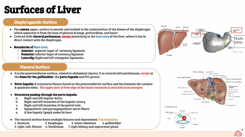
● The convex upper, surface is smooth and molded to the undersurface of the domes of the diaphragm which separates it from the base of pleurae & lungs, pericardium, and heart.
● Covered with visceral peritoneum, except posteriorly in the bare area of the liver, where it lies in direct contact with the diaphragm.
● Boundaries of Bare area:○ Anterior: superior layer of coronary ligament.○ Posterior: inferior layer of coronary ligament.○ Laterally: Right and left triangular ligaments.
4
Surfaces of Liver
● It is the posteroinferior surface, related to abdominal viscera. It is covered with peritoneum, except at the fossa for the gallbladder, the porta hepatis and IVC groove.
● Porta hepatis: A transverse fissure found on the posteroinferior surface and lies between the caudate & quadrate lobes. The upper part of free edge of the lesser omentum is attached to its margins
● Structures passing through the porta hepatis:a. Right and left hepatic ducts. b. Right and left branches of the hepatic arteryc. Right and left branches of the portal veind. Sympathetic and parasympathetic nerve fiberse. A few hepatic lymph nodes lie here
● The visceral surface bears multiple fissures and impressions It is related to:1. Stomach. 2. Esophagus 3. lesser omentum 4. gallbladder 5. right colic flexure 6. Duodenum. 7. right kidney and suprarenal gland
2
3
4
5
7
7
6
1
Visceral Surface
Diaphragmatic Surface
5
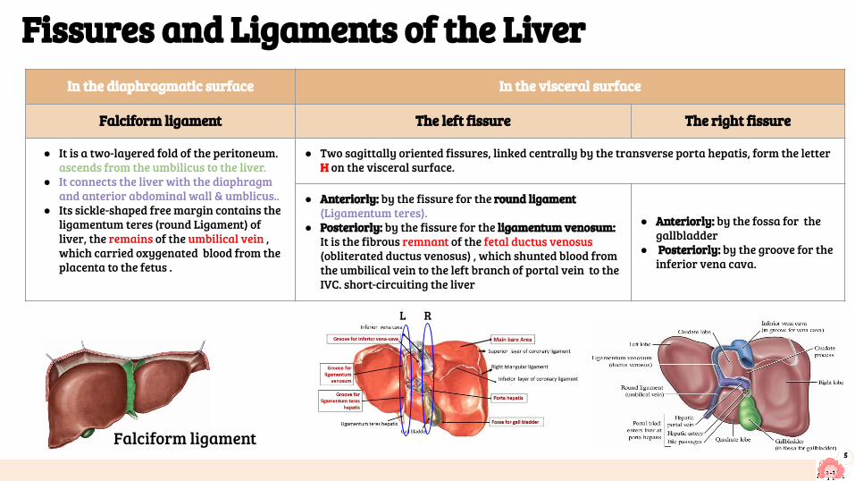
Falciform ligament
Fissures and Ligaments of the LiverIn the diaphragmatic surface In the visceral surface
Falciform ligament The left fissure The right fissure
● It is a two-layered fold of the peritoneum. ascends from the umbilicus to the liver.
● It connects the liver with the diaphragm and anterior abdominal wall & umblicus..
● Its sickle-shaped free margin contains the ligamentum teres (round Ligament) of liver, the remains of the umbilical vein , which carried oxygenated blood from the placenta to the fetus .
● Two sagittally oriented fissures, linked centrally by the transverse porta hepatis, form the letter H on the visceral surface.
● Anteriorly: by the fissure for the round ligament (Ligamentum teres).
● Posteriorly: by the fissure for the ligamentum venosum: It is the fibrous remnant of the fetal ductus venosus (obliterated ductus venosus) , which shunted blood from the umbilical vein to the left branch of portal vein to the IVC. short-circuiting the liver
● Anteriorly: by the fossa for the gallbladder
● Posteriorly: by the groove for the inferior vena cava.
RL
6
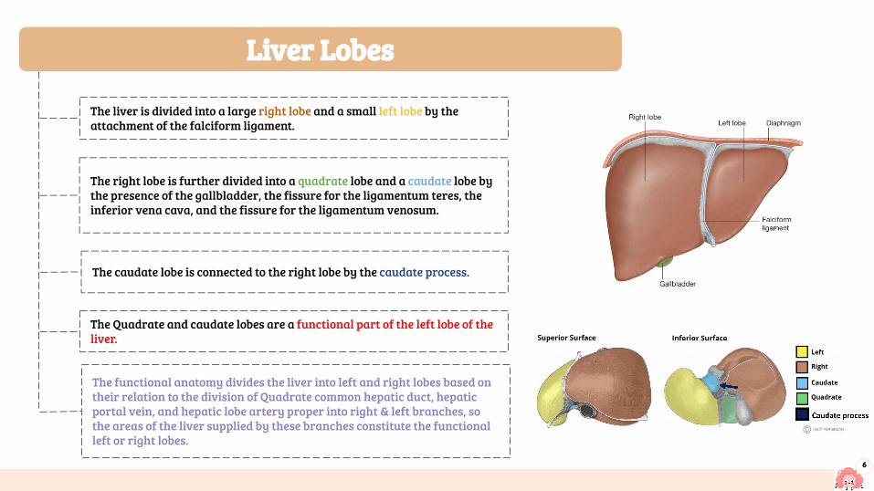
The liver is divided into a large right lobe and a small left lobe by the attachment of the falciform ligament.
The right lobe is further divided into a quadrate lobe and a caudate lobe by the presence of the gallbladder, the fissure for the ligamentum teres, the inferior vena cava, and the fissure for the ligamentum venosum.
The caudate lobe is connected to the right lobe by the caudate process.
The Quadrate and caudate lobes are a functional part of the left lobe of the liver.
The functional anatomy divides the liver into left and right lobes based on their relation to the division of Quadrate common hepatic duct, hepatic portal vein, and hepatic lobe artery proper into right & left branches, so the areas of the liver supplied by these branches constitute the functional left or right lobes.
Liver Lobes

7
Liver blood circulation ★ The blood vessels conveying blood to the liver are the hepatic
artery (30%) a branch of celiac trunk, and portal vein (70%).
At or close to the porta hepatis, the hepatic artery and portal vein terminate by dividing into right and left primary branches which supply the right and left parts of liver, respectively.
Within the liver, the primary branches divide to give secondary and Portal vein tertiary to supply the hepatic segments independently
The hepatic artery brings oxygenated blood to the liver hepatic Vein.
The venous blood is drained by right & left hepatic veins into the IVC
1.
5.
6. The hepatic veins, are intersegmental in their distribution and function, draining parts of adjacent segments, and they open in the posterior surface on the groove for IVC.
The portal vein brings venous blood rich in the products of digestion, which have been absorbed from the gastrointestinal tract to the liver.
3.
2.
4.
7.The attachment of the two hepatic veins to the IVC helps hold the liver in position. (The peritoneal ligaments and the tone of the abdominal muscles play a minor role in the support of liver).
Portal- systemic (portacaval) anastomosis
8
Lymph drainage of GIT
● The liver produces a large amount of lymph about one third to one half of all body lymph.
● The lymph vessels leave the liver and enter several lymph nodes in the porta hepatis.
● The efferent vessels pass to the celiac nodes. ● A few vessels pass from the bare area of the liver
through the diaphragm to the posterior mediastinal lymph nodes.
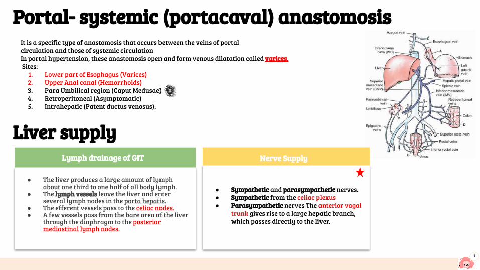
It is a specific type of anastomosis that occurs between the veins of portalcirculation and those of systemic circulation In portal hypertension, these anastomosis open and form venous dilatation called varices. Sites:
1. Lower part of Esophagus (Varices) 2. Upper Anal canal (Hemorrhoids) 3. Para Umbilical region (Caput Medusae)4. Retroperitoneal (Asymptomatic) 5. Intrahepatic (Patent ductus venosus).
Liver supply Nerve Supply
● Sympathetic and parasympathetic nerves. ● Sympathetic from the celiac plexus● Parasympathetic nerves The anterior vagal
trunk gives rise to a large hepatic branch, which passes directly to the liver.
9
Spleen
Location SizeShape
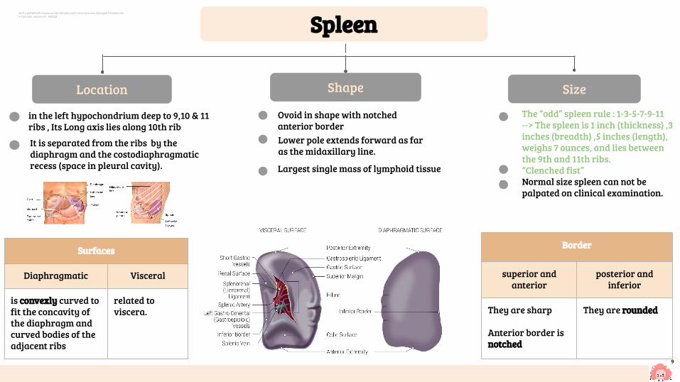
in the left hypochondrium deep to 9,10 & 11 ribs , Its Long axis lies along 10th rib
It is separated from the ribs by the diaphragm and the costodiaphragmatic recess (space in pleural cavity).
● The “odd” spleen rule : 1-3-5-7-9-11 --> The spleen is 1 inch (thickness) ,3 inches (breadth) ,5 inches (length), weighs 7 ounces, and lies between the 9th and 11th ribs.
● “Clenched fist”● Normal size spleen can not be
palpated on clinical examination.
Lower pole extends forward as far as the midaxillary line.
Ovoid in shape with notched anterior border
Largest single mass of lymphoid tissue
Surfaces
Diaphragmatic Visceral
is convexly curved to fit the concavity of the diaphragm andcurved bodies of the adjacent ribs
related to viscera.
Border
superior and anterior
posterior and inferior
They are sharp
Anterior border is notched
They are rounded
so if a girl fell off a horse on her left side and a structure was damaged between the 9-11th ribs, what is it? - NVSLR

10
Anterior Posterior
1. Stomach2. tail of pancreas3. left colic flexure4. left kidney
1. Diaphragm,that separates it from the left pleura (left costo diaphragmatic recess)
2. left lung 3. 9, 10 & 11 ribs.
Medially Inferior
Left kidney. Left colic flexure.
Attached to the left kidney carrying the splenic vessels and the tail of pancreas
Attached to the greater curvature of stomach carrying the short gastric and left gastroepiploic vessels
Lienorenal ligament (splenorenal) Gastrosplenic ligament
Ligaments of the spleen:Spleen is completely surrounded by peritoneum EXCEPT at the hilum where its margins give attachement to :
Relations of the spleen :

11
Spleen supply
Venous DrainageSplenic vein
● Leaves the hilus● Runs behind the tail &body of the pancreas● Reaches behind the neck of pancreas, where it joins the ● superior mesenteric vein to form the portal vein ● Tributaries:
1. Short gastric vein2. left gastroepiploic vein3. Pancreatic veins 4. Inferior mesenteric vein
Nerve Supply
● Derived from the celiac plexus (Innervation is purely sympathetic)
● Are distributed mainly along branches of the splenic artery, and are vasomotor in function.
Lymph drainage of GIT
● Lymphatics emerge from the hilus and drain into several nodes lying at the hilum
● Efferents from the hilar nodes pass along the course of splenic artery, and drain into the celiac lymph nodes
Arterial SupplySplenic artery❖ Largest branch of the celiac artery ❖ Runs a tortuous course along:
➔ upper border of the pancreas (behind the stomach)➔ Passes within the lienorenal ligament
❖ Divides into 4-5 terminal branches, which enter the spleen at the hilus❖ The lack of anastomosis of thesearterial vessels within the spleen,❖ results in the formation of vascular segments of the spleen with ❖ relatively avascular planes between them, enabling subtotal splenectomy.
branches:branches to the pancreasshort gastricleft gastroepiploicposterior gastric (not always present)

QUIZQ1: The lateral boundaries of the Bare area include:
A. Superior triangular ligament
B. Left coronary ligament
C. Posterior triangular Ligament
D. Right triangular ligament
Q2: Which of the following lies posterior to the liver:
A. The body of the stomach
B. The hepatic flexure
C. The jejunum
D. The splenic flexure
Q3: The liver lies mainly in the :
A. Right lumbar region
B. Left hypochondrium
C. Right hypogastrium
D. Right hypochondrium
Q4: If there was an erosion of the ulcer in the posterior wall of the stomach,
which artery would be injured?
A. Gastroduodenal
B. Right gastric
C. Splenic
Q5: The portal vein is form by joining of :
A. Splenic vein and inferior mesenteric vein
B. Splenic vein and superior mesenteric vein
C.Hepatic vein and superior mesenteric vein
D. Superior and inferior mesenteric veins
Q6: The left colic flexure is related to the spleen :
A.laterally
B.Medially
C.Posteriorly
D.Anteriorly
Q7: Which arteries could bleed if the posterior gastric artery was injured?
A. Left gastroepiploic artery
B. Short gastric arteries
C. Splenic artery
D. Hepatic artery
Q8: The ligament that attaches the hilum of spleen to the great curvature of stomach :
A. Gastrosplenic ligament
B.Splenorenal ligament
C. Gastrohepatic ligament
D. Lienorenal ligament12
Q1 Q2 Q3 Q4 Q5 Q6 Q7 Q8
D B D C B D C A
Members board
● Abdulrahman Shadid ● Ateen Almutairi
Girls team :
● Ajeed Al Rashoud● Taif Alotaibi● Noura Al Turki● Amirah Al-Zahrani● Alhanouf Al-haluli● Sara Al-Abdulkarem● Renad Al Haqbani● Nouf Al Humaidhi● Jude Al Khalifah● Nouf Al Hussaini● Danah Al Halees● Rema Al Mutawa● Maha Al Nahdi ● Razan Al zohaifi ● Ghalia Alnufaei
Team leaders
Editing file
Contact us:
Boys team:
● Mohammed Al-huqbani● Salman Alagla● Ziyad Al-jofan● Ali Aldawood● Khalid Nagshabandi● Sameh nuser● Abdullah Basamh● Alwaleed Alsaleh● Mohaned Makkawi● Abdullah Alghamdi