LIVER, PANCREAS, SPLEEN - univet.hu · b. incisura lig. teretis . lobes of the liver (lobus...
88
LIVER, PANCREAS, SPLEEN Andrea Heinzlmann Veterinary University Department of Anatomy and Histology 8th April 2019
Transcript of LIVER, PANCREAS, SPLEEN - univet.hu · b. incisura lig. teretis . lobes of the liver (lobus...
LIVER, PANCREAS, SPLEEN
Andrea Heinzlmann
Veterinary University
Department of Anatomy and Histology
8th April 2019
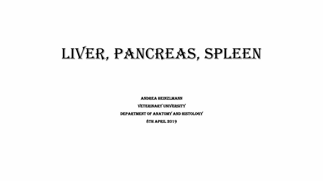
LIVER (HEPAR)
• the largest gland of the body
• intraperitoneal organ
FUNCTIONS:
1. secretion of bile
2. in embryonic live – hemopoetic center
3. storage of glycogen
4. converts end products of protein catabolism to urea and uric acid
5. end products of hemoglobin catabolism discharged in the bile as bile pigments
6. disintoxication
DOG
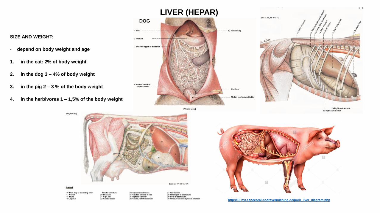
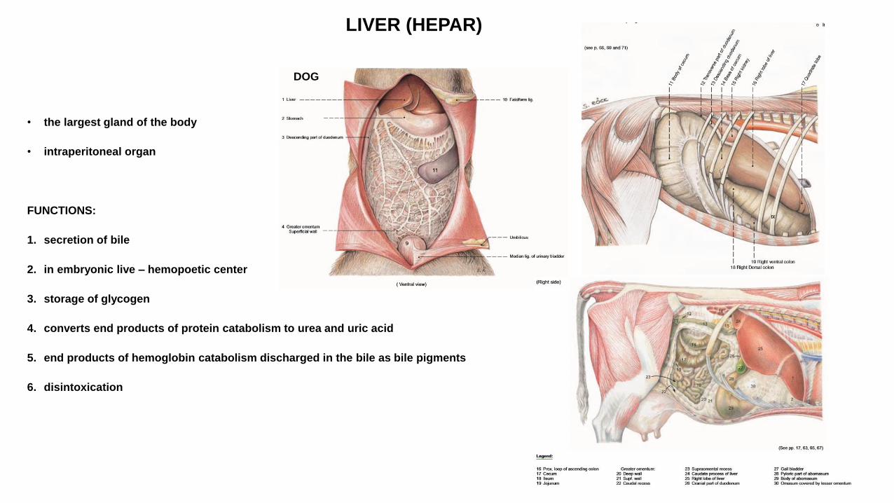
LIVER (HEPAR)
SIZE AND WEIGHT:
- depend on body weight and age
1. in the cat: 2% of body weight
2. in the dog 3 – 4% of body weight
3. in the pig 2 – 3 % of the body weight
4. in the herbivores 1 – 1,5% of the body weight
DOG
http://18.hyt.capecoral-bootsvermietung.de/pork_liver_diagram.php
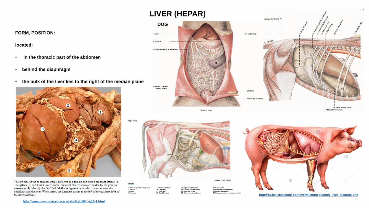
LIVER (HEPAR)
FORM, POSITION:
located:
• in the thoracic part of the abdomen
• behind the diaphragm
• the bulk of the liver lies to the right of the median plane
DOG
http://18.hyt.capecoral-bootsvermietung.de/pork_liver_diagram.php
http://vanat.cvm.umn.edu/carnLabs/Lab16/Img16-1.html
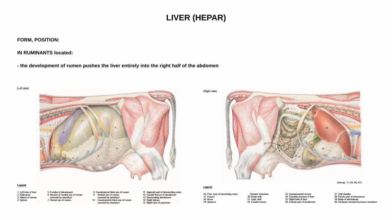
LIVER (HEPAR)
FORM, POSITION:
IN RUMINANTS located:
- the development of rumen pushes the liver entirely into the right half of the abdomen
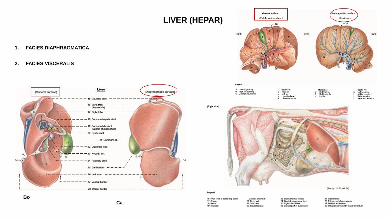
LIVER (HEPAR)
1. FACIES DIAPHRAGMATICA
2. FACIES VISCERALIS
Bo Ca
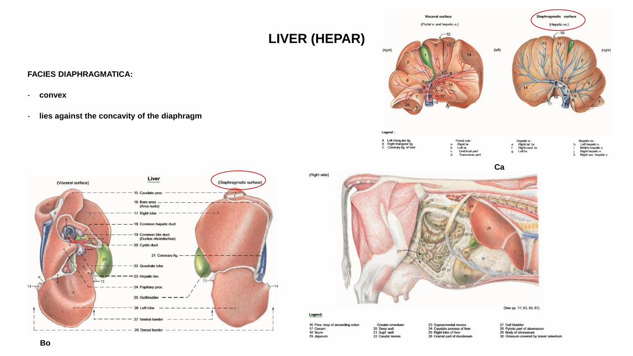
LIVER (HEPAR)
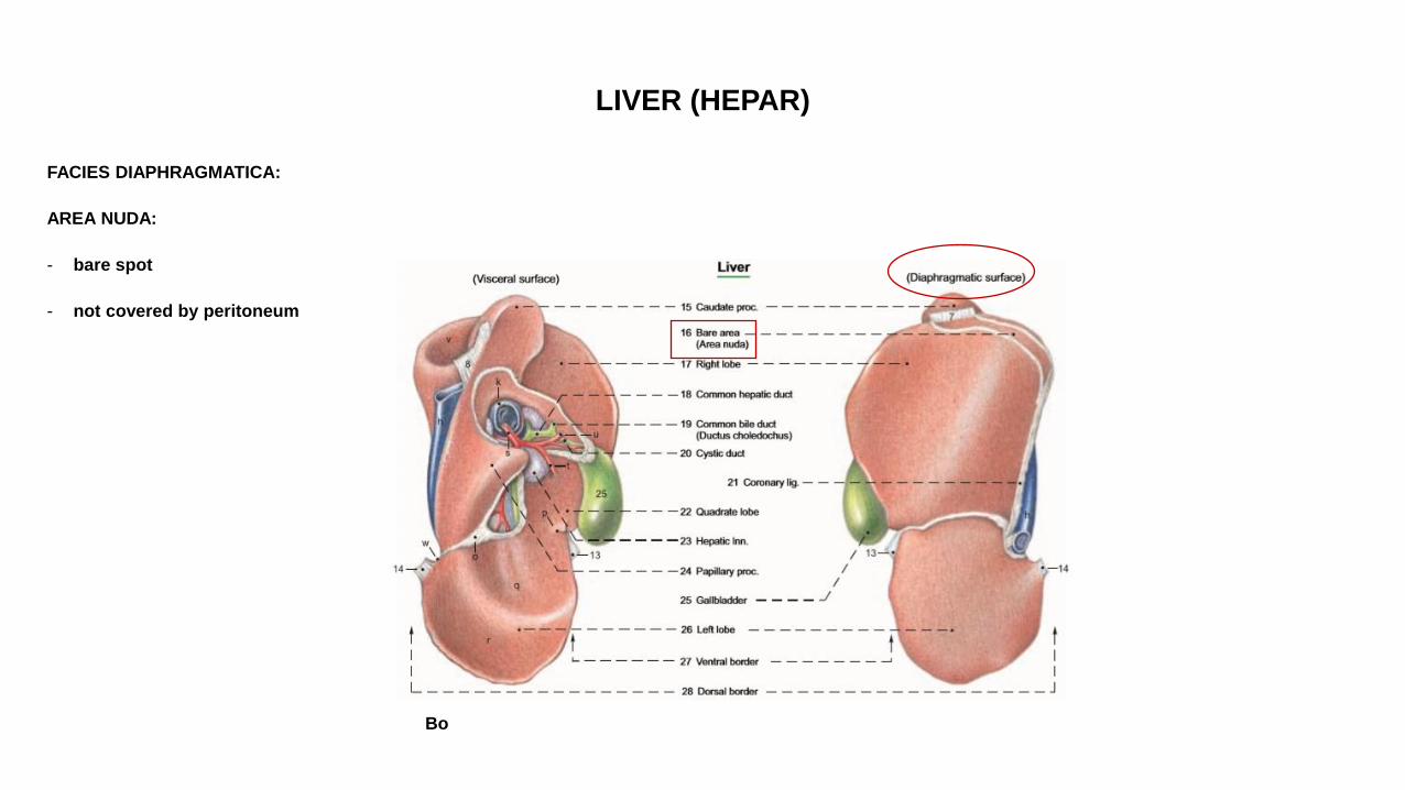
FACIES DIAPHRAGMATICA:
- convex
- lies against the concavity of the diaphragm
Ca
Bo
LIVER (HEPAR)
FACIES DIAPHRAGMATICA:
AREA NUDA:
- bare spot
- not covered by peritoneum
Bo
LIVER (HEPAR)
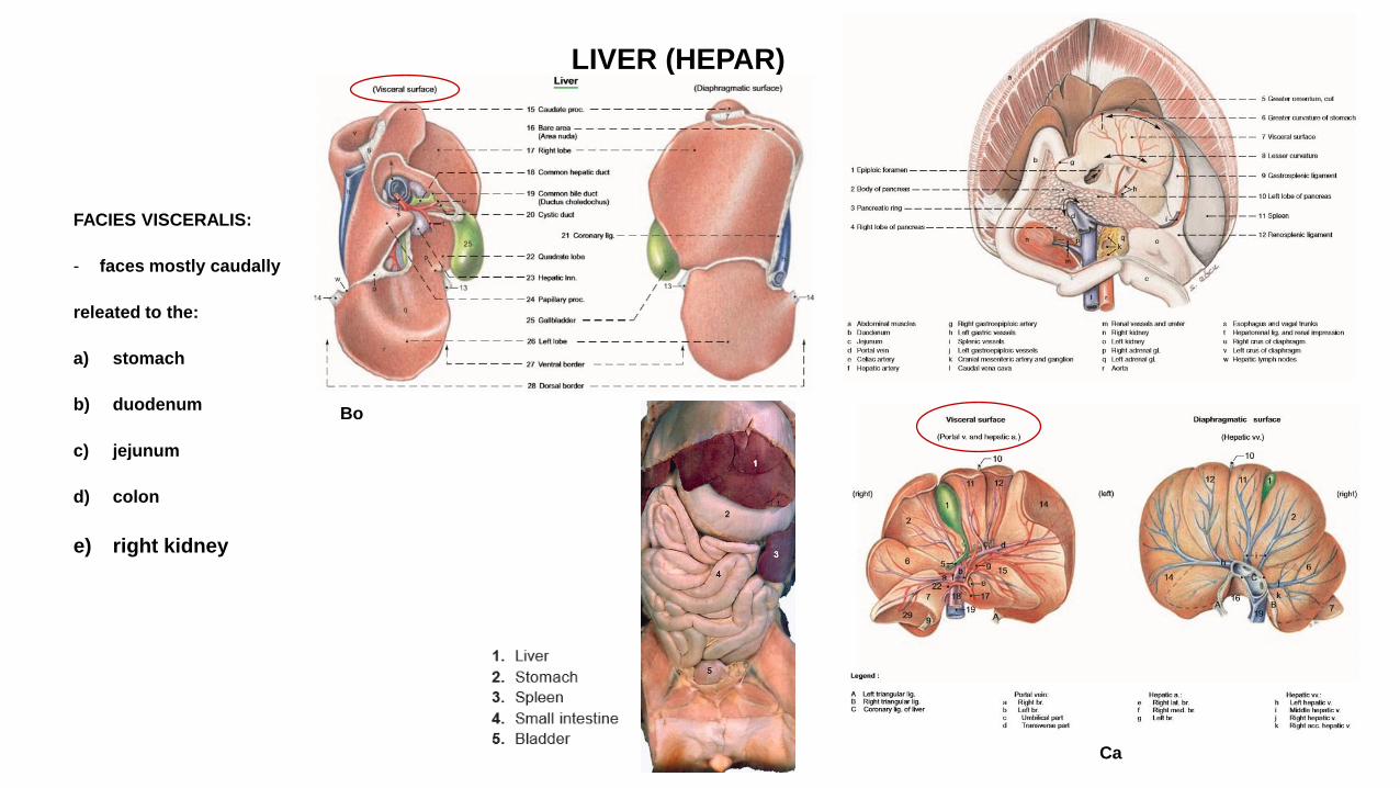
FACIES VISCERALIS:
- faces mostly caudally
releated to the:
a) stomach
b) duodenum
c) jejunum
d) colon
e) right kidney
Bo
Ca
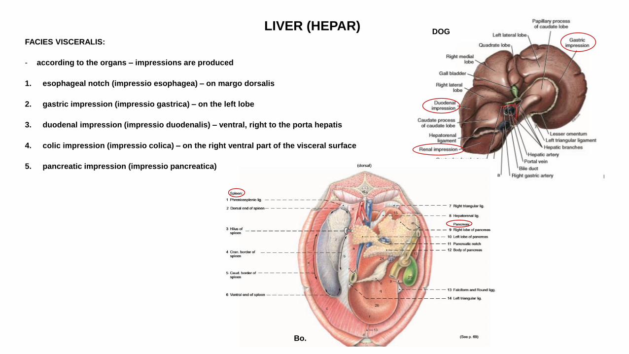
LIVER (HEPAR) FACIES VISCERALIS:
- according to the organs – impressions are produced
1. esophageal notch (impressio esophagea) – on margo dorsalis
2. gastric impression (impressio gastrica) – on the left lobe
3. duodenal impression (impressio duodenalis) – ventral, right to the porta hepatis
4. colic impression (impressio colica) – on the right ventral part of the visceral surface
5. pancreatic impression (impressio pancreatica)
DOG
Bo.
LIVER (HEPAR)
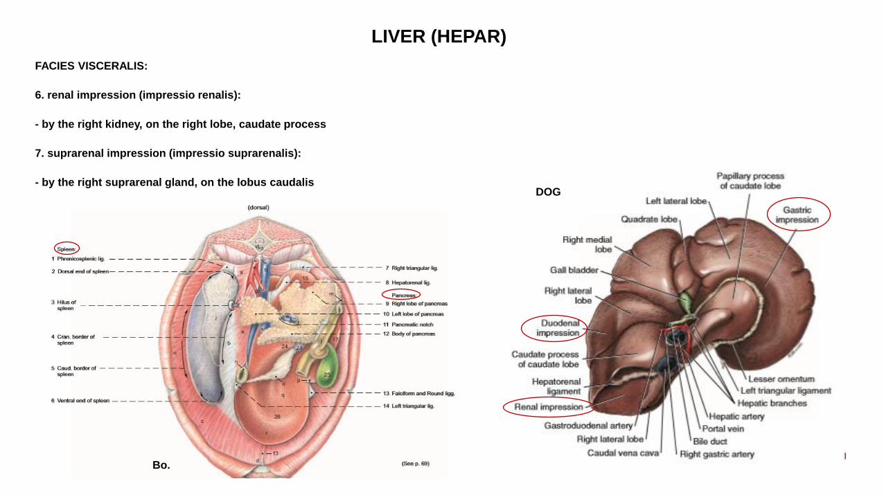
FACIES VISCERALIS:
6. renal impression (impressio renalis):
- by the right kidney, on the right lobe, caudate process
7. suprarenal impression (impressio suprarenalis):
- by the right suprarenal gland, on the lobus caudalis
DOG
Bo.

LIVER (HEPAR)
FACIES VISCERALIS:
a. reticular impression (impressio reticularis) – on the left lobe – in Bo.
b. omasal impression (impressio omasica) – occupies a large part of the visceral surface – in Bo.
c. cecal impression (impressio cecalis) – in Eq – on the right lobe
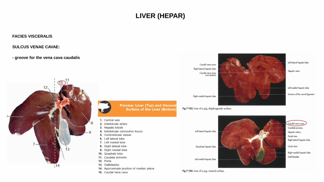
LIVER (HEPAR)
FACIES VISCERALIS
SULCUS VENAE CAVAE:
- groove for the vena cava caudalis
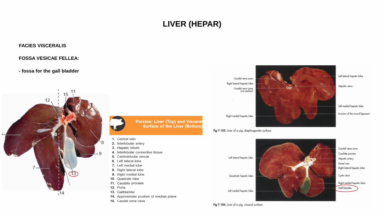
LIVER (HEPAR)
FACIES VISCERALIS
FOSSA VESICAE FELLEA:
- fossa for the gall bladder
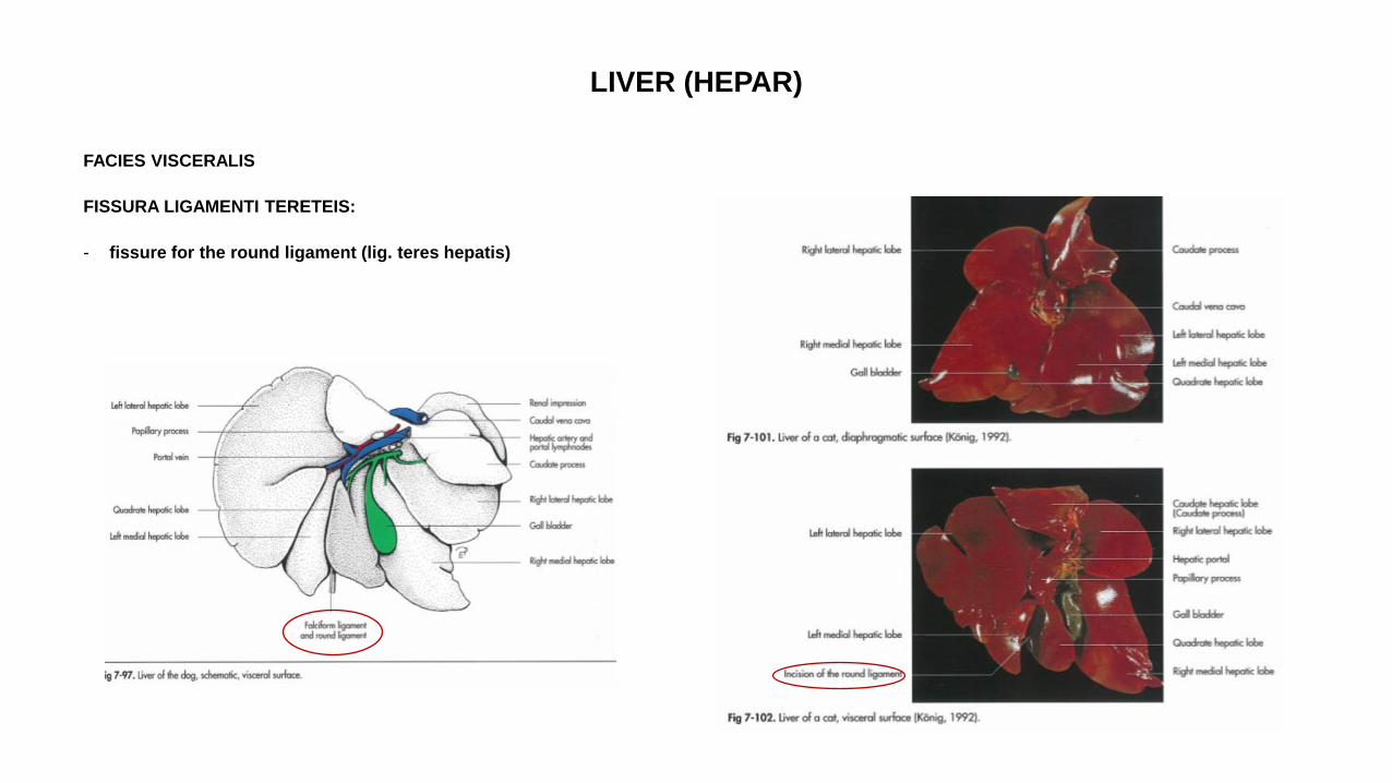
LIVER (HEPAR)
FACIES VISCERALIS
FISSURA LIGAMENTI TERETEIS:
- fissure for the round ligament (lig. teres hepatis)
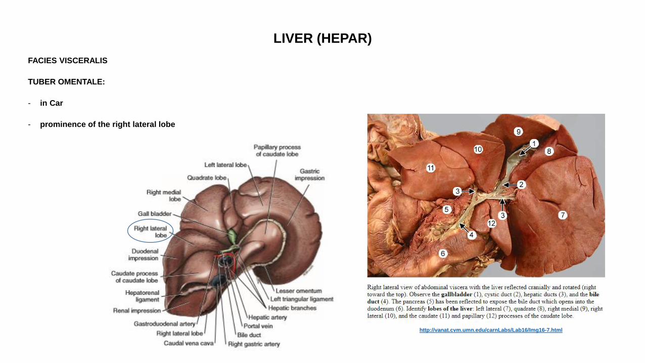
LIVER (HEPAR)
FACIES VISCERALIS
TUBER OMENTALE:
- in Car
- prominence of the right lateral lobe
http://vanat.cvm.umn.edu/carnLabs/Lab16/Img16-7.html
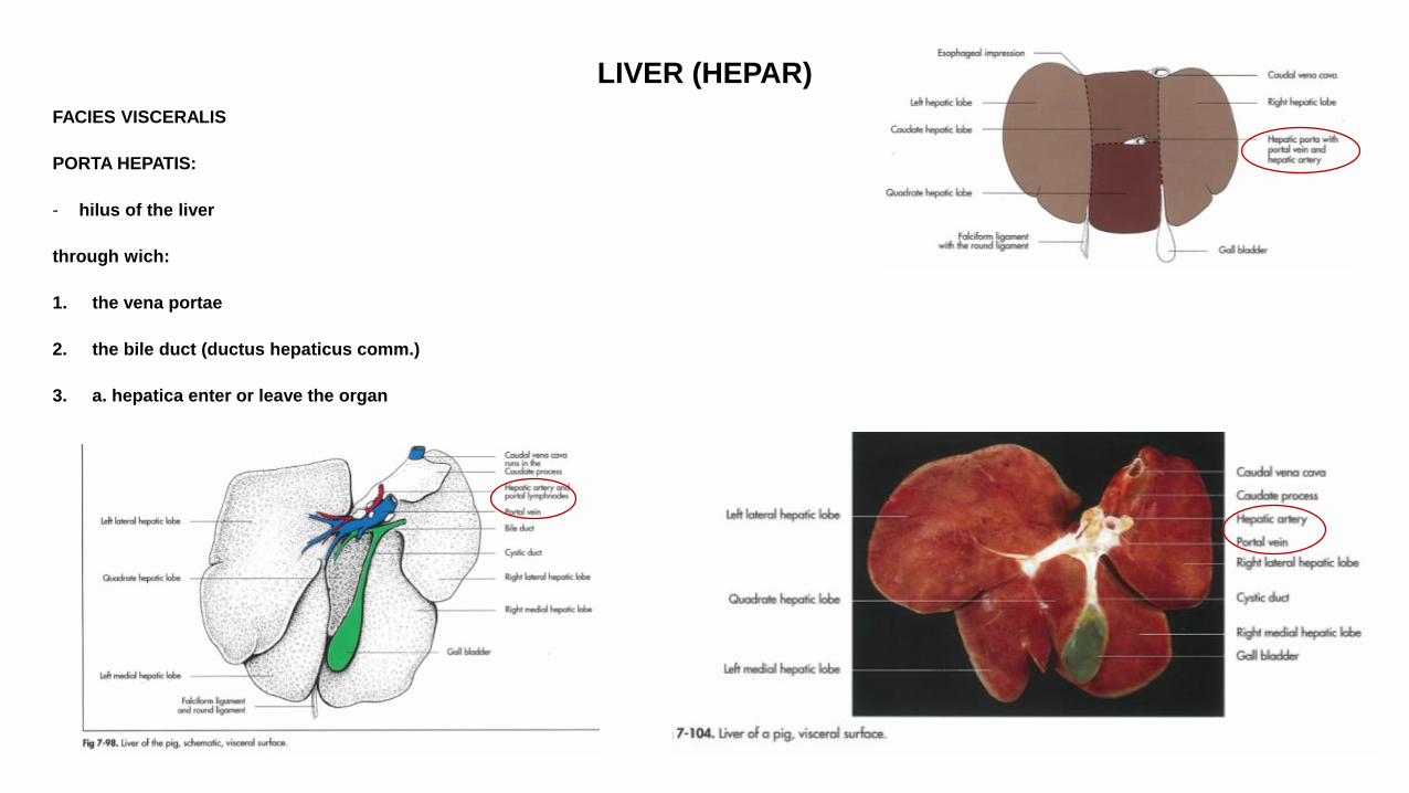
LIVER (HEPAR)
FACIES VISCERALIS
PORTA HEPATIS:
- hilus of the liver
through wich:
1. the vena portae
2. the bile duct (ductus hepaticus comm.)
3. a. hepatica enter or leave the organ
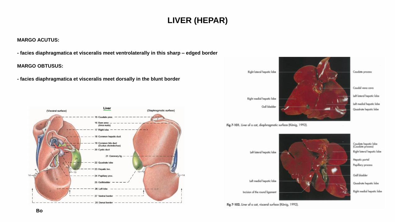
LIVER (HEPAR)
MARGO ACUTUS:
- facies diaphragmatica et visceralis meet ventrolaterally in this sharp – edged border
MARGO OBTUSUS:
- facies diaphragmatica et visceralis meet dorsally in the blunt border
Bo
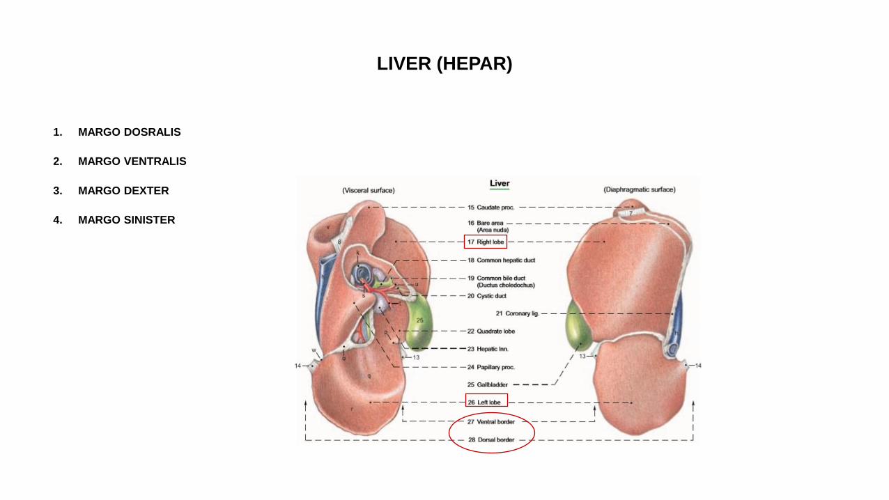
LIVER (HEPAR)
1. MARGO DOSRALIS
2. MARGO VENTRALIS
3. MARGO DEXTER
4. MARGO SINISTER
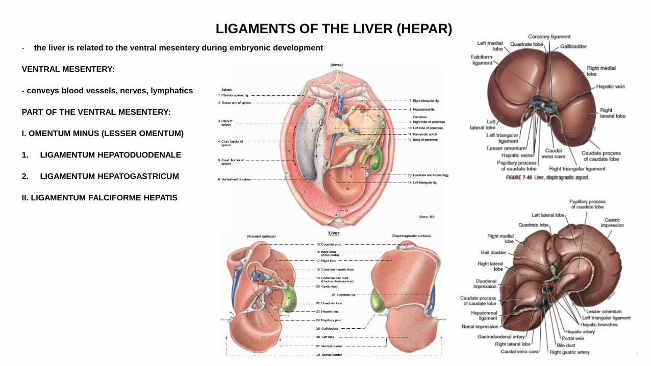
LIGAMENTS OF THE LIVER (HEPAR)
- the liver is related to the ventral mesentery during embryonic development
VENTRAL MESENTERY:
- conveys blood vessels, nerves, lymphatics
PART OF THE VENTRAL MESENTERY:
I. OMENTUM MINUS (LESSER OMENTUM)
1. LIGAMENTUM HEPATODUODENALE
2. LIGAMENTUM HEPATOGASTRICUM
II. LIGAMENTUM FALCIFORME HEPATIS
LIGAMENTS OF THE LIVER (HEPAR)
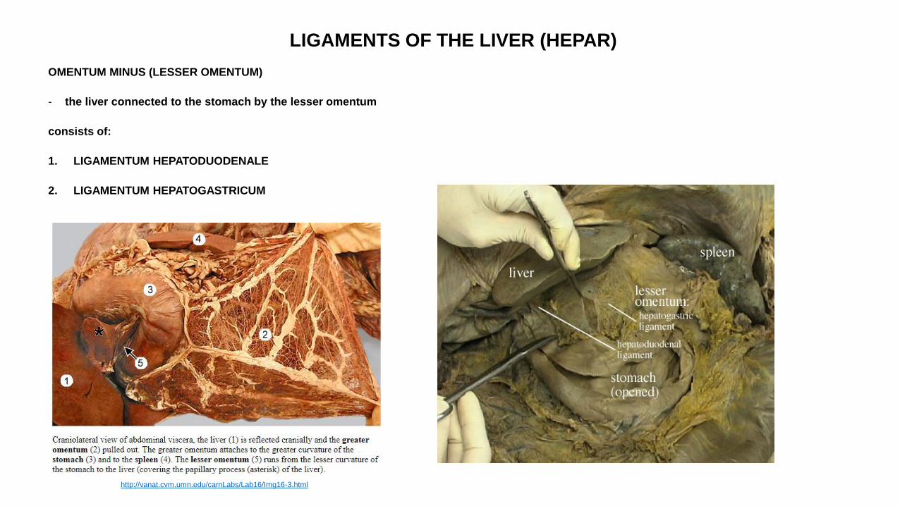
OMENTUM MINUS (LESSER OMENTUM)
- the liver connected to the stomach by the lesser omentum
consists of:
1. LIGAMENTUM HEPATODUODENALE
2. LIGAMENTUM HEPATOGASTRICUM
http://vanat.cvm.umn.edu/carnLabs/Lab16/Img16-3.html
LIGAMENTS OF THE LIVER (HEPAR)
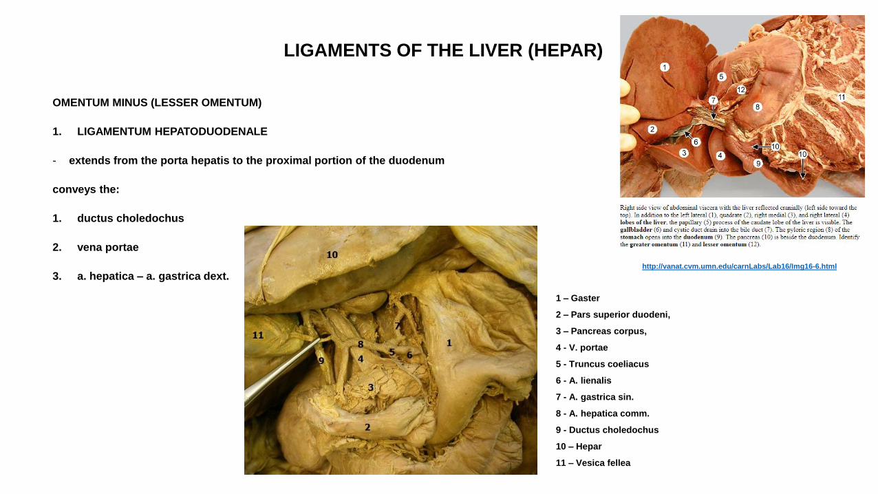
OMENTUM MINUS (LESSER OMENTUM)
1. LIGAMENTUM HEPATODUODENALE
- extends from the porta hepatis to the proximal portion of the duodenum
conveys the:
1. ductus choledochus
2. vena portae
3. a. hepatica – a. gastrica dext.
1 – Gaster
2 – Pars superior duodeni,
3 – Pancreas corpus,
4 - V. portae
5 - Truncus coeliacus
6 - A. lienalis
7 - A. gastrica sin.
8 - A. hepatica comm.
9 - Ductus choledochus
10 – Hepar
11 – Vesica fellea
http://vanat.cvm.umn.edu/carnLabs/Lab16/Img16-6.html
LIGAMENTS OF THE LIVER (HEPAR)
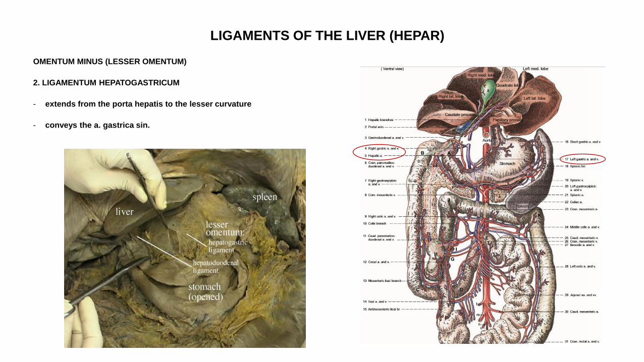
OMENTUM MINUS (LESSER OMENTUM)
2. LIGAMENTUM HEPATOGASTRICUM
- extends from the porta hepatis to the lesser curvature
- conveys the a. gastrica sin.
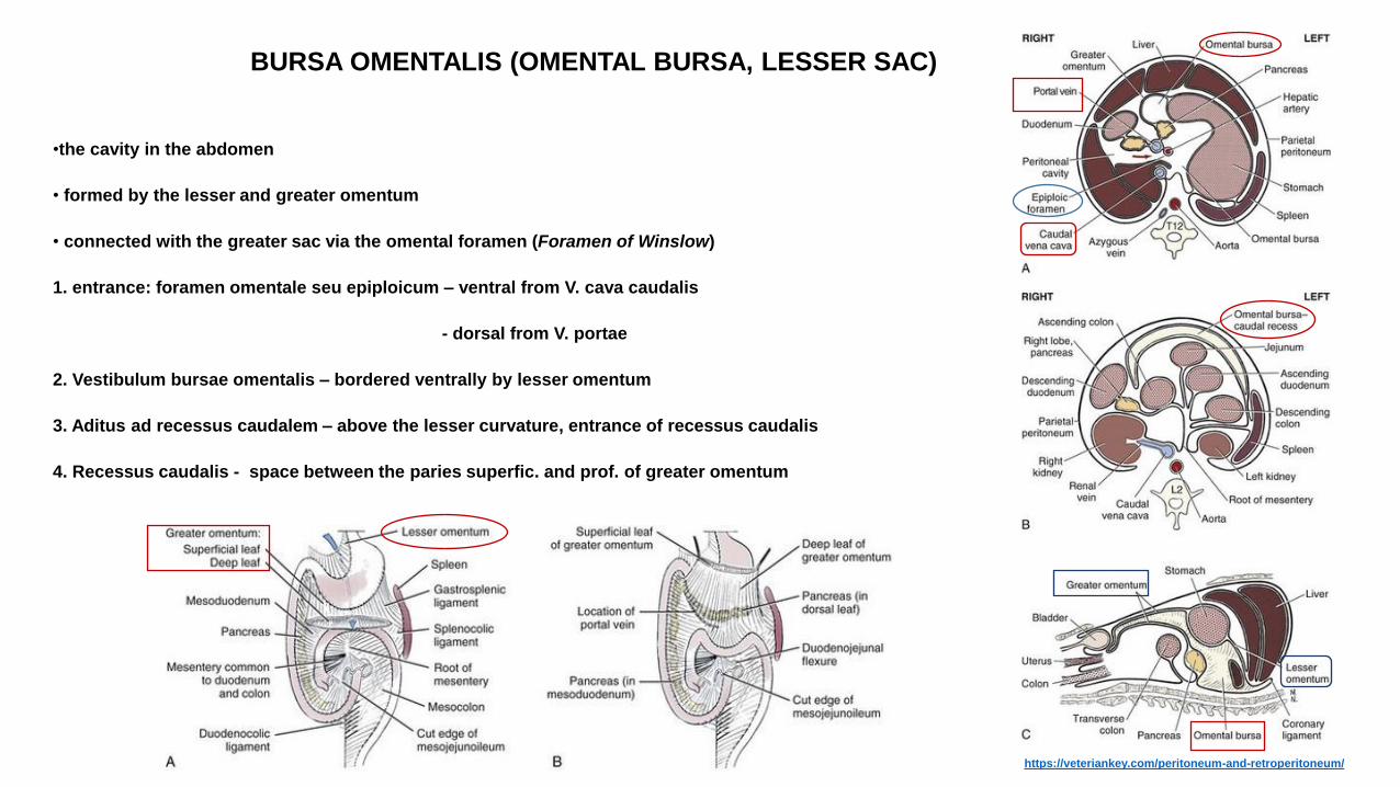
BURSA OMENTALIS (OMENTAL BURSA, LESSER SAC)
•the cavity in the abdomen
• formed by the lesser and greater omentum
• connected with the greater sac via the omental foramen (Foramen of Winslow)
1. entrance: foramen omentale seu epiploicum – ventral from V. cava caudalis
- dorsal from V. portae
2. Vestibulum bursae omentalis – bordered ventrally by lesser omentum
3. Aditus ad recessus caudalem – above the lesser curvature, entrance of recessus caudalis
4. Recessus caudalis - space between the paries superfic. and prof. of greater omentum
https://veteriankey.com/peritoneum-and-retroperitoneum/

LIGAMENTS OF THE LIVER (HEPAR)
II. LIGAMENTUM FALCIFORME HEPATIS (FALCIFORM LIGAMENT):
- remnant of the ventral mesentery
extends:
a) between the liver and the diaphragm
b) between liver and the ventral abdominal wall
• includes the v. umbilicalis in fetal life
• v. umbilicalis obliterates after the birth froming the round ligament (lig. teres hepatis)
http://vanat.cvm.umn.edu/carnLabs/Lab16/Img16-1.html
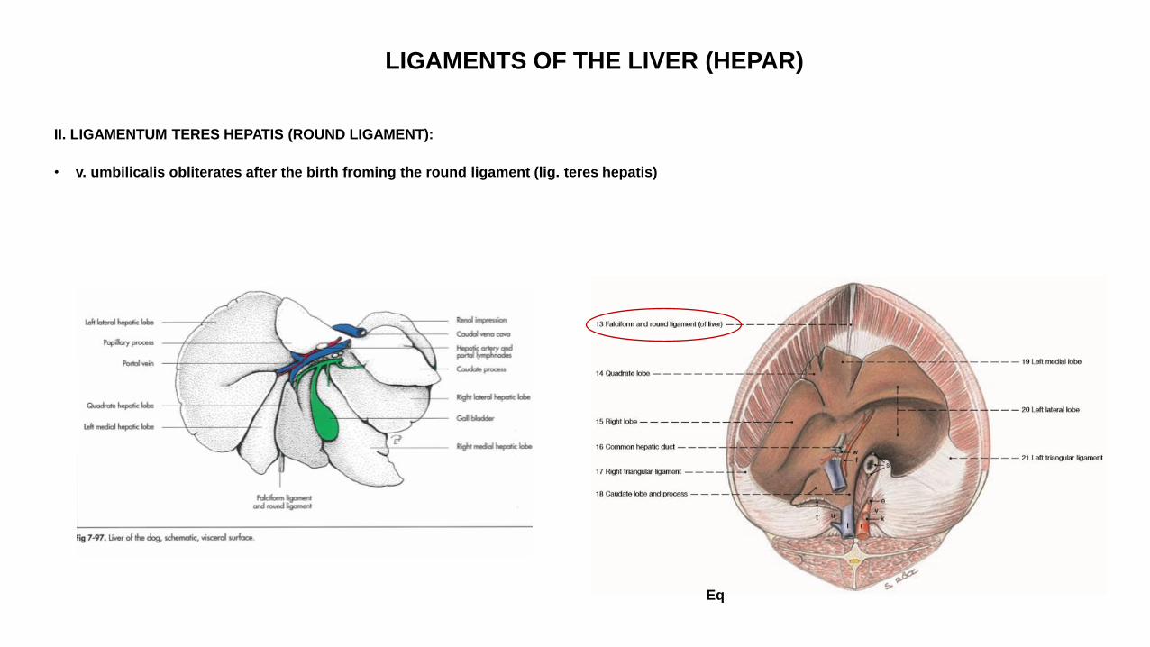
LIGAMENTS OF THE LIVER (HEPAR)
II. LIGAMENTUM TERES HEPATIS (ROUND LIGAMENT):
• v. umbilicalis obliterates after the birth froming the round ligament (lig. teres hepatis)
Eq
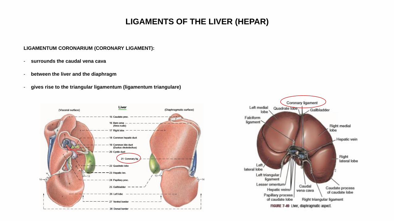
LIGAMENTS OF THE LIVER (HEPAR)
LIGAMENTUM CORONARIUM (CORONARY LIGAMENT):
- surrounds the caudal vena cava
- between the liver and the diaphragm
- gives rise to the triangular ligamentum (ligamentum triangulare)

LIGAMENTS OF THE LIVER (HEPAR)
LIGAMENTUM TRIANGULARE DEXTRUM et SINISTRUM (TRIANGULAR LIGAMENT):
- the right and the left lobe attechedto the diaphragm by these ligaments
- continue medially with the coronary ligament
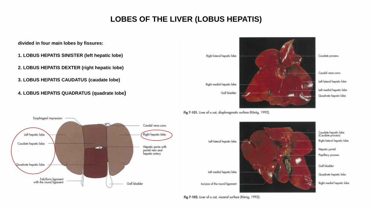
LOBES OF THE LIVER (LOBUS HEPATIS)
divided in four main lobes by fissures:
1. LOBUS HEPATIS SINISTER (left hepatic lobe)
2. LOBUS HEPATIS DEXTER (right hepatic lobe)
3. LOBUS HEPATIS CAUDATUS (caudate lobe)
4. LOBUS HEPATIS QUADRATUS (quadrate lobe)
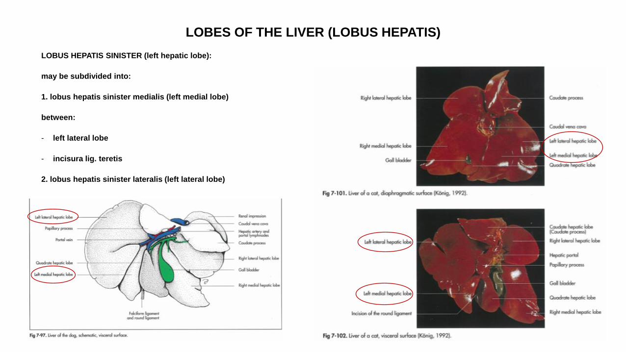
LOBES OF THE LIVER (LOBUS HEPATIS)
LOBUS HEPATIS SINISTER (left hepatic lobe):
may be subdivided into:
1. lobus hepatis sinister medialis (left medial lobe)
between:
- left lateral lobe
- incisura lig. teretis
2. lobus hepatis sinister lateralis (left lateral lobe)
LOBES OF THE LIVER (LOBUS HEPATIS)
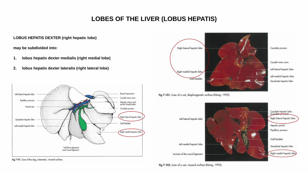
LOBUS HEPATIS DEXTER (right hepatic lobe)
may be subdivided into:
1. lobus hepatis dexter medialis (right medial lobe)
2. lobus hepatis dexter lateralis (right lateral lobe)
LOBES OF THE LIVER (LOBUS HEPATIS)
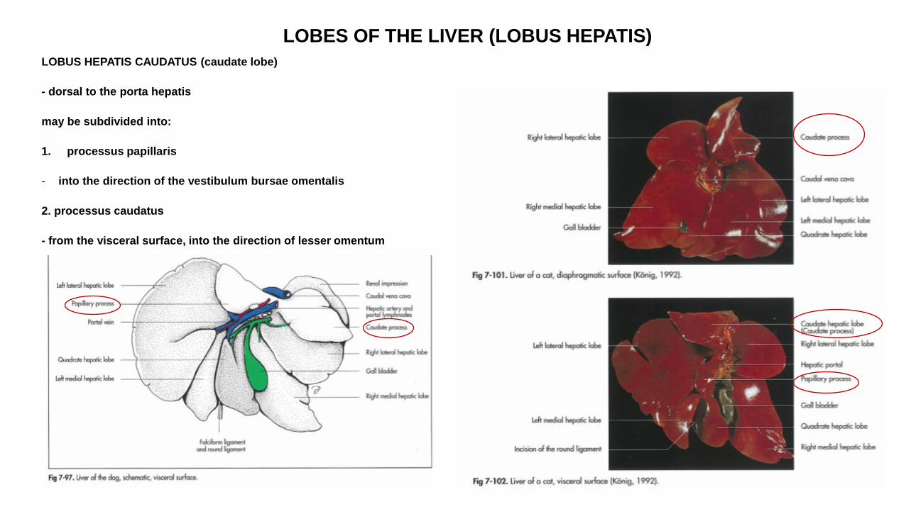
LOBUS HEPATIS CAUDATUS (caudate lobe)
- dorsal to the porta hepatis
may be subdivided into:
1. processus papillaris
- into the direction of the vestibulum bursae omentalis
2. processus caudatus
- from the visceral surface, into the direction of lesser omentum
LOBES OF THE LIVER (LOBUS HEPATIS)
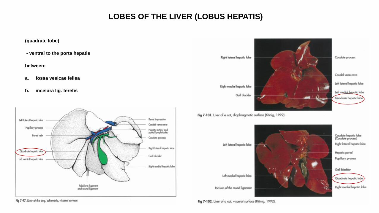
(quadrate lobe)
- ventral to the porta hepatis
between:
a. fossa vesicae fellea
b. incisura lig. teretis
LOBES OF THE LIVER
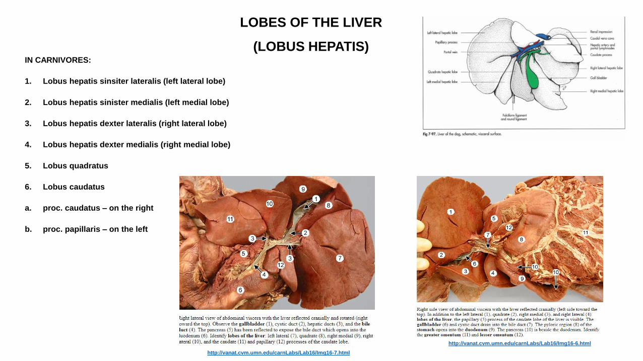
(LOBUS HEPATIS) IN CARNIVORES:
1. Lobus hepatis sinsiter lateralis (left lateral lobe)
2. Lobus hepatis sinister medialis (left medial lobe)
3. Lobus hepatis dexter lateralis (right lateral lobe)
4. Lobus hepatis dexter medialis (right medial lobe)
5. Lobus quadratus
6. Lobus caudatus
a. proc. caudatus – on the right
b. proc. papillaris – on the left
http://vanat.cvm.umn.edu/carnLabs/Lab16/Img16-7.html
http://vanat.cvm.umn.edu/carnLabs/Lab16/Img16-6.html
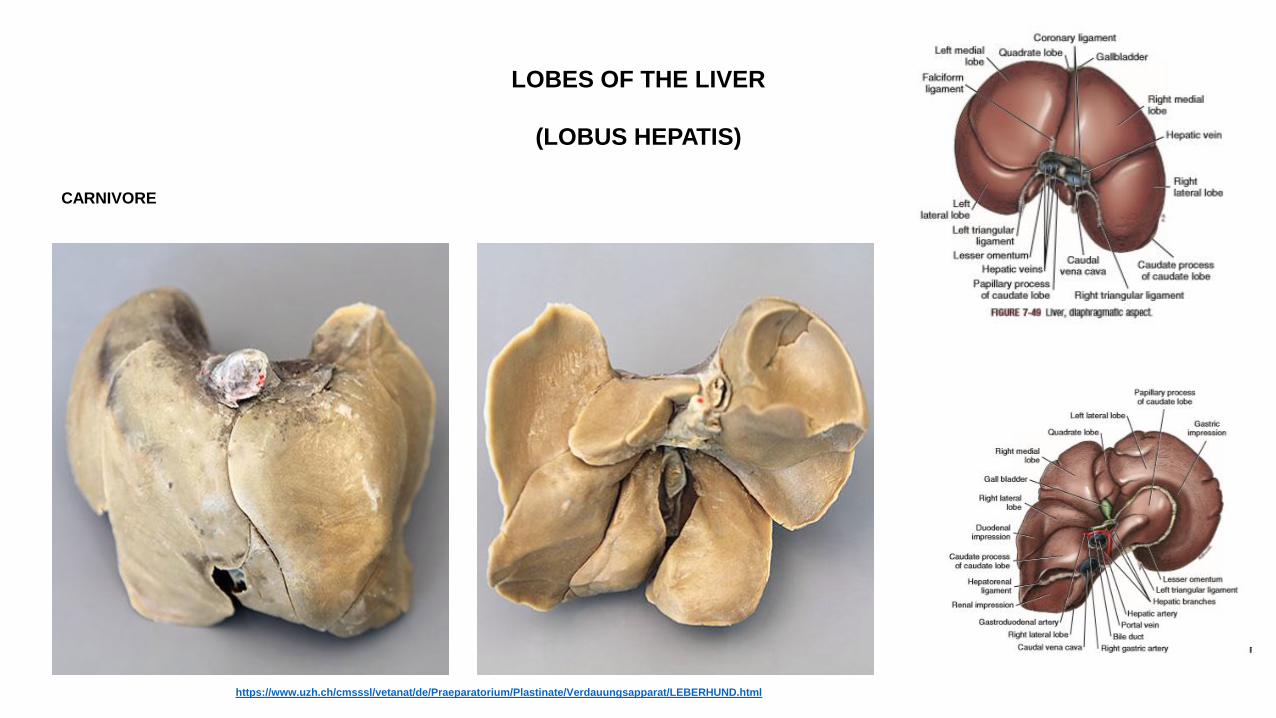
LOBES OF THE LIVER
(LOBUS HEPATIS)
CARNIVORE
https://www.uzh.ch/cmsssl/vetanat/de/Praeparatorium/Plastinate/Verdauungsapparat/LEBERHUND.html
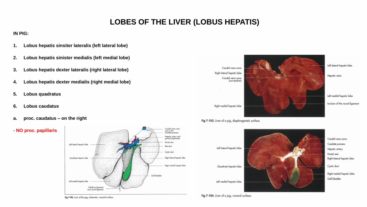
LOBES OF THE LIVER (LOBUS HEPATIS)
IN PIG:
1. Lobus hepatis sinsiter lateralis (left lateral lobe)
2. Lobus hepatis sinister medialis (left medial lobe)
3. Lobus hepatis dexter lateralis (right lateral lobe)
4. Lobus hepatis dexter medialis (right medial lobe)
5. Lobus quadratus
6. Lobus caudatus
a. proc. caudatus – on the right
- NO proc. papillaris
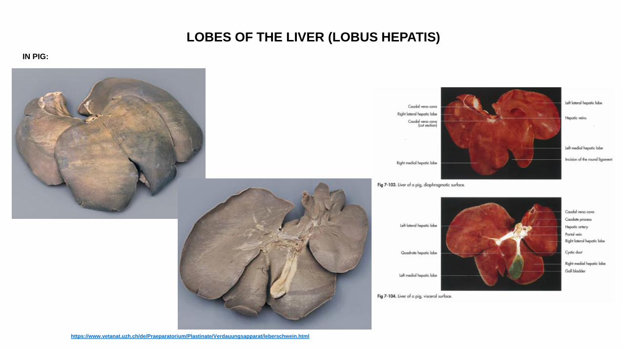
LOBES OF THE LIVER (LOBUS HEPATIS)
IN PIG:
https://www.vetanat.uzh.ch/de/Praeparatorium/Plastinate/Verdauungsapparat/leberschwein.html
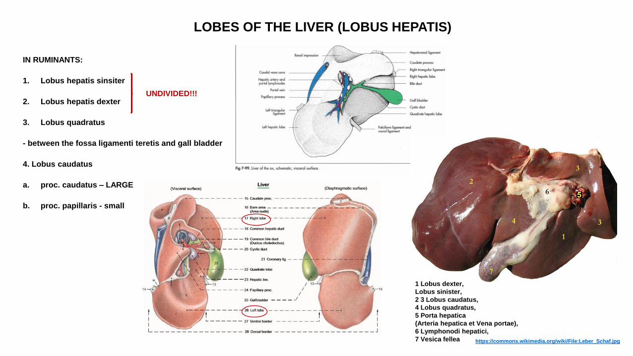
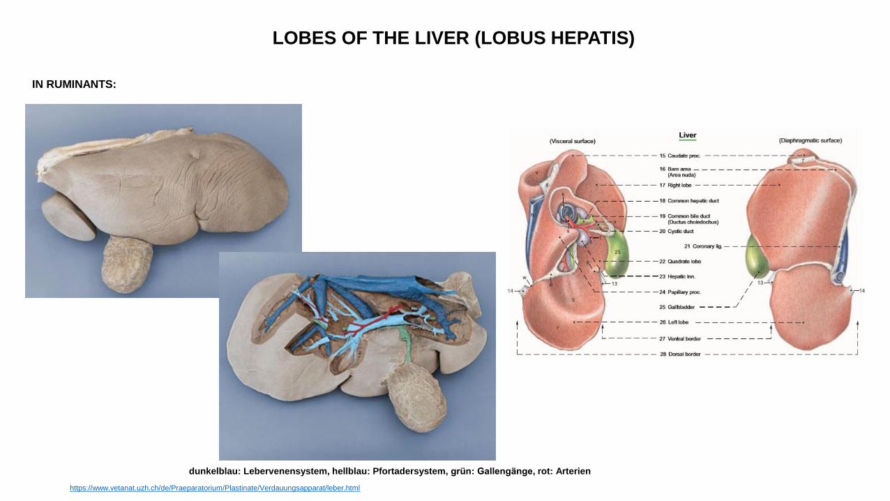
LOBES OF THE LIVER (LOBUS HEPATIS)
IN RUMINANTS:
1. Lobus hepatis sinsiter
2. Lobus hepatis dexter
3. Lobus quadratus
- between the fossa ligamenti teretis and gall bladder
4. Lobus caudatus
a. proc. caudatus – LARGE
b. proc. papillaris - small
UNDIVIDED!!!
1 Lobus dexter,
Lobus sinister,
2 3 Lobus caudatus,
4 Lobus quadratus,
5 Porta hepatica
(Arteria hepatica et Vena portae),
6 Lymphonodi hepatici,
7 Vesica fellea https://commons.wikimedia.org/wiki/File:Leber_Schaf.jpg
LOBES OF THE LIVER (LOBUS HEPATIS)
IN RUMINANTS:
dunkelblau: Lebervenensystem, hellblau: Pfortadersystem, grün: Gallengänge, rot: Arterien
https://www.vetanat.uzh.ch/de/Praeparatorium/Plastinate/Verdauungsapparat/leber.html
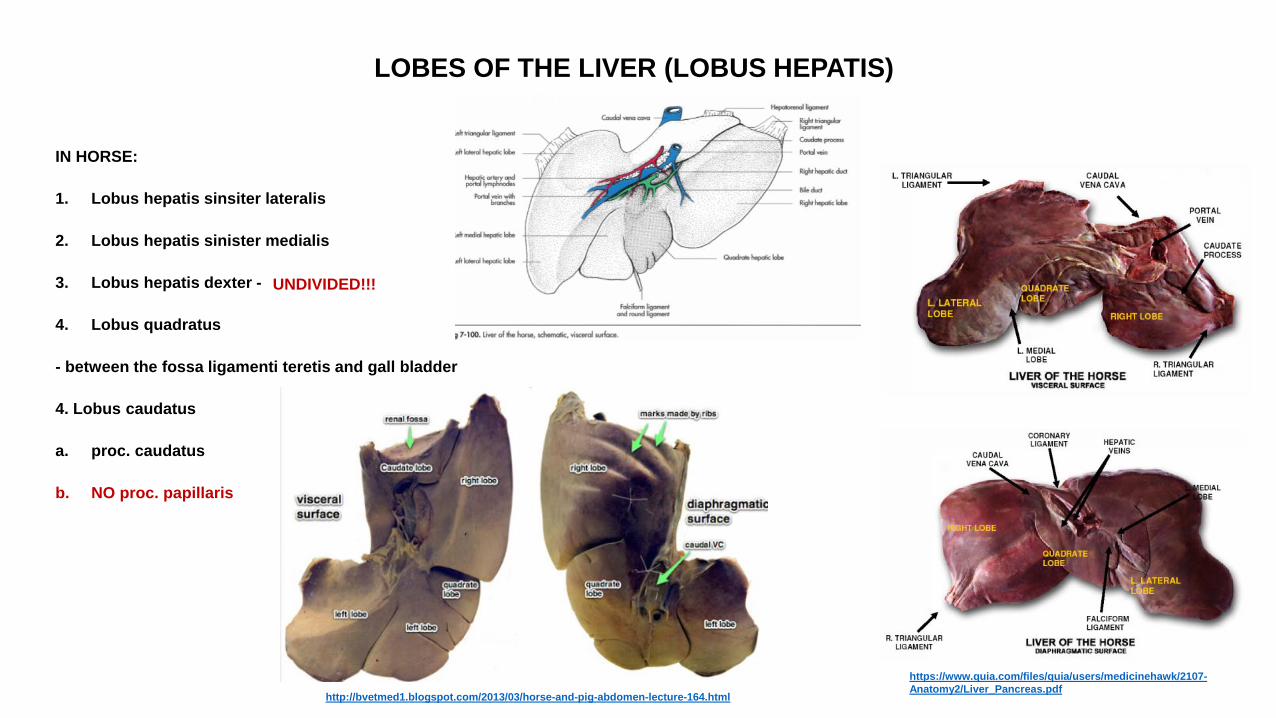
LOBES OF THE LIVER (LOBUS HEPATIS)
IN HORSE:
1. Lobus hepatis sinsiter lateralis
2. Lobus hepatis sinister medialis
3. Lobus hepatis dexter -
4. Lobus quadratus
- between the fossa ligamenti teretis and gall bladder
4. Lobus caudatus
a. proc. caudatus
b. NO proc. papillaris
UNDIVIDED!!!
http://bvetmed1.blogspot.com/2013/03/horse-and-pig-abdomen-lecture-164.html
https://www.quia.com/files/quia/users/medicinehawk/2107-
Anatomy2/Liver_Pancreas.pdf
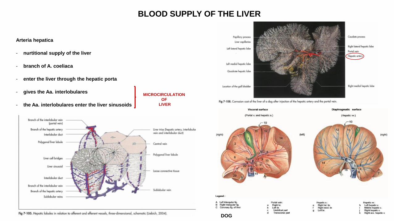
BLOOD SUPPLY OF THE LIVER
Arteria hepatica
- nurtitional supply of the liver
- branch of A. coeliaca
- enter the liver through the hepatic porta
- gives the Aa. interlobulares
- the Aa. interlobulares enter the liver sinusoids
MICROCIRCULATION
OF
LIVER
DOG
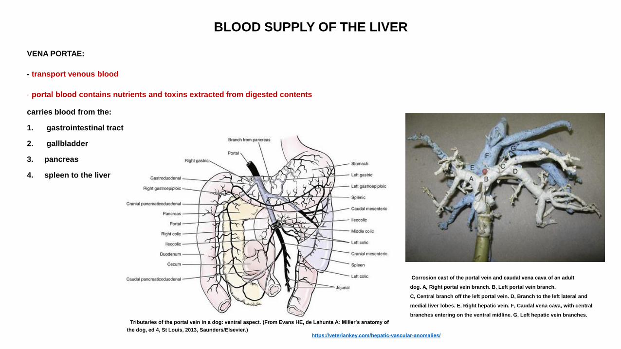
BLOOD SUPPLY OF THE LIVER
VENA PORTAE:
- transport venous blood
- portal blood contains nutrients and toxins extracted from digested contents
carries blood from the:
1. gastrointestinal tract
2. gallbladder
3. pancreas
4. spleen to the liver
Corrosion cast of the portal vein and caudal vena cava of an adult
dog. A, Right portal vein branch. B, Left portal vein branch.
C, Central branch off the left portal vein. D, Branch to the left lateral and
medial liver lobes. E, Right hepatic vein. F, Caudal vena cava, with central
branches entering on the ventral midline. G, Left hepatic vein branches.
Tributaries of the portal vein in a dog: ventral aspect. (From Evans HE, de Lahunta A: Miller’s anatomy of
the dog, ed 4, St Louis, 2013, Saunders/Elsevier.) https://veteriankey.com/hepatic-vascular-anomalies/

BLOOD SUPPLY OF THE LIVER
VEINS DRAIN INTO VENA PORTAE:
1. VENA LIENALIS
2. VENA MESENTERICA CRANIALIS
3. VENA MESENTERICA CAUDALIS
https://onlinelibrary.wiley.com/doi/pdf/10.1111/jsap.12392
Stylized view of the splanchnic vasculature; arrows show direction of blood flow.
https://www.sciencedirect.com/science/article/pii/S0022030206722007

MICROCIRCULATION OF THE LIVER
VENA PORTAE:
- enter the liver through porta hepatis
- gives Vv. interlobulares
- Vv. interlobulares enter the liver sinusoids
LIVER SINUSOIDS CONTAIN MIXED BLOOD:
a. blood from Aa. interlobulares
b. blood from Vv. Interlobulares
- liver sinusoids empty into the central veins
https://en.wikipedia.org/wiki/Liver#/media/File:2423_Microscopic_Anatomy_of_Liver.jpg Microscopic anatomy of the liver. (From Levy MN: Berne & Levy principles of physiology, ed 4, St Louis, 2005, Mosby/Elsevier.)
https://veteriankey.com/hepatic-vascular-anomalies/
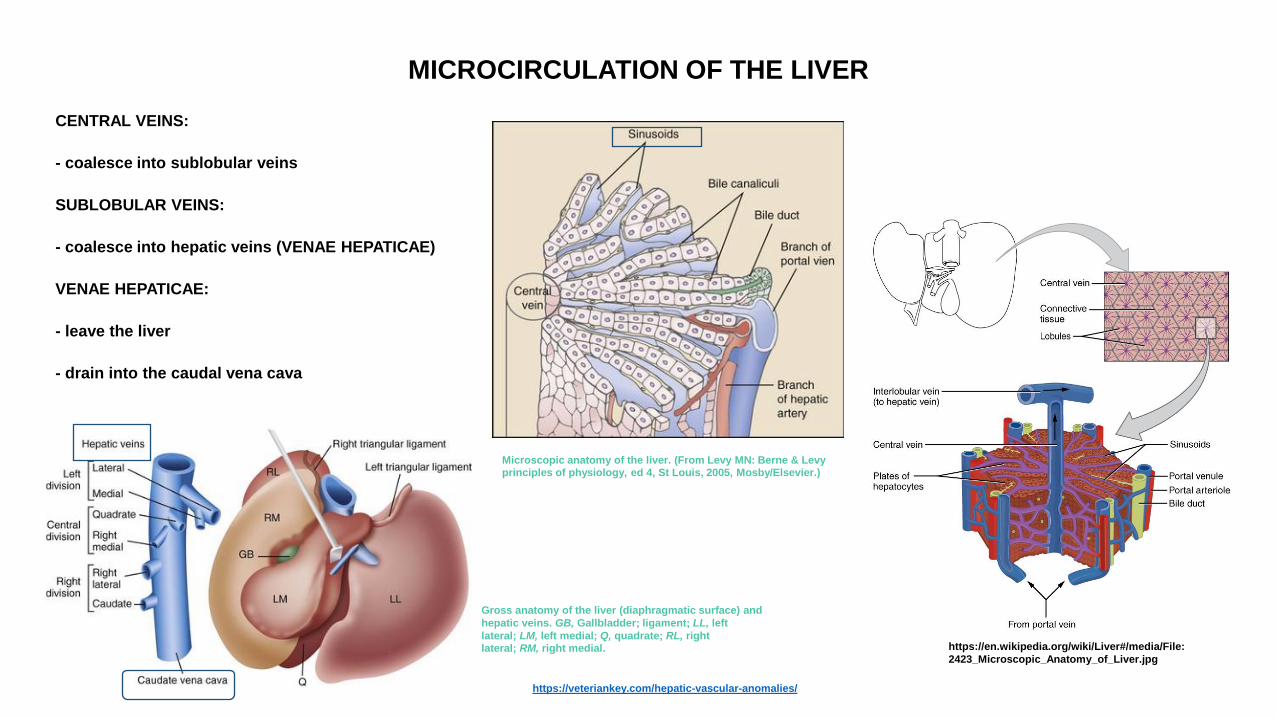
MICROCIRCULATION OF THE LIVER
CENTRAL VEINS:
- coalesce into sublobular veins
SUBLOBULAR VEINS:
- coalesce into hepatic veins (VENAE HEPATICAE)
VENAE HEPATICAE:
- leave the liver
- drain into the caudal vena cava
https://en.wikipedia.org/wiki/Liver#/media/File:
2423_Microscopic_Anatomy_of_Liver.jpg
Microscopic anatomy of the liver. (From Levy MN: Berne & Levy principles of physiology, ed 4, St Louis, 2005, Mosby/Elsevier.)
https://veteriankey.com/hepatic-vascular-anomalies/
Gross anatomy of the liver (diaphragmatic surface) and
hepatic veins. GB, Gallbladder; ligament; LL, left
lateral; LM, left medial; Q, quadrate; RL, right lateral; RM, right medial.
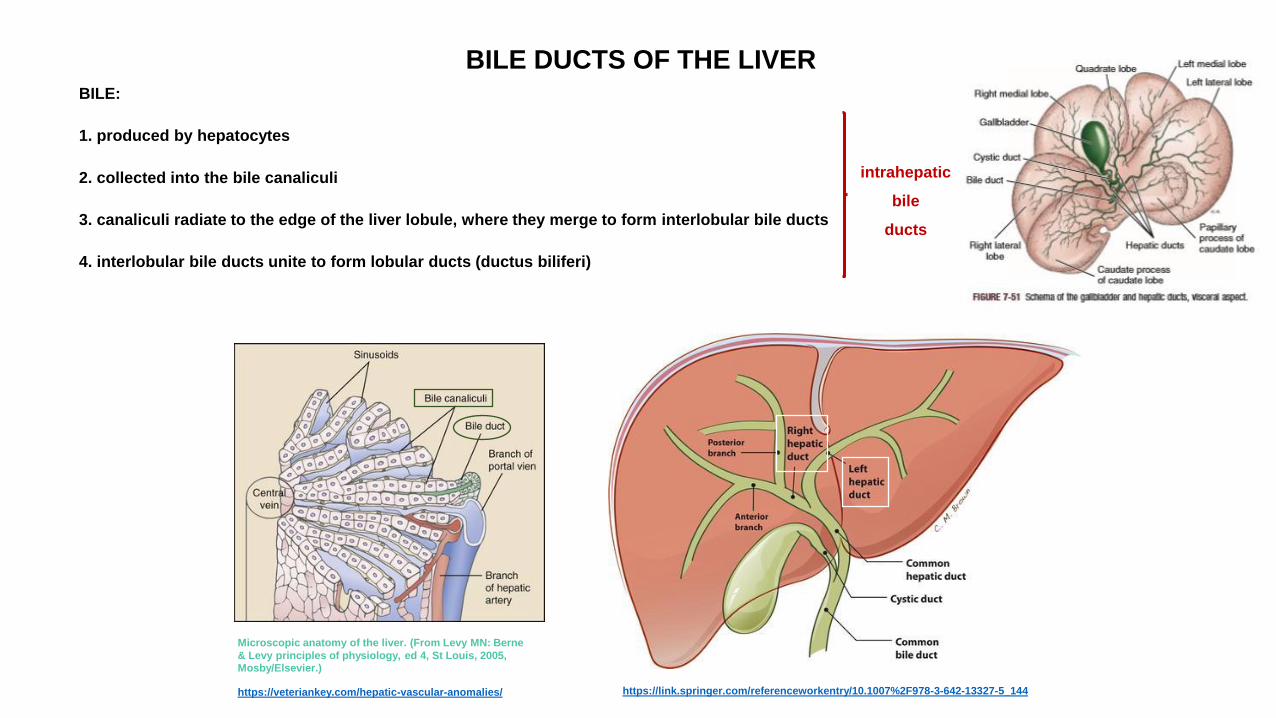
BILE DUCTS OF THE LIVER BILE:
1. produced by hepatocytes
2. collected into the bile canaliculi
3. canaliculi radiate to the edge of the liver lobule, where they merge to form interlobular bile ducts
4. interlobular bile ducts unite to form lobular ducts (ductus biliferi)
intrahepatic
bile
ducts
Microscopic anatomy of the liver. (From Levy MN: Berne
& Levy principles of physiology, ed 4, St Louis, 2005, Mosby/Elsevier.)
https://veteriankey.com/hepatic-vascular-anomalies/ https://link.springer.com/referenceworkentry/10.1007%2F978-3-642-13327-5_144
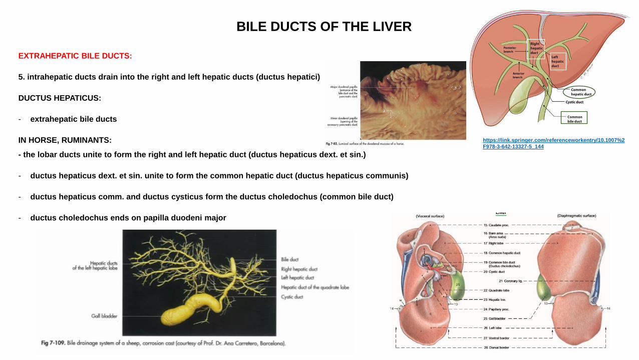
BILE DUCTS OF THE LIVER
EXTRAHEPATIC BILE DUCTS:
5. intrahepatic ducts drain into the right and left hepatic ducts (ductus hepatici)
DUCTUS HEPATICUS:
- extrahepatic bile ducts
IN HORSE, RUMINANTS:
- the lobar ducts unite to form the right and left hepatic duct (ductus hepaticus dext. et sin.)
- ductus hepaticus dext. et sin. unite to form the common hepatic duct (ductus hepaticus communis)
- ductus hepaticus comm. and ductus cysticus form the ductus choledochus (common bile duct)
- ductus choledochus ends on papilla duodeni major
https://link.springer.com/referenceworkentry/10.1007%2
F978-3-642-13327-5_144
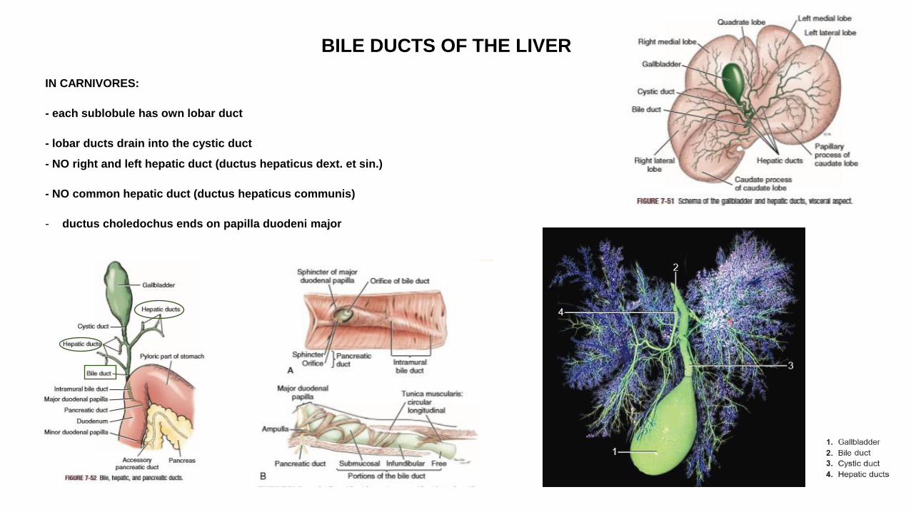
BILE DUCTS OF THE LIVER
IN CARNIVORES:
- each sublobule has own lobar duct
- lobar ducts drain into the cystic duct
- NO right and left hepatic duct (ductus hepaticus dext. et sin.)
- NO common hepatic duct (ductus hepaticus communis)
- ductus choledochus ends on papilla duodeni major
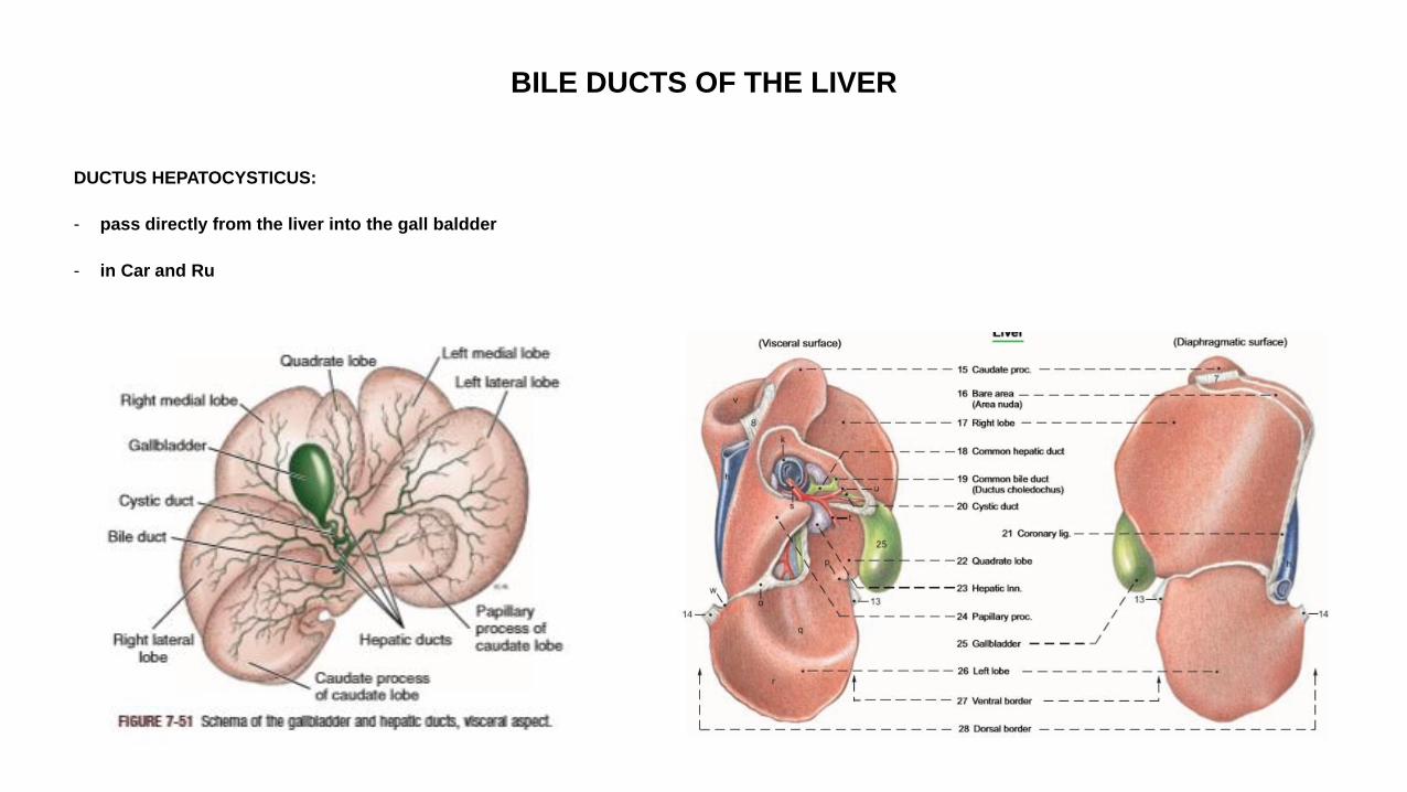
BILE DUCTS OF THE LIVER
DUCTUS HEPATOCYSTICUS:
- pass directly from the liver into the gall baldder
- in Car and Ru
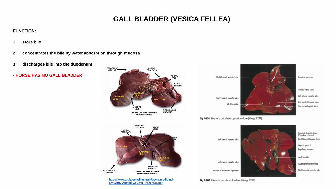
GALL BLADDER (VESICA FELLEA)
FUNCTION:
1. store bile
2. concentrates the bile by water absorption through mucosa
3. discharges bile into the duodenum
- HORSE HAS NO GALL BLADDER
https://www.quia.com/files/quia/users/medicineh
awk/2107-Anatomy2/Liver_Pancreas.pdf
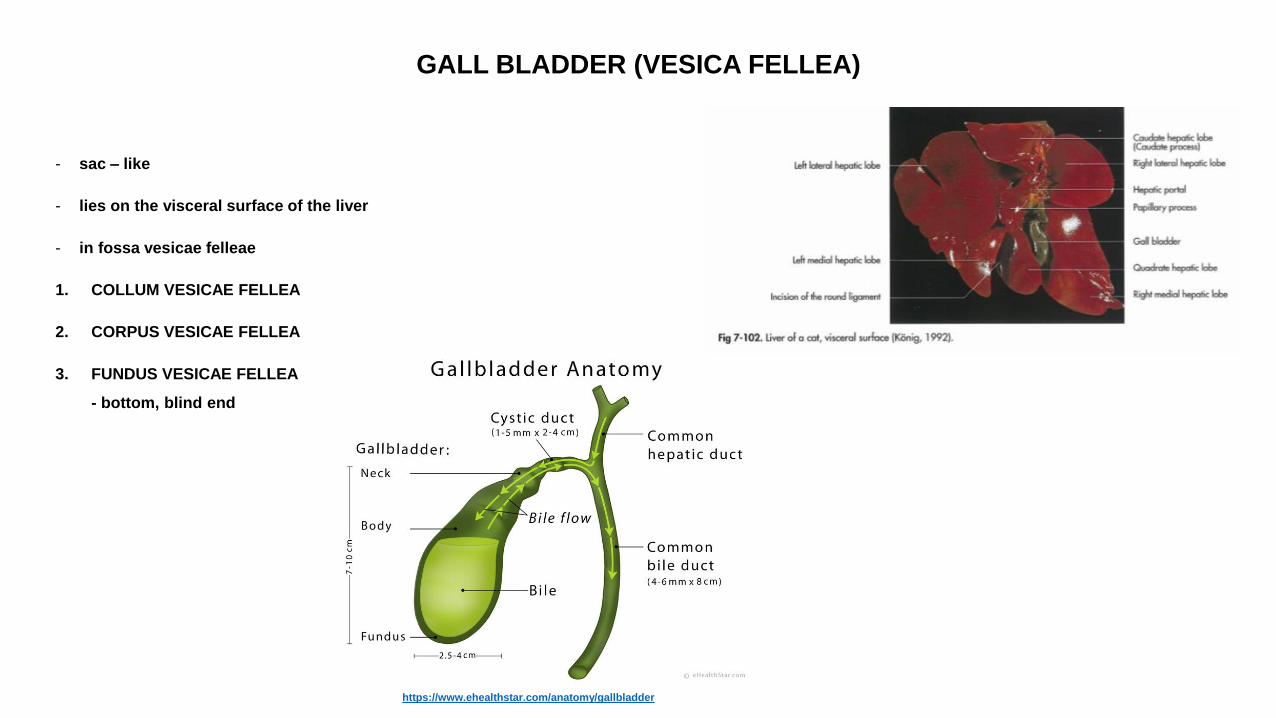
GALL BLADDER (VESICA FELLEA)
- sac – like
- lies on the visceral surface of the liver
- in fossa vesicae felleae
1. COLLUM VESICAE FELLEA
2. CORPUS VESICAE FELLEA
3. FUNDUS VESICAE FELLEA
- bottom, blind end
https://www.ehealthstar.com/anatomy/gallbladder
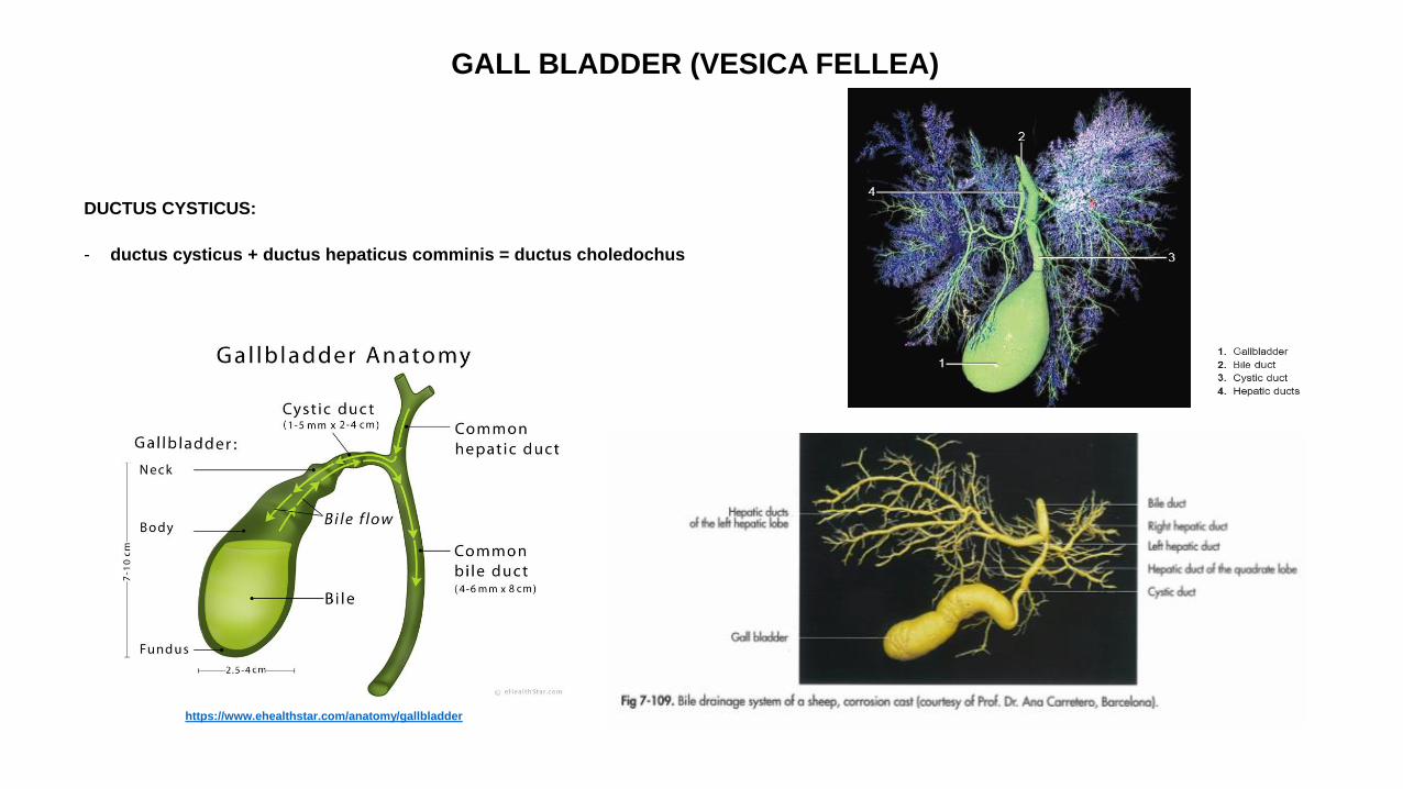
GALL BLADDER (VESICA FELLEA)
DUCTUS CYSTICUS:
- ductus cysticus + ductus hepaticus comminis = ductus choledochus
https://www.ehealthstar.com/anatomy/gallbladder
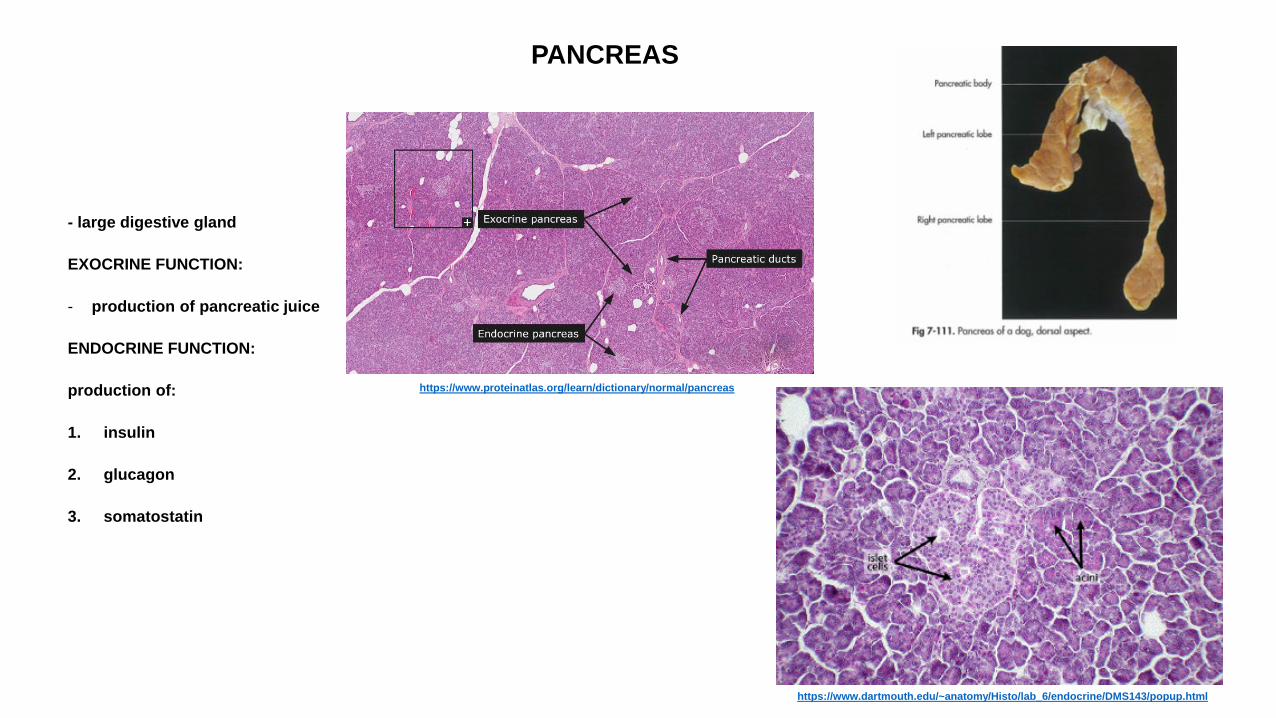
PANCREAS
- large digestive gland
EXOCRINE FUNCTION:
- production of pancreatic juice
ENDOCRINE FUNCTION:
production of:
1. insulin
2. glucagon
3. somatostatin
https://www.proteinatlas.org/learn/dictionary/normal/pancreas
https://www.dartmouth.edu/~anatomy/Histo/lab_6/endocrine/DMS143/popup.html
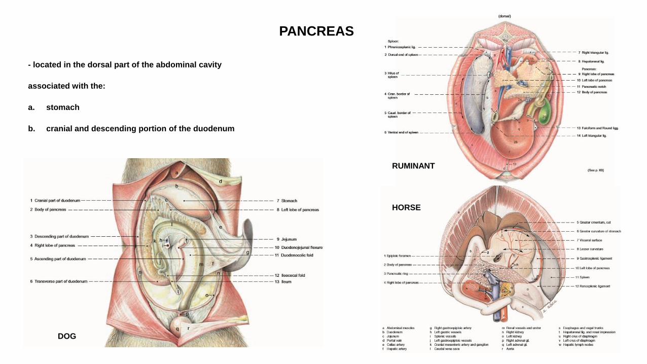
PANCREAS
- located in the dorsal part of the abdominal cavity
associated with the:
a. stomach
b. cranial and descending portion of the duodenum
DOG
HORSE
RUMINANT
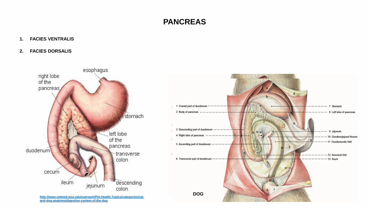
PANCREAS
1. FACIES VENTRALIS
2. FACIES DORSALIS
http://www.vetmed.wsu.edu/outreach/Pet-Health-Topics/categories/cat-
and-dog-anatomy/digestive-system-of-the-dog
DOG DOG
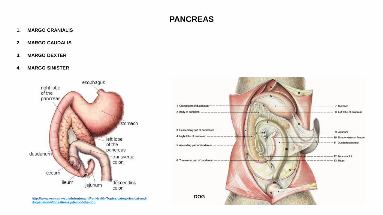
PANCREAS
1. MARGO CRANIALIS
2. MARGO CAUDALIS
3. MARGO DEXTER
4. MARGO SINISTER
DOG http://www.vetmed.wsu.edu/outreach/Pet-Health-Topics/categories/cat-and-
dog-anatomy/digestive-system-of-the-dog
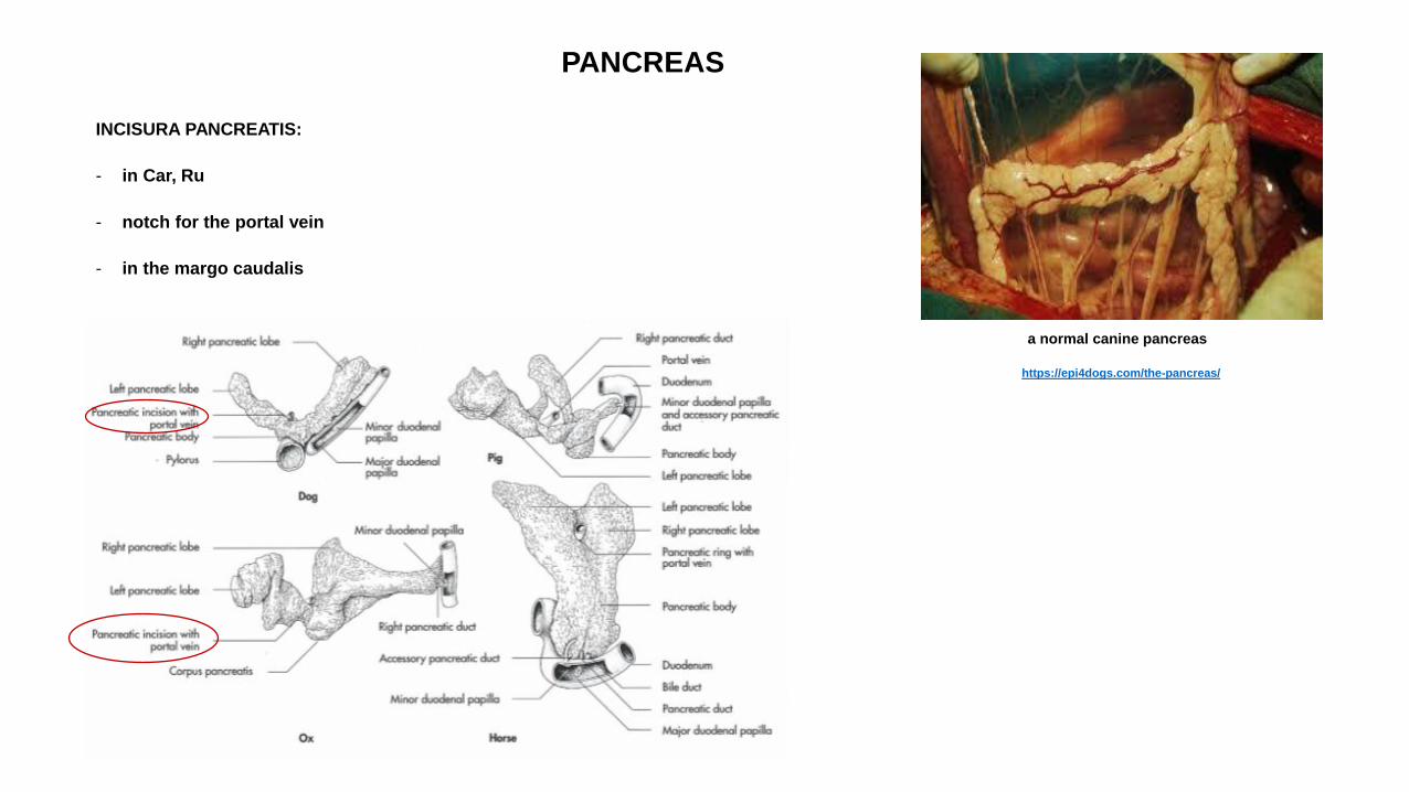
PANCREAS
INCISURA PANCREATIS:
- in Car, Ru
- notch for the portal vein
- in the margo caudalis
a normal canine pancreas
https://epi4dogs.com/the-pancreas/
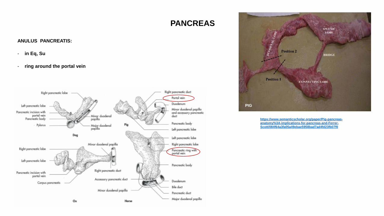
PANCREAS
ANULUS PANCREATIS:
- in Eq, Su
- ring around the portal vein
https://www.semanticscholar.org/paper/Pig-pancreas-
anatomy%3A-implications-for-pancreas-and-Ferrer-
Scott/084f64a3fa05a49ebae5958bad7ad4fd23fb07f0
PIG
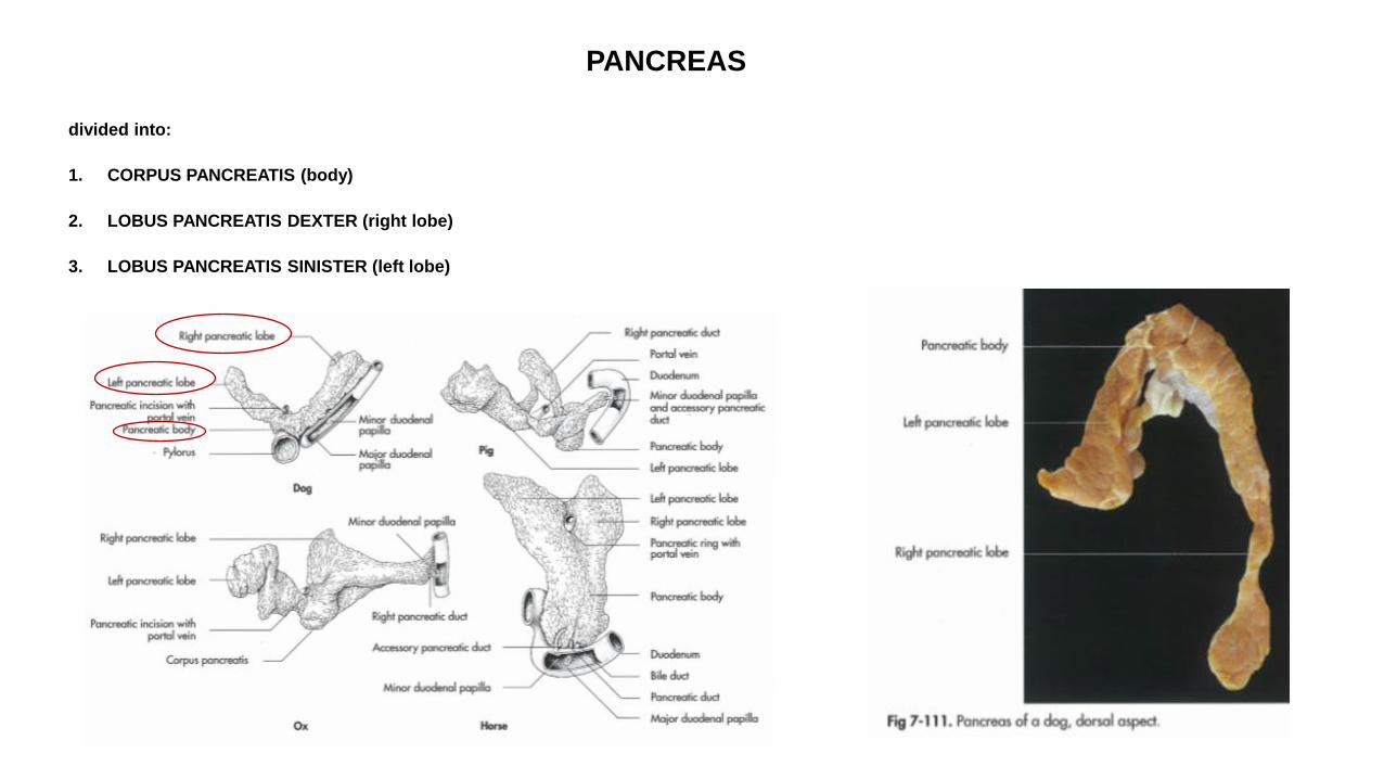
PANCREAS
divided into:
1. CORPUS PANCREATIS (body)
2. LOBUS PANCREATIS DEXTER (right lobe)
3. LOBUS PANCREATIS SINISTER (left lobe)
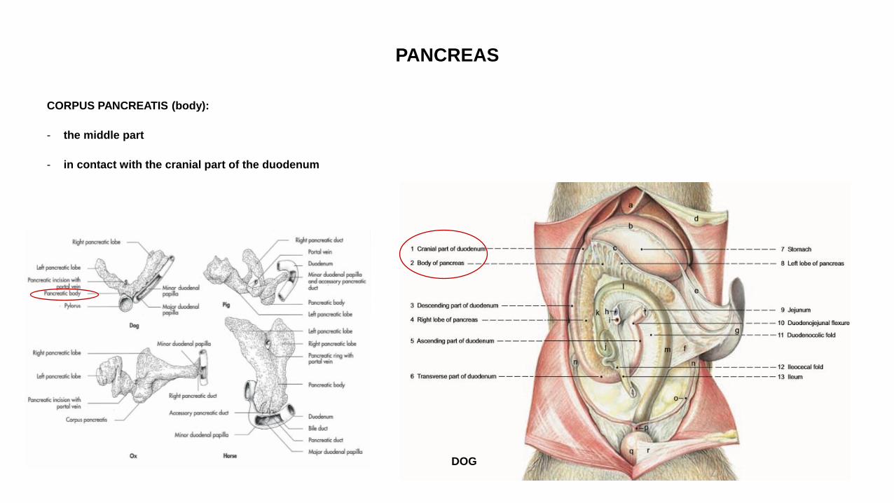
PANCREAS
CORPUS PANCREATIS (body):
- the middle part
- in contact with the cranial part of the duodenum
DOG
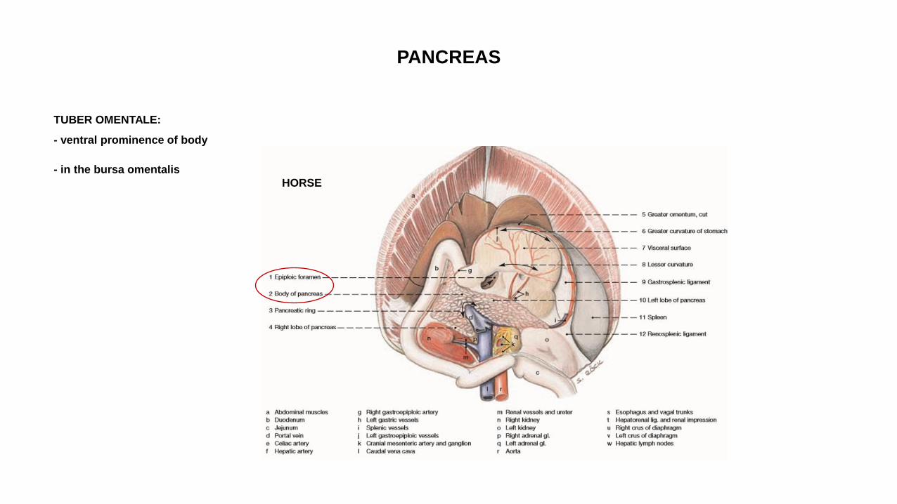
PANCREAS
TUBER OMENTALE:
- ventral prominence of body
- in the bursa omentalis
HORSE
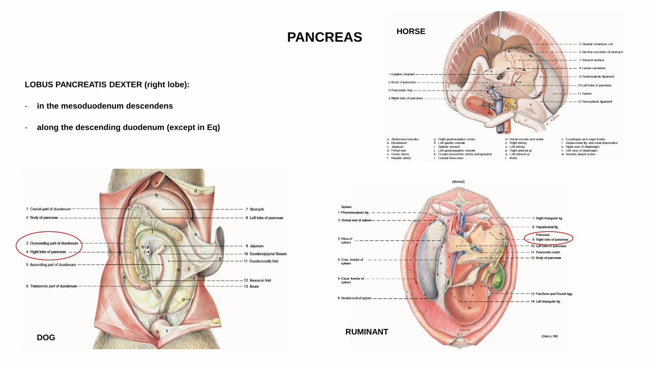
PANCREAS
LOBUS PANCREATIS DEXTER (right lobe):
- in the mesoduodenum descendens
- along the descending duodenum (except in Eq)
DOG RUMINANT
HORSE
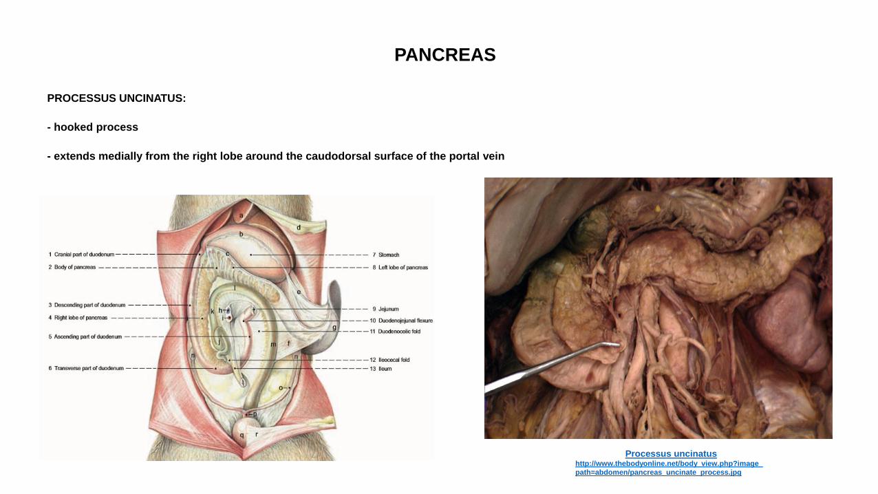
PANCREAS
PROCESSUS UNCINATUS:
- hooked process
- extends medially from the right lobe around the caudodorsal surface of the portal vein
Processus uncinatus http://www.thebodyonline.net/body_view.php?image_
path=abdomen/pancreas_uncinate_process.jpg
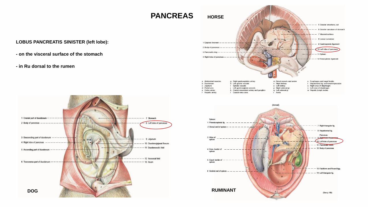
PANCREAS
LOBUS PANCREATIS SINISTER (left lobe):
- on the visceral surface of the stomach
- in Ru dorsal to the rumen
DOG RUMINANT
HORSE
PANCREAS
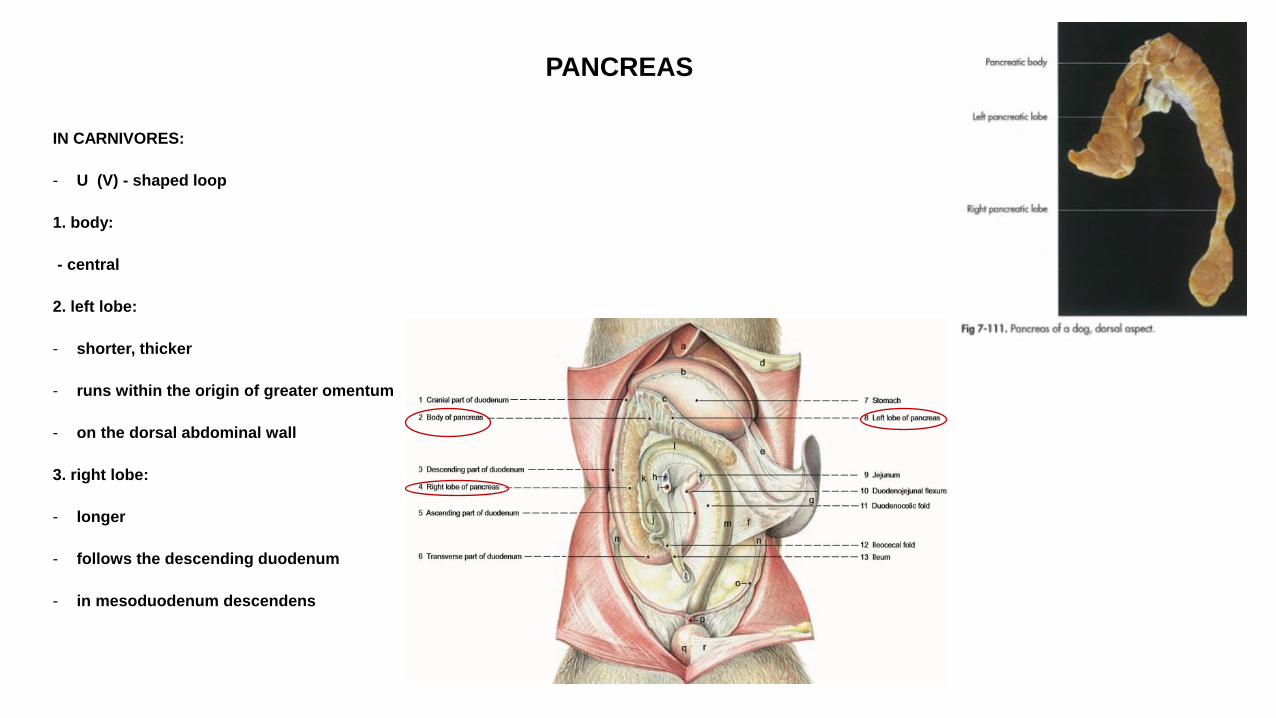
IN CARNIVORES:
- U (V) - shaped loop
1. body:
- central
2. left lobe:
- shorter, thicker
- runs within the origin of greater omentum
- on the dorsal abdominal wall
3. right lobe:
- longer
- follows the descending duodenum
- in mesoduodenum descendens
PANCREAS
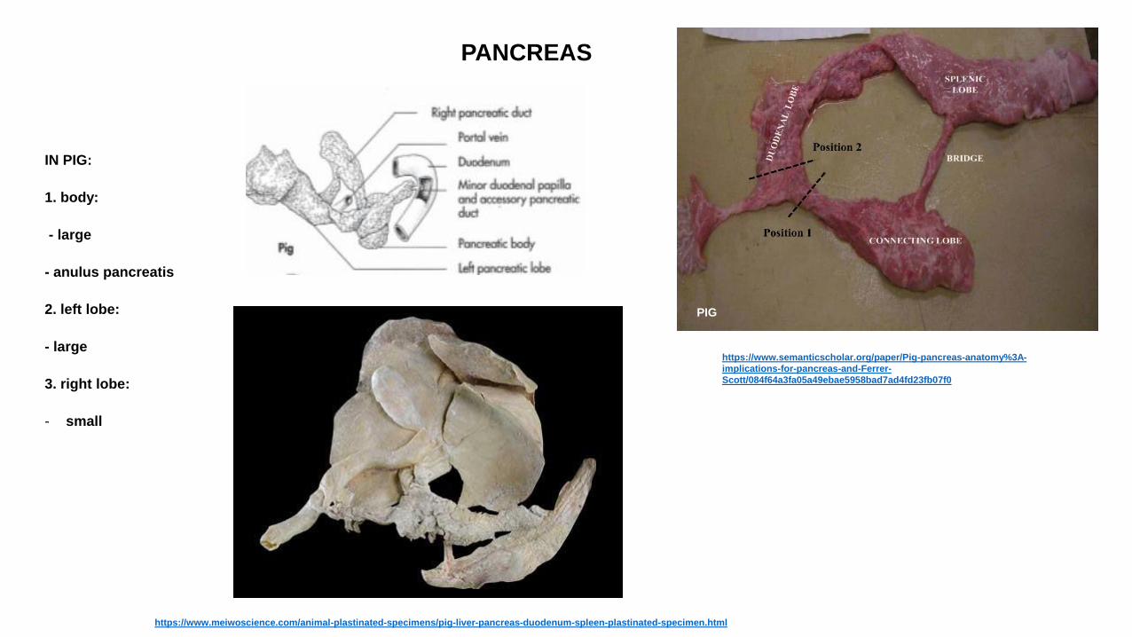
IN PIG:
1. body:
- large
- anulus pancreatis
2. left lobe:
- large
3. right lobe:
- small
https://www.semanticscholar.org/paper/Pig-pancreas-anatomy%3A-
implications-for-pancreas-and-Ferrer-
Scott/084f64a3fa05a49ebae5958bad7ad4fd23fb07f0
PIG
https://www.meiwoscience.com/animal-plastinated-specimens/pig-liver-pancreas-duodenum-spleen-plastinated-specimen.html

PANCREAS
IN HORSE:
- triangular - shaped
1. body:
- large, compact
- anulus pancreatis
2. left lobe:
- long
3. right lobe:
- short
HORSE
PANCREAS
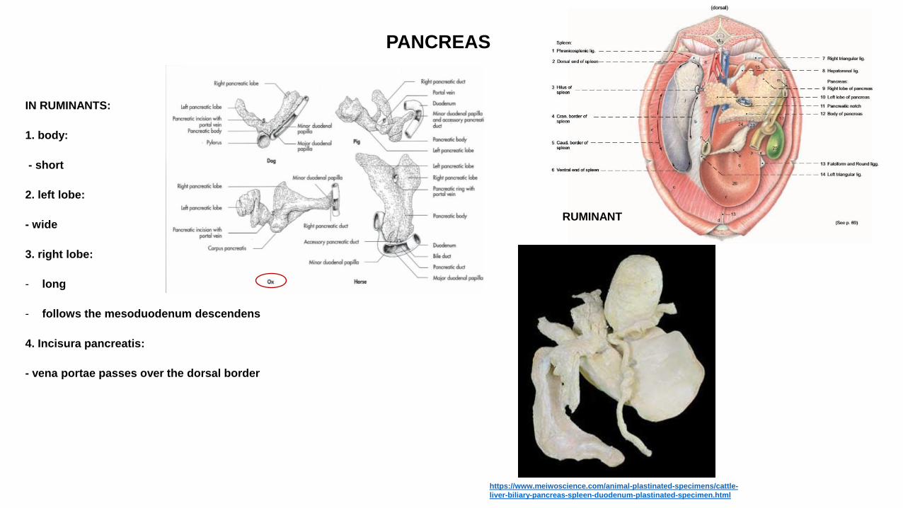
IN RUMINANTS:
1. body:
- short
2. left lobe:
- wide
3. right lobe:
- long
- follows the mesoduodenum descendens
4. Incisura pancreatis:
- vena portae passes over the dorsal border
RUMINANT
https://www.meiwoscience.com/animal-plastinated-specimens/cattle-
liver-biliary-pancreas-spleen-duodenum-plastinated-specimen.html
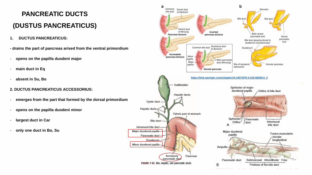
PANCREATIC DUCTS
(DUSTUS PANCREATICUS)
1. DUCTUS PANCREATICUS:
- drains the part of pancreas arised from the ventral primordium
- opens on the papilla duodeni major
- main duct in Eq
- absent in Su, Bo
2. DUCTUS PANCREATICUS ACCESSORIUS:
- emerges from the part that formed by the dorsal primordium
- opens on the papilla duodeni minor
- largest duct in Car
- only one duct in Bo, Su
https://link.springer.com/chapter/10.1007/978-3-319-58256-6_2
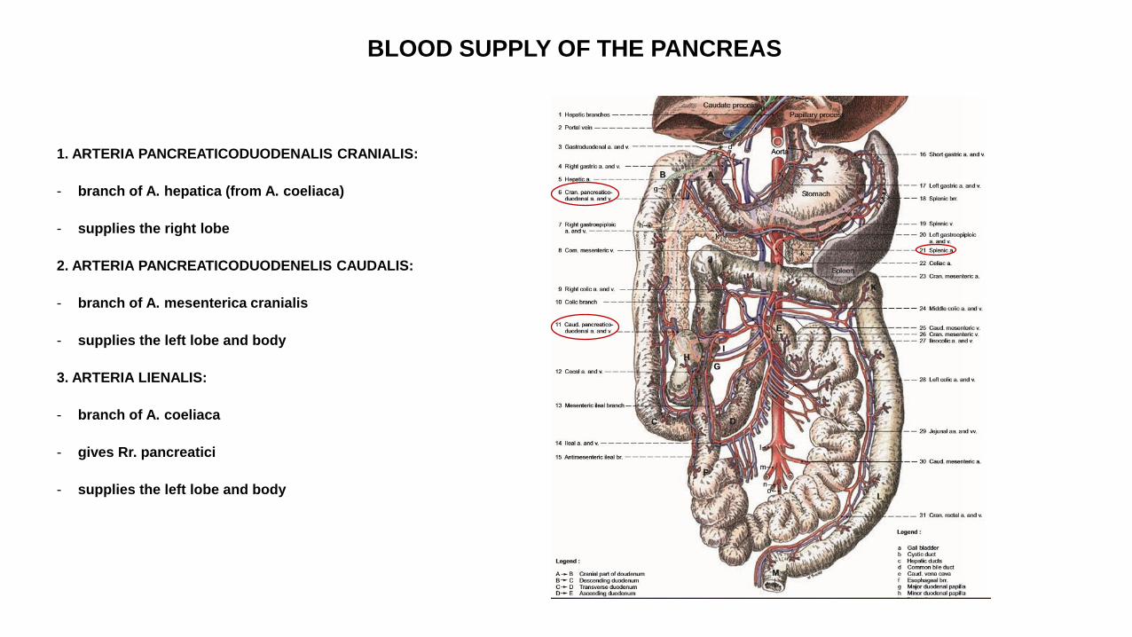
BLOOD SUPPLY OF THE PANCREAS
1. ARTERIA PANCREATICODUODENALIS CRANIALIS:
- branch of A. hepatica (from A. coeliaca)
- supplies the right lobe
2. ARTERIA PANCREATICODUODENELIS CAUDALIS:
- branch of A. mesenterica cranialis
- supplies the left lobe and body
3. ARTERIA LIENALIS:
- branch of A. coeliaca
- gives Rr. pancreatici
- supplies the left lobe and body
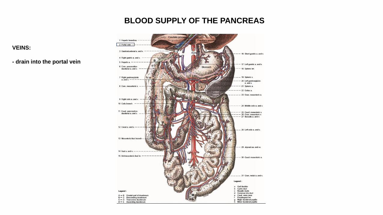
BLOOD SUPPLY OF THE PANCREAS
VEINS:
- drain into the portal vein
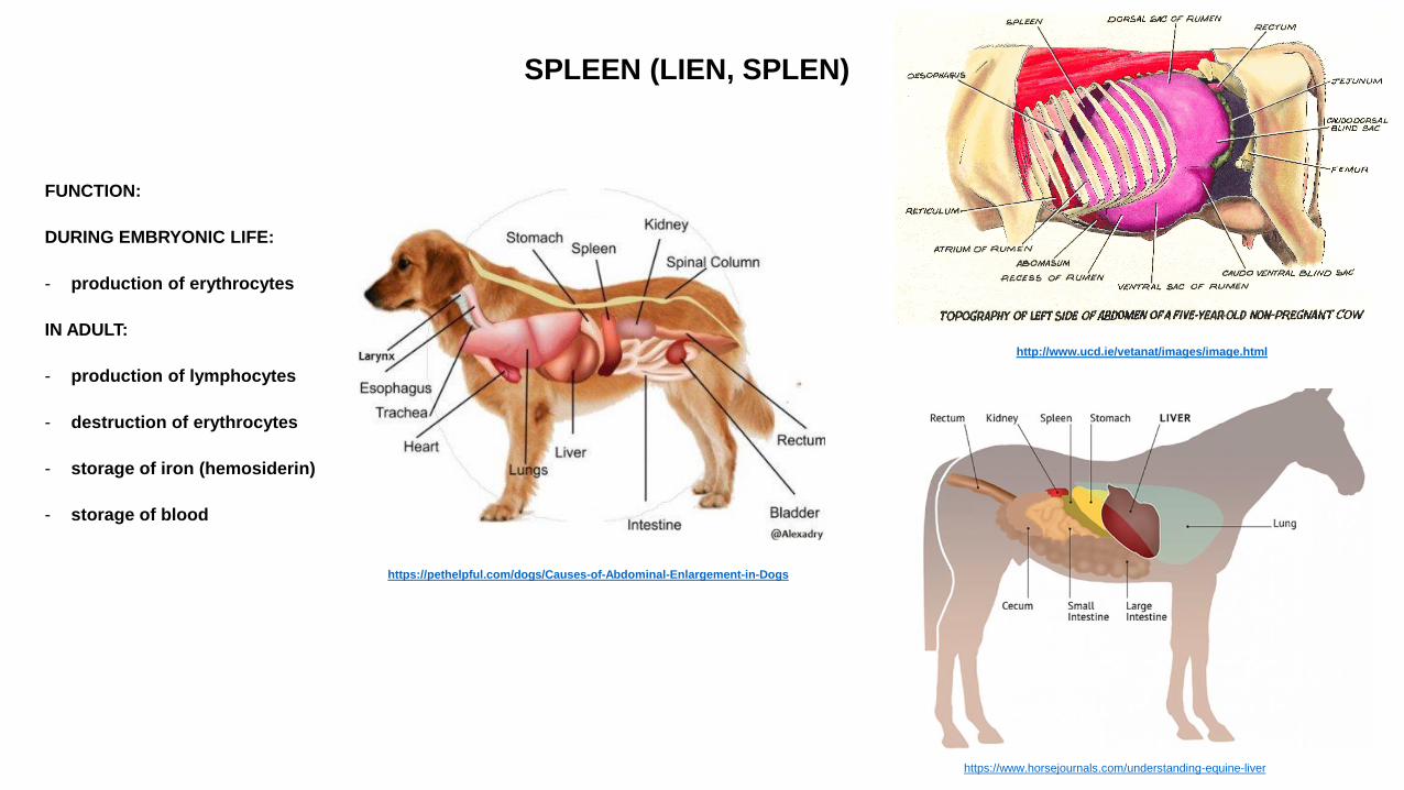
SPLEEN (LIEN, SPLEN)
FUNCTION:
DURING EMBRYONIC LIFE:
- production of erythrocytes
IN ADULT:
- production of lymphocytes
- destruction of erythrocytes
- storage of iron (hemosiderin)
- storage of blood
https://pethelpful.com/dogs/Causes-of-Abdominal-Enlargement-in-Dogs
http://www.ucd.ie/vetanat/images/image.html
https://www.horsejournals.com/understanding-equine-liver
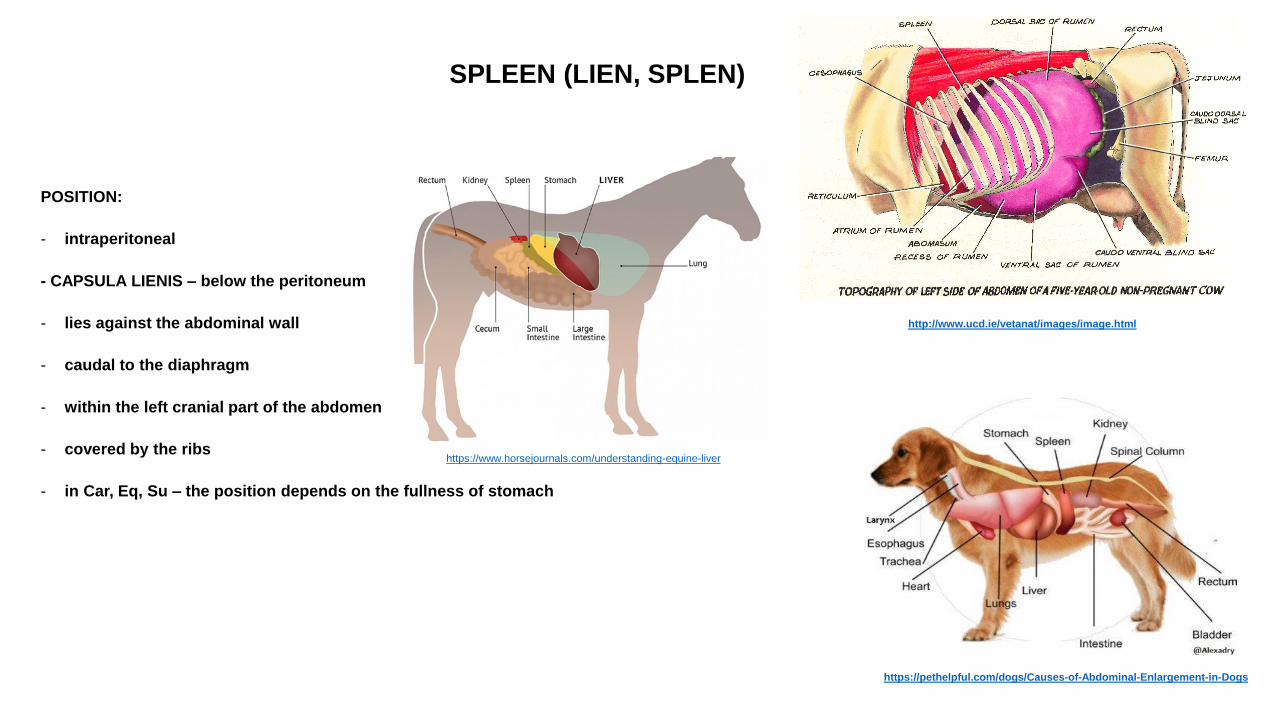
SPLEEN (LIEN, SPLEN)
POSITION:
- intraperitoneal
- CAPSULA LIENIS – below the peritoneum
- lies against the abdominal wall
- caudal to the diaphragm
- within the left cranial part of the abdomen
- covered by the ribs
- in Car, Eq, Su – the position depends on the fullness of stomach
https://pethelpful.com/dogs/Causes-of-Abdominal-Enlargement-in-Dogs
http://www.ucd.ie/vetanat/images/image.html
https://www.horsejournals.com/understanding-equine-liver
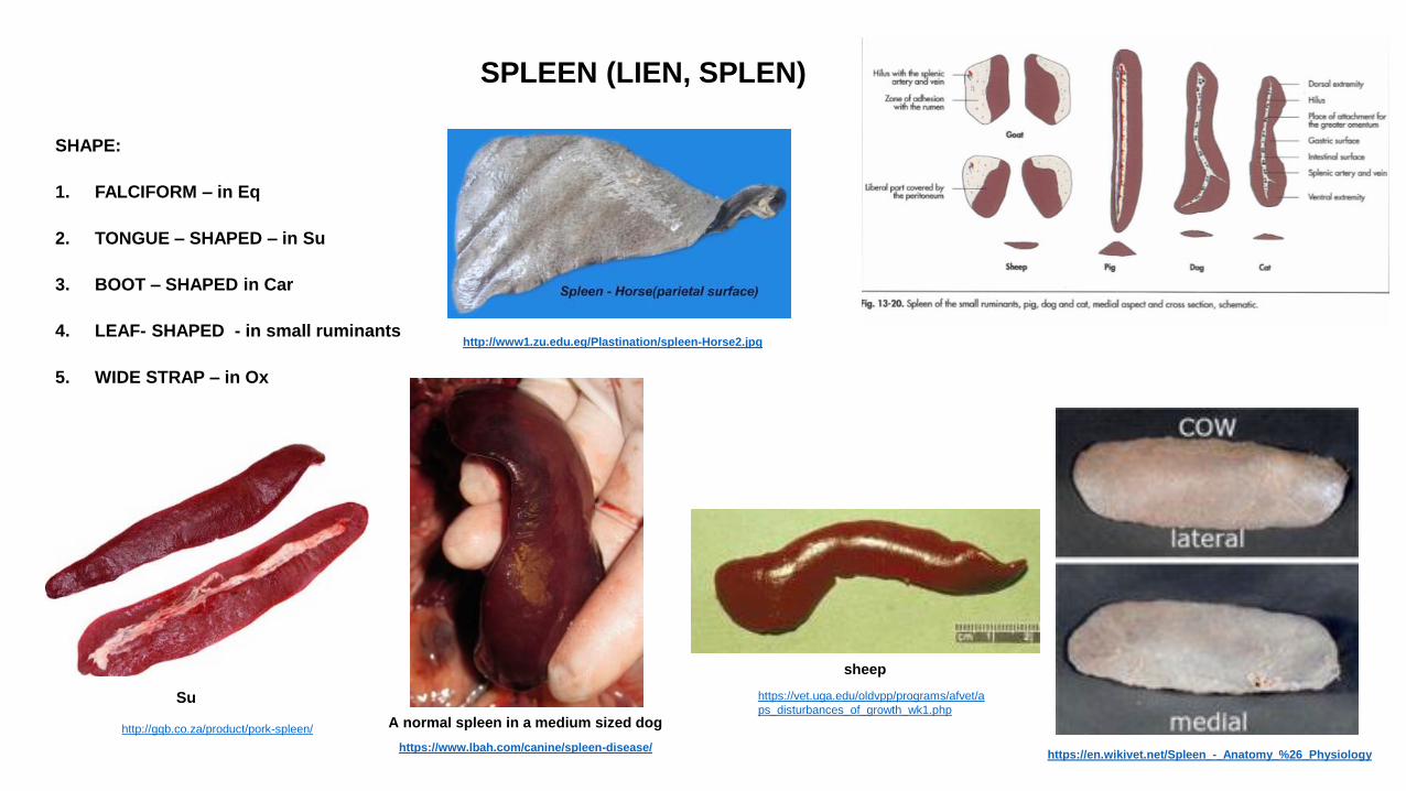
SPLEEN (LIEN, SPLEN)
SHAPE:
1. FALCIFORM – in Eq
2. TONGUE – SHAPED – in Su
3. BOOT – SHAPED in Car
4. LEAF- SHAPED - in small ruminants
5. WIDE STRAP – in Ox
A normal spleen in a medium sized dog
https://www.lbah.com/canine/spleen-disease/
Su
http://gqb.co.za/product/pork-spleen/
https://vet.uga.edu/oldvpp/programs/afvet/a
ps_disturbances_of_growth_wk1.php
sheep
http://www1.zu.edu.eg/Plastination/spleen-Horse2.jpg
https://en.wikivet.net/Spleen_-_Anatomy_%26_Physiology
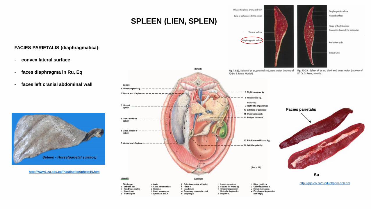
SPLEEN (LIEN, SPLEN)
FACIES PARIETALIS (diaphragmatica):
- convex lateral surface
- faces diaphragma in Ru, Eq
- faces left cranial abdominal wall
http://www1.zu.edu.eg/Plastination/photo16.htm Su
http://gqb.co.za/product/pork-spleen/
Facies parietalis
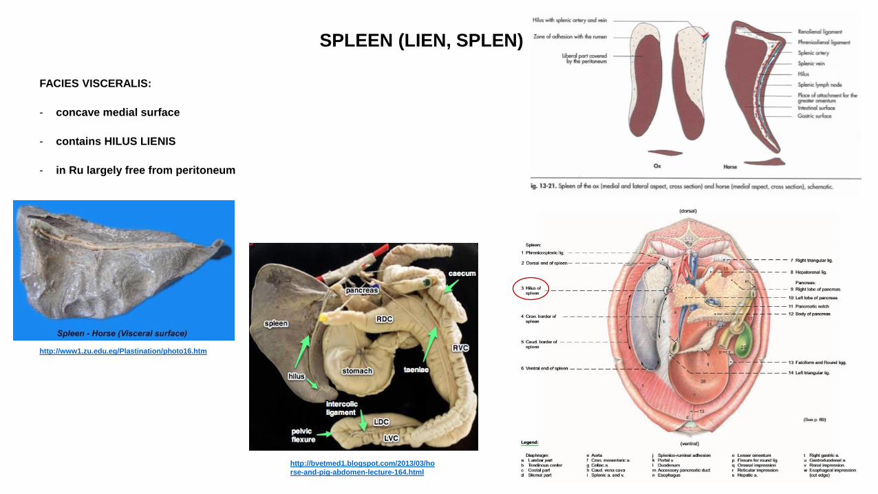
SPLEEN (LIEN, SPLEN)
FACIES VISCERALIS:
- concave medial surface
- contains HILUS LIENIS
- in Ru largely free from peritoneum
http://bvetmed1.blogspot.com/2013/03/ho
rse-and-pig-abdomen-lecture-164.html
http://www1.zu.edu.eg/Plastination/photo16.htm
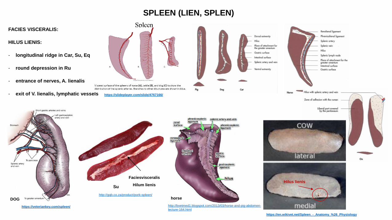
SPLEEN (LIEN, SPLEN)
FACIES VISCERALIS:
HILUS LIENIS:
- longitudinal ridge in Car, Su, Eq
- round depression in Ru
- entrance of nerves, A. lienalis
- exit of V. lienalis, lymphatic vessels
https://slideplayer.com/slide/4767166/
http://bvetmed1.blogspot.com/2013/03/horse-and-pig-abdomen-
lecture-164.html
horse
Su
http://gqb.co.za/product/pork-spleen/
Faciesvisceralis
Hilum lienis
https://veteriankey.com/spleen/
DOG
https://en.wikivet.net/Spleen_-_Anatomy_%26_Physiology
Hilus lienis
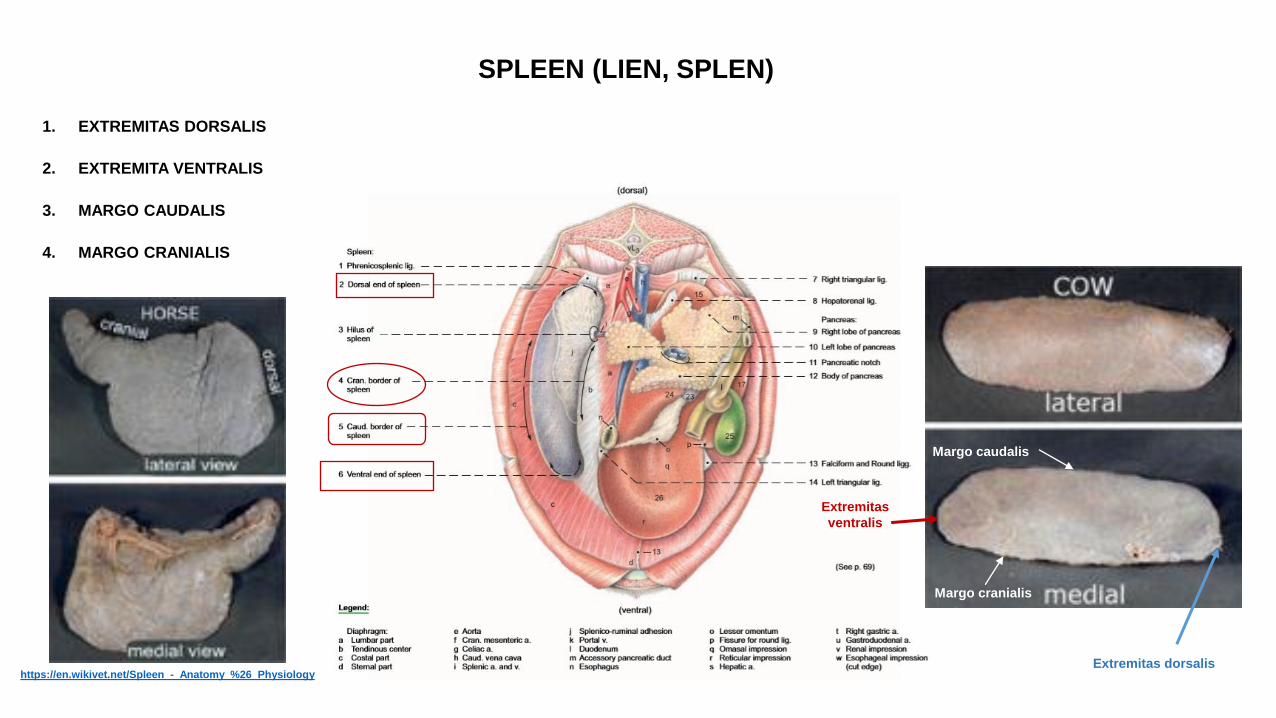
SPLEEN (LIEN, SPLEN)
1. EXTREMITAS DORSALIS
2. EXTREMITA VENTRALIS
3. MARGO CAUDALIS
4. MARGO CRANIALIS
https://en.wikivet.net/Spleen_-_Anatomy_%26_Physiology
Margo cranialis
Margo caudalis
Extremitas dorsalis
Extremitas
ventralis
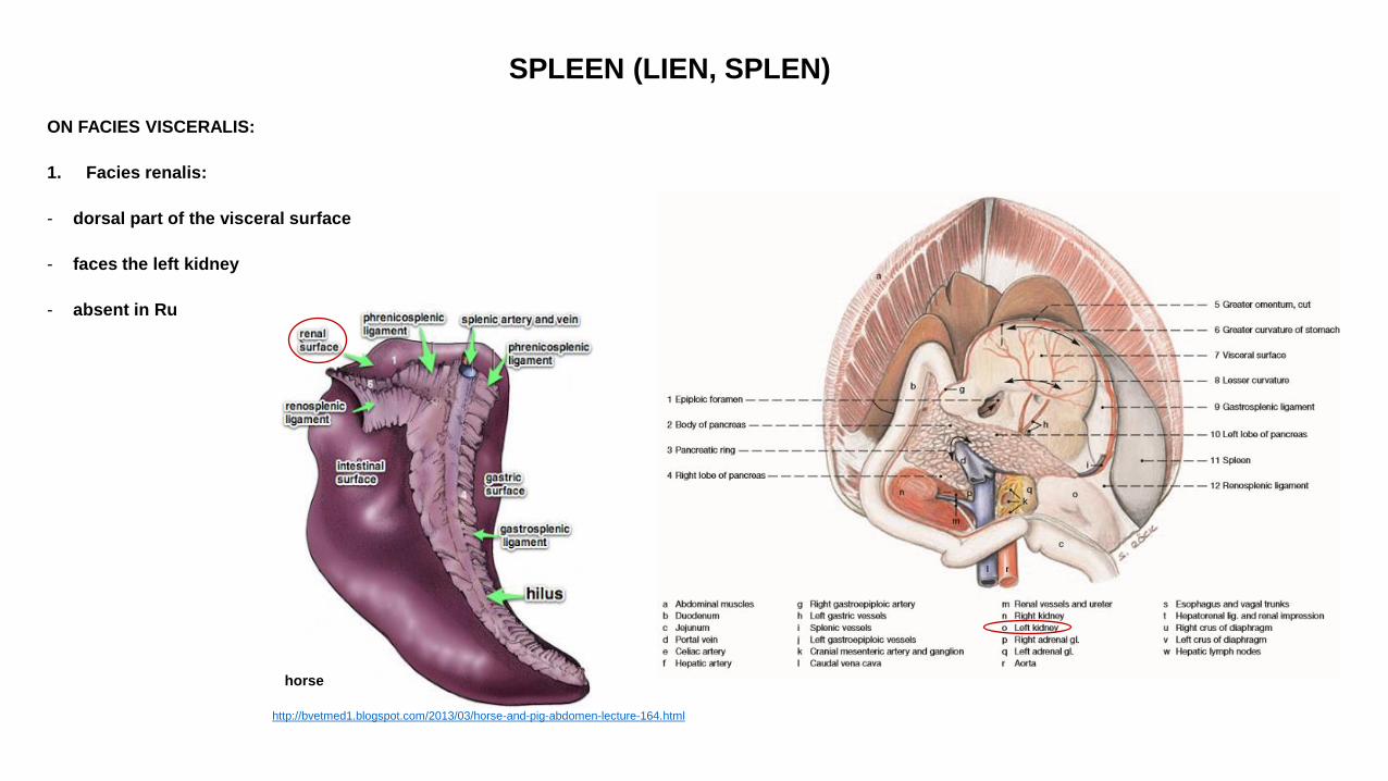
SPLEEN (LIEN, SPLEN)
ON FACIES VISCERALIS:
1. Facies renalis:
- dorsal part of the visceral surface
- faces the left kidney
- absent in Ru
http://bvetmed1.blogspot.com/2013/03/horse-and-pig-abdomen-lecture-164.html
horse
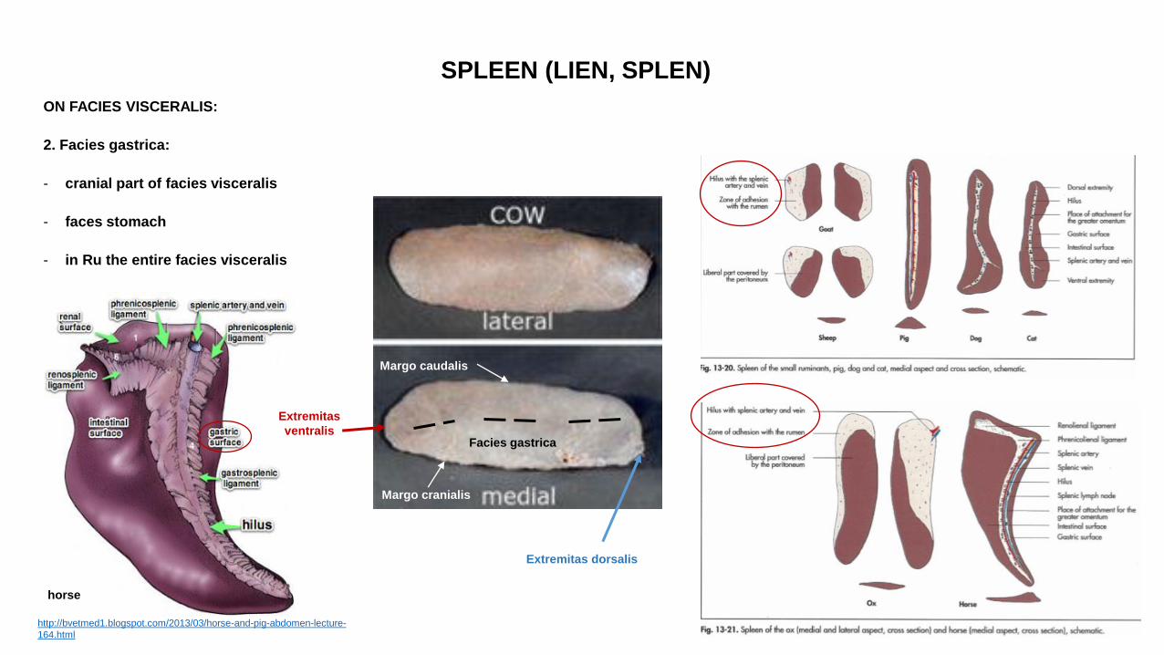
SPLEEN (LIEN, SPLEN)
ON FACIES VISCERALIS:
2. Facies gastrica:
- cranial part of facies visceralis
- faces stomach
- in Ru the entire facies visceralis
http://bvetmed1.blogspot.com/2013/03/horse-and-pig-abdomen-lecture-
164.html
horse
Margo cranialis
Margo caudalis
Extremitas dorsalis
Extremitas
ventralis Facies gastrica
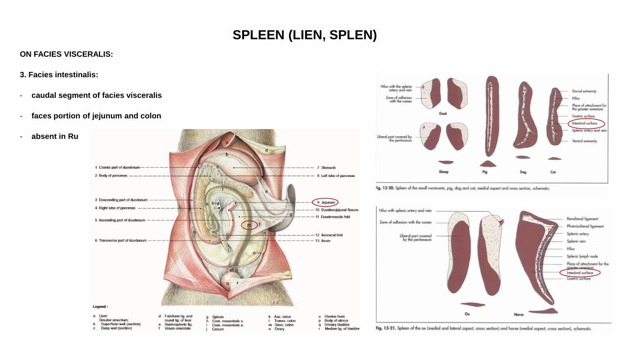
SPLEEN (LIEN, SPLEN)
ON FACIES VISCERALIS:
3. Facies intestinalis:
- caudal segment of facies visceralis
- faces portion of jejunum and colon
- absent in Ru
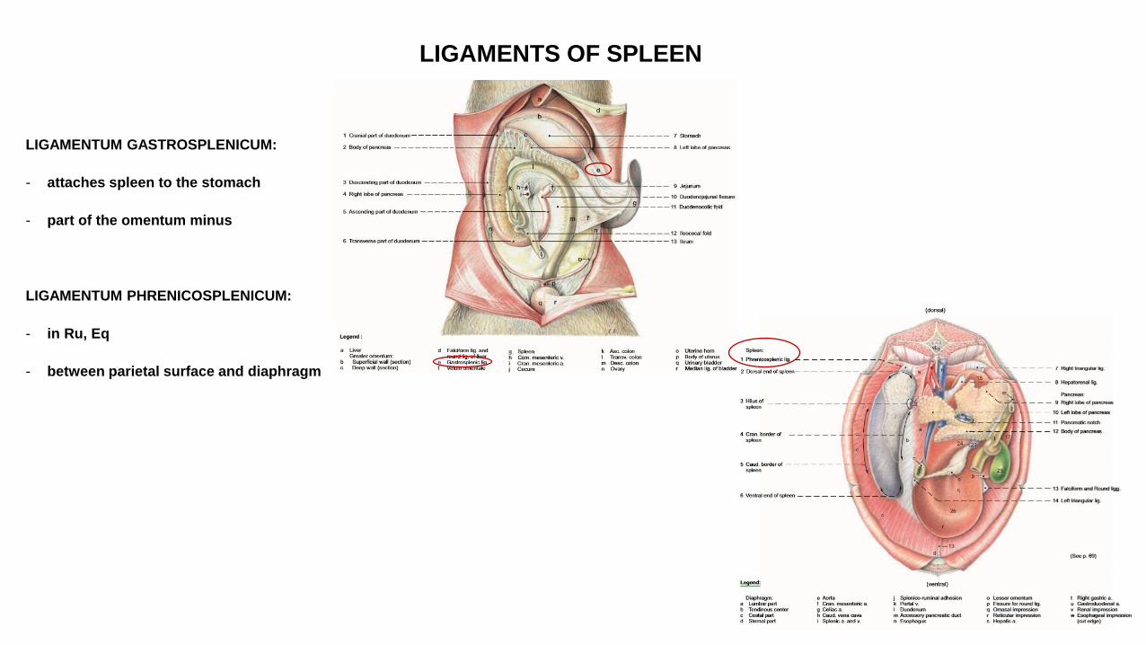
LIGAMENTS OF SPLEEN
LIGAMENTUM GASTROSPLENICUM:
- attaches spleen to the stomach
- part of the omentum minus
LIGAMENTUM PHRENICOSPLENICUM:
- in Ru, Eq
- between parietal surface and diaphragm
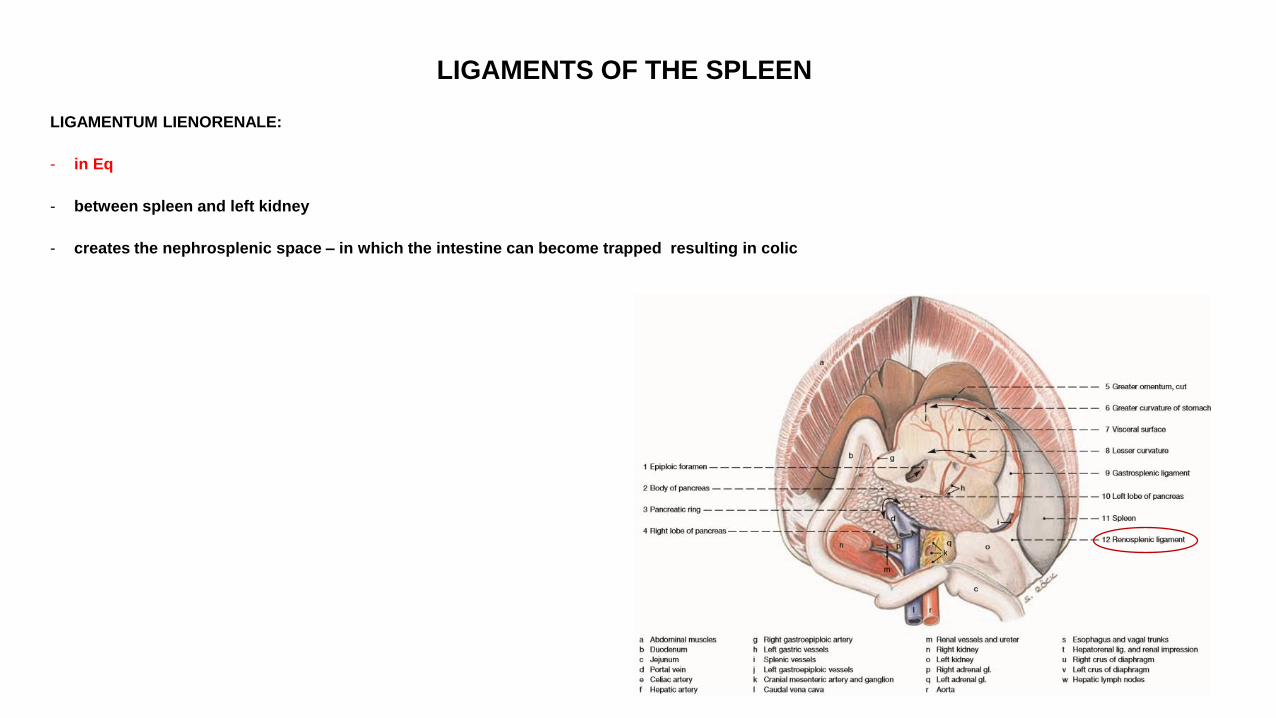
LIGAMENTS OF THE SPLEEN
LIGAMENTUM LIENORENALE:
- in Eq
- between spleen and left kidney
- creates the nephrosplenic space – in which the intestine can become trapped resulting in colic
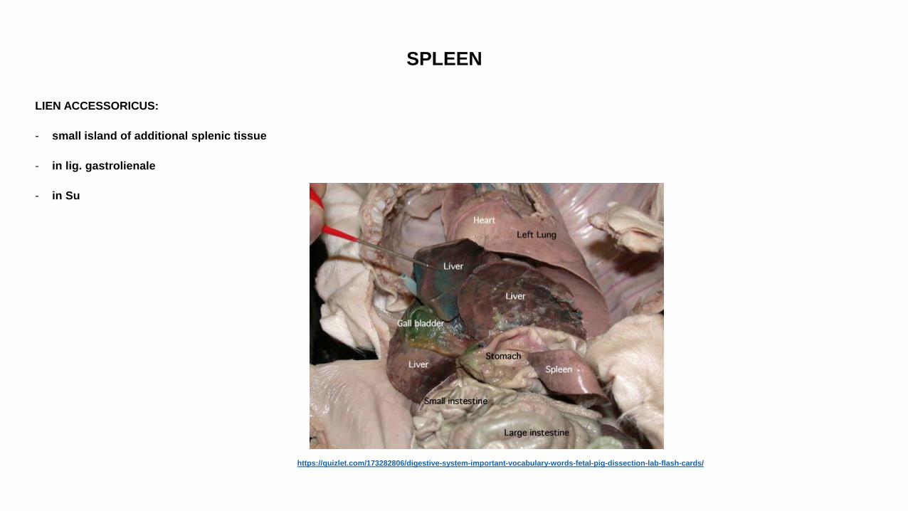
SPLEEN
LIEN ACCESSORICUS:
- small island of additional splenic tissue
- in lig. gastrolienale
- in Su
https://quizlet.com/173282806/digestive-system-important-vocabulary-words-fetal-pig-dissection-lab-flash-cards/
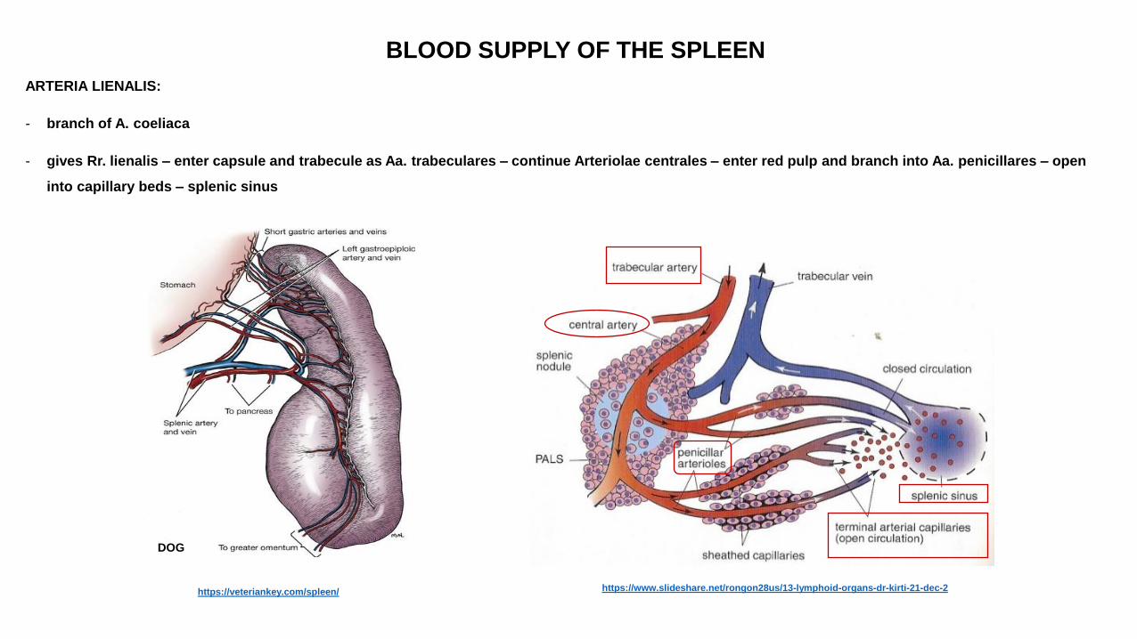
BLOOD SUPPLY OF THE SPLEEN
ARTERIA LIENALIS:
- branch of A. coeliaca
- gives Rr. lienalis – enter capsule and trabecule as Aa. trabeculares – continue Arteriolae centrales – enter red pulp and branch into Aa. penicillares – open
into capillary beds – splenic sinus
https://veteriankey.com/spleen/
DOG
https://www.slideshare.net/rongon28us/13-lymphoid-organs-dr-kirti-21-dec-2
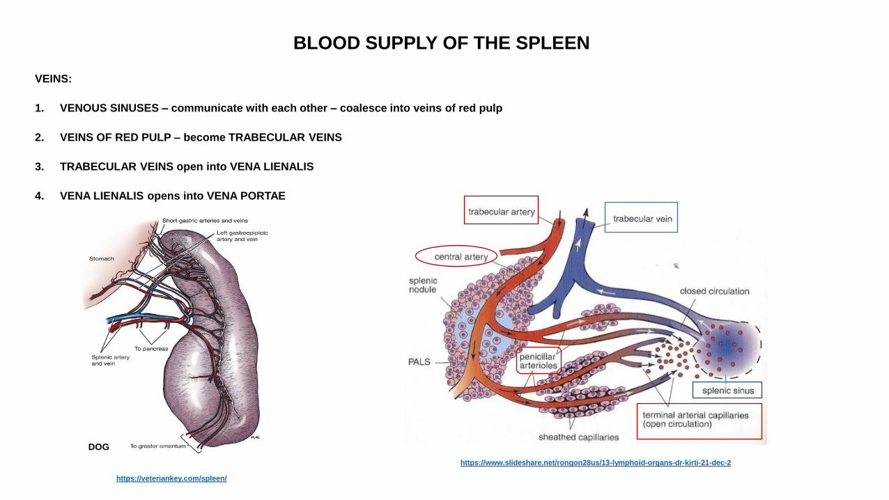
BLOOD SUPPLY OF THE SPLEEN
VEINS:
1. VENOUS SINUSES – communicate with each other – coalesce into veins of red pulp
2. VEINS OF RED PULP – become TRABECULAR VEINS
3. TRABECULAR VEINS open into VENA LIENALIS
4. VENA LIENALIS opens into VENA PORTAE
https://veteriankey.com/spleen/
DOG
https://www.slideshare.net/rongon28us/13-lymphoid-organs-dr-kirti-21-dec-2

THANK YOU FOR YOUR ATTENTION!

BIBLIOGRAPHIE
R. Nickel, A. Schummer, E. Seiferle: The Viscera of the Domestic Animals, 2nd revised edition
Klaus-Dieter Budras, Patrick H. McCarthy , Wolfgang Fricke : Renate Richter Anatomy of the Dog, 5th revised Edition
Klaus-Dieter Budras , W.O.Sack, Sabine Röck : Anatomy of the Horse 5th revised Edition
Klaus – Dieter Budras, Rober E. Habel: Bovine Anatomy, 1st Edition
Miller’s Anatomy of the dog, 4th Edition
König – Liebich: Anatomie der Haussäugetiere, 4. Auflage
König – Liebich: Veterinary Anatomy of Domestic Mammals, 4th Edition
Saunders W.B: Veterinary Anatomy Flash Cards, 2nd Revised edition