Liver, Pancreas and Biliary System - med.swu.ac.thmed.swu.ac.th/radiology/images/sheet_radio/2003...
135
Liver, Pancreas and Biliary System Wirana Angthong, M.D.
Transcript of Liver, Pancreas and Biliary System - med.swu.ac.thmed.swu.ac.th/radiology/images/sheet_radio/2003...

Liver, Pancreas and Biliary System
Wirana Angthong, M.D.

Objectives
•
•
•
•

Outline
• Anatomy
• Imaging Techniques
• Common Diseases

Outline
• Anatomy
• Imaging Techniques
• Common Diseases

Liver anatomy
Morphological anatomy: 3 lobes
Right lobe
Left lobe
Caudate lobe
Median fissure Middle hepatic vein
Ligamentum venosum

Liver anatomy
RL
LL

Liver anatomy
LL
RL

Caudate lobe
CC

Hepatic vasculature
Splenic V
IMV
SMV
Portal V

Pancreas anatomy
HN
BT

Pancreas anatomy

Pancreas anatomy

Pancreas anatomy

Biliary system anatomy

Outline
• Anatomy
• Imaging Techniques
• Common Diseases
Plain film radiograph
Ultrasonography
Computed tomography
Magnetic resonance imaging
ERCPEndoscopic retrograde cholangiography

Outline
• Anatomy
• Imaging Techniques
• Common Diseases
Plain film radiograph Ultrasonography Computed tomography Magnetic resonance imaging ERCP
Endoscopic retrograde cholangiography

Plain film radiographCalcification MetallicSoft tissueFatAir
Organomegaly
Abnormal air
Abnormal calcification
Foreign bodies

Plain film radiograph
•
•
Low sensitivity, low specificity
Radiation
•
Pregnancy

Outline
• Anatomy
• Imaging Techniques
• Common Diseases
Plain film radiograph Ultrasonography Computed tomography Magnetic resonance imaging ERCP
Endoscopic retrograde cholangiography

Ultrasonography
Convex probe: 2-6 MHz Abdomen Pelvis
Linear probe:7-12 MHz Breast Thyroid Testis

Ultrasonography
• Preparation
Upper abdomen
Lower abdomen
“ Fasting 4-6 hr”
Whole abdomen = upper + lower

Ultrasonography
• Preparation
Upper abdomen
Lower abdomen
Whole abdomen = upper + lower
“ No fasting
But full bladder ”

Ultrasonography
• Preparation
Upper abdomen
Lower abdomen
Whole abdomen = upper + lower

Ultrasonography

Ultrasonography
Ultrasonography

Ultrasonography

Ultrasonography
• Indication
RUQ mass
Abnormal liver function test
Abdominal pain
Jaundice
Abnormal vascular structures
Abdominal trauma
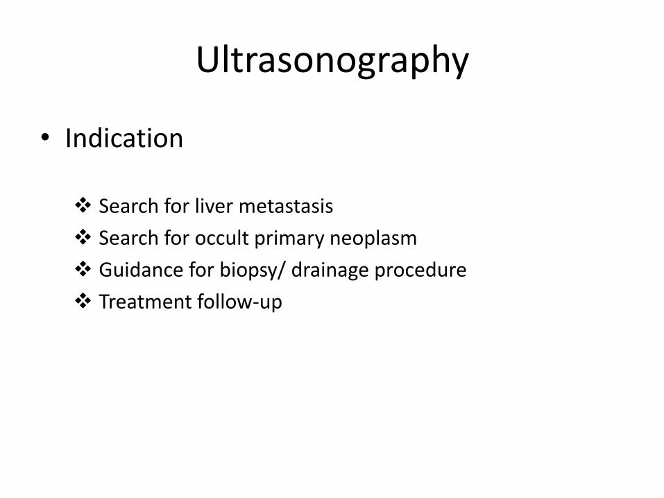
Ultrasonography
• Indication
Search for liver metastasis
Search for occult primary neoplasm
Guidance for biopsy/ drainage procedure
Treatment follow-up

Ultrasonography
•
No radiation
No intravenous contrast media administration
Portable
•
obesity
Gas
Operator dependent
Distal common bile duct: difficult to evaluate

Ultrasonography

Endoscopic Ultrasound (EUS)
High resolution because of proximity of pancreas to the probe
Utility: Diagnosis small tumors, and for which a biopsy is needed.

Outline
• Anatomy
• Imaging Techniques
• Common Diseases
Plain film radiograph Ultrasonography Computed tomography Magnetic resonance imaging ERCP
Endoscopic retrograde cholangiography

Computed tomography

Computed tomography

Computed tomography
• Contrast agent: iodine based contrast media
• Preparation Fasting 4-6 hr
History
allergy, asthma, renal insufficiency
• Indication
Mass
Infiltrative disease
Abdominal pain
Abnormal calcification

Computed tomography
• Indication
Biliary tract obstruction
Abnormal vascular structure
Abdominal trauma
Tumor staging, treatment planning
Treatment follow-up

Computed tomography
•
Fast Multiplanar imagingHigh sensitive to detection of calcification and gas
•
Radiation Risk of contrast allergy Risk in patient with renal insufficiency
•
Pregnancy

Multiphase or Dynamic contrast enhanced CT scan
• Intravenous contrast: marker for where blood travels in tissue
Extracellular interstitial space
Intravascular space

Multiphase or Dynamic contrast enhanced CT scan
• Intravenous contrast: marker for where blood travels in tissue
• Disease alter blood flow to affected tissues
• Contrast enhancement:
character of disease
character of underlying organ

Multiphase or Dynamic contrast enhanced CT scan
• Alter in blood flow characteristic can be imaged by different times following contrast media
• Liver: dual blood supply
Multiphase or dynamic contrast enhanced CT scan
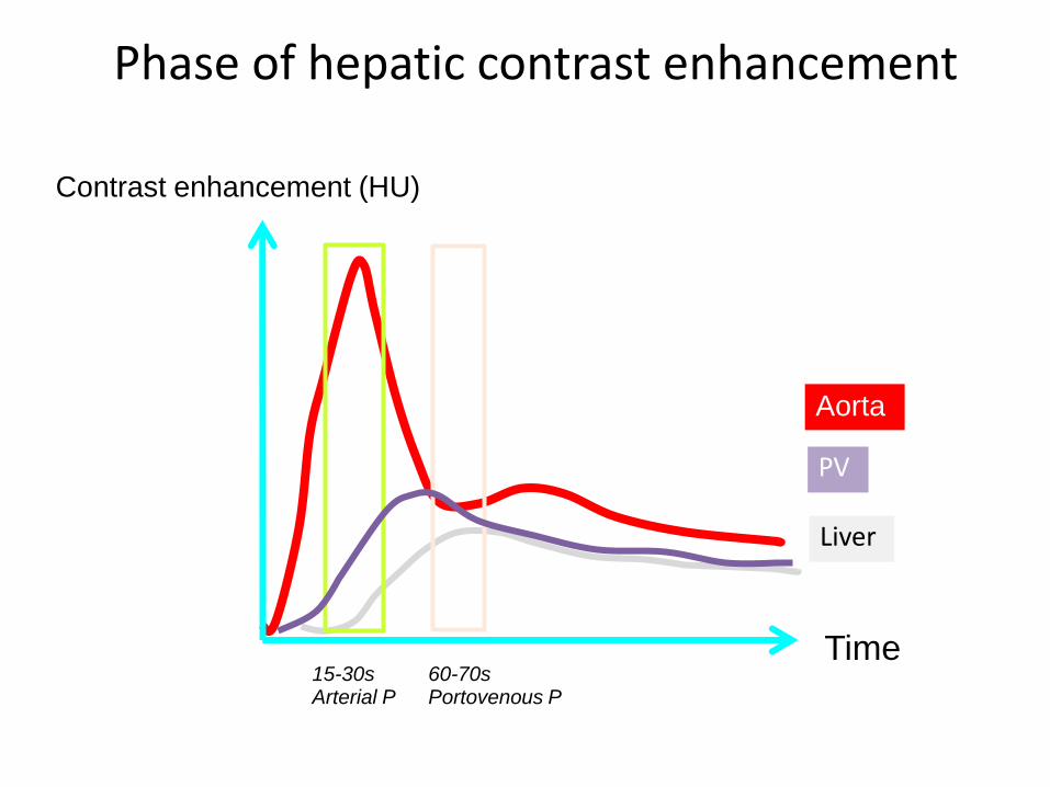
Phase of hepatic contrast enhancement
Time (s)
Contrast enhancement (HU)
15-30sArterial P
60-70sPortovenous P
Aorta
PV
Liver

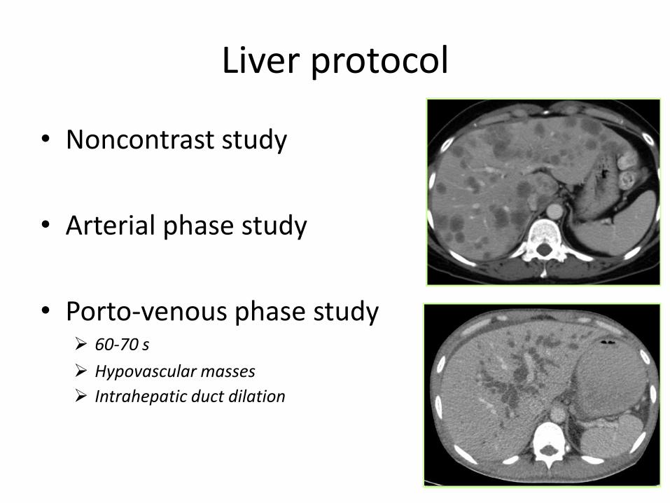
Liver protocol
• Noncontrast study
• Arterial phase study
• Porto-venous phase study

Liver protocol
• Noncontrast study Calcification, hemorrhage, iron deposit
Determination of precontrast attenuation
• Arterial phase study
• Porto-venous phase study

Liver protocol
• Noncontrast study
• Arterial phase study 15-30 s
Hypervascular masses: HCC,
hemangioma, hypervascular metastasis
• Porto-venous phase study
Liver protocol
• Noncontrast study
• Arterial phase study
• Porto-venous phase study 60-70 s
Hypovascular masses
Intrahepatic duct dilation

CT: liver mass
Hypervascular mass in cirrhosis: HCC
Multiple hypovascular masses: metastases

Outline
• Anatomy
• Imaging Techniques
• Common Diseases
Plain film radiograph Ultrasonography Computed tomography Magnetic resonance imaging

MRI upper abdomen with MRCP
• MRI upper abdomen: pre and postcontrast
• MRCP “ Magnetic Resonance Cholangiopancreatography ”

MRI
• Contrast agent: Gadolinium based contrast agent
• Preparation
Fasting 4-6 hr
Remove all metallic objects
History: contraindication for Gadolinium
Severe renal insufficiency, acute renal failure,
Pregnancy

Stratta P et al. Rheumatology 2010;49:821-823
GBCA in Severe Renal Insufficiency
and AKI

• Systemic disorder characterized by widespread tissue fibrosis, affects many organs (lungs, heart, MSK etc.) but predominantly skin
• Relatively spares the face and neck unlike systemic sclerosis
• Strongly correlated with exposure to GBCAs
• Usually manifests within 2-10 wks of exposure
• Diagnosis: Clinical presentation in the setting of severe renal insufficiency ( GFR < 30) + Confirmatory cutaneous histopathological findings.
Shellock FG et al. AJR 2008;191:1129-39
Cowper SE et al. Lancet 2000; 356:1000-1001
Daram SR et al. Am J Kidney Dis 2005; 46:754-759
Kuo PH et al. Radiology 2007; 242:647-649
Nephrogenic systemic fibrosis (NSF)

GBCAs administrations:
Chronic kidney disease
Acute kidney injury of any severity
Hepatic sufficiency/ Hepatorenal syndrome
Vascular injury, venous thrombosis* * Fuluru K Radiographics 2009
ACR 2013 Manual Version 9
Kaewalia AJR 2012;199:17-23
Causes and Associations Stability: Linear < Macrocyclic
Non-ionic < Ionic
High-dose and multiple exposure Most unconfounded cases:
Gadodiamide (Omniscan) Gadopentetate (Magnevist) Gadoversetamide (Optimark)
Stage 4 Severe CKD GFR 15 – 29
Stage 5 End-stage CKD GFR < 15 (or dialysis)

Who should we screen to identify at risk patients?
Recommendation
Serum Cr NOT necessary in all patients
eGFR is recommended for following patients
Within 6 weeks of the MRI study
Patient > 60
History of renal disease including Dialysis Kidney transplant, kidney surgery Single kidney History of know cancer involving kidneys
History of HT requiring medical therapy
History of DMACR 2013 Manual Version 9 Kaewalia AJR 2012;199:17-23

MRI
• Indication
Biliary tract stone: MRCP
Tumor staging, treatment planning
Treatment follow-up
CT contrast media
ERCP ERCP

MRI
• No radiation Multiplanar imaging Superior soft tissue contrast: better detection,
characterization Safer contrast agent than CT: much lessor
associated with nephrotoxicity and allergic reaction
• Expensive Not widely available Longer acquisition time

MR superior soft tissue contrast

MRI
•
Cardiac pacemaker
Foreign body
Metallic clip: ferromagnetic aneurysm clip
Cochlear implant
Avoiding in first trimester pregnancy (teratogenic effect)

Outline
• Anatomy
• Imaging Techniques
• Common Diseases
Plain film radiograph Ultrasonography Computed tomography Magnetic resonance imaging ERCP
Endoscopic retrograde cholangiography

ERCP(Endoscopic retrograde cholangiopancreatography)
• Luminal endoscopy and fluoroscopic imaging
• Cholangiogram (CBD) + Pancreatogram (pancreatic duct)

ERCP(Endoscopic retrograde cholangiopancreatography)
• Diagnose and treat conditions associated with pancreatobiliary system
• Indication
Inconclusive ultrasound, CT or MRI/ MRCP findings
Therapeutic procedure: stone removal, treatment bile duct stricture and biopsy
•
Acute pancreatitis

Outline
• Anatomy
• Imaging Techniques
• Common Diseases

Common disease
Hepatomegaly
Liver cirrhosis and portal hypertension
Liver abscess
Gallstone and bile duct stone
Biliary tract obstruction
Acute cholecystitis
Acute and chronic pancreatitis

Hepatomegaly
> 15.5 cm

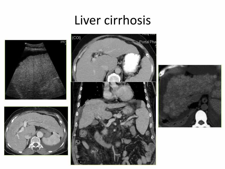
Liver cirrhosis

Liver cirrhosis
• Irreversible remodeling of hepatic architecture with fibrosis and hepatic nodules
• Most are regenerative nodule (RN): localized proliferation of hepatocytes and supporting stroma in response to liver injury (stimuli, alter circulation)

Liver cirrhosis
• Role of imaging
Evaluate liver size
Evaluate portal hypertension
Screening hepatocellular carcinoma (HCC)
Liver cirrhosis

Portal hypertension
• Increased portal flow
• Increase resistant to portal flow
Prehepatic portal hypertension Portal vein thrombosis Splenic vein, SMV, IMV thrombosis
Intrahepatic portal hypertension Liver cirrhosis Hepatic vein thrombosis
Posthepatic portal hypertension IVC thrombosis Cardiac disease: constrictive pericarditis

Portal hypertension

Portal hypertension

Portal hypertension
• Collateral circulation or portosystemic circulation
Coronary vein or gastric vein esophageal vein
azygos/ hemiazygos vein gastroesophageal
varices
Recanalization of paraumbilical vein caput medusae
Inferior mesenteric vein hemorrhoidal venous plexus
internal iliac vein internal hemorrhoid

Portal hypertension

Portal hypertension
• Plain film
Ascites
Splenomegaly

Portal hypertension
• Plain film Ascites
Loss of definition of edge of liver/ spleen
Medial displacement of solid organ and bowel
loops away from properitoneal fat stripe
Separation of bowel loops
Centrally located bowel loops
Bulging of the flank
Increased density of abdomen
Fluid accumulation in pelvis
Splenomegaly

Portal hypertension
• Plain film
Ascites
Splenomegaly
Tip of spleen extends below rib 12 th
Displacement of splenic flexure of colon or displacement of stomach medially

Portal hypertension
• US Enlargement portal vein > 13 mm
Hepatofugal blood flow
Ascites
Splenomegaly
Portosystemic circulation: varices
Normal: Hepatopedal
Hepatofugal

Portal hypertension
• CT Liver cirrhosis
Ascites
Enlarged portal vein > 13 vein
Portosystemic circulation

Liver abscess
• Hepatic pyogenic abscess
Bacterial infection: E coli, K pneumoniae
• Hepatic amebic abscess
Entamoeba histolytica
• Hepatic fungal abscess Candida albicans

Pyogenic abscess
• 5 Routes:
Biliary route: Ascending cholangitis
Portal route: Intra-abdominal infection (diverticulitis, appendicitis)
Hepatic artery route: Septicemia from bacterial endocarditis
Direct extension from adjacent organ
Trauma: Blunt, penetrating injury

Pyogenic abscess
• Plain film
Elevation of right hemidiaphragm
Right pleural effusion
Hepatomegaly
Gas, air-fluid level

Pyogenic abscess
• US
Nonspecific
Cannot distinguish from other hepatic mass (HCC, metastasis)

Pyogenic abscess
• CT and MR
Hypodensity mass
Smooth rim enhancement
Cluster
Gas or air-fluid level

Hepatic amebic abscess
• Route: portal system
• Usually solitary
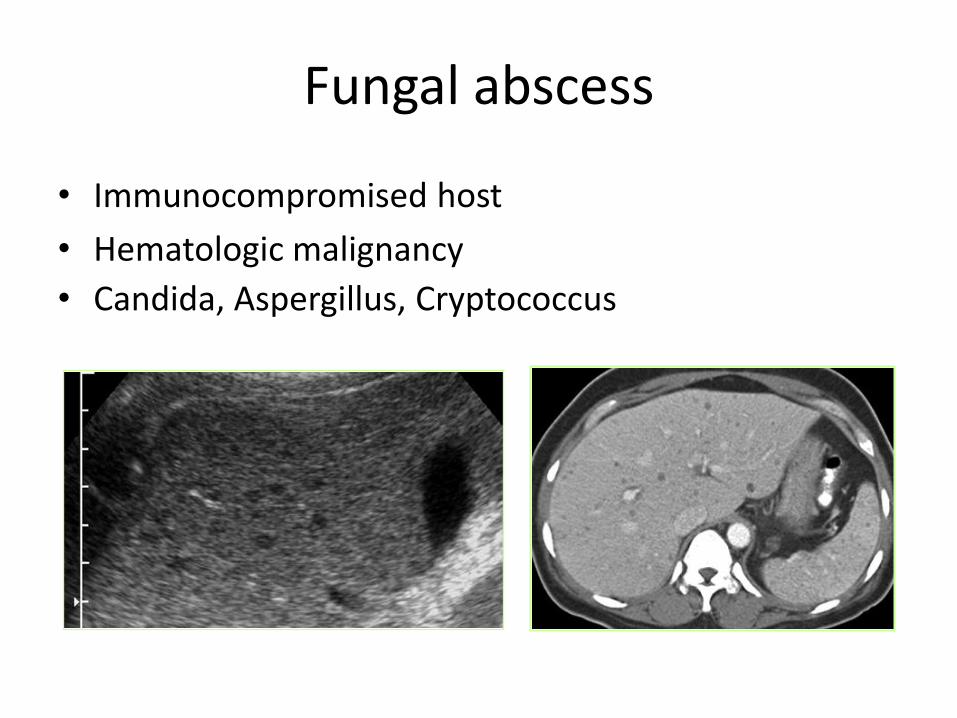
Fungal abscess
• Immunocompromised host
• Hematologic malignancy
• Candida, Aspergillus, Cryptococcus

Gallstone
• Cholesterol stones: Most common
Obesity, female > male
• Pigment or calcium bilirubinate stone
Excessive hemolysis, thalassemia
• Mixed stone
• Calcium carbonate stones
Calcium

Gallstone
• Plain film
• Depend on calcium composition
80-85% miss gallstone
Pigment, mixed, calcium carbonate stones:
radio-opaque
Cholesterol stone: not visible

Gallstone

Gallstone
• US
• The best way to detect gallstone
• All stones appear similar on US, independent on stone composition.
Mobile
Echogenic intraluminal structure
Posterior acoustic shadow

Gallstone
Gallstone
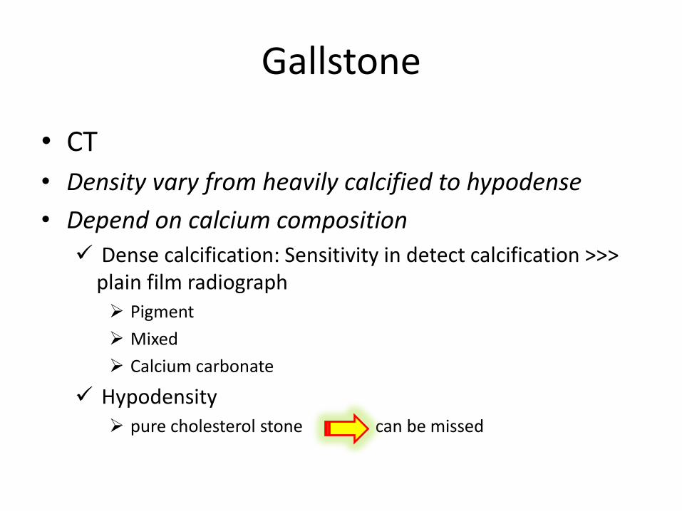
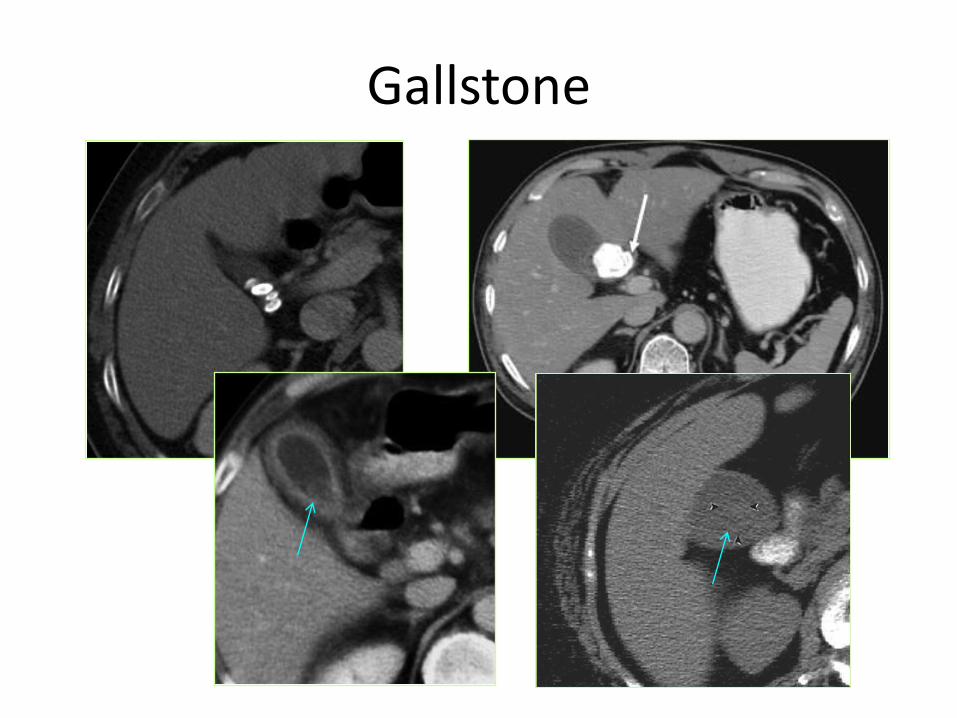
• CT
• Density vary from heavily calcified to hypodense
• Depend on calcium composition
Dense calcification: Sensitivity in detect calcification >>> plain film radiograph Pigment
Mixed
Calcium carbonate
Hypodensity pure cholesterol stone can be missed
Gallstone

Gallstone
• MRCP• Similar ERCP
• All stones appear similar on US, independent on stone composition.

Porcelain gallbladder
• Calcification in GB wall
• Associated with chronic inflammation
• Increased risk of malignancy

Common bile duct stone
• Most located at distal CBD, near ampulla of Vater
• Plain film
Not useful, very low sensitivity

Common bile duct stone
• US
Similar gallstone
Hyperechoic structure with posterior acoustic shadow
Proximal ductal dilation
> 25% may not be visualized distal CBD

Normal CBD in US
< 7 mm

CBD stones

CBD stones
• CT and MRI
Similar gallstone
Proximal ductal dilation

CBD stones

CBD stones

Biliary tract obstruction
• CBD stone
• Malignancy CholangioCA
Ampullary neoplasm
Pancreatic malignancy
Duodenal carcinoma
• Disorder of sphincter of Oddi Ampullary stenosis
• Inflammatory strictures Prior Sx
Prior choledocholithiasis Prior liary infection (ascending cholangitis)

Biliary tract obstruction
• Role of imaging
• Confirm the presence of obstruction
• Location of obstruction
• Cause of obstruction Stone
Stricture
Tumor
US is a screening modality
IHD dilation EHD dilation

IHD dilation
• > 2 mm in diameter, parallel channel sign
• Lack of Doppler signal
• Irregular, tortuous wall
• Stellate configuration centrally

Normal IHD and CBD

IHD dilation
Normal CT
Normal MRI

IHD dilation

EHD dilation
• > 7 mm
Longitudinal normal CBD

EHD dilationTransverse
normal CBD

EHD dilation

EHD dilation
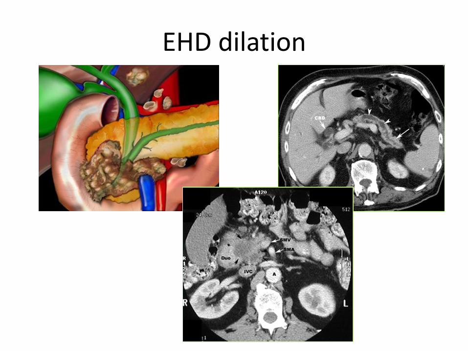
EHD dilation

EHD dilation

Biliary tract obstruction
• Investigation
US
CT or MRI with MRCP
ERCP

Acute cholecystitis
• Obstruction of GB by stone at neck or cystic duct
• Plain film
Not useful
may detect radio-opaque stone
Acute cholecystitis
• US
Gallstone
Gallbladder wall thickening
Distend gallbladder
Pericholecystic fluid
Tender at gallbladder; US Murphy’s sign positive

Normal gallbladder vs wall thickening
Normal GB wall < 3 mm
Wall thickening > 3 mm

Acute cholecystitis

Acute cholecystitis

Emphysematous cholecystitis
• Less common
• Vascular insufficiency of cystic artery
• Facilitating infection of gas-forming organisms (eg, Clostridium or Escherichia coli)
• Considered as surgical emergency

Emphysematous cholecystitis

Acute pancreatitis
Activation of pancreatic enzymes
within the pancreas
leads to organ injury
Autodigestion
Parenchymal edema and peripancreatic fat necrosis
Pancreatic necrosis, hemorrhage

Acute pancreatitis
• Most common: Alcohol abuse
Gallstone
• Less common:
Trauma
Sepsis
Medication
Post ERCP

Acute pancreatitis
• Plain film “Colon cutoff sign”
Abrupt termination of gas at splenic flexure
Inflammation extends to phrenicocolic ligament
Function spasm/ luminal narrowing

Acute pancreatitis

Acute pancreatitis
• USNot be used for exclude the diagnosis
Enlarge, decreased echogenicity (compare with liver)
Peripancreatic fluid collection
Detect gallstone
US normal pancreas

Acute pancreatitis
CT normal pancreas

Chronic pancreatitis
• Permanent impairment of pancreatic function
• Permanent morphologic change as a result of persistent inflammation
• Findings:
Pancreatic calcification
Ductal dilation
Parenchymal atrophy

Chronic pancreatitis

Summary
• Acute abdominal pain
• Palpable mass
• Jaundice
• Abnormal liver function test

Summary
• Acute abdominal pain Plain film
US
CT or MRI with MRCP
• Palpable mass
• Jaundice
• Abnormal liver function test

Summary
• Acute abdominal pain
• Palpable mass
US
CT or MRI
• Jaundice
• Abnormal liver function test

Summary
• Acute abdominal pain
• Palpable mass
• Jaundice US
CT or MRI with MRCP
• Abnormal liver function test
Summary
• Acute abdominal pain
• Palpable mass
• Jaundice
• Abnormal liver function test US
CT or MRI

THANK YOU FOR YOUR ATTENTION