Liver Fibrosis in an Extremely Small Infant for ...
5
Severe SGA and Liver Fibrosis 181 181 Received January 12, 2010; revision accepted for publication May 7, 2010. doi:10.1620/tjem.221.181 Correspondence: Hirokazu Arai, M.D., Ph.D., Department of Pediatrics, Akita University Graduate School of Medicine, Hondo 1-1-1, Akita, Akita 010-8543, Japan. e-mail: [email protected] Tohoku J. Exp. Med., 2010, 221, 181-185 Liver Fibrosis in an Extremely Small Infant for Gestational Age Hirokazu Arai, 1,2 Atsuko Noguchi, 1 Ryoji Goto, 2 Takefumi Matsuda, 2 Hatsushi Nakajima 2 and Tsutomu Takahashi 1 1 Department of Pediatrics, Akita University Graduate School of Medicine, Akita, Japan 2 Department of Pediatrics, Akita Red Cross Hospital, Akita, Japan Premature infants with intrauterine growth restriction (IUGR) are at greater risk for an adverse perinatal outcome. IUGR affects hepatocyte function, but the histopathological changes in the postnatal liver are not well known. We report a male infant with severe IUGR. His mother was transferred to our hospital at 26 weeks of gestation because of preterm labor and severe IUGR. An emergency cesarean section was carried out because of a non-reassuring fetal status. The birth weight of the infant was 332 g. He showed congestive heart failure and marked hepatomegaly from birth. After 1 week, blood examinations showed hyperbilirubinemia with high direct bilirubin. Because of liver dysfunction, he received the minimal total parenteral nutrition for 7 days. After 1 month, he progressively developed ascites and coagulopathy, and died 3 months after birth. Liver autopsy showed diffuse perisinusoidal fibrosis. Fibrosis was also prominent around the central vein. Immunohistochemical study revealed many α -smooth muscle actin-positive cells, which represent activated hepatic stellate cells, and a few transforming growth factor- β 1-positive cells in the perisinusoidal fibrotic area. These results indicate that the infant developed chronic (end stage) liver failure by 3 months of age. We excluded congenital infection, metabolic syndrome and citrin deficiency. It is therefore conceivable that intrauterine cardiac failure may be responsible for liver fibrosis. Early detection of liver dysfunction soon after birth is a key to predict the prognosis of severe IUGR in preterm infants. Keywords: liver failure; IUGR; preterm infant; liver fibrosis; extremely low birth weight Tohoku J. Exp. Med., 2010, 221 (3), 181-185. © 2010 Tohoku University Medical Press Premature infants with intrauterine growth restriction (IUGR) are at greater risk for postnatal growth and develop- ment as well as acute and chronic morbidities (Rosenberg 2008). IUGR influences hepatocyte function (Alonso et al. 2007). Compared to appropriate for gestational age (AGA) infants of similar birth weight, small for gestational age (SGA) infants show a higher rate of neonatal cholestasis (Boehm et al. 1990); however, only one report has shown postnatal liver histopathological changes for SGA infants that received prolonged total parenteral nutrition (TPN) (Baserga and Sola 2004). Herein, we report the unusual case of an extremely low birth weight (ELBW) infant with severe IUGR. At birth, the infant showed congestive heart failure. Severe coagu- lopathy, thrombocytopenia, and jaundice persisted. The infant received the minimal TPN for only 7 days, and sub- sequently developed severe perisinusoidal liver fibrosis. Patient Report A 36-year-old woman, gravida 2, para 1, was referred to another hospital at 25 weeks of gestation with suspected IUGR. IUGR was first suspected at 23 weeks of gestation at another clinic. She was transferred to our hospital at 26 weeks of gestation because of preterm labor and severe IUGR. Ultrasound revealed severe growth restriction of the fetal biparietal diameter and abdominal circumference (each below -2 standard deviation). A first-trimester ultrasound had confirmed gestational age. She had reliable date param- eters confirmed by the crown-rump length at 8 weeks’ ges- tation. During her first pregnancy at 26 years old, she had undergone an artificial abortion. There had been no compli- cations in her second pregnancy; a healthy child was born spontaneously at term. There was no family history of hereditary disorders. She had not taken medicine during the pregnancy. On admission, she did not have premature rup- ture of the membranes. Fetal ultrasound showed reverse end-diastolic flow in the umbilical artery and umbilical venous pulsations. Middle cerebral artery flow pattern showed redistribution (resistance index = 0.66). We did not examine the fetal karyotype because her family refused. At 26 weeks of gestation, an emergency cesarean sec- tion was carried out because of a non-reassuring fetal status (minimal fetal heart rate variability). The birth weight of the male infant was 332 g. The infant had asphyxia, with Apgar scores of 3 at one minute and 5 at five minutes, and required resuscitation with tracheal intubation. On admis-
Transcript of Liver Fibrosis in an Extremely Small Infant for ...

Severe SGA and Liver Fibrosis 181
181
Received January 12, 2010; revision accepted for publication May 7, 2010. doi:10.1620/tjem.221.181Correspondence: Hirokazu Arai, M.D., Ph.D., Department of Pediatrics, Akita University Graduate School of Medicine, Hondo 1-1-1,
Akita, Akita 010-8543, Japan.e-mail: [email protected]
Tohoku J. Exp. Med., 2010, 221, 181-185
Liver Fibrosis in an Extremely Small Infant for Gestational Age
Hirokazu Arai,1,2 Atsuko Noguchi,1 Ryoji Goto,2 Takefumi Matsuda,2 Hatsushi Nakajima2 and Tsutomu Takahashi1
1Department of Pediatrics, Akita University Graduate School of Medicine, Akita, Japan2Department of Pediatrics, Akita Red Cross Hospital, Akita, Japan
Premature infants with intrauterine growth restriction (IUGR) are at greater risk for an adverse perinatal outcome. IUGR affects hepatocyte function, but the histopathological changes in the postnatal liver are not well known. We report a male infant with severe IUGR. His mother was transferred to our hospital at 26 weeks of gestation because of preterm labor and severe IUGR. An emergency cesarean section was carried out because of a non-reassuring fetal status. The birth weight of the infant was 332 g. He showed congestive heart failure and marked hepatomegaly from birth. After 1 week, blood examinations showed hyperbilirubinemia with high direct bilirubin. Because of liver dysfunction, he received the minimal total parenteral nutrition for 7 days. After 1 month, he progressively developed ascites and coagulopathy, and died 3 months after birth. Liver autopsy showed diffuse perisinusoidal fibrosis. Fibrosis was also prominent around the central vein. Immunohistochemical study revealed many α -smooth muscle actin-positive cells, which represent activated hepatic stellate cells, and a few transforming growth factor-β1-positive cells in the perisinusoidal fibrotic area. These results indicate that the infant developed chronic (end stage) liver failure by 3 months of age. We excluded congenital infection, metabolic syndrome and citrin deficiency. It is therefore conceivable that intrauterine cardiac failure may be responsible for liver fibrosis. Early detection of liver dysfunction soon after birth is a key to predict the prognosis of severe IUGR in preterm infants.
Keywords: liver failure; IUGR; preterm infant; liver fibrosis; extremely low birth weightTohoku J. Exp. Med., 2010, 221 (3), 181-185. © 2010 Tohoku University Medical Press
Premature infants with intrauterine growth restriction (IUGR) are at greater risk for postnatal growth and develop-ment as well as acute and chronic morbidities (Rosenberg 2008). IUGR influences hepatocyte function (Alonso et al. 2007). Compared to appropriate for gestational age (AGA) infants of similar birth weight, small for gestational age (SGA) infants show a higher rate of neonatal cholestasis (Boehm et al. 1990); however, only one report has shown postnatal liver histopathological changes for SGA infants that received prolonged total parenteral nutrition (TPN) (Baserga and Sola 2004).
Herein, we report the unusual case of an extremely low birth weight (ELBW) infant with severe IUGR. At birth, the infant showed congestive heart failure. Severe coagu-lopathy, thrombocytopenia, and jaundice persisted. The infant received the minimal TPN for only 7 days, and sub-sequently developed severe perisinusoidal liver fibrosis.
Patient ReportA 36-year-old woman, gravida 2, para 1, was referred
to another hospital at 25 weeks of gestation with suspected IUGR. IUGR was first suspected at 23 weeks of gestation at another clinic. She was transferred to our hospital at 26
weeks of gestation because of preterm labor and severe IUGR. Ultrasound revealed severe growth restriction of the fetal biparietal diameter and abdominal circumference (each below -2 standard deviation). A first-trimester ultrasound had confirmed gestational age. She had reliable date param-eters confirmed by the crown-rump length at 8 weeks’ ges-tation. During her first pregnancy at 26 years old, she had undergone an artificial abortion. There had been no compli-cations in her second pregnancy; a healthy child was born spontaneously at term. There was no family history of hereditary disorders. She had not taken medicine during the pregnancy. On admission, she did not have premature rup-ture of the membranes. Fetal ultrasound showed reverse end-diastolic flow in the umbilical artery and umbilical venous pulsations. Middle cerebral artery flow pattern showed redistribution (resistance index = 0.66). We did not examine the fetal karyotype because her family refused.
At 26 weeks of gestation, an emergency cesarean sec-tion was carried out because of a non-reassuring fetal status (minimal fetal heart rate variability). The birth weight of the male infant was 332 g. The infant had asphyxia, with Apgar scores of 3 at one minute and 5 at five minutes, and required resuscitation with tracheal intubation. On admis-

H. Arai et al.182
sion, he had respiratory distress syndrome, and received artificial surfactant replacement therapy. A chest radiograph showed cardiomegaly. Echocardiography showed left ven-tricular dysfunction with ejection fraction of 46% (Murase et al. 2002), a dilated inferior vena cava, and tricuspid and mitral regurgitation (Fig. 1). Soon after birth, the patient showed congestive heart failure. Congenital cardiac mal-formation was absent. The patient was treated with inotro-pic and vasodilatory agents, and his cardiopulmonary condi-tion improved gradually. No renal anomaly could be found on ultrasonography. Ophthalmological examination yielded normal findings. We could not examine his karyotype as the parents did not consent, but his face, appearance, and physical examination did not suggest trisomy 21 or 18.
Initial laboratory studies (Table 1) indicated a platelet count of 0.2 × 10⁴ /µL. Blood examinations showed meta-bolic acidosis (pH 7.202, BE -13.3 mmol/L), and coagulop-athy was identified: fibrinogen (< 50 mg/dL) and prothrom-bin time (PT) % (34%) were low. Blood ammonia and blood glucose levels were within the normal ranges.
He showed marked hepatomegaly from birth. After 1 week, blood examinations showed hyperbilirubinemia with high direct bilirubin (total and direct bilirubin of 17.7 and
13.3 mg/dL at 17 days of life, respectively) (Fig. 2). Because of liver dysfunction, TPN (amino acids 0.3-1 g/kg/day; glucose infusion rate, max 6 mg/kg/minute; and vita-mins) was used for only 7 days and we did not use intrave-nous lipid infusion; however, cholestasis did not improve, and we started ursodeoxycholic acid (UDC) (15 mg/kg/day).
After 1 month, he progressively presented with severe liver dysfunction with cholestasis, which led to the develop-ment of ascites, coagulopathy, and hypoalbuminemia. We administered fresh frozen plasma and vitamin K infusion frequently to improve coagulopathy, but these therapies were unsuccessful; his condition became unstable and he progressed to grade IV intraventricular hemorrhage. Plasma amino acid examination on day 91 showed hypertyrosin-emia (1,113 nmol/mL). We suspected tyrosinemia type I, but no accumulation of upstream metabolites and succinyl-acetone was detected. Alpha-fetoprotein was 8,264.6 ng/mL (normal range) on the same day. Total bile acids were 119 µmol/L (day 91). We used special milk formulas (medium chain triglyceride (MCT)-enriched formulas or tyrosine-free milk), but his condition did not improve. He died 3 months after birth because of sepsis.
Fig. 1. Infant echocardiography. Echocardiography showed dilated inferior vena cava (left) and tricuspid regurgitation (right) at day 0.
Table1. Laboratory findings on admission (day 0).
pH 7.202 AST 52 U/L Na 140 mmol/LpCO2 35.7 mmHg ALT 3 U/L K 3.6 mmol/LBE −13.3 mEq/L LDH 851 U/L Cl 99 mmol/LBS 127 mg/dL GGT 204 U/L Ca 9.4 mg/dL
T.Bil 2.4 mg/dL iP 5.2 mg/dLWBC 3,600/µL D.Bil 0.5 mg/dL Fe 79 µg/dLHb 13.6 g/dL ALP 199 U/L IgG 353 mg/dLPlt 0.2 × 10⁴/µL CK 309 U/L IgM 1 mg/dL
TP 3.2 g/dL CRP 0.0 mg/dLAPTT 69.9 sec Alb 2.2 g/dLPT 34% T.Chol 21 mg/dLFib < 50 mg/dL TG 27 mg/dLD-dimer 2.0 mg/dL BUN 9.7 mg/dLAT-III < 20% Cr 0.66 mg/dLHPT 24%
Severe SGA and Liver Fibrosis 183
After death, congenital infection (varicela zoster, her-pes, cytomegalovirus, toxoplasmosis, syphilis, and rubella), extrahepatic obstruction, citrin deficiency (neonatal intrahe-patic cholestasis), and inborn errors of bile acid metabolism (e.g., primary 3-oxo-delta 4-steroid 5 beta-reductase defi-ciency) were excluded. The results of neonatal screening for metabolic diseases (galactosemia, homocystiuria, phe-nylketouria, and maple syrup urine disease) and endocrine diseases (hypothyroidism and adrenal hyperplasia) were negative. A liver sample was the only specimen obtained after death with informed consent from the parents. No extrahepatic bilary atresia was noted. Microscopically, dif-fuse perisinusoidal liver fibrosis with increased collagen fibers was observed (Fig. 3). Fibrosis was prominent around the central vein, and the hepatocytes showed fre-quent disruption. Iron deposits were mildly present. Cellular infiltration in the parenchyma was absent. Fatty liver change was also absent (Fig. 3).
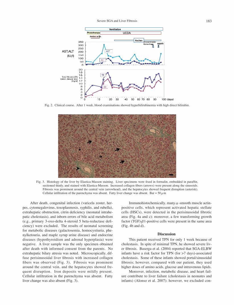
Immunohistochemically, many α -smooth muscle actin-positive cells, which represent activated hepatic stellate cells (HSCs), were detected in the perisinusoidal fibrotic area (Fig. 4a and c); moreover, a few transforming growth factor (TGF)-β1-positive cells were present in the same area (Fig. 4b and d).
DiscussionThis patient received TPN for only 1 week because of
cholestasis. In spite of minimal TPN, he showed severe liv-er fibrosis. Baserga et al. (2004) reported that SGA-ELBW infants have a risk factor for TPN (for >7 days)-associated cholestasis. Some of these infants showed portal/sinusoidal fibrosis; however, compared with our patient, they used higher doses of amino acids, glucose and intravenous lipids.
Moreover, infection, metabolic disease, and heart fail-ure contribute to liver failure (cholestasis in neonates and infants) (Alonso et al. 2007); however, we excluded con-
Fig. 2. Clinical course. After 1 week, blood examinations showed hyperbilirubinemia with high direct bilirubin.
Fig. 3. Histology of the liver by Elastica-Masson staining. Liver specimens were fixed in formalin, embedded in paraffin, sectioned thinly, and stained with Elastica-Masson. Increased collagen fibers (arrows) were present along the sinusoids. Fibrosis was prominent around the central vein (arrowhead), and the hepatocytes showed frequent disruption (asterisk). Cellular infiltration of the parenchyma was absent. Fatty liver change was absent. Bar = 50 µm
H. Arai et al.184
genital infection, metabolic syndrome, and neonatal intrahe-patic cholestasis.
Our patient showed hypertyrosinemia, but we excluded tyrosinemia type I because of no accumulation of upstream metabolites and succinylacetone. SGA infants are more sensitive to metabolic imbalances than AGA infants (Baserga et al. 2004). Also, Robles et al. (1984) reported hypertyro-sinemia in small-for-date (SFD) infants. Hypertyrosinemia results from immature function of the enzyme 4-hydroxy-phenylpyruvate dioxygenase, most frequently in premature infants (Russo et al. 2001).
A cause of IUGR is uteroplacental insufficiency sec-ondary to maternal morbidities such as hypertension (Baserga et al. 2004). IUGR fetuses demonstrate progres-sive hemodynamic changes, and develop earlier and more pronounced right than left ventricular function deterioration (Bahtiyar and Copel 2008). Soon after birth, this patient showed congestive heart failure, which is associated with the development of liver fibrosis (Naschitz et al. 2000). An animal-based study of right ventricular pressure overload showed liver fibrosis and α -smooth muscle actin expression (Gieling et al. 2004). Although it could not be excluded that liver failure had been caused by another etiology, we sug-gested that intrauterine cardiac failure contributed to liver fibrosis.
An immunohistochemical study showed many α -smooth muscle actin-positive cells (myofibroblasts) and a few TGF-β1-positive cells in the perisinusoidal fibrotic area. Liver fibrosis contributes to the differentiation of HSCs into myofibroblasts (Hinz et al. 2007). TGF-β1 is the
most potent profibrogenic cytokine that activates HSC, but the present patient showed only a few TGF-β1-positive cells, despite that fibrotic changes developed in the liver at 3 months of age.
Early detection of liver dysfunction (including the examination of markers of liver fibrosis) soon after birth is a key to predict the prognosis of severe IUGR in preterm infants.
ReferencesAlonso, E.A., Squires, R.H. & Whitington, P.F. (2007) Acute liver
failure in children. In Liver disease in children, edited by F.J. Suchy. Cambridge University Press, NY, pp. 71-96.
Bahtiyar, M.O. & Copel, J.A. (2008) Cardiac changes in the intra-uterine growth-restricted fetus. Semin. Perinatol., 32, 190-193.
Baserga, M.C. & Sola, A. (2004) Intrauterine growth restriction impacts tolerance to total parenteral nutrition in extremely low birth weight infants. J. Perinatol., 24, 476-481.
Boehm, G., Müller, D.M., Teichmann, B. & Krumbiegel, P. (1990) Influence of intrauterine growth retardation on parameters of liver function in low birth weight infants. Eur. J. Pediatr., 149, 396-398.
Gieling, R.G., Ruijter, J.M., Maas, A.A., Van Den Bergh Weerman, M.A., Dingemans, K.P., ten Kate, F.J., Lekanne dit Deprez, R.H., Moorman, A.F. & Lamers, W.H. (2004) Hepatic response to right ventricular pressure overload. Gastroenterol-ogy, 127, 1210-1221.
Hinz, B., Phan, S.H., Thannickal, V.J., Galli, A., Bochaton-Piallat, M.L. & Gabbiani, G. (2007) The myofibroblast: one function, multiple origins. Am. J. Pathol., 170, 1807-1816.
Murase, M., Ishida, A. & Momota, T. (2002) Serial pulsed Doppler assessment of early left ventricular output in critically ill very low-birth-weight infants. Pediatr. Cardiol., 23, 442-448.
Fig. 4. Immunohistochemical staining of the liver. Immunohistochemical staining was performed using frozen sections. Antibodies against α -smooth muscle actin (a) (Boehringer Mannheim, Mannheim, Germany, dilution: 1:10) and TGF-β1 (b) (Santa Cruz Biotechnology, Inc., CA, USA, dilution: 1:200) were obtained commercially. (a) Immunoreactivity of spindle-like cells (arrows) was found along sinusoids. (b) Immunoreactivity was detected in round cells (arrows) in si-nusoidal areas. (c) Negative control section for (a). (d) Negative control section for (b). Bar = 50 µm

Severe SGA and Liver Fibrosis 185
Naschitz, J.E., Slobodin, G., Lewis, R.J., Zuckerman, E. & Yeshurun, D. (2000) Heart diseases affecting the liver and liv-er diseases affecting the heart. Am. Heart J., 140, 111-120.
Robles, R., Gil, A., Faus, M.J., Periago, J.L., Sánchez-Pozo, A., Pita, M.L. & Sánchez-Medina, F. (1984) Serum and urine amino acid patterns during the first month of life in small-for-
date infants. Biol. Neonate, 45, 209-217.Rosenberg, A. (2008) The IUGR newborn. Semin. Perinatol., 32,
219-224.Russo, P.A., Mitchell, G.A. & Tanguay, R.M. (2001) Tyrosinemia:
a review. Pediatr. Dev. Pathol., 4, 212-221.