Liver Disease in Pregnancy - wesley ob/gyn · Liver Disease in Pregnancy ... • Risks –PTB,...
308
Liver Disease in Pregnancy Darren Farley, MD Clinical Assistant Professor Division of Maternal-Fetal Medicine Dept. of Obstetrics and Gynecology University of Kansas School of Medicine – Wichita
Transcript of Liver Disease in Pregnancy - wesley ob/gyn · Liver Disease in Pregnancy ... • Risks –PTB,...

Liver Disease in Pregnancy
Darren Farley, MD
Clinical Assistant Professor
Division of Maternal-Fetal Medicine
Dept. of Obstetrics and Gynecology
University of Kansas School of Medicine – Wichita

Hepatobiliary Disease in
Pregnancy • Physiologic changes
• Hepatitis
• AFLP
• HELLP
• Cholestasis
• Chronic liver disease

Case
• 31 yo P2002
• Chronic liver disease, cirrhosis
• Portal HTN
• Treatment?
• Delivery Recommendations ?

Case
• 38 yo P2002
• IVF pregnancy
• New onset pruritis, LFTs 300s at 20
weeks
• Evaluation?
• Treatment?

Physiologic Changes
• Elevated alkaline phosphatase, palmar erythema,
spider angiomas, which might suggest liver disease, are
commonly found during normal pregnancy.
• Metabolism - altered expression of the cytochrome
P450 system that is mediated by higher levels of
estrogen, progesterone, and other hormones.
• Hepatic CYP1A2 expression is decreased, whereas that
of CYP2D6 and CYP3A4 is increased.
• Importantly, cytochrome enzymes are expressed in
many organs besides the liver, most notably the
placenta.
• No major hepatic histological changes are induced by
normal pregnancy

• 50-1. Of the following findings that
can be associated with liver dysfunction,
which is a normal physiologic change in
pregnancy?
• Elevation of hepatic transaminases
• Spider angiomas *****
• Esophageal varices
• Asterixis

Hepatitis - viralFeature Hep A Hep B Hep C Hep D Hep E
Viral type RNA DNA RNA RNA RNA
Inc period 14-50d 30-180d 30-160d 30-180d 14-63d
Transmission Fecal/oral Parenteral Parenteral Parenteral Fecal/oral
Diagnosis IgM anti
HAV ab
HBsAg,
anti-HBs,
anti-HBc,
HBeAg,
HBV DNA
Hep C ab Delta Ag,
IgM-specific
Ab
IgM anti-
HEV Ab
Carrier risk
of chronic
infection
0 10-15% 50-85% Up to 80%
when
superinfection
with Hep D
occurs
0
Vertical
transmission
risk
No Yes Yes Yes Yes
Vaccination
available
Yes Yes No No No
Creasy – MFM

Date of download: 3/7/2015 Copyright © 2015 McGraw-Hill Education. All rights reserved.
Sequence of various antigens and antibodies in acute hepatitis B. ALT = alanine aminotransferase; anti-HBc = antibody to hepatitis B core
antigen; anti-HBe = antibody to hepatitis B e antigen; anti-HBs = antibody to hepatitis B surface antigen; HBeAg = hepatitis B e antigen; HBsAg
= hepatitis B surface antigen. (Redrawn from Dienstag, 2012a.)
Legend:
From: Hepatic, Biliary, and Pancreatic Disorders
Williams Obstetrics, 24e, 2013

Interpretations of serologic testing in patients with hepatitis B virus
HBsAg HBsAb HBcAb HBeAg HBeAb Possible interpretation
- - - - - Never infected
+ - - - - -Early acute infxn
-Transient (up to 18d) after vaccination
+ - IgM + - Acute HBV infxn, highly infectious
+ - IgG + - Chronic HBV infxn, highly infectious
+ - IgG - + Late acute or chronic HBV infxn, low
infectivity
- - IgM +/- +/- Acute HBV infxn
- - IgG - +/- -Low-level HBsAg carrier or remote
past infxn
-Passive transfer to infant of HBsAg-
positive mother
- + IgG - +/- Recovery from HBV infxn and immune
- + - - - -Immune if concentration >/=
10mIU/mL
-Passive transfer after hepatitis B
immune globulin
Creasy – MFM
Hepatitis B Vaccination Recommendations (CDC, 2005)
• Maternal HBsAg Testing - All pregnant women should be tested routinely for HBsAg.
• Vaccination of Infants
• At Birth
– Infants born to HBsAg-positive mothers should receive hepatitis B vaccine and
HBIG within 12 hours of birth.
– Infants who are born to mothers whose HBsAg status is unknown should receive
hepatitis B vaccine within 12 hours of birth.
– Term infants who weigh 2000 g or more at birth, are medically stable, and are born
to HBsAg-negative mothers should receive hepatitis B vaccine before hospital
discharge.
– Preterm infants who weigh 2000 g or less at birth and are born to HBsAg-negative
mothers should receive the first dose of hepatitis B vaccine 1 month after birth.
• After the Birth
– All infants should complete the hepatitis B vaccine series
– Infants born to HBsAg-posivite mothers should be tested for HBsAg and HBsAb
after completion of the hepatitis B vaccine series at age 9 to 18 months
Williams, Creasy

Pregnancy related liver disease
• Hypertensive disease
• HELLP
• AFLP

AFLP
• Most common cause of acute liver failure
in pregnancy
• 1 in 10,000 pregnancies
• 3rd trimester

AFLP
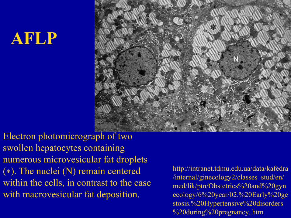
Electron photomicrograph of two
swollen hepatocytes containing
numerous microvesicular fat droplets
(∗). The nuclei (N) remain centered
within the cells, in contrast to the case
with macrovesicular fat deposition.
http://intranet.tdmu.edu.ua/data/kafedra
/internal/ginecology2/classes_stud/en/
med/lik/ptn/Obstetrics%20and%20gyn
ecology/6%20year/02.%20Early%20ge
stosis.%20Hypertensive%20disorders
%20during%20pregnancy..htm

AFLP - Pathogenesis
• Association with recessively inherited mitochondrial
abnormalities of fatty acid oxidation.
• Similar to those in children with Reye-like syndromes.
• Mutations - mitochondrial trifunctional protein enzyme
complex that catalyzes the last oxidative steps in the
pathway; most common - G1528C and E474Q
mutations of the gene on chromosome 2 that code for
long-chain-3-hydroxyacyl-CoA-dehydrogenase—
known as LCHAD.

• 50-7. Mutations in enzymes involved in
fatty acid oxidation have been classically
associated with maternal acute fatty liver of
pregnancy when they occur in what pattern?
• Homozygous mutation in the fetus and the
mother
• Homozygous mutation in the fetus;
heterozygous mutation in the mother *****
• Heterozygous mutation in the fetus and the
mother
• Heterozygous mutation in the fetus;
homozygous mutation in the mother

AFLP• Parkland study – n=51 - 37wk (31-40.9)
• 41% nulliparous, 10-20% multifetal pregnancy
• N/v, malaise, anorexia, epigastric pain, progressive jaundice
• ½ have HTN, unclear overlap
• Elevated LFTs (<1000)
• Hypofibrinogenemia, hypoalbuminemia, hypocholesterolemia, and
prolonged clotting times.
• Serum bilirubin levels usually are < 10 mg/dL,
• Hypoglycemia
• Endothelial cell activation with capillary leakage causing
hemoconcentration, acute kidney injury, ascites, and sometimes
pulmonary permeability edema (Bernal, 2013)
• Severe hemoconcentration, uteroplacental perfusion is reduced,
maternal acidosis can cause fetal death; high CD rate due to NR
testing
• Peripheral blood smear – echinocytosis; hemolysis due
hypocholesterolemia

• A 33-year old G2P1 presents at 35 weeks’ gestation with
complaints of nausea and vomiting. Laboratory analysis reveals
elevated transaminases and creatinine levels and coagulopathy. A
peripheral smear is performed with the results shown.
• 50-9. What is the underlying etiology of these hemolyzed cells
found on the blood smear in this patient?
• Increase destruction in the spleen
• Intense vasospasm
• Occlusive pain crisis
• Decreased cholesterol production *****

Peripheral smear
Echinocytes
http://www.pclv.net/casestudies/mi
keybloodsmears.html
SchistocytesA schistocyte
count of >1% is
most often found
in thrombotic
thrombocytopeni
c purpura
AFLP - Hemolysis is
caused by the effects of
hypocholesterolemia on
erythrocyte membranes
(Cunningham, 1985)
Normal smear
http://library.med.
utah.edu/WebPath/
TUTORIAL/IRO
N/IRON002.html
Target cells – liver
disease, etc;
http://www.medical-
labs.net
?

AFLP • Swansea Diagnostic Criteria for Acute Fatty Liver of Pregnancy
• Six or more criteria are required in the absence of another cause:
• Vomiting
• Abdominal pain
• Polydipsia or polyuria
• Encephalopathy
• Elevated bilirubin level (>14 µmol/L)
• Hypoglycemia (<4 mmol/L)
• Elevated urea level (>340 µmol/L)
• Leukocytosis (>11 × 109/L)
• Ascites or bright liver on ultrasound scan
• Elevated transaminase levels (AST or ALT >42 IU/L)
• Elevated ammonia level (>47 µmol/L)
• Renal impairment (creatinine >150 µmol/L)
• Coagulopathy (PT >14 sec or aPTT >34 sec)
• Microvesicular steatosis on liver biopsy
• Adapted from Ch’ng CL, Morgan M, Hainsworth I, et al: Prospective study
of liver dysfunction in pregnancy in Southwest Wales, Gut 51:876–880,
2002.

AFLP
• Imaging – Not helpful – sonogram, CT, MRI, poor sensitivity
(Castro, 1996; Ch’ng, 2002; Knight, 2008; Nelson 2013)
– Maternal ascites, echogenic hepatic appearance
• Diagnosis – Clinical
• Syndrome worsens – Encephalopathy, DIC, renal failure, liver
failure (in 50%), delivery arrests liver deterioration
• Parkland – severeal women have isolated hemolysis,
hypofibrinogenemia as only findings (if LDH is increased, check
fibrinogen)
• Cases diagnosed as severe preeclampsia
• Coagulopathy, thrombocytopenia (10-20% with plt <50k)

AFLP - Management
• Delivery –
• Mode – vaginal preferred; cesarean if remote
from delivery, to expedite maternal recovery
• Blood product replacement
• Seizure ppx if unclear diagnosis
• N-Acetylcysteine
• Regional Anesthetic – CI/use with caution due
to coagulopathy
• GI consult – in event that liver failure
necessitates orthoptic liver transplantation, so
this can be arranged

• 50-12 Postpartum acute pancreatitis
occurs in up to what percentage of
women who have acute fatty liver of
pregnancy?
• 10%
• 25%
• 50% *****
• 75%

• 50-13 All EXCEPT which of the
following acute liver diseases of
pregnancy can be associated with
thrombocytopenia?
• Viral hepatitis
• Preeclampsia
• Acute fatty liver of pregnancy
• Cholestasis of pregnancy *****

Date of download: 3/7/2015 Copyright © 2015 McGraw-Hill Education. All rights reserved.
Acute fatty liver of pregnancy. Cross section of the liver from a woman who died as the result of pulmonary aspiration and respiratory failure.
The liver has a greasy yellow appearance, which was present throughout the entire specimen. Inset: Electron photomicrograph of two swollen
hepatocytes containing numerous microvesicular fat droplets (∗). The nuclei (N) remain centered within the cells, in contrast to the case with
macrovesicular fat deposition. (Photograph contributed by Dr. Don Wheeler.)
Legend:
From: Hepatic, Biliary, and Pancreatic Disorders
Williams Obstetrics, 24e, 2013


HELLP syndrome
• Hemolysis – peripheral smear, LDH >600
• Elevated LFTs - >70 (mean LFTs of a
young healthy pregnant patient – 19-20)
• Low Platelets - <100,000

HELLP Syndrome Management
• Delivery regardless of GA
• Seizure prophylaxis/HTN control
• +/- corticosteroids
– Increase the chance for a regional
anesthetic?
– Data on improved outcomes otherwise are
lacking

HELLP syndrome
ACOG Executive Summary – Hypertension in Pregnancy – November 2013

Fonseca 2005
Review of studies of
steroid ‘rescue’ in HELLP sd

Katz 2008, n = 145, prospective study,
randomized, placebo controlled


Liver Disease Coincidental to
Pregnancy

Liver Disease Coincidental To
Pregnancy
• Liver Rupture

Liver Rupture and Infarction
• 95% of cases in pregnancy occur in severe preeclampsia and
HELLP syndrome
– Subcapsular hematoma occurs in <1% of HELLP sd patients
• Biliary disease, infection, aneurysm, hepatic neoplasm
• Subcapsular hemorrhage seen commonly in maternal autopsies
from preeclampsia
• Diagnosis – CT/MRI
• Lab – Coagulopathy, thrombocytopenia, anemia, hemolysis,
elevated LFTs, bilirubin
• DDX for unruptured liver hematoma – AFLP, abruption with
coagulopathy, TTP, HELLP, severe pree, cholangitis with sepsis

• Management
• Unruptured hematoma –hemodynamically stable –
conservative, Trauma consult, surveillance in ICU,
blood product replacement
• Liver rupture/Ruptured hematoma – Hepatic resection,
hepatic artery ligation, embolization of hepatic artery,
exploratory laparotomy, digital compression of hepatic
artery/portal vein (Pringle); evacuation of hematoma,
packing
Liver Rupture and Infarction

Chronic Liver Disease

Cirrhosis patients
• Risks – PTB, IUFD, Maternal mortality,
anemia, preeclampsia, bleeding from
esophageal varices, PPH
• Endoscopy to eval for varices
• Vaginal delivery, shortened second stage,
forceps if varices present
• Prep for PPH – blood products, etc

Portal Hypertension• Normal – 5-10mmHg,
increased if >10mmHg
• Diagnosis – Endoscopy,
Catheterization of hepatic
vein, Pulsed wave Dopp
• Exclude splenic artery
aneurysm (Pulsed-wave
Doppler and CT imaging)
– Rec surgery to ligate
before pregnancy if this is
present
• Beta blockers,
sclerotherapy, TIPS
procedure (transjugular
intrahepatic
protosystemic shunt
procedure)
• Propranolol 80mg/d
• Assisted second stage

Liver Failure• Hepatomegaly is abnormal in pregnancy
• Labs – LFTs >2000 – liver ischemia, rupture, infarction;
hypoglycemia, coagulopathy
• Causes – Viral (hepatitis viruses, CMV, EBV, HIV, HSV), APAP,
drug reactions (PTU), pregnancy related (AFLP, HELLP, HTN),
Budd-Chiari (hepatic vein thrombosis), ischemic necrosis, Wilson
disease, autoimmune hepatitis, toxin, malignancy)
• Management –
– Exclude pregnancy related causes – HTN, HELLP, AFLP;
delivery if these are cause;
– If viral hepatitis, supportive care is management, delivery is
considered if fetus is viable and risks of prematurity are low
due to high fetal mortality rate
– Supportive care, correct coagulopathy, hypoglycemia, PPI,
mannitol (cerebral edema tx)
• Acetaminophen overdose – NAC -

Liver Failure - Acetaminophen• Acetaminophen overdose
– NAC – Nomogram
• Tylenol level – 4hr after
injestion – if >120 – Tx
• If unknown tylenol level –
est gram ingestion – if
>7.5g – Tx
• If unknown ingestion and
level – empiric treatment
esp if liver failure or
dysfunction is noted
• NAC – 140mg/kg load;
Maintenance 17 doses of
70mg/kg, q4hr x72hr
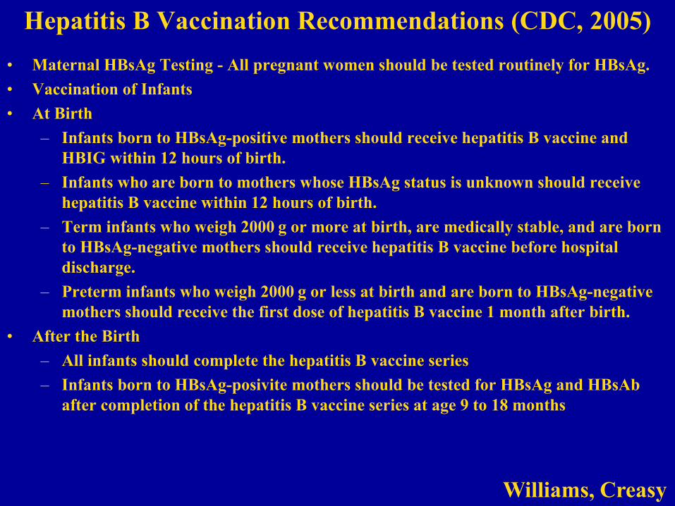
Liver transplant patients
• N >200 cases
• Wait 12 months before pregnancy
• Tacrolimus, cyclosporine – monitor drug
levels
• Risks – Preeclampsia, PTB, GDM, LBW,
SGA
• GDM screening

Tacrolimus (Prograf)
• MOA – Inhibits T-lymphocytes;
macrolide antibiotic from streptomyces
• CI – Hypersensitivity, Kidney disease
(relative)
• Pregnancy – Class C – Risk of PTB,
neonatal hyperkalemia, renal
dysfunction; animal studies show
increased risk of anomalies, APO
• Breast feeding – Excreted; UnsafeDrugs.com
Critical Care Obstetrics, 4th Ed (Dr. Scott)

Cyclosporine• MOA – inhibits T-lymphocytes
– Fungal metabolite, inhibits Tcell response by
inhibiting IL2, inhibits cell mediated immunity
• CI - Hypersensitivity, Renal dysfunction, liver
dysfunction, severe HTN; A/E lymphoma
• Pregnancy – C, PTB, LBW; crosses, no evidence of
teratogenicity; levels drop in pregnancy
• Breast feeding – Excreted; usafe; immunosuppression,
neutropenia, growth impairment, carcinogenesis
Drugs.com
Critical Care Obstetrics, 4th Ed (Dr. Scott)

Cholestasis

Intrahepatic cholestasis of
pregnancy
• Prurititis and mild jaundice (+/-)
• 3rd trimester (possible earlier)
• Incidence 1 in 1,000-10,000 deliveries
– Increased in chilean and swedish populations
– Rare in blacks
• Recurrence risk is significant
– Gonzalez study of chilean pop was 70% recurrence rate
– Varying degrees

Cholestasis – risk factors
• Chilean, Swedish descent
• Multiples
• Hepatitis C infection

• 50-4. Which of the following chronic
viral infections has been associated with a
marked increased risk for cholestasis of
pregnancy?
• Hepatitis B
• Hepatitis C *****
• Human immunodeficiency virus (HIV)
• Cytomegalovirus (CMV)

Natural history
• Initial symptom is nocturnal pruritus
followed by continual pruritus
• Clinical jaundice 2 weeks later in 50%
– Mild, constant until delivery
– Pruritus can worsen, excoriation possible
• Symptoms resolve quickly after delivery
(~2 days)

Differential diagnoses
• Viral hepatitis (cholestasis usually does
not have fevers, abdominal pain or as
high of an elevation of transaminases)
• Gallbladder disease (cholestasis usually is
not associated with nausea or vomiting)\
Differential diagnoses
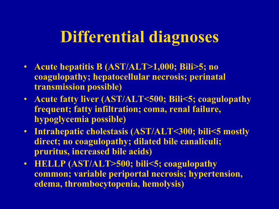
• Acute hepatitis B (AST/ALT>1,000; Bili>5; no coagulopathy; hepatocellular necrosis; perinatal transmission possible)
• Acute fatty liver (AST/ALT<500; Bili<5; coagulopathy frequent; fatty infiltration; coma, renal failure, hypoglycemia possible)
• Intrahepatic cholestasis (AST/ALT<300; bili<5 mostly direct; no coagulopathy; dilated bile canaliculi; pruritus, increased bile acids)
• HELLP (AST/ALT>500; bili<5; coagulopathy common; variable periportal necrosis; hypertension, edema, thrombocytopenia, hemolysis)
• 50-3. Which of the following clinical
features are characteristic of intrahepatic
cholestasis?
• Serum transaminases
• Maculopapular rash
• Generalized pruritus *****
• Nausea and vomiting

Lab findings
• Alkaline phosphatase levels 5-10x normal elevation of pregnancy– Normally increased b/c of placental production
– Fractionation reveals increased hepatic source
• 5’-nucleotidase increased?
• Urinary excretion of total sulfated progesterone metabolites are increased– Primary changes in reductase metabolism of progesterone
• Bilirubin is elevated, usually <5mg/dl (mostly direct, conjugated form)
• If chronic, liver dysfunction leads to decreased vit K reabsorption, decreased prothrombin production, prolonged prothrombin time
• Transaminases normal or moderately elevated, well below viral hepatitis levels
• Elevated cholesterol, triglyceride levels

Lab findings
• Serum bile acids (chenodeoxycholic acid, deoxycholic acid, cholic acid) increased 10x normal
– Deposited in skin, cause of pruritis
– Variable levels and symptoms
– To diagnose – fasting levels of serum bile acids elevated 3x upper limit of normal (random levels should not be used alone) + symptoms
• Copper and selenium increased
• CHO metabolism impaired (screen or re-screen for gestational diabetes)

Histologically
• Periportal, hepatocellular architecture
unchanged
• Centrilobular areas with dilated bile
canaliculi with bile plugs
– destruction, atrophy of microvilli of bile
canaliculi
• Regress PP
• 50-5. Liver biopsy in women who
suffer from cholestasis of pregnancy
would be expected to show which of the
following findings?
• Bile plugs in hepatocytes *****
• Inflammatory changes
• Periportal necrosis
• All of the above

Perinatal outcome
• Risk of preterm birth, IUFD increased
• Fisk study of 10 yr period– Meconium staining - 45%
– Spontaneous PTL - 44%
– Intrapartum fetal distress - 22%
– Of 86 infants• 2 iufds, 1 neonatal death
– Overall perinatal mortality 35 per 1,000
– Nsts, serial sonograms for fluid volume, estriol levels failed to predict fetal distress
– Early intervention (delivery) indicated in 49 pregnancies (12 b/c of fetal distress)
– Reason for increased fetal surveillance
– Induction at term, +FLM

Perinatal outcome
• Heinonen study of 91 cases
– CD rate 10% higher in cholestasis
pregnancies
– risk of preterm delivery (OR 2.73)
– risk of NICU admission (OR 2.15)
• case report of intracerebral hemorrhage
in pt with cholestasis (b/c delayed PT and
vit K factor production of mother?)

Management
• Reduce pruritus – start as soon as
diagnosis is made
• Antihistamines (diphenhydramine,
hydroxizine) help little
• Anion-binding resin (cholestyramine)
• Corticosteroids (dexamethasone)
• S-adenyl-methionine (sam-e)
• Ursodeoxycholic acid (UDCA)
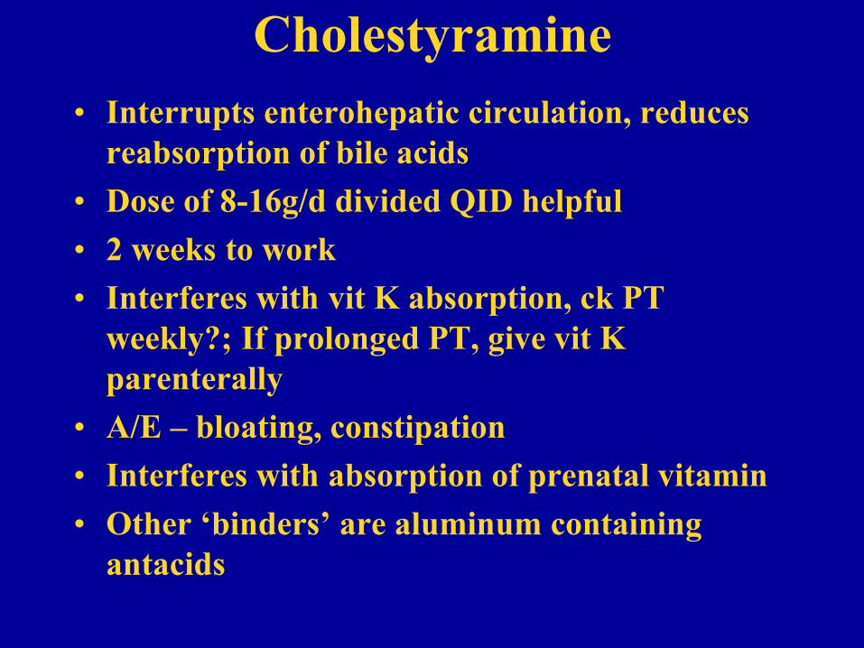
Cholestyramine
• Interrupts enterohepatic circulation, reduces
reabsorption of bile acids
• Dose of 8-16g/d divided QID helpful
• 2 weeks to work
• Interferes with vit K absorption, ck PT
weekly?; If prolonged PT, give vit K
parenterally
• A/E – bloating, constipation
• Interferes with absorption of prenatal vitamin
• Other ‘binders’ are aluminum containing
antacids

Other meds
• Phenobarbital can increase bile salt
secretion and increase bile flow
– No change in bile acid concentration
• Guar gum – gel forming fiber increases
fecal elimination of bile acids
– Finland study ?RCT showed stabilization of
bile acids and symptoms
• SAMe (Sadenosyl methionine) -Reverses
estrogen-induced impairment of bile
secretion

Corticosteroids
• Dexamethasone
– suppresses fetal/placental estrogen
production (out of balance in cholestasis
pregs)

SAM-e
• Reverses estrogen-induced impairment of
bile secretion
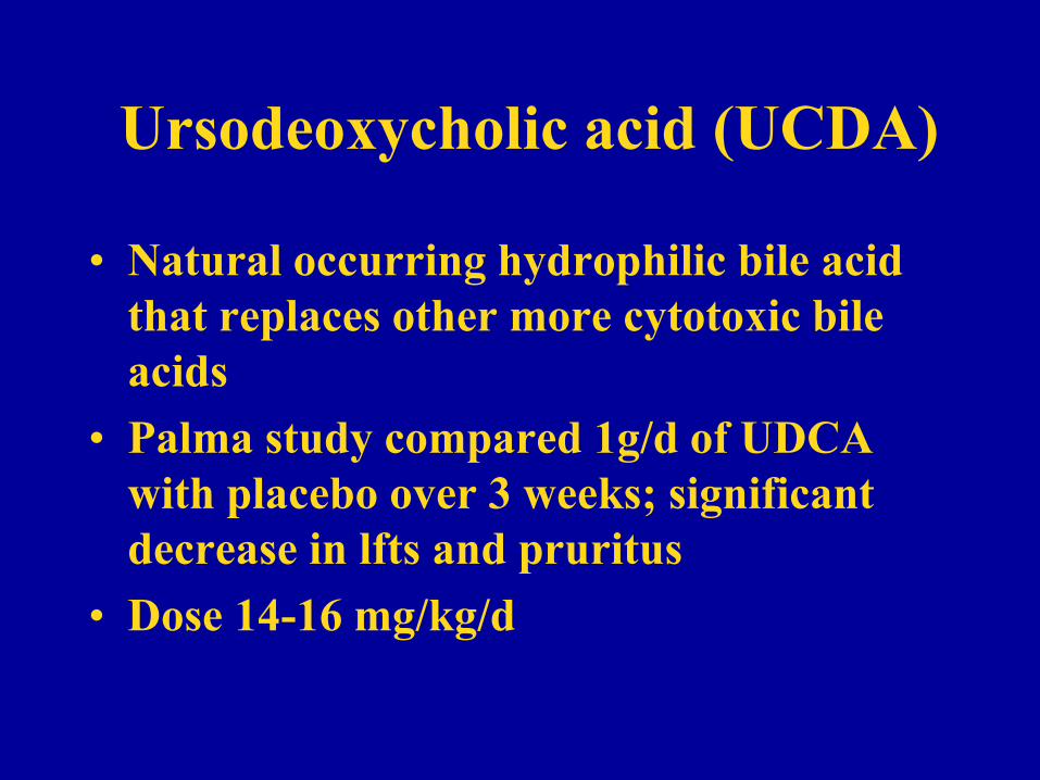
Ursodeoxycholic acid (UCDA)
• Natural occurring hydrophilic bile acid
that replaces other more cytotoxic bile
acids
• Palma study compared 1g/d of UDCA
with placebo over 3 weeks; significant
decrease in lfts and pruritus
• Dose 14-16 mg/kg/d
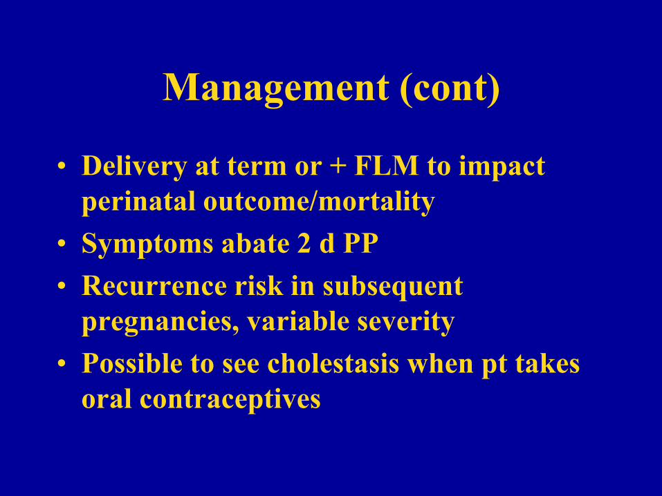
Management (cont)
• Delivery at term or + FLM to impact
perinatal outcome/mortality
• Symptoms abate 2 d PP
• Recurrence risk in subsequent
pregnancies, variable severity
• Possible to see cholestasis when pt takes
oral contraceptives

Conclusions
• AFLP – LFTs, coagulopathy – supportive
care, delivery
• Chronic liver disease/cirrhosis – check
for portal HTN, if present – beta
blockers, avoid valsalva efforts
• Cholestasis – elevated bile acids, >40
(worse prognosis) – ursodiol, delivery at
37 weeks (36 weeks per UTD)

Misc
• ANCS at 34-36 weeks
• No rescue courses
• Dispositioned for delivery at 34-36 weeks
• PPROM – OK
• Severe preeclampsia – per ACOG, ANCS
administration with delivery; per late
preterm steroid trial ~1/3 had severe
preeclampsia as indication for delivery
(completed ANCS course, followed by
delivery)
• No tocolysis

End

Add the following for the
resident question session

Objectives for Resident
Sessions
• AFLP
• HELLP
• Cholestasis

Creasy

Williams Questions – Ch 50

• See notes, ppt –
• See notes from previous Willams Review

Physiologic Changes
• Elevated alkaline phosphatase, palmar erythema,
spider angiomas, which might suggest liver disease, are
commonly found during normal pregnancy.
• Metabolism - altered expression of the cytochrome
P450 system that is mediated by higher levels of
estrogen, progesterone, and other hormones.
• Hepatic CYP1A2 expression is decreased, whereas that
of CYP2D6 and CYP3A4 is increased.
• Importantly, cytochrome enzymes are expressed in
many organs besides the liver, most notably the
placenta.
• No major hepatic histological changes are induced by
normal pregnancy

Pregnancy related liver disease

Liver Disease Coincidental to
Pregnancy

Chronic Liver Disease


Pancreatitis

Pancreatitis• 1 in 1000-3000 pregnancies
• Etiology –
– Gallstones (pregnancy),
– ETOH (nonpregnant)
– Hyperlipidemia
– Familial hypertriglyceridemia
– AFLP (complication in ½)
– Hyperparathyroidism
• Primary – Hypercalcemia
• Secondary – Initially low calcium
– Hypercalcemia
– CF
– Malignancy
• 20% have severe pancreatitis
• 25% mortality rate

Pancreatitis• Diagnosis /Symptoms
• N/v, pain, fever, tachycardia, SIRS
• Lab – Amylase, lipase (level does not
correlate with severity
• Cholesterol
• Triglycerides
• Leukocytosis
• Hyperbilirubinemia, elevated LFTs (Gall
stone disease)
• Hypocalcemia

Pancreatitis
• Predictive risk factors
- Shock
- Need for massive colloid replacement
- Hypocalcemia (<8mg/dL)
- Dark hemorrhagic fluid on paracentesis
If 3 of 4 present – 30% survival
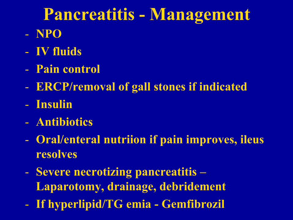
Pancreatitis - Management- NPO
- IV fluids
- Pain control
- ERCP/removal of gall stones if indicated
- Insulin
- Antibiotics
- Oral/enteral nutriion if pain improves, ileus
resolves
- Severe necrotizing pancreatitis –
Laparotomy, drainage, debridement
- If hyperlipid/TG emia - Gemfibrozil

Chronic Liver Disease
• Primary Biliary Cirrhosis –
• AI liver disease
• UCDA
• Cholestyramine
• Vitamin K 100mg BID
• Fetal surveillance

Primay Sclerosing Cholangitis• Chronic cholestatic disease,
unclear origin,
fibrosis/inflammation of
intrahepatic and extrahepatic
bile ducts
• Biliary cirrhosis >hepatic failure
>death
• Ulcerative colitis pts increased
risk of PSC (Crohn disease less)
• UCDA
• Liver failure > transplant
• Increased fetal bile acids
• Meconium passage
• Increased rate of fetal death –
Similar to ICP

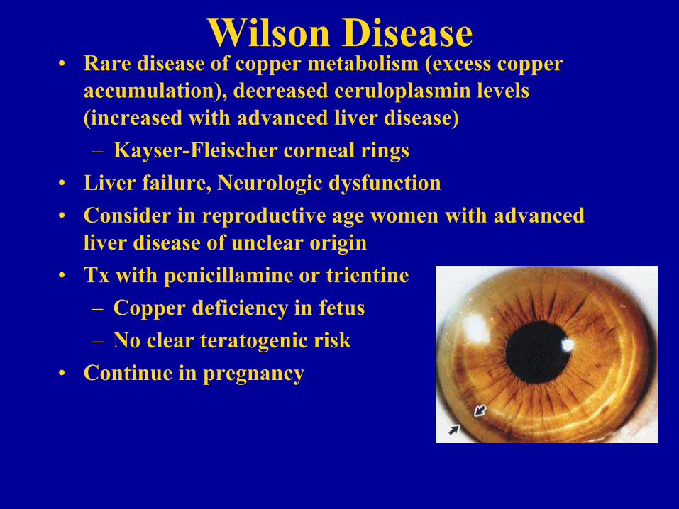
Wilson Disease• Rare disease of copper metabolism (excess copper
accumulation), decreased ceruloplasmin levels
(increased with advanced liver disease)
– Kayser-Fleischer corneal rings
• Liver failure, Neurologic dysfunction
• Consider in reproductive age women with advanced
liver disease of unclear origin
• Tx with penicillamine or trientine
– Copper deficiency in fetus
– No clear teratogenic risk
• Continue in pregnancy
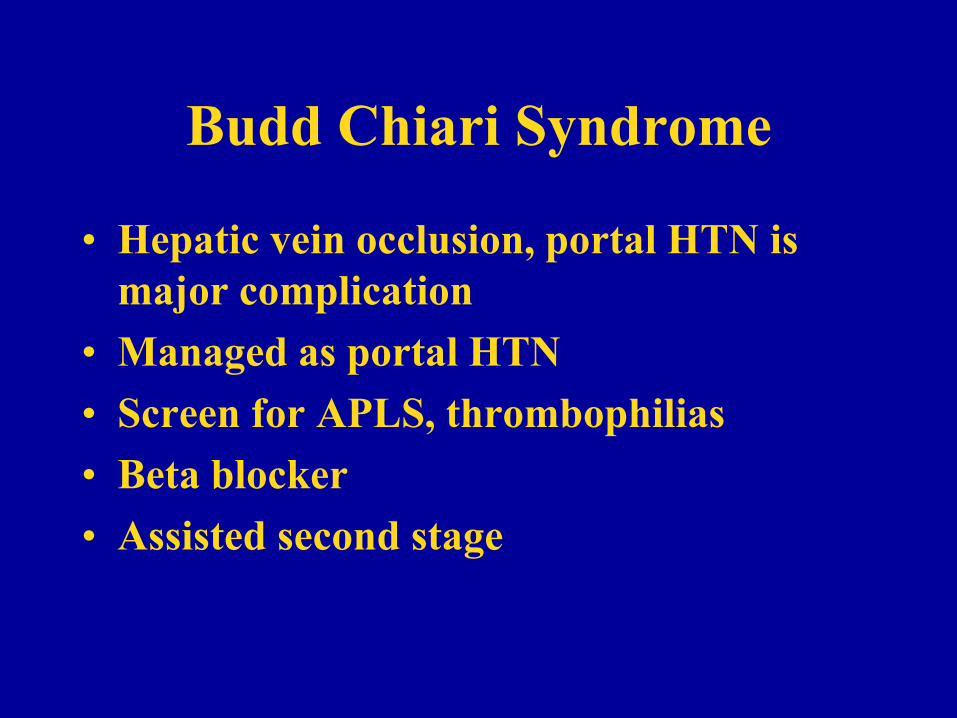
Budd Chiari Syndrome
• Hepatic vein occlusion, portal HTN is
major complication
• Managed as portal HTN
• Screen for APLS, thrombophilias
• Beta blocker
• Assisted second stage

Hepatobiliary Disesae in
Pregnancy
• April 2015
• END END END END END


Pulmonary edema

• Wedge and CVP Do Not Correlate
• SVR is Low Initially, and then Becomes Very High (along with BP)
• Pulmonary Artery Catheter Findings
– Elevated SBP, SVR
– Hyperdynamic LV Function
– Normal to Increased PCWP
– Low CVP
– High Wedge with Low CVP May be Due to Increased Afterload with Volume Depletion
Cardiac Manifestations
of Preeclampsia

Pulmonary Edema
in Preeclampsia
• Occurs in 3% of Women with Preeclampsia
• 70% Occurs Postpartum (Fluid Overload)
• Antepartum Pulmonary Edema Associated
with Chronic HTN in 90% Cases
• Risk Factors: Older Women, Multigravidas,
Chronic Hypertension
• Associated with Fluid Overload, either Colloid
or Crystalloid

Pulmonary Edema
in Preeclampsia
• Pathophysiology of Pulmonary Edema
– Reduced COP
– Alteration of Capillary Membrane
Permeability and Integrity
– Elevated Pulmonary Vascular Hydrostatic
Pressures
• Extravasation of Fluids in Pulmonary
Interstitium
Pulmonary Edema
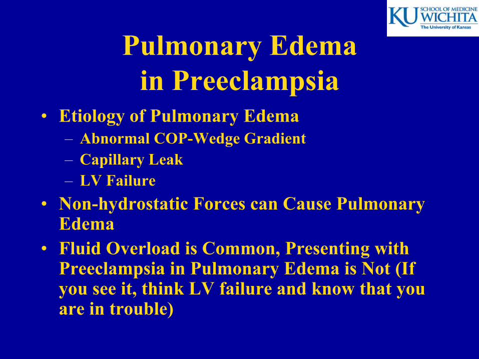
in Preeclampsia• Etiology of Pulmonary Edema
– Abnormal COP-Wedge Gradient
– Capillary Leak
– LV Failure
• Non-hydrostatic Forces can Cause Pulmonary Edema
• Fluid Overload is Common, Presenting with Preeclampsia in Pulmonary Edema is Not (If you see it, think LV failure and know that you are in trouble)

Pulmonary Edema
in Preeclampsia
• Risk factors – fluid overload, preeclampsia,
tocolysis, uncontrolled hypertension
• Diagnosis of Pulmonary Edema
– Clinical Diagnosis: Progressive Dyspnea and Chest
Discomfort
– Tachypnea, Tachycardia, Bilateral crackles
– Confirm with CXR and ABG
– Don’t Forget about Pulmonary Embolism

Case
• 34 yo P0, admitted for preeclampsia
– IVF pregnancy
• HD #3, developed progressive dyspnea, crackles on physical exam, oxygen requirements
– CXR revealed bilateral pleural effusions
• Fluid restriction, diuretics (Lasix 20mgIV), delivery, seizure prophylaxis

CXR of pulmonary edema

Pulmonary Edema
in Preeclampsia
• Management
– Oxygen, Fluid Restriction, Semi-Fowler
– Accurate intake/output
– If Fluid Overload, then Lasix, Increasing
Doses as Needed
– Consider PA Catheter: Fluid Overload vs.
LV Dysfunction vs. Nonhydrostatic
Pulmonary Edema

Indications for PA Catheter in
Hypertensive Disease
• Severe preeclampsia with refractory
oliguria or pulmonary edema
• Ineffective IV antihypertensive therapy
• Intraoperative or intrapartum cardiac
failure
p1169, Creasy

• Management
– Intravascular volume depletion (oliguria), low PCWP, high CO, high SVR, low CVP –
• fluids
– Renal Vasoconstriction (High PCWP, Normal CO and SVR, uroconcentration):
• Dopamine – 1-5µg/kg/min; furosemide
– LV Dysfunction/Failure with Vasospasm (high PCWP, high SVR, low CO <5 L/min) :
• Needs Afterload Reduction (Sodium nitroprusside 0.25-0.5µg/kg/min IV infusion)
• Volume Restriction
• Diuretics (max acute dose of furosemide is 120mg, start with 20-40mg)
– Mechanical Ventilation for Respiratory Failure (If still Pregnant, Intubate Early rather than Late)
Pulmonary Edema
in Preeclampsia – 3 subsets

Medications

NSAIDS
• NSAIDS – inhibits cyclooxygenase, lipoxygenase, reduces prostaglandin synthesis
• Class D
• Avoid especially in 3rd trimester
– Cross placenta, blocks prostaglandin synthesis in fetal tissue
– Premature closure of ductus arteriosis, fetal pulmonary hypertension, NEC, fetal renal insufficiency
– Occurs with selective COX-II inhibitors
– ASA crosses placenta and can affect fetal platelet function and is associated with intracranial fetal hemorrhage in 3rd
trimester; avoid in pregnancy
• Used outside of pregnancy – most common anti-inflammatory agent

Hydroxychloroquine
• Hydroxychloroquine (antimalarial/antirheumatic; binds DNA, interferes with vesicle functions, inhibits phospholipid metabolism; immunosuppressive by inhibiting rheumatoid factor, acute phase reactants, enzymes)
– Stopping this in pregnancy is associated with increased risk of lupus flares, continuing this drug is recommended if needed to control lupus (prospective study by cortes-hernandez showed the increased risk)
– Large series show no increased risk of anomalies
– Used in prevention of malaria with increase of fetal anomalies
– Not associated with increased r/o fetal malformations
• Class C
• Chloroquine possible teratogenic in initial studies
– Ototoxicity, eye development
Buchanan, 1996; Khamashta 1996
Klinger 2001; Motta 2002

Glucocorticoids
• Glucocorticoids (antiinflammatory, glucocorticoid, mineralocorticoid)
• Preg class C
• Avoid fluorinated glucocorticoids b/c they cross the placenta– Hydrocortisone, prednisone, prednisolone inactivated by 11-beta
hydroxysteroid dehydrogenase in the placenta allowing <10% of active drug to reach fetus
• High dose associated with maternal/fetal A/E– Osteoporosis (tx with vit D, ca2+); glucose intolerance, sodium, h2o
retention; hypertension, infection; avascular necrosis
– Preg complications – GDM, preeclampsia, PPROM, IUGR
– Incidence of fetal adrenal suppression with maternal tx is low
• Avoid empiric treatment, use at lowest possible dose
• Stress dose steroids (hydrocortisone 100mg IV q8hr in labor and for 24 hr PP)– Use if chronic steroids (>5mg/day for >2-4 weeks prior to delivery)

Azathioprine• Azathioprine (inhibits T lymphocytes)
• Class D
• Teratogenic in animals, appears safe in
humans
• Associated with IUGR
• Neonatal immunosuppression
• Indicated in pregnancy if chronic high
doses of steroids is not controlling
symptoms or to lower steroid dose

Cyclophosphamide
• Cyclophosphamide (alkylates and cross links DNA)
• Preg class D
– Cleft palate, skeletal abnormalities, abnormal renal function
– Avoid, esp in first trimester
– May be needed in cases of severe proliferative nephritis (drug of choice in nonpregnant patients with proliferative lupus nephritis)
– Crosses placenta

Methotrexate
• Methotrexate (inhibits dihydrofolate
reductase; inhibits lymphocyte
proliferation) (folate antagonist)
• Preg class X
• Avoid
• Embryolethal, IUFD
• Congenital anomalies
Cyclosporine
• Cyclosporine A (inhibits T lymphocytes)
• Preg class C
• Data comes from use in renal transplant
patients, not an animal teratogen,
appears safe in humans, long term follow
up studies are limited

Tacrolimus
• Tacrolimus (inhibits T lymphocyte activation,
immunosuppressant)
• Dose in liver transplant
– 0.1-0.15mg/kg/d po divide q12 hr
• Preg class C
• Therapeutic drug levels 5-20 ng/ml just before
next dose; time to steady state 3 days
• Monitor creatinine, K, fasting blood glucose,
serum drug levels

Pregnancy - FDA classes
• A – controlled studies show no fetal risk in any trimester, probability of fetal harm is remote
• B – animal studies, no risk; if risk in animal studies, controlled human studies do not confirm harm
• C – harm in animal studies with no controlled human studies; no available human or animal studies
• D – human studies show fetal risk but r/b relative to medical state of mother may support use
• X – animal/human studies show fetal risk or abnormalities, use is contraindicated during pregnancy or in women who may become pregnant


Lupus meds - NSAIDS
• used outside of pregnancy – most common anti-inflammatory agent
• inhibits cyclooxygenase, lipoxygenase, reduces prostaglandin synthesis
• Preg class B
• Avoid in 3rd trimester
– cross placenta, blocks prostaglandin synthesis in fetal tissue
– premature closure of ductus arteriosis, fetal pulmonary hypertension, NEC, fetal renal insufficiency
– ASA crosses placenta and can affect fetal platelet function and is associated with intracranial fetal hemorrhage in 3rd
trimester; avoid in pregnancy

Lupus meds -
Hydroxychloroquine• Hydroxychloroquine (antimalarial/antirheumatic;
binds DNA, interferes with vesicle functions, inhibits phospholipid metabolism; immunosuppressive by inhibiting rheumatoid factor, acute phase reactants, enzymes)
– stopping this in pregnancy - increased risk of lupus flares, continue if needed for control
– limited data
– not associated with increased r/o fetal malformations
• Preg class C
• Chloroquine is teratogenic

Lupus meds - steroids
• Preg class B
• avoid fluorinated glucocorticoids b/c they cross the placenta
– hydrocortisone, prednisone, prednisolone inactivated by 11-beta hydroxysteroid dehydrogenase in the placenta allowing <10% of active drug to reach fetus
• high dose associated with maternal/fetal side effects
– Maternal
• Osteoporosis, glucose intolerance, sodium/water retention, hypertension, infection
– Adverse pregnancy outcomes - GDM, preeclampsia, PPROM, IUGR
– Incidence of fetal adrenal suppression with maternal tx is low
• Avoid empiric treatment, use at lowest possible dose
• Stress dose steroids (hydrocortisone 100mg IV q8hr in labor and for 24 hr PP)
– use if chronic steroids (20mg or more of prednisone for >= 3 weeks during last 6 mos)

Lupus meds - Azathioprine
• Azathioprine (inhibits T lymphocytes)
• Preg class D
• teratogenic in animals, appears safe in
humans
• associated with IUGR
• indicated in pregnancy if chronic high
doses of steroids is not controlling
symptoms or to lower steroid dose
Lupus meds
• cyclosporine A (inhibits T lymphocytes)
• preg class C
• data comes from use in renal transplant
patients, not an animal teratogen,
appears safe in humans, long term follow
up studies are limited

Lupus meds
• cyclophosphamide (alkylates and cross links DNA)
• preg class D
– cleft palate, skeletal abnormalities; avoid if possible
– may be needed in cases of severe proliferative nephritis (drug of choice in nonpregnant patients with prolif lupus nephritis)
– crosses placenta

Lupus meds
• methotrexate (inhibits dihydrofolate
reductase; inhibits lymphocyte
proliferation (folate antagonist)
• preg class X
• avoid, embryolethal, congenital
anomalies


SLE
• Chronic autoimmune d/o with disease
flares and remissions
• Can affect all organs
– Mild cases – skin, musculoskeletal system
– More severe – kidney, brain
– Possible manifestations are arthralgias,
rashes, renal abnormalities, neurologic
complications, thromboemboli, myocarditis,
serositis

Medications

NSAIDS
• NSAIDS – inhibits cyclooxygenase, lipoxygenase, reduces prostaglandin synthesis
• Class D
• Avoid especially in 3rd trimester
– Cross placenta, blocks prostaglandin synthesis in fetal tissue
– Premature closure of ductus arteriosis, fetal pulmonary hypertension, NEC, fetal renal insufficiency
– Occurs with selective COX-II inhibitors
– ASA crosses placenta and can affect fetal platelet function and is associated with intracranial fetal hemorrhage in 3rd
trimester; avoid in pregnancy
• Used outside of pregnancy – most common anti-inflammatory agent

Hydroxychloroquine
• Hydroxychloroquine (antimalarial/antirheumatic; binds DNA, interferes with vesicle functions, inhibits phospholipid metabolism; immunosuppressive by inhibiting rheumatoid factor, acute phase reactants, enzymes)
– Stopping this in pregnancy is associated with increased risk of lupus flares, continuing this drug is recommended if needed to control lupus (prospective study by cortes-hernandez showed the increased risk)
– Large series show no increased risk of anomalies
– Used in prevention of malaria with increase of fetal anomalies
– Not associated with increased r/o fetal malformations
• Class C
• Chloroquine possible teratogenic in initial studies
– Ototoxicity, eye development
Buchanan, 1996; Khamashta 1996
Klinger 2001; Motta 2002

Glucocorticoids
• Glucocorticoids (antiinflammatory, glucocorticoid, mineralocorticoid)
• Preg class C
• Avoid fluorinated glucocorticoids b/c they cross the placenta– Hydrocortisone, prednisone, prednisolone inactivated by 11-beta
hydroxysteroid dehydrogenase in the placenta allowing <10% of active drug to reach fetus
• High dose associated with maternal/fetal A/E– Osteoporosis (tx with vit D, ca2+); glucose intolerance, sodium, h2o
retention; hypertension, infection; avascular necrosis
– Preg complications – GDM, preeclampsia, PPROM, IUGR
– Incidence of fetal adrenal suppression with maternal tx is low
• Avoid empiric treatment, use at lowest possible dose
• Stress dose steroids (hydrocortisone 100mg IV q8hr in labor and for 24 hr PP)– Use if chronic steroids (>5mg/day for >2-4 weeks prior to delivery)

Azathioprine• Azathioprine (inhibits T lymphocytes)
• Class D
• Teratogenic in animals, appears safe in
humans
• Associated with IUGR
• Neonatal immunosuppression
• Indicated in pregnancy if chronic high
doses of steroids is not controlling
symptoms or to lower steroid dose

Cyclophosphamide
• Cyclophosphamide (alkylates and cross links DNA)
• Preg class D
– Cleft palate, skeletal abnormalities, abnormal renal function
– Avoid, esp in first trimester
– May be needed in cases of severe proliferative nephritis (drug of choice in nonpregnant patients with proliferative lupus nephritis)
– Crosses placenta

Methotrexate
• Methotrexate (inhibits dihydrofolate
reductase; inhibits lymphocyte
proliferation) (folate antagonist)
• Preg class X
• Avoid
• Embryolethal, IUFD
• Congenital anomalies

Cyclosporine
• Cyclosporine A (inhibits T lymphocytes)
• Preg class C
• Data comes from use in renal transplant
patients, not an animal teratogen,
appears safe in humans, long term follow
up studies are limited

Tacrolimus
• Tacrolimus (inhibits T lymphocyte activation,
immunosuppressant)
• Dose in liver transplant
– 0.1-0.15mg/kg/d po divide q12 hr
• Preg class C
• Therapeutic drug levels 5-20 ng/ml just before
next dose; time to steady state 3 days
• Monitor creatinine, K, fasting blood glucose,
serum drug levels

• 54-21 Which of the following drugs
would be a choice of last resort in the
treatment of lupus during pregnancy?
• Aspirin
• Corticosteroids
• Azathioprine
• Cyclophosphamide****
Williams Obstetrics – Study Guide

Pregnancy - FDA classes
• A – controlled studies show no fetal risk in any trimester, probability of fetal harm is remote
• B – animal studies, no risk; if risk in animal studies, controlled human studies do not confirm harm
• C – harm in animal studies with no controlled human studies; no available human or animal studies
• D – human studies show fetal risk but r/b relative to medical state of mother may support use
• X – animal/human studies show fetal risk or abnormalities, use is contraindicated during pregnancy or in women who may become pregnant

Med list – good from GI –
complete remicade

Medications

Immunosuppressants (Scott CC OB)
• All drugs cross placenta, diffuse to fetus
• No convincing evidence that prednisone, azathioprine,
cyclosporine, tacrolimus produce congenital
abnormalities;
• Drugs of choice in preg transplant pts
• A/e IUGR, PTB , o/w neonates do well
– Short term ‘prematurity issues of infection,
hypoglycemia, bone marrow hypoplasia, leukopenia,
reduced IGM, IGG, elevated serum cr’ hard to
know if it’s the drug or prematurity
• Long term – poss infertility, autoimmune disease,
neoplasia – need for long term f/u

Medication
• MOA –
• CI -
• Pregnancy -
• Breast feeding -

Sulfasalazine (Azulfidine)• MOA – Sulfasalazine, metabolites (5-ASA,
sulfapyridine) - anti-inflammatory and/or
immunomodulatory properties, main effect in UC is
from 5-ASA
• CI – Intestinal or urinary obstruction, patients with
porphyria, allergy to sulfasalzine, metobolites,
sulfonamides, salicylates
• Pregnancy – Class B, crosses placenta;; no increased
rate of defects
– Impairs folate absorption/metabolism – so take 1-4mg folate
/day, esp periconceptionally
• Breastfeeding – Excreted; unsafe, especially if infant is
preterm, <1month old, or FHX of G6PD deficiencyDrugs.com
Critical Care Obstetrics, 4th Ed (Dr. Scott)

Azathioprine (Imuran)• MOA – inhibits T-lymphocytes; More toxic metabolite 6-
mercaptopurine- purine analog that decreases delayed hypersensitivity
and cellular cytotoxicity
• CI – Hypersensitivity, relative (pregnancy), increased risk of cancer,
esp if previously used alkylating agents (eg, chlorambucil,
cyclophosphamide, melphalan), liver disease, need to follow CBC,
CMP
• Pregnancy – Class D - D b/c increased anomaly rate of 9 and 6.4%, not
found in recent series, no specific pattern, possible bone marrow
suppression in fetus, with anemia, leukopenia, thrombocytopenia
• 64-90% of azathioprine crosses the placenta, majority is inactive
thiouric acid
• Breast feeding – Excreted; UK, unsafe; US – Caution is rec ;Relative
CI – Neutropenia, unknown risk of carcinogenesis; Women with
decreased activity of enzyme that detoxifies azathioprine metabolites
may pass on higher levels of drug to their infants via breast milk; if
used, check CBC/diff, CMP in exclusively breastfed infants; wait to
breastfeed 4-6hr after dose Drugs.com
Critical Care Obstetrics, 4th Ed (Dr. Scott)

Tacrolimus (Prograf)
• MOA – Inhibits T-lymphocytes;
macrolide antibiotic from streptomyces
• CI – Hypersensitivity, Kidney disease
(relative)
• Pregnancy – Class C – Risk of PTB,
neonatal hyperkalemia, renal
dysfunction; animal studies show
increased risk of anomalies, APO
• Breast feeding – Excreted; UnsafeDrugs.com
Critical Care Obstetrics, 4th Ed (Dr. Scott)

Cyclosporine• MOA – inhibits T-lymphocytes
– Fungal metabolite, inhibits Tcell response by
inhibiting IL2, inhibits cell mediated immunity
• CI - Hypersensitivity, Renal dysfunction, liver
dysfunction, severe HTN; A/E lymphoma
• Pregnancy – C, PTB, LBW; crosses, no evidence of
teratogenicity; levels drop in pregnancy
• Breast feeding – Excreted; usafe; immunosuppression,
neutropenia, growth impairment, carcinogenesis
Drugs.com
Critical Care Obstetrics, 4th Ed (Dr. Scott)

Infliximab (Remicade)
• MOA –
• CI –
• Pregnancy –
• Breastfeeding

Prednisone• MOA – inhibit humoral and cell mediated
immune response
• CI – Uncontrolled DM, hypersensitity
• Pregnancy – C; Prolonged courses of fluorinated steroids
(dexa, beta) may cause decreased brain, somatic growth, adrenal
suppression, neonatal sepsis, CLD, psychomotor delay, behavioral
prob; metabolized by 11B HSD, fetus exposed to 10%
– Vitamin D/calcium
– Stress dosing at delivery
• Breast feeding – Risks increased
Drugs.com
Critical Care Obstetrics, 4th Ed (Dr. Scott)

Glucocorticoids
• Glucocorticoids (antiinflammatory, glucocorticoid, mineralocorticoid)
• Preg class C
• Avoid fluorinated glucocorticoids b/c they cross the placenta– Hydrocortisone, prednisone, prednisolone inactivated by 11-beta
hydroxysteroid dehydrogenase in the placenta allowing <10% of active drug to reach fetus
• High dose associated with maternal/fetal A/E– Osteoporosis (tx with vit D, ca2+); glucose intolerance, sodium, h2o
retention; hypertension, infection; avascular necrosis
– Preg complications – GDM, preeclampsia, PPROM, IUGR
– Incidence of fetal adrenal suppression with maternal tx is low
• Avoid empiric treatment, use at lowest possible dose
• Stress dose steroids (hydrocortisone 100mg IV q8hr in labor and for 24 hr PP)– Use if chronic steroids (>5mg/day for >2-4 weeks prior to delivery)

Immunosuppressants-
Cyclophosphamide per Dr. Scott in CC in OB
book 4th ed
• Cyclophosphamide – MOA – alkylating agent; D
– cancer chemotherapy and as an immunosuppressant
– In human pregnancies, cyclophosphamide exposures that
occurred during the first trimester have been associated with
skeletal and palate defects, as well as malformations of the
limbs and eyes
– Cyclophosphamide is excreted into human milk (34). Two
reports indicates that the platelet and leukocyte counts of a
nursing infants were reversibly depressed during maternal
cyclophosphamide therapy (35,48). Cyclophosphamide was
classified among the cytotoxic drugs that may interfere with
cellular metabolism of a nursing infant by the American
Academy of Pediatrics (36).

Cyclophosphamide
• Cyclophosphamide (alkylates and cross links DNA)
• Preg class D
– Cleft palate, skeletal abnormalities, abnormal renal function
– Avoid, esp in first trimester
– May be needed in cases of severe proliferative nephritis (drug of choice in nonpregnant patients with proliferative lupus nephritis)
– Crosses placenta

• Which is not safe to use in pregnancy for
reflux esophagitis?
• Cimetidine, PPIs, calcium carbonate –
OK
NO misoprostol - -
• What is constellation of defects that
misoprostol is associated with if it does
not cause SAB? – Moebius sequence –
Williams Obstetrics
Pregnancy - FDA classes
• A – controlled studies show no fetal risk in any trimester, probability of fetal harm is remote
• B – animal studies, no risk; if risk in animal studies, controlled human studies do not confirm harm
• C – harm in animal studies with no controlled human studies; no available human or animal studies
• D – human studies show fetal risk but r/b relative to medical state of mother may support use
• X – animal/human studies show fetal risk or abnormalities, use is contraindicated during pregnancy or in women who may become pregnant


Glucocorticoids
• Glucocorticoids (antiinflammatory, glucocorticoid, mineralocorticoid)
• Preg class C
• Avoid fluorinated glucocorticoids b/c they cross the placenta– Hydrocortisone, prednisone, prednisolone inactivated by 11-beta
hydroxysteroid dehydrogenase in the placenta allowing <10% of active drug to reach fetus
• High dose associated with maternal/fetal A/E– Osteoporosis (tx with vit D, ca2+); glucose intolerance, sodium, h2o
retention; hypertension, infection; avascular necrosis
– Preg complications – GDM, preeclampsia, PPROM, IUGR
– Incidence of fetal adrenal suppression with maternal tx is low
• Avoid empiric treatment, use at lowest possible dose
• Stress dose steroids (hydrocortisone 100mg IV q8hr in labor and for 24 hr PP)– Use if chronic steroids (>5mg/day for >2-4 weeks prior to delivery)

Azathioprine• Azathioprine (inhibits T lymphocytes)
• Class D
• Teratogenic in animals, appears safe in
humans
• Associated with IUGR
• Neonatal immunosuppression
• Indicated in pregnancy if chronic high
doses of steroids is not controlling
symptoms or to lower steroid dose

Cyclophosphamide
• Cyclophosphamide (alkylates and cross links DNA)
• Preg class D
– Cleft palate, skeletal abnormalities, abnormal renal function
– Avoid, esp in first trimester
– May be needed in cases of severe proliferative nephritis (drug of choice in nonpregnant patients with proliferative lupus nephritis)
– Crosses placenta

Methotrexate
• Methotrexate (inhibits dihydrofolate
reductase; inhibits lymphocyte
proliferation) (folate antagonist)
• Preg class X
• Avoid
• Embryolethal, IUFD
• Congenital anomalies

Cyclosporine
• Cyclosporine A (inhibits T lymphocytes)
• Preg class C
• Data comes from use in renal transplant
patients, not an animal teratogen,
appears safe in humans, long term follow
up studies are limited

Tacrolimus
• Tacrolimus (inhibits T lymphocyte activation,
immunosuppressant)
• Dose in liver transplant
– 0.1-0.15mg/kg/d po divide q12 hr
• Preg class C
• Therapeutic drug levels 5-20 ng/ml just before
next dose; time to steady state 3 days
• Monitor creatinine, K, fasting blood glucose,
serum drug levels

References
• Email – [email protected]
• Provided on request
• Friedman 2008 PRIDE study
• Izmirly 2010 Hydroxychloroquine study
• Creasy – Resnik Maternal Fetal Medicine, Principles and Practice


Liver Disease in Pregnancy
Darren Farley, MD
Clinical Assistant Professor
Division of Maternal-Fetal Medicine
Dept. of Obstetrics and Gynecology
University of Kansas School of Medicine – Wichita

Objectives for Resident
Sessions
• AFLP
• HELLP
• Cholestasis


Hepatobiliary Disease in
Pregnancy • April 2015
• Physiologic changes
• Hepatitis
• Chronic liver disease
• AFLP
• Pancreatitis
• Gall bladder
• Cholestasis

Physiologic Changes
• Elevated alkaline phosphatase, palmar erythema,
spider angiomas, which might suggest liver disease, are
commonly found during normal pregnancy.
• Metabolism - altered expression of the cytochrome
P450 system that is mediated by higher levels of
estrogen, progesterone, and other hormones.
• Hepatic CYP1A2 expression is decreased, whereas that
of CYP2D6 and CYP3A4 is increased.
• Importantly, cytochrome enzymes are expressed in
many organs besides the liver, most notably the
placenta.
• No major hepatic histological changes are induced by
normal pregnancy

• 50-1. Of the following findings that
can be associated with liver dysfunction,
which is a normal physiologic change in
pregnancy?
• Elevation of hepatic transaminases
• Spider angiomas *****
• Esophageal varices
• Asterixis

Hepatitis - viralFeature Hep A Hep B Hep C Hep D Hep E
Viral type RNA DNA RNA RNA RNA
Inc period 14-50d 30-180d 30-160d 30-180d 14-63d
Transmission Fecal/oral Parenteral Parenteral Parenteral Fecal/oral
Diagnosis IgM anti
HAV ab
HBsAg,
anti-HBs,
anti-HBc,
HBeAg,
HBV DNA
Hep C ab Delta Ag,
IgM-specific
Ab
IgM anti-
HEV Ab
Carrier risk
of chronic
infection
0 10-15% 50-85% Up to 80%
when
superinfection
with Hep D
occurs
0
Vertical
transmission
risk
No Yes Yes Yes Yes
Vaccination
available
Yes Yes No No No
Creasy – MFM

Date of download: 3/7/2015 Copyright © 2015 McGraw-Hill Education. All rights reserved.
Sequence of various antigens and antibodies in acute hepatitis B. ALT = alanine aminotransferase; anti-HBc = antibody to hepatitis B core
antigen; anti-HBe = antibody to hepatitis B e antigen; anti-HBs = antibody to hepatitis B surface antigen; HBeAg = hepatitis B e antigen; HBsAg
= hepatitis B surface antigen. (Redrawn from Dienstag, 2012a.)
Legend:
From: Hepatic, Biliary, and Pancreatic Disorders
Williams Obstetrics, 24e, 2013

Interpretations of serologic testing in patients with hepatitis B virus
HBsAg HBsAb HBcAb HBeAg HBeAb Possible interpretation
- - - - - Never infected
+ - - - - -Early acute infxn
-Transient (up to 18d) after vaccination
+ - IgM + - Acute HBV infxn, highly infectious
+ - IgG + - Chronic HBV infxn, highly infectious
+ - IgG - + Late acute or chronic HBV infxn, low
infectivity
- - IgM +/- +/- Acute HBV infxn
- - IgG - +/- -Low-level HBsAg carrier or remote
past infxn
-Passive transfer to infant of HBsAg-
positive mother
- + IgG - +/- Recovery from HBV infxn and immune
- + - - - -Immune if concentration >/=
10mIU/mL
-Passive transfer after hepatitis B
immune globulin
Creasy – MFM

Hepatitis B Vaccination Recommendations (CDC, 2005)
• Maternal HBsAg Testing - All pregnant women should be tested routinely for HBsAg.
• Vaccination of Infants
• At Birth
– Infants born to HBsAg-positive mothers should receive hepatitis B vaccine and
HBIG within 12 hours of birth.
– Infants who are born to mothers whose HBsAg status is unknown should receive
hepatitis B vaccine within 12 hours of birth.
– Term infants who weigh 2000 g or more at birth, are medically stable, and are born
to HBsAg-negative mothers should receive hepatitis B vaccine before hospital
discharge.
– Preterm infants who weigh 2000 g or less at birth and are born to HBsAg-negative
mothers should receive the first dose of hepatitis B vaccine 1 month after birth.
• After the Birth
– All infants should complete the hepatitis B vaccine series
– Infants born to HBsAg-posivite mothers should be tested for HBsAg and HBsAb
after completion of the hepatitis B vaccine series at age 9 to 18 months
Williams, Creasy

Pregnancy related liver disease
• Hypertensive disease
• HELLP
• AFLP

AFLP
• Most common cause of acute liver failure
in pregnancy
• 1 in 10,000 pregnancies
• 3rd trimester
AFLP
Electron photomicrograph of two
swollen hepatocytes containing
numerous microvesicular fat droplets
(∗). The nuclei (N) remain centered
within the cells, in contrast to the case
with macrovesicular fat deposition.
http://intranet.tdmu.edu.ua/data/kafedra
/internal/ginecology2/classes_stud/en/
med/lik/ptn/Obstetrics%20and%20gyn
ecology/6%20year/02.%20Early%20ge
stosis.%20Hypertensive%20disorders
%20during%20pregnancy..htm

AFLP - Etiopathogenesis
• Association with recessively inherited mitochondrial
abnormalities of fatty acid oxidation.
• Similar to those in children with Reye-like syndromes.
• Mutations - mitochondrial trifunctional protein enzyme
complex that catalyzes the last oxidative steps in the
pathway; most common - G1528C and E474Q
mutations of the gene on chromosome 2 that code for
long-chain-3-hydroxyacyl-CoA-dehydrogenase—
known as LCHAD.

• 50-7. Mutations in enzymes involved in
fatty acid oxidation have been classically
associated with maternal acute fatty liver of
pregnancy when they occur in what pattern?
• Homozygous mutation in the fetus and the
mother
• Homozygous mutation in the fetus;
heterozygous mutation in the mother *****
• Heterozygous mutation in the fetus and the
mother
• Heterozygous mutation in the fetus;
homozygous mutation in the mother

AFLP• Parkland study – n=51 - 37wk (31-40.9)
• 41% nulliparous, 10-20% multifetal pregnancy
• N/v, malaise, anorexia, epigastric pain, progressive jaundice
• ½ have HTN, unclear overlap
• Elevated LFTs (<1000)
• Hypofibrinogenemia, hypoalbuminemia, hypocholesterolemia, and
prolonged clotting times.
• Serum bilirubin levels usually are < 10 mg/dL,
• Hypoglycemia
• Endothelial cell activation with capillary leakage causing
hemoconcentration, acute kidney injury, ascites, and sometimes
pulmonary permeability edema (Bernal, 2013)
• Severe hemoconcentration, uteroplacental perfusion is reduced,
maternal acidosis can cause fetal death; high CD rate due to NR
testing
• Peripheral blood smear – echinocytosis; hemolysis due
hypocholesterolemia

• A 33-year old G2P1 presents at 35 weeks’ gestation with
complaints of nausea and vomiting. Laboratory analysis reveals
elevated transaminases and creatinine levels and coagulopathy. A
peripheral smear is performed with the results shown.
• 50-9. What is the underlying etiology of these hemolyzed cells
found on the blood smear in this patient?
• Increase destruction in the spleen
• Intense vasospasm
• Occlusive pain crisis
• Decreased cholesterol production *****

Peripheral smear
Echinocytes
http://www.pclv.net/casestudies/mi
keybloodsmears.html
SchistocytesA schistocyte
count of >1% is
most often found
in thrombotic
thrombocytopeni
c purpura
AFLP - Hemolysis is
caused by the effects of
hypocholesterolemia on
erythrocyte membranes
(Cunningham, 1985)
Normal smear
http://library.med.
utah.edu/WebPath/
TUTORIAL/IRO
N/IRON002.html
Target cells – liver
disease, etc;
http://www.medical-
labs.net
?

AFLP • Swansea Diagnostic Criteria for Acute Fatty Liver of Pregnancy
• Six or more criteria are required in the absence of another cause:
• Vomiting
• Abdominal pain
• Polydipsia or polyuria
• Encephalopathy
• Elevated bilirubin level (>14 µmol/L)
• Hypoglycemia (<4 mmol/L)
• Elevated urea level (>340 µmol/L)
• Leukocytosis (>11 × 109/L)
• Ascites or bright liver on ultrasound scan
• Elevated transaminase levels (AST or ALT >42 IU/L)
• Elevated ammonia level (>47 µmol/L)
• Renal impairment (creatinine >150 µmol/L)
• Coagulopathy (PT >14 sec or aPTT >34 sec)
• Microvesicular steatosis on liver biopsy
• Adapted from Ch’ng CL, Morgan M, Hainsworth I, et al: Prospective study
of liver dysfunction in pregnancy in Southwest Wales, Gut 51:876–880,
2002.
AFLP
• Imaging – Not helpful – sonogram, CT, MRI, poor sensitivity
(Castro, 1996; Ch’ng, 2002; Knight, 2008; Nelson 2013)
– Maternal ascites, echogenic hepatic appearance
• Diagnosis – Clinical
• Syndrome worsens – Encephalopathy, DIC, renal failure, liver
failure (in 50%), delivery arrests liver deterioration
• Parkland – severeal women have isolated hemolysis,
hypofibrinogenemia as only findings (if LDH is increased, check
fibrinogen)
• Cases diagnosed as severe preeclampsia
• Coagulopathy, thrombocytopenia (10-20% with plt <50k)

• A 33-year old G2P1 presents at 35 weeks’ gestation with complaints of nausea and vomiting. Laboratory analysis reveals elevated transaminases and creatinine levels and coagulopathy. A peripheral smear is performed with the results shown.
• 50-10 As a part of the evaluation, you want to confirm the suspected diagnosis with imaging. Which of the following modalities is most appropriate?
• Sonography
• Computed tomography
• Magnetic resonance imaging
• None of the above *****

• A 33-year old G2P1 presents at 35 weeks’ gestation with complaints of nausea and vomiting. Laboratory analysis reveals elevated transaminases and creatinine levels and coagulopathy. A peripheral smear is performed with the results shown.
• 50-11 After you stabilize the patient and correct her coagulopathy, you induce labor and she has a vaginal delivery. On postpartum day 2, she appears to be doing well, but you notice that her urine output has increased to approximately 800 cc per hour. What is the most likely cause of this condition?
• Hypothalamic dysfunction
• Elevated serum vasopressinase concentrations *****
• Acute tubular necrosis
• Pituitary tumor

AFLP - Management
• Delivery –
• Mode – vaginal preferred; cesarean if remote
from delivery, to expedite maternal recovery
• Blood product replacement
• Seizure ppx if unclear diagnosis
• N-Acetylcysteine
• Regional Anesthetic – CI/use with caution due
to coagulopathy
• GI consult – in event that liver failure
necessitates orthoptic liver transplantation, so
this can be arranged
• 50-12 Postpartum acute pancreatitis
occurs in up to what percentage of
women who have acute fatty liver of
pregnancy?
• 10%
• 25%
• 50% *****
• 75%

• 50-13 All EXCEPT which of the
following acute liver diseases of
pregnancy can be associated with
thrombocytopenia?
• Viral hepatitis
• Preeclampsia
• Acute fatty liver of pregnancy
• Cholestasis of pregnancy *****

Date of download: 3/7/2015 Copyright © 2015 McGraw-Hill Education. All rights reserved.
Acute fatty liver of pregnancy. Cross section of the liver from a woman who died as the result of pulmonary aspiration and respiratory failure.
The liver has a greasy yellow appearance, which was present throughout the entire specimen. Inset: Electron photomicrograph of two swollen
hepatocytes containing numerous microvesicular fat droplets (∗). The nuclei (N) remain centered within the cells, in contrast to the case with
macrovesicular fat deposition. (Photograph contributed by Dr. Don Wheeler.)
Legend:
From: Hepatic, Biliary, and Pancreatic Disorders
Williams Obstetrics, 24e, 2013

Liver Disease Coincidental to
Pregnancy

Liver Disease Coincidental To
Pregnancy
• Liver Rupture

Liver Rupture and Infarction
• 95% of cases in pregnancy occur in severe preeclampsia and
HELLP syndrome
– Subcapsular hematoma occurs in <1% of HELLP sd patients
• Biliary disease, infection, aneurysm, hepatic neoplasm
• Subcapsular hemorrhage seen commonly in maternal autopsies
from preeclampsia
• Diagnosis – CT/MRI
• Lab – Coagulopathy, thrombocytopenia, anemia, hemolysis,
elevated LFTs, bilirubin
• DDX for unruptured liver hematoma – AFLP, abruption with
coagulopathy, TTP, HELLP, severe pree, cholangitis with sepsis

• Management
• Unruptured hematoma –hemodynamically stable –
conservative, Trauma consult, surveillance in ICU,
blood product replacement
• Liver rupture/Ruptured hematoma – Hepatic resection,
hepatic artery ligation, embolization of hepatic artery,
exploratory laparotomy, digital compression of hepatic
artery/portal vein (Pringle); evacuation of hematoma,
packing
Liver Rupture and Infarction

Chronic Liver Disease

Cirrhosis patients
• Risks – PTB, IUFD, Maternal mortality,
anemia, preeclampsia, bleeding from
esophageal varices, PPH
• Endoscopy to eval for varices
• Vaginal delivery, shortened second stage,
forceps if varices present
• Prep for PPH – blood products, etc

Portal Hypertension• Normal – 5-10mmHg,
increased if >10mmHg
• Diagnosis – Endoscopy,
Catheterization of hepatic
vein, Pulsed wave Dopp
• Exclude splenic artery
aneurysm (Pulsed-wave
Doppler and CT imaging)
– Rec surgery to ligate
before pregnancy if this is
present
• Beta blockers,
sclerotherapy, TIPS
procedure (transjugular
intrahepatic
protosystemic shunt
procedure)
• Propranolol 80mg/d
• Assisted second stage

Liver Failure• Hepatomegaly is abnormal in pregnancy
• Labs – LFTs >2000 – liver ischemia, rupture, infarction;
hypoglycemia, coagulopathy
• Causes – Viral (hepatitis viruses, CMV, EBV, HIV, HSV), APAP,
drug reactions (PTU), pregnancy related (AFLP, HELLP, HTN),
Budd-Chiari (hepatic vein thrombosis), ischemic necrosis, Wilson
disease, autoimmune hepatitis, toxin, malignancy)
• Management –
– Exclude pregnancy related causes – HTN, HELLP, AFLP;
delivery if these are cause;
– If viral hepatitis, supportive care is management, delivery is
considered if fetus is viable and risks of prematurity are low
due to high fetal mortality rate
– Supportive care, correct coagulopathy, hypoglycemia, PPI,
mannitol (cerebral edema tx)
• Acetaminophen overdose – NAC -

Liver Failure - Acetaminophen• Acetaminophen overdose
– NAC – Nomogram
• Tylenol level – 4hr after
injestion – if >120 – Tx
• If unknown tylenol level –
est gram ingestion – if
>7.5g – Tx
• If unknown ingestion and
level – empiric treatment
esp if liver failure or
dysfunction is noted
• NAC – 140mg/kg load;
Maintenance 17 doses of
70mg/kg, q4hr x72hr

Liver transplant patients
• N >200 cases
• Wait 12 months before pregnancy
• Tacrolimus, cyclosporine – monitor drug
levels
• Risks – Preeclampsia, PTB, GDM, LBW,
SGA
• GDM screening

Tacrolimus (Prograf)
• MOA – Inhibits T-lymphocytes;
macrolide antibiotic from streptomyces
• CI – Hypersensitivity, Kidney disease
(relative)
• Pregnancy – Class C – Risk of PTB,
neonatal hyperkalemia, renal
dysfunction; animal studies show
increased risk of anomalies, APO
• Breast feeding – Excreted; UnsafeDrugs.com
Critical Care Obstetrics, 4th Ed (Dr. Scott)

Cyclosporine• MOA – inhibits T-lymphocytes
– Fungal metabolite, inhibits Tcell response by
inhibiting IL2, inhibits cell mediated immunity
• CI - Hypersensitivity, Renal dysfunction, liver
dysfunction, severe HTN; A/E lymphoma
• Pregnancy – C, PTB, LBW; crosses, no evidence of
teratogenicity; levels drop in pregnancy
• Breast feeding – Excreted; usafe; immunosuppression,
neutropenia, growth impairment, carcinogenesis
Drugs.com
Critical Care Obstetrics, 4th Ed (Dr. Scott)

Immunosuppressants
per Dr. Scott in CC in OB book 4th ed
• Tacrolimus – MOA – inhibits T-
lymphocytes; C
– FK506 – macrolide abx from streptomyces;
– Incidence of post transplant DM with
tacrolimus is 11-20%; median time to onset
is 68d; 50% reversible
– Nephrotoxicity, hyperkalemia in 1/3; HA,
tremor, motor fxn, sensory fxn are neuro a/e
– Cord blood concentrations 50% of maternal
levels, no proven association with anomalies
to date

Immunosuppressants -
Cyclosporine per Dr. Scott in CC in OB book 4th ed
• Cyclosporine – MOA – inhibits T-lymphocytes; C
– Fungal metabolite, inhibits Tcell response by
inhibiting IL2
– Improved survival in transplant pt, in most regimen
– a/e – nephrotoxicity, HTN; others hirsutism, tremor,
gingival hyperplasia, hepatotoxicity, risk of
lymphomas
– Cyclosporine levels drop in pregnancy, but graft
function remains stable in most pts (Bumgardner
Matas 1992)
– Readily crosses placenta, no evidence of
teratogenicity

Chronic Liver Disease
• Primary Biliary Cirrhosis –
• AI liver disease
• UCDA
• Cholestyramine
• Vitamin K 100mg BID
• Fetal surveillance

Primay Sclerosing Cholangitis• Chronic cholestatic disease,
unclear origin,
fibrosis/inflammation of
intrahepatic and extrahepatic
bile ducts
• Biliary cirrhosis >hepatic failure
>death
• Ulcerative colitis pts increased
risk of PSC (Crohn disease less)
• UCDA
• Liver failure > transplant
• Increased fetal bile acids
• Meconium passage
• Increased rate of fetal death –
Similar to ICP
Wilson Disease• Rare disease of copper metabolism (excess copper
accumulation), decreased ceruloplasmin levels
(increased with advanced liver disease)
– Kayser-Fleischer corneal rings
• Liver failure, Neurologic dysfunction
• Consider in reproductive age women with advanced
liver disease of unclear origin
• Tx with penicillamine or trientine
– Copper deficiency in fetus
– No clear teratogenic risk
• Continue in pregnancy

Budd Chiari Syndrome
• Hepatic vein occlusion, portal HTN is
major complication
• Managed as portal HTN
• Screen for APLS, thrombophilias
• Beta blocker
• Assisted second stage

Pancreatitis

Pancreatitis• 1 in 1000-3000 pregnancies
• Etiology –
– Gallstones (pregnancy),
– ETOH (nonpregnant)
– Hyperlipidemia
– Familial hypertriglyceridemia
– AFLP (complication in ½)
– Hyperparathyroidism
• Primary – Hypercalcemia
• Secondary – Initially low calcium
– Hypercalcemia
– CF
– Malignancy
• 20% have severe pancreatitis
• 25% mortality rate

Pancreatitis• Diagnosis /Symptoms
• N/v, pain, fever, tachycardia, SIRS
• Lab – Amylase, lipase (level does not
correlate with severity
• Cholesterol
• Triglycerides
• Leukocytosis
• Hyperbilirubinemia, elevated LFTs (Gall
stone disease)
• Hypocalcemia

Pancreatitis
• Predictive risk factors
- Shock
- Need for massive colloid replacement
- Hypocalcemia (<8mg/dL)
- Dark hemorrhagic fluid on paracentesis
If 3 of 4 present – 30% survival

Pancreatitis - Management- NPO
- IV fluids
- Pain control
- ERCP/removal of gall stones if indicated
- Insulin
- Antibiotics
- Oral/enteral nutriion if pain improves, ileus
resolves
- Severe necrotizing pancreatitis –
Laparotomy, drainage, debridement
- If hyperlipid/TG emia - Gemfibrozil

Cholestasis

Intrahepatic cholestasis of
pregnancy
• Prurititis and mild jaundice (+/-)
• 3rd trimester (possible earlier)
• Incidence 1 in 1,000-10,000 deliveries
– Increased in chilean and swedish populations
– Rare in blacks
• Recurrence risk is significant
– Gonzalez study of chilean pop was 70% recurrence rate
– Varying degrees

Cholestasis – risk factors
• Chilean, swedish descent
• Multiples
• Hepatitis C infection

• 50-4. Which of the following chronic
viral infections has been associated with a
marked increased risk for cholestasis of
pregnancy?
• Hepatitis B
• Hepatitis C *****
• Human immunodeficiency virus (HIV)
• Cytomegalovirus (CMV)

Natural history
• Initial symptom is nocturnal pruritus
followed by continual pruritus
• Clinical jaundice 2 weeks later in 50%
– mild, constant until delivery
– pruritus can worsen, excoriation possible
• symptoms resolve quickly after delivery
(~2 days)

Differential diagnoses
• Viral hepatitis (cholestasis usually does
not have fevers, abdominal pain or as
high of an elevation of transaminases)
• Gallbladder disease (cholestasis usually is
not associated with nausea or vomiting)\

Differential diagnoses
• Acute hepatitis B (AST/ALT>1,000; Bili>5; no coagulopathy; hepatocellular necrosis; perinatal transmission possible)
• Acute fatty liver (AST/ALT<500; Bili<5; coagulopathy frequent; fatty infiltration; coma, renal failure, hypoglycemia possible)
• Intrahepatic cholestasis (AST/ALT<300; bili<5 mostly direct; no coagulopathy; dilated bile canaliculi; pruritus, increased bile acids)
• HELLP (AST/ALT>500; bili<5; coagulopathy common; variable periportal necrosis; hypertension, edema, thrombocytopenia, hemolysis)

• 50-3. Which of the following clinical
features are characteristic of intrahepatic
cholestasis?
• Serum transaminases
• Maculopapular rash
• Generalized pruritus *****
• Nausea and vomiting

Lab findings
• Alkaline phosphatase levels 5-10x normal elevation of pregnancy– normally increased b/c of placental production
– fractionation reveals increased hepatic source
• 5’-nucleotidase increased?
• urinary excretion of total sulfated progesterone metabolites are increased– primary changes in reductase metabolism of progesterone
• bilirubin is elevated, usually <5mg/dL (mostly direct, conjugated form)
• if chronic, liver dysfunction leads to decreased vit K reabsorption, decreased prothrombin production, prolonged prothrombin time
• transaminases normal or moderately elevated, well below viral hepatitis levels
• elevated cholesterol, triglyceride levels

Lab findings
• serum bile acids (chenodeoxycholic acid, deoxycholic acid, cholic acid) increased 10x normal
– deposited in skin, cause of pruritis
– variable levels and symptoms
– to diagnose – fasting levels of serum bile acids elevated 3x upper limit of normal (random levels should not be used alone) + symptoms
• copper and selenium increased
• CHO metabolism impaired (screen or re-screen for gestational diabetes)

Histologically
• periportal, hepatocellular architecture
unchanged
• centrilobular areas with dilated bile
canaliculi with bile plugs
– destruction, atrophy of microvilli of bile
canaliculi
• regress PP

• 50-5. Liver biopsy in women who
suffer from cholestasis of pregnancy
would be expected to show which of the
following findings?
• Bile plugs in hepatocytes *****
• Inflammatory changes
• Periportal necrosis
• All of the above

Perinatal outcome
• risk of preterm birth, IUFD increased
• Fisk study of 10 yr period– meconium staining - 45%
– spontaneous PTL - 44%
– intrapartum fetal distress - 22%
– of 86 infants• 2 IUFDs, 1 neonatal death
– overall perinatal mortality 35 per 1,000
– NSTs, serial sonograms for fluid volume, estriol levels failed to predict fetal distress
– early intervention (delivery) indicated in 49 pregnancies (12 b/c of fetal distress)
– reason for increased fetal surveillance
– induction at term, +FLM

Perinatal outcome
• Heinonen study of 91 cases
– CD rate 10% higher in cholestasis
pregnancies
– risk of preterm delivery (OR 2.73)
– risk of NICU admission (OR 2.15)
• case report of intracerebral hemorrhage
in pt with cholestasis (b/c delayed PT and
vit K factor production of mother?)

Management
• Reduce pruritus – start as soon as
diagnosis is made
• Antihistamines (diphenhydramine,
hydroxizine) help little
• Anion-binding resin (cholestyramine)
• Corticosteroids (dexamethasone)
• S-adenyl-methionine (SAM-e)
• ursodeoxycholic acid (UDCA)

Cholestyramine
• interrupts enterohepatic circulation, reduces
reabsorption of bile acids
• dose of 8-16g/d divided QID helpful
• 2 weeks to work
• interferes with vit K absorption, ck PT
weekly?; if prolonged PT, give vit K
parenterally
• A/E – bloating, constipation
• interferes with absorption of prenatal vitamin
• other ‘binders’ are aluminum containing
antacids

Other meds
• phenobarbital can increase bile salt
secretion and increase bile flow
– no change in bile acid concentration
• guar gum – gel forming fiber increases
fecal elimination of bile acids
– Finland study ?RCT showed stabilization of
bile acids and symptoms

Corticosteroids
• Dexamethasone
– suppresses fetal/placental estrogen
production (out of balance in cholestasis
pregs)

SAM-e
• reverses estrogen-induced impairment of
bile secretion

ursodeoxycholic acid (UCDA)
• natural occurring hydrophilic bile acid
that replaces other more cytotoxic bile
acids
• Palma study compared 1g/d of UDCA
with placebo over 3 weeks; significant
decrease in LFTs and pruritus
• dose 14-16 mg/kg/d

Management (cont)
• delivery at Term or + FLM to impact
perinatal outcome/mortality
• symptoms abate 2 d PP
• Recurrence risk in subsequent
pregnancies, variable severity
• Possible to see cholestasis when pt takes
oral contraceptives
Gallbladder disease

Date of download: 3/7/2015 Copyright © 2015 McGraw-Hill Education. All rights reserved.
This sonogram shows multiple hyperechoic gallstones filling an anechoic gallbladder.
Legend:
From: Hepatic, Biliary, and Pancreatic Disorders
Williams Obstetrics, 24e, 2013

Cholelithiasis
• Et of 7% of jaundice in pregnancy
• Present in 12% of all pregnancies
• Increased development of cholelithiasis in pregnancy, but not choleycystitis
• GB function altered– After 14 weeks
• Fasting GB volume 2x normal
• Rate of GB emptying is delayed
• % Of emptying is delayed
• Higher residual compared to nonpregnant state
• Cholecystokinin major stimulus for GB contractions
• Estrogen/progesterone make CTX less effective, increased residual

Diagnosis
• H&P
• ultrasound

Management
• Medical
– IV hydration
– analgesics
– NG suction
– antibiotics
– TPN
• Surgical
– if possible, postpone until PP
– cholecystectomy, cholangiography
– common bile duct exploration sometimes needed

Indications For
Cholecystectomy• Ascending cholangitis
• Common bile duct obstuction
• Severe pancreatitis
• Acute abdomen
• Cholecystectomy in 2nd/3rd trimester associated with fetal mortality of < 5%– If pancreatitis secondary to biliary tract stones
remains untreated, fetal mortality approaches 60% (printen et al)
• Laparoscopic cholecystectomy safe in pregnancy– Studies note insufflation pressures of 8-10mmhg

Liver disease
END of ppt slides, notes are f/u
slides

Creasy

Williams Questions – Ch 50

• See notes, ppt –
• See notes from previous Willams Review
Physiologic Changes
• Elevated alkaline phosphatase, palmar erythema,
spider angiomas, which might suggest liver disease, are
commonly found during normal pregnancy.
• Metabolism - altered expression of the cytochrome
P450 system that is mediated by higher levels of
estrogen, progesterone, and other hormones.
• Hepatic CYP1A2 expression is decreased, whereas that
of CYP2D6 and CYP3A4 is increased.
• Importantly, cytochrome enzymes are expressed in
many organs besides the liver, most notably the
placenta.
• No major hepatic histological changes are induced by
normal pregnancy

Pregnancy related liver disease

Liver Disease Coincidental to
Pregnancy
Chronic Liver Disease


Hepatobiliary Disesae in
Pregnancy
• April 2015
• END END END END END

Pulmonary edema

• Wedge and CVP Do Not Correlate
• SVR is Low Initially, and then Becomes Very High (along with BP)
• Pulmonary Artery Catheter Findings
– Elevated SBP, SVR
– Hyperdynamic LV Function
– Normal to Increased PCWP
– Low CVP
– High Wedge with Low CVP May be Due to Increased Afterload with Volume Depletion
Cardiac Manifestations
of Preeclampsia

Pulmonary Edema
in Preeclampsia
• Occurs in 3% of Women with Preeclampsia
• 70% Occurs Postpartum (Fluid Overload)
• Antepartum Pulmonary Edema Associated
with Chronic HTN in 90% Cases
• Risk Factors: Older Women, Multigravidas,
Chronic Hypertension
• Associated with Fluid Overload, either Colloid
or Crystalloid

Pulmonary Edema
in Preeclampsia
• Pathophysiology of Pulmonary Edema
– Reduced COP
– Alteration of Capillary Membrane
Permeability and Integrity
– Elevated Pulmonary Vascular Hydrostatic
Pressures
• Extravasation of Fluids in Pulmonary
Interstitium

Pulmonary Edema
in Preeclampsia• Etiology of Pulmonary Edema
– Abnormal COP-Wedge Gradient
– Capillary Leak
– LV Failure
• Non-hydrostatic Forces can Cause Pulmonary Edema
• Fluid Overload is Common, Presenting with Preeclampsia in Pulmonary Edema is Not (If you see it, think LV failure and know that you are in trouble)

Pulmonary Edema
in Preeclampsia
• Risk factors – fluid overload, preeclampsia,
tocolysis, uncontrolled hypertension
• Diagnosis of Pulmonary Edema
– Clinical Diagnosis: Progressive Dyspnea and Chest
Discomfort
– Tachypnea, Tachycardia, Bilateral crackles
– Confirm with CXR and ABG
– Don’t Forget about Pulmonary Embolism

Case
• 34 yo P0, admitted for preeclampsia
– IVF pregnancy
• HD #3, developed progressive dyspnea, crackles on physical exam, oxygen requirements
– CXR revealed bilateral pleural effusions
• Fluid restriction, diuretics (Lasix 20mgIV), delivery, seizure prophylaxis

CXR of pulmonary edema

Pulmonary Edema
in Preeclampsia
• Management
– Oxygen, Fluid Restriction, Semi-Fowler
– Accurate intake/output
– If Fluid Overload, then Lasix, Increasing
Doses as Needed
– Consider PA Catheter: Fluid Overload vs.
LV Dysfunction vs. Nonhydrostatic
Pulmonary Edema

Indications for PA Catheter in
Hypertensive Disease
• Severe preeclampsia with refractory
oliguria or pulmonary edema
• Ineffective IV antihypertensive therapy
• Intraoperative or intrapartum cardiac
failure
p1169, Creasy

• Management
– Intravascular volume depletion (oliguria), low PCWP, high CO, high SVR, low CVP –
• fluids
– Renal Vasoconstriction (High PCWP, Normal CO and SVR, uroconcentration):
• Dopamine – 1-5µg/kg/min; furosemide
– LV Dysfunction/Failure with Vasospasm (high PCWP, high SVR, low CO <5 L/min) :
• Needs Afterload Reduction (Sodium nitroprusside 0.25-0.5µg/kg/min IV infusion)
• Volume Restriction
• Diuretics (max acute dose of furosemide is 120mg, start with 20-40mg)
– Mechanical Ventilation for Respiratory Failure (If still Pregnant, Intubate Early rather than Late)
Pulmonary Edema
in Preeclampsia – 3 subsets

Medications

NSAIDS
• NSAIDS – inhibits cyclooxygenase, lipoxygenase, reduces prostaglandin synthesis
• Class D
• Avoid especially in 3rd trimester
– Cross placenta, blocks prostaglandin synthesis in fetal tissue
– Premature closure of ductus arteriosis, fetal pulmonary hypertension, NEC, fetal renal insufficiency
– Occurs with selective COX-II inhibitors
– ASA crosses placenta and can affect fetal platelet function and is associated with intracranial fetal hemorrhage in 3rd
trimester; avoid in pregnancy
• Used outside of pregnancy – most common anti-inflammatory agent

Hydroxychloroquine
• Hydroxychloroquine (antimalarial/antirheumatic; binds DNA, interferes with vesicle functions, inhibits phospholipid metabolism; immunosuppressive by inhibiting rheumatoid factor, acute phase reactants, enzymes)
– Stopping this in pregnancy is associated with increased risk of lupus flares, continuing this drug is recommended if needed to control lupus (prospective study by cortes-hernandez showed the increased risk)
– Large series show no increased risk of anomalies
– Used in prevention of malaria with increase of fetal anomalies
– Not associated with increased r/o fetal malformations
• Class C
• Chloroquine possible teratogenic in initial studies
– Ototoxicity, eye development
Buchanan, 1996; Khamashta 1996
Klinger 2001; Motta 2002

Glucocorticoids
• Glucocorticoids (antiinflammatory, glucocorticoid, mineralocorticoid)
• Preg class C
• Avoid fluorinated glucocorticoids b/c they cross the placenta– Hydrocortisone, prednisone, prednisolone inactivated by 11-beta
hydroxysteroid dehydrogenase in the placenta allowing <10% of active drug to reach fetus
• High dose associated with maternal/fetal A/E– Osteoporosis (tx with vit D, ca2+); glucose intolerance, sodium, h2o
retention; hypertension, infection; avascular necrosis
– Preg complications – GDM, preeclampsia, PPROM, IUGR
– Incidence of fetal adrenal suppression with maternal tx is low
• Avoid empiric treatment, use at lowest possible dose
• Stress dose steroids (hydrocortisone 100mg IV q8hr in labor and for 24 hr PP)– Use if chronic steroids (>5mg/day for >2-4 weeks prior to delivery)

Azathioprine• Azathioprine (inhibits T lymphocytes)
• Class D
• Teratogenic in animals, appears safe in
humans
• Associated with IUGR
• Neonatal immunosuppression
• Indicated in pregnancy if chronic high
doses of steroids is not controlling
symptoms or to lower steroid dose
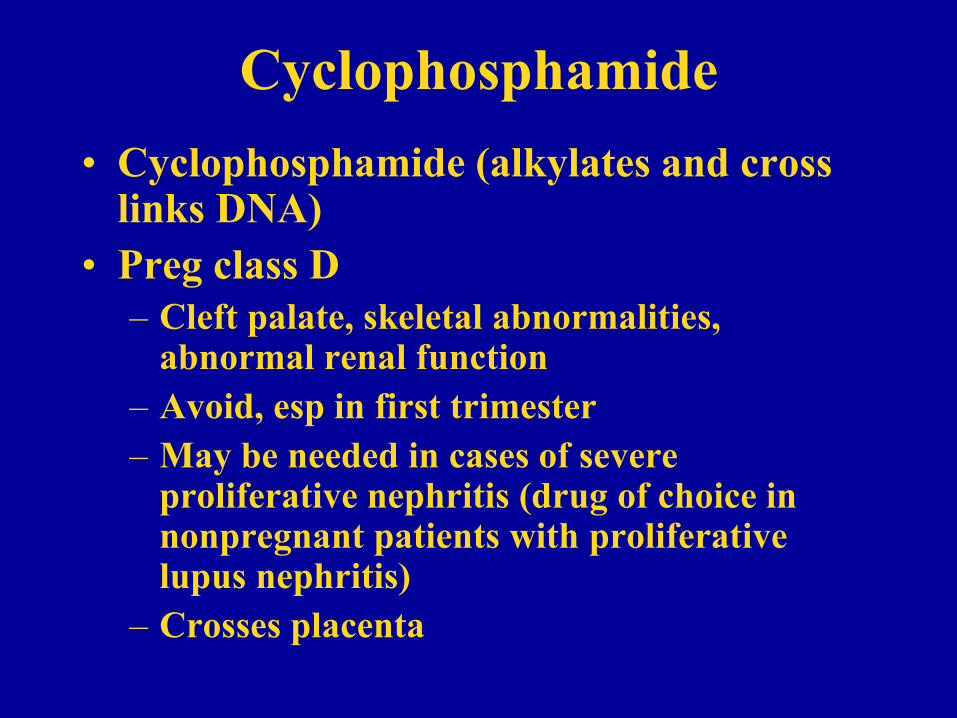
Cyclophosphamide
• Cyclophosphamide (alkylates and cross links DNA)
• Preg class D
– Cleft palate, skeletal abnormalities, abnormal renal function
– Avoid, esp in first trimester
– May be needed in cases of severe proliferative nephritis (drug of choice in nonpregnant patients with proliferative lupus nephritis)
– Crosses placenta

Methotrexate
• Methotrexate (inhibits dihydrofolate
reductase; inhibits lymphocyte
proliferation) (folate antagonist)
• Preg class X
• Avoid
• Embryolethal, IUFD
• Congenital anomalies

Cyclosporine
• Cyclosporine A (inhibits T lymphocytes)
• Preg class C
• Data comes from use in renal transplant
patients, not an animal teratogen,
appears safe in humans, long term follow
up studies are limited

Tacrolimus
• Tacrolimus (inhibits T lymphocyte activation,
immunosuppressant)
• Dose in liver transplant
– 0.1-0.15mg/kg/d po divide q12 hr
• Preg class C
• Therapeutic drug levels 5-20 ng/ml just before
next dose; time to steady state 3 days
• Monitor creatinine, K, fasting blood glucose,
serum drug levels

Pregnancy - FDA classes
• A – controlled studies show no fetal risk in any trimester, probability of fetal harm is remote
• B – animal studies, no risk; if risk in animal studies, controlled human studies do not confirm harm
• C – harm in animal studies with no controlled human studies; no available human or animal studies
• D – human studies show fetal risk but r/b relative to medical state of mother may support use
• X – animal/human studies show fetal risk or abnormalities, use is contraindicated during pregnancy or in women who may become pregnant


Lupus meds - NSAIDS
• used outside of pregnancy – most common anti-inflammatory agent
• inhibits cyclooxygenase, lipoxygenase, reduces prostaglandin synthesis
• Preg class B
• Avoid in 3rd trimester
– cross placenta, blocks prostaglandin synthesis in fetal tissue
– premature closure of ductus arteriosis, fetal pulmonary hypertension, NEC, fetal renal insufficiency
– ASA crosses placenta and can affect fetal platelet function and is associated with intracranial fetal hemorrhage in 3rd
trimester; avoid in pregnancy

Lupus meds -
Hydroxychloroquine• Hydroxychloroquine (antimalarial/antirheumatic;
binds DNA, interferes with vesicle functions, inhibits phospholipid metabolism; immunosuppressive by inhibiting rheumatoid factor, acute phase reactants, enzymes)
– stopping this in pregnancy - increased risk of lupus flares, continue if needed for control
– limited data
– not associated with increased r/o fetal malformations
• Preg class C
• Chloroquine is teratogenic

Lupus meds - steroids
• Preg class B
• avoid fluorinated glucocorticoids b/c they cross the placenta
– hydrocortisone, prednisone, prednisolone inactivated by 11-beta hydroxysteroid dehydrogenase in the placenta allowing <10% of active drug to reach fetus
• high dose associated with maternal/fetal side effects
– Maternal
• Osteoporosis, glucose intolerance, sodium/water retention, hypertension, infection
– Adverse pregnancy outcomes - GDM, preeclampsia, PPROM, IUGR
– Incidence of fetal adrenal suppression with maternal tx is low
• Avoid empiric treatment, use at lowest possible dose
• Stress dose steroids (hydrocortisone 100mg IV q8hr in labor and for 24 hr PP)
– use if chronic steroids (20mg or more of prednisone for >= 3 weeks during last 6 mos)
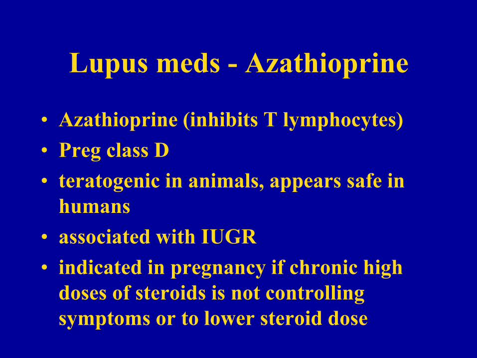
Lupus meds - Azathioprine
• Azathioprine (inhibits T lymphocytes)
• Preg class D
• teratogenic in animals, appears safe in
humans
• associated with IUGR
• indicated in pregnancy if chronic high
doses of steroids is not controlling
symptoms or to lower steroid dose

Lupus meds
• cyclosporine A (inhibits T lymphocytes)
• preg class C
• data comes from use in renal transplant
patients, not an animal teratogen,
appears safe in humans, long term follow
up studies are limited

Lupus meds
• cyclophosphamide (alkylates and cross links DNA)
• preg class D
– cleft palate, skeletal abnormalities; avoid if possible
– may be needed in cases of severe proliferative nephritis (drug of choice in nonpregnant patients with prolif lupus nephritis)
– crosses placenta

Lupus meds
• methotrexate (inhibits dihydrofolate
reductase; inhibits lymphocyte
proliferation (folate antagonist)
• preg class X
• avoid, embryolethal, congenital
anomalies


SLE
• Chronic autoimmune d/o with disease
flares and remissions
• Can affect all organs
– Mild cases – skin, musculoskeletal system
– More severe – kidney, brain
– Possible manifestations are arthralgias,
rashes, renal abnormalities, neurologic
complications, thromboemboli, myocarditis,
serositis

Medications

NSAIDS
• NSAIDS – inhibits cyclooxygenase, lipoxygenase, reduces prostaglandin synthesis
• Class D
• Avoid especially in 3rd trimester
– Cross placenta, blocks prostaglandin synthesis in fetal tissue
– Premature closure of ductus arteriosis, fetal pulmonary hypertension, NEC, fetal renal insufficiency
– Occurs with selective COX-II inhibitors
– ASA crosses placenta and can affect fetal platelet function and is associated with intracranial fetal hemorrhage in 3rd
trimester; avoid in pregnancy
• Used outside of pregnancy – most common anti-inflammatory agent

Hydroxychloroquine
• Hydroxychloroquine (antimalarial/antirheumatic; binds DNA, interferes with vesicle functions, inhibits phospholipid metabolism; immunosuppressive by inhibiting rheumatoid factor, acute phase reactants, enzymes)
– Stopping this in pregnancy is associated with increased risk of lupus flares, continuing this drug is recommended if needed to control lupus (prospective study by cortes-hernandez showed the increased risk)
– Large series show no increased risk of anomalies
– Used in prevention of malaria with increase of fetal anomalies
– Not associated with increased r/o fetal malformations
• Class C
• Chloroquine possible teratogenic in initial studies
– Ototoxicity, eye development
Buchanan, 1996; Khamashta 1996
Klinger 2001; Motta 2002

Glucocorticoids
• Glucocorticoids (antiinflammatory, glucocorticoid, mineralocorticoid)
• Preg class C
• Avoid fluorinated glucocorticoids b/c they cross the placenta– Hydrocortisone, prednisone, prednisolone inactivated by 11-beta
hydroxysteroid dehydrogenase in the placenta allowing <10% of active drug to reach fetus
• High dose associated with maternal/fetal A/E– Osteoporosis (tx with vit D, ca2+); glucose intolerance, sodium, h2o
retention; hypertension, infection; avascular necrosis
– Preg complications – GDM, preeclampsia, PPROM, IUGR
– Incidence of fetal adrenal suppression with maternal tx is low
• Avoid empiric treatment, use at lowest possible dose
• Stress dose steroids (hydrocortisone 100mg IV q8hr in labor and for 24 hr PP)– Use if chronic steroids (>5mg/day for >2-4 weeks prior to delivery)

Azathioprine• Azathioprine (inhibits T lymphocytes)
• Class D
• Teratogenic in animals, appears safe in
humans
• Associated with IUGR
• Neonatal immunosuppression
• Indicated in pregnancy if chronic high
doses of steroids is not controlling
symptoms or to lower steroid dose

Cyclophosphamide
• Cyclophosphamide (alkylates and cross links DNA)
• Preg class D
– Cleft palate, skeletal abnormalities, abnormal renal function
– Avoid, esp in first trimester
– May be needed in cases of severe proliferative nephritis (drug of choice in nonpregnant patients with proliferative lupus nephritis)
– Crosses placenta

Methotrexate
• Methotrexate (inhibits dihydrofolate
reductase; inhibits lymphocyte
proliferation) (folate antagonist)
• Preg class X
• Avoid
• Embryolethal, IUFD
• Congenital anomalies

Cyclosporine
• Cyclosporine A (inhibits T lymphocytes)
• Preg class C
• Data comes from use in renal transplant
patients, not an animal teratogen,
appears safe in humans, long term follow
up studies are limited

Tacrolimus
• Tacrolimus (inhibits T lymphocyte activation,
immunosuppressant)
• Dose in liver transplant
– 0.1-0.15mg/kg/d po divide q12 hr
• Preg class C
• Therapeutic drug levels 5-20 ng/ml just before
next dose; time to steady state 3 days
• Monitor creatinine, K, fasting blood glucose,
serum drug levels

• 54-21 Which of the following drugs
would be a choice of last resort in the
treatment of lupus during pregnancy?
• Aspirin
• Corticosteroids
• Azathioprine
• Cyclophosphamide****
Williams Obstetrics – Study Guide

Pregnancy - FDA classes
• A – controlled studies show no fetal risk in any trimester, probability of fetal harm is remote
• B – animal studies, no risk; if risk in animal studies, controlled human studies do not confirm harm
• C – harm in animal studies with no controlled human studies; no available human or animal studies
• D – human studies show fetal risk but r/b relative to medical state of mother may support use
• X – animal/human studies show fetal risk or abnormalities, use is contraindicated during pregnancy or in women who may become pregnant

Med list – good from GI –
complete remicade

Medications
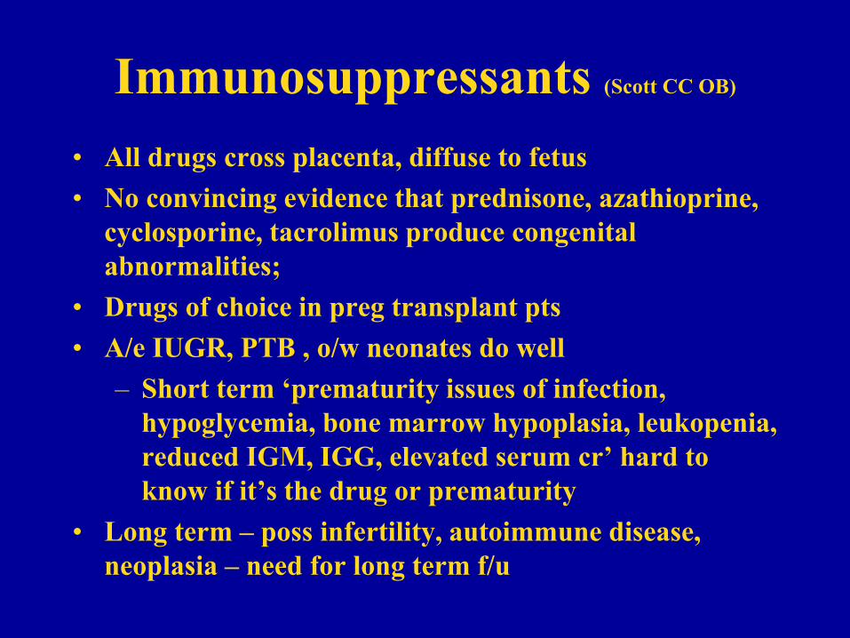
Immunosuppressants (Scott CC OB)
• All drugs cross placenta, diffuse to fetus
• No convincing evidence that prednisone, azathioprine,
cyclosporine, tacrolimus produce congenital
abnormalities;
• Drugs of choice in preg transplant pts
• A/e IUGR, PTB , o/w neonates do well
– Short term ‘prematurity issues of infection,
hypoglycemia, bone marrow hypoplasia, leukopenia,
reduced IGM, IGG, elevated serum cr’ hard to
know if it’s the drug or prematurity
• Long term – poss infertility, autoimmune disease,
neoplasia – need for long term f/u

Medication
• MOA –
• CI -
• Pregnancy -
• Breast feeding -

Sulfasalazine (Azulfidine)• MOA – Sulfasalazine, metabolites (5-ASA,
sulfapyridine) - anti-inflammatory and/or
immunomodulatory properties, main effect in UC is
from 5-ASA
• CI – Intestinal or urinary obstruction, patients with
porphyria, allergy to sulfasalzine, metobolites,
sulfonamides, salicylates
• Pregnancy – Class B, crosses placenta;; no increased
rate of defects
– Impairs folate absorption/metabolism – so take 1-4mg folate
/day, esp periconceptionally
• Breastfeeding – Excreted; unsafe, especially if infant is
preterm, <1month old, or FHX of G6PD deficiencyDrugs.com
Critical Care Obstetrics, 4th Ed (Dr. Scott)

Azathioprine (Imuran)• MOA – inhibits T-lymphocytes; More toxic metabolite 6-
mercaptopurine- purine analog that decreases delayed hypersensitivity
and cellular cytotoxicity
• CI – Hypersensitivity, relative (pregnancy), increased risk of cancer,
esp if previously used alkylating agents (eg, chlorambucil,
cyclophosphamide, melphalan), liver disease, need to follow CBC,
CMP
• Pregnancy – Class D - D b/c increased anomaly rate of 9 and 6.4%, not
found in recent series, no specific pattern, possible bone marrow
suppression in fetus, with anemia, leukopenia, thrombocytopenia
• 64-90% of azathioprine crosses the placenta, majority is inactive
thiouric acid
• Breast feeding – Excreted; UK, unsafe; US – Caution is rec ;Relative
CI – Neutropenia, unknown risk of carcinogenesis; Women with
decreased activity of enzyme that detoxifies azathioprine metabolites
may pass on higher levels of drug to their infants via breast milk; if
used, check CBC/diff, CMP in exclusively breastfed infants; wait to
breastfeed 4-6hr after dose Drugs.com
Critical Care Obstetrics, 4th Ed (Dr. Scott)

Tacrolimus (Prograf)
• MOA – Inhibits T-lymphocytes;
macrolide antibiotic from streptomyces
• CI – Hypersensitivity, Kidney disease
(relative)
• Pregnancy – Class C – Risk of PTB,
neonatal hyperkalemia, renal
dysfunction; animal studies show
increased risk of anomalies, APO
• Breast feeding – Excreted; UnsafeDrugs.com
Critical Care Obstetrics, 4th Ed (Dr. Scott)
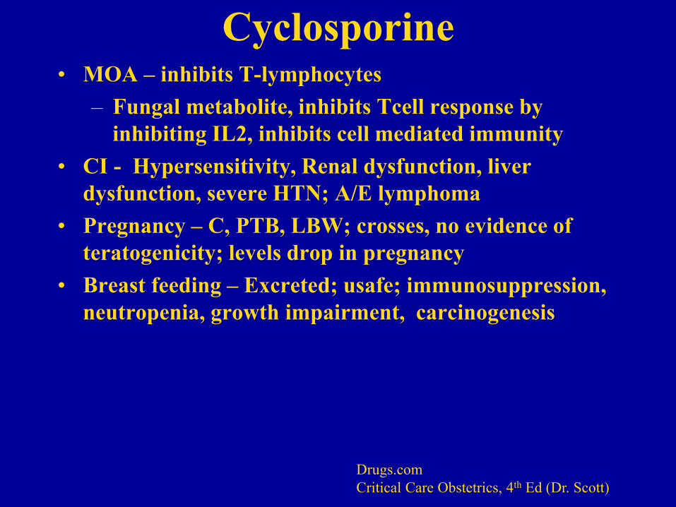
Cyclosporine• MOA – inhibits T-lymphocytes
– Fungal metabolite, inhibits Tcell response by
inhibiting IL2, inhibits cell mediated immunity
• CI - Hypersensitivity, Renal dysfunction, liver
dysfunction, severe HTN; A/E lymphoma
• Pregnancy – C, PTB, LBW; crosses, no evidence of
teratogenicity; levels drop in pregnancy
• Breast feeding – Excreted; usafe; immunosuppression,
neutropenia, growth impairment, carcinogenesis
Drugs.com
Critical Care Obstetrics, 4th Ed (Dr. Scott)

Infliximab (Remicade)
• MOA –
• CI –
• Pregnancy –
• Breastfeeding

Prednisone• MOA – inhibit humoral and cell mediated
immune response
• CI – Uncontrolled DM, hypersensitity
• Pregnancy – C; Prolonged courses of fluorinated steroids
(dexa, beta) may cause decreased brain, somatic growth, adrenal
suppression, neonatal sepsis, CLD, psychomotor delay, behavioral
prob; metabolized by 11B HSD, fetus exposed to 10%
– Vitamin D/calcium
– Stress dosing at delivery
• Breast feeding – Risks increased
Drugs.com
Critical Care Obstetrics, 4th Ed (Dr. Scott)

Glucocorticoids
• Glucocorticoids (antiinflammatory, glucocorticoid, mineralocorticoid)
• Preg class C
• Avoid fluorinated glucocorticoids b/c they cross the placenta– Hydrocortisone, prednisone, prednisolone inactivated by 11-beta
hydroxysteroid dehydrogenase in the placenta allowing <10% of active drug to reach fetus
• High dose associated with maternal/fetal A/E– Osteoporosis (tx with vit D, ca2+); glucose intolerance, sodium, h2o
retention; hypertension, infection; avascular necrosis
– Preg complications – GDM, preeclampsia, PPROM, IUGR
– Incidence of fetal adrenal suppression with maternal tx is low
• Avoid empiric treatment, use at lowest possible dose
• Stress dose steroids (hydrocortisone 100mg IV q8hr in labor and for 24 hr PP)– Use if chronic steroids (>5mg/day for >2-4 weeks prior to delivery)

Immunosuppressants-
Cyclophosphamide per Dr. Scott in CC in OB
book 4th ed
• Cyclophosphamide – MOA – alkylating agent; D
– cancer chemotherapy and as an immunosuppressant
– In human pregnancies, cyclophosphamide exposures that
occurred during the first trimester have been associated with
skeletal and palate defects, as well as malformations of the
limbs and eyes
– Cyclophosphamide is excreted into human milk (34). Two
reports indicates that the platelet and leukocyte counts of a
nursing infants were reversibly depressed during maternal
cyclophosphamide therapy (35,48). Cyclophosphamide was
classified among the cytotoxic drugs that may interfere with
cellular metabolism of a nursing infant by the American
Academy of Pediatrics (36).

Cyclophosphamide
• Cyclophosphamide (alkylates and cross links DNA)
• Preg class D
– Cleft palate, skeletal abnormalities, abnormal renal function
– Avoid, esp in first trimester
– May be needed in cases of severe proliferative nephritis (drug of choice in nonpregnant patients with proliferative lupus nephritis)
– Crosses placenta

• Which is not safe to use in pregnancy for
reflux esophagitis?
• Cimetidine, PPIs, calcium carbonate –
OK
NO misoprostol - -
• What is constellation of defects that
misoprostol is associated with if it does
not cause SAB? – Moebius sequence –
Williams Obstetrics

Pregnancy - FDA classes
• A – controlled studies show no fetal risk in any trimester, probability of fetal harm is remote
• B – animal studies, no risk; if risk in animal studies, controlled human studies do not confirm harm
• C – harm in animal studies with no controlled human studies; no available human or animal studies
• D – human studies show fetal risk but r/b relative to medical state of mother may support use
• X – animal/human studies show fetal risk or abnormalities, use is contraindicated during pregnancy or in women who may become pregnant


Glucocorticoids
• Glucocorticoids (antiinflammatory, glucocorticoid, mineralocorticoid)
• Preg class C
• Avoid fluorinated glucocorticoids b/c they cross the placenta– Hydrocortisone, prednisone, prednisolone inactivated by 11-beta
hydroxysteroid dehydrogenase in the placenta allowing <10% of active drug to reach fetus
• High dose associated with maternal/fetal A/E– Osteoporosis (tx with vit D, ca2+); glucose intolerance, sodium, h2o
retention; hypertension, infection; avascular necrosis
– Preg complications – GDM, preeclampsia, PPROM, IUGR
– Incidence of fetal adrenal suppression with maternal tx is low
• Avoid empiric treatment, use at lowest possible dose
• Stress dose steroids (hydrocortisone 100mg IV q8hr in labor and for 24 hr PP)– Use if chronic steroids (>5mg/day for >2-4 weeks prior to delivery)
Azathioprine• Azathioprine (inhibits T lymphocytes)
• Class D
• Teratogenic in animals, appears safe in
humans
• Associated with IUGR
• Neonatal immunosuppression
• Indicated in pregnancy if chronic high
doses of steroids is not controlling
symptoms or to lower steroid dose

Cyclophosphamide
• Cyclophosphamide (alkylates and cross links DNA)
• Preg class D
– Cleft palate, skeletal abnormalities, abnormal renal function
– Avoid, esp in first trimester
– May be needed in cases of severe proliferative nephritis (drug of choice in nonpregnant patients with proliferative lupus nephritis)
– Crosses placenta

Methotrexate
• Methotrexate (inhibits dihydrofolate
reductase; inhibits lymphocyte
proliferation) (folate antagonist)
• Preg class X
• Avoid
• Embryolethal, IUFD
• Congenital anomalies

Cyclosporine
• Cyclosporine A (inhibits T lymphocytes)
• Preg class C
• Data comes from use in renal transplant
patients, not an animal teratogen,
appears safe in humans, long term follow
up studies are limited

Tacrolimus
• Tacrolimus (inhibits T lymphocyte activation,
immunosuppressant)
• Dose in liver transplant
– 0.1-0.15mg/kg/d po divide q12 hr
• Preg class C
• Therapeutic drug levels 5-20 ng/ml just before
next dose; time to steady state 3 days
• Monitor creatinine, K, fasting blood glucose,
serum drug levels

References
• Email – [email protected]
• Provided on request
• Friedman 2008 PRIDE study
• Izmirly 2010 Hydroxychloroquine study
• Creasy – Resnik Maternal Fetal Medicine, Principles and Practice


![Liver disease in pregnancy: diagnosis and treatment disease in pregnancy: diagnosis and treatment XIII Falk Liver Week, ... Lucena et al. Gastroenterology 2003; ... CA [µmol/l] CDCA](https://static.fdocuments.net/doc/165x107/5af19da87f8b9a8b4c8efb74/liver-disease-in-pregnancy-diagnosis-and-treatment-disease-in-pregnancy-diagnosis.jpg)