
Liver biopsy assessment in chronic viral hepatitis: a ... · Liver biopsy assessment in chronic...
12
Liver biopsy assessment in chronic viral hepatitis: a personal, practical approach Neil D Theise 1,2 1 Department of Pathology, Beth Israel Medical Center of Albert Einstein College of Medicine, New York, NY, USA and 2 Department of Medicine, Division of Digestive Diseases, Beth Israel Medical Center of Albert Einstein College of Medicine, New York, NY, USA The terminology for assessment of chronic viral hepatitis in liver biopsy specimens has become confusing with the proliferation of grading and staging schemes that have paralleled the rise of the hepatitis C epidemic and the importance of mixed viral infections. This review represents a personal approach to the interpretation of these biopsy specimens, aiming at clarifying and simplifying the important points for the general pathologist confronted by these diagnostic dilemmas. The most commonly used schemes—Ishak modification of the Knodell ‘hepatic activity index’, Scheuer, Metavir, Batts–Ludwig classifications—are presented with evaluation of their pros and cons. Which scheme is selected is less important than the consistent use of a single scheme and the clear naming of that scheme in pathology reports. The importance and clinical implications of identifying severe necroinflammatory activity in the form of ‘confluent necrosis’ is discussed. Pathologists must also be clear about assessing concomitant diseases, in particular, alcoholic or non-alcoholic fatty liver disease, and be aware that grading/staging schemes for chronic hepatitis do not apply to mixed disease conditions. Other important features to be evaluated in all chronic hepatitis biopsy specimens include iron (which may represent hereditary hemochromatosis or secondary uptake) and neoplasia-associated changes, namely large cell change and small cell change; these findings and their clinical import are updated and reviewed. Sample approaches to composing useful diagnostic reports are also presented. Modern Pathology (2007) 20, S3–S14. doi:10.1038/modpathol.3800693 Keywords: grading; staging; chronic hepatitis; viral hepatitis; liver biopsy Once upon a time, the assessment of liver biopsy specimens from patients with chronic viral hepatitis was very simple. There were three categories created for us by the Gnomes (not fairytale, mythical, woodland folk, but ‘an International Working Group’ of liver pathologists): chronic persistent, active, and lobular hepatitis (CPH, CAH, and CLH, respectively). 1 In addition, one would describe the scarring and the progression toward cirrhosis. These categories were sufficient because there were only three likely diseases to be dealt with: autoimmune hepatitis, hepatitis B, and non-A-non-B hepatitis. For the first two, these categories of histopathology were reasonably prognostic. Also, only autoimmune hepatitis had a treatment, which was immune suppression. As for non-A, non-B hepatitis, welly there wasn’t much to be said for that beyond describing the histologic changes, knowing that CPH, CAH, and CLH were not predictive in those cases. Then things changed. Hepatitis C was discovered and the true extent of the epidemic was made clear. Antiviral treatments were developed that required assessments in clinical trials and detailed compar- ison between pre- and post-treatment biopsies. Continuing intravenous drug use in urban centers made mixed viral infections more common. Most liver biopsies were not performed for autoimmune or hepatitis B anymore, but rather were for a steady stream of hepatitis C patients and patients with mixed viral infections (hepatotropic viruses with or without HIV). Suddenly, the gift to Hepatology bequeathed by the Gnomes was no longer of use and something new had to take its place. 2,3 Thus, it was that descriptive terminology, quantitatively, or semi-quantitatively ‘grading’ necroinflammatory activity and ‘staging’ scarring and architectural distortion, that is progression toward cirrhosis, became the dominant trend. The same features that were recognized in the original Gnomes’ descriptions remained unchanged, but were now simply described (in greater detail) rather Received 14 July 2006; accepted 26 July 2006 Correspondence: Dr ND Theise, MD, Department of Medicine, Division of Digestive Diseases, Beth Israel Medical Center, First Avenue at 16th Street, New York, NY 10003, USA. E-mail: [email protected] Modern Pathology (2007) 20, S3–S14 & 2007 USCAP, Inc All rights reserved 0893-3952/07 $30.00 www.modernpathology.org
-
Upload
hoangkhanh -
Category
Documents
-
view
215 -
download
0
Transcript of Liver biopsy assessment in chronic viral hepatitis: a ... · Liver biopsy assessment in chronic...
Liver biopsy assessment in chronic viralhepatitis: a personal, practical approach
Neil D Theise1,2
1Department of Pathology, Beth Israel Medical Center of Albert Einstein College of Medicine, New York, NY,USA and 2Department of Medicine, Division of Digestive Diseases, Beth Israel Medical Center of AlbertEinstein College of Medicine, New York, NY, USA
The terminology for assessment of chronic viral hepatitis in liver biopsy specimens has become confusing withthe proliferation of grading and staging schemes that have paralleled the rise of the hepatitis C epidemic andthe importance of mixed viral infections. This review represents a personal approach to the interpretation ofthese biopsy specimens, aiming at clarifying and simplifying the important points for the general pathologistconfronted by these diagnostic dilemmas. The most commonly used schemes—Ishak modification of theKnodell ‘hepatic activity index’, Scheuer, Metavir, Batts–Ludwig classifications—are presented with evaluationof their pros and cons. Which scheme is selected is less important than the consistent use of a single schemeand the clear naming of that scheme in pathology reports. The importance and clinical implications ofidentifying severe necroinflammatory activity in the form of ‘confluent necrosis’ is discussed. Pathologistsmust also be clear about assessing concomitant diseases, in particular, alcoholic or non-alcoholic fatty liverdisease, and be aware that grading/staging schemes for chronic hepatitis do not apply to mixed diseaseconditions. Other important features to be evaluated in all chronic hepatitis biopsy specimens include iron(which may represent hereditary hemochromatosis or secondary uptake) and neoplasia-associated changes,namely large cell change and small cell change; these findings and their clinical import are updated andreviewed. Sample approaches to composing useful diagnostic reports are also presented.Modern Pathology (2007) 20, S3–S14. doi:10.1038/modpathol.3800693
Keywords: grading; staging; chronic hepatitis; viral hepatitis; liver biopsy
Once upon a time, the assessment of liver biopsyspecimens from patients with chronic viral hepatitiswas very simple. There were three categories createdfor us by the Gnomes (not fairytale, mythical,woodland folk, but ‘an International WorkingGroup’ of liver pathologists): chronic persistent,active, and lobular hepatitis (CPH, CAH, and CLH,respectively).1 In addition, one would describe thescarring and the progression toward cirrhosis. Thesecategories were sufficient because there were onlythree likely diseases to be dealt with: autoimmunehepatitis, hepatitis B, and non-A-non-B hepatitis.For the first two, these categories of histopathologywere reasonably prognostic. Also, only autoimmunehepatitis had a treatment, which was immunesuppression. As for non-A, non-B hepatitis, wellythere wasn’t much to be said for that beyonddescribing the histologic changes, knowing that
CPH, CAH, and CLH were not predictive in thosecases.
Then things changed. Hepatitis C was discoveredand the true extent of the epidemic was made clear.Antiviral treatments were developed that requiredassessments in clinical trials and detailed compar-ison between pre- and post-treatment biopsies.Continuing intravenous drug use in urban centersmade mixed viral infections more common. Mostliver biopsies were not performed for autoimmuneor hepatitis B anymore, but rather were for a steadystream of hepatitis C patients and patients withmixed viral infections (hepatotropic viruses with orwithout HIV). Suddenly, the gift to Hepatologybequeathed by the Gnomes was no longer of useand something new had to take its place.2,3
Thus, it was that descriptive terminology,quantitatively, or semi-quantitatively ‘grading’necroinflammatory activity and ‘staging’ scarringand architectural distortion, that is progressiontoward cirrhosis, became the dominant trend. Thesame features that were recognized in the originalGnomes’ descriptions remained unchanged, butwere now simply described (in greater detail) ratherReceived 14 July 2006; accepted 26 July 2006
Correspondence: Dr ND Theise, MD, Department of Medicine,Division of Digestive Diseases, Beth Israel Medical Center,First Avenue at 16th Street, New York, NY 10003, USA.E-mail: [email protected]
Modern Pathology (2007) 20, S3–S14& 2007 USCAP, Inc All rights reserved 0893-3952/07 $30.00
www.modernpathology.org
than used to suggest prognosis. The Gnomes were atthe forefront of this reconsideration, but manyothers also joined in. All were agreed that a newsystem was needed, but none of the proposedsystems rose to the fore as the selection of acommunity consensus.
This abundance of choices coupled with thehepatitis C epidemic has proved to be tremendouslyconfusing to the general surgical pathologist. Forthem, liver biopsies had once been occasionalproblems and there was a satisfyingly simpleclassification. But how to make sense of all thenew options and directives from a multitude ofexperts to deal with an increasing rate of liverspecimens from the huge reservoir of patients withhepatitis C infections? The Gnomes’ gentle wood-land has become something of a ‘scoring jungle’.4
The goal of this paper is, in short, to allay theanxiety caused by this situation for the generalpathologist by setting forth some basic principles forassessment of liver biopsy specimens from chronichepatitis patients. There will be elements of review,of course, but this will not be academicallycomplete; such papers have already been publishedby a number of wise and skilled hepatopatho-logists.5–11 Instead, this paper will briefly describethe histopathologic features of note, some prosand cons of the different grading/staging systems,and the extra things that one must look out forto know that one has confidently squeezed every bitof clinically important information out of everybiopsy.
Grading and staging systems galore
Stepping back from the simplicity of CPH vs CAH(and from the increasingly common, and not verypredictive CLH) meant that a system for describingthese changes needed to be developed. Knodell et alpresented the first system to accomplish thissystematically.12 They described three categories ofnecroinflammatory activity to be semiquantitativelygraded: periportal injury with or without bridgingnecrosis, lobular injury, and portal inflammation(Figure 1). A fourth category assessed stage ofscarring and architectural distortion, ranging fromportal fibrosis to cirrhosis. The numbers assigned tothe different categories were then to be addedtogether to yield an hepatic activity index (HAI).
This system was quickly seized upon for use inclinical trials and clinical–pathologic correlationstudies as they provided numerical data that couldbe used for statistical analysis. However, it wasquickly recognized that there was a flaw in thisapproach. The HAI combined the grading andstaging into a single number, but these were entirelydifferent categories of observation, which could notlogically be combined. It became particularly evi-dent at descriptive extremes: a biopsy with verylittle necroinflammatory activity, but with cirrhosis,
might have the same numerical value as a biopsyspecimen with moderate activity and early scarring.Same number, but clearly different disease states.
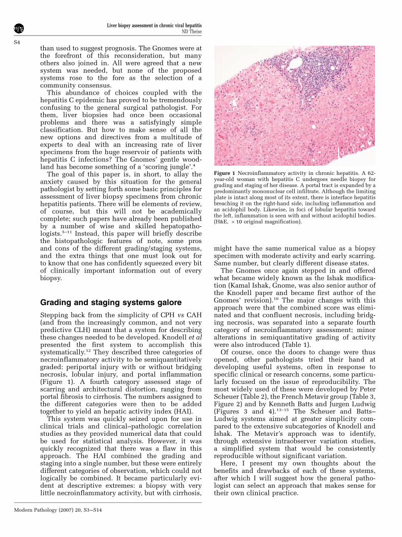
The Gnomes once again stepped in and offeredwhat became widely known as the Ishak modifica-tion (Kamal Ishak, Gnome, was also senior author ofthe Knodell paper and became first author of theGnomes’ revision).10 The major changes with thisapproach were that the combined score was elimi-nated and that confluent necrosis, including bridg-ing necrosis, was separated into a separate fourthcategory of necroinflammatory assessment; minoralterations in semiquantitative grading of activitywere also introduced (Table 1).
Of course, once the doors to change were thusopened, other pathologists tried their hand atdeveloping useful systems, often in response tospecific clinical or research concerns, some particu-larly focused on the issue of reproducibility. Themost widely used of these were developed by PeterScheuer (Table 2), the French Metavir group (Table 3,Figure 2) and by Kenneth Batts and Jurgen Ludwig(Figures 3 and 4).13–15 The Scheuer and Batts–Ludwig systems aimed at greater simplicity com-pared to the extensive subcategories of Knodell andIshak. The Metavir’s approach was to identify,through extensive intraobserver variation studies,a simplified system that would be consistentlyreproducible without significant variation.
Here, I present my own thoughts about thebenefits and drawbacks of each of these systems,after which I will suggest how the general patho-logist can select an approach that makes sense fortheir own clinical practice.
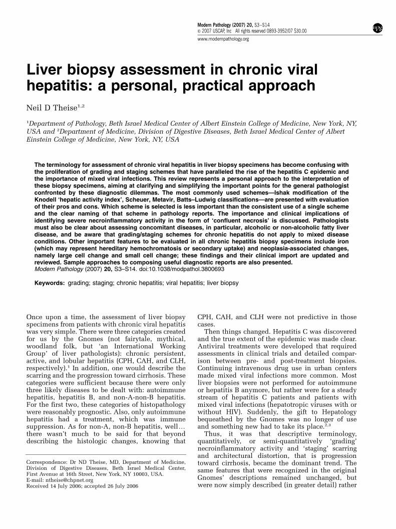
Figure 1 Necroinflammatory activity in chronic hepatitis. A 62-year-old woman with hepatitis C undergoes needle biopsy forgrading and staging of her disease. A portal tract is expanded by apredominantly mononuclear cell infiltrate. Although the limitingplate is intact along most of its extent, there is interface hepatitisbreaching it on the right-hand side, including inflammation andan acidophil body. Likewise, in foci of lobular hepatitis towardthe left, inflammation is seen with and without acidophil bodies.(H&E, � 10 original magnification).
Liver biopsy assessment in chronic viral hepatitisND Theise
S4
Modern Pathology (2007) 20, S3–S14
Ishak
Pros: Great detail! This abundance of descriptivedetail is extremely useful for performing clinical-pathologic research and for guiding the pathologistto take note of all aspects of viral injury. It has been
shown to be fairly reproducible, although more so(as is true for most of the systems) in terms of stagingthan for grading. It also recognizes the importance ofconfluent necrosis as a separate category of injury.This is particularly important in that the presence ofconfluent necrosis, truly severe disease, has specificand quite important clinical implications. Thesewill be discussed in detail below. Also, this systemhas a stage of ‘developing cirrhosis’, absent in
Table 2 Scheuer classification for grading and staging of chronic hepatitis
Grade Portal/periportal activity Lobular activity0 None None1 Portal inflammation Inflammation but no necrosis2 Mild piecemeal necrosis Focal necrosis or acidophil bodies3 Moderate piecemeal necrosis Severe focal cell damage4 Severe piecemeal necrosis Damage includes bridging necrosis
Stage Fibrosis0 None1 Enlarged, fibrotic portal tracts2 Periportal or portal-portal septa, but intact architecture3 Fibrosis with architectural distortion, but no obvious cirrhosis4 Probable or definite cirrhosis
Table 3 Metavir classification for staging of hepatitis C liver disease
No scarring 0Minimal scarring 1Scarring has occurred and extends outside the areas in the liver that contains blood vessels 2Bridging fibrosis is spreading and connecting to other areas that contain fibrosis 3Cirrhosis or advanced scarring of the liver 4
Table 1 Ishak modification for hepatic activity index (HAI) forscoring of necroinflammatory activity in chronic hepatitis
(A) Periportal or periseptal interface hepatitis (piecemealnecrosis)
Absent 0Mild (focal, few portal areas) 1Mild/moderate (focal, most portal areas) 2Moderate (continuous around o50% of tracts or septa) 3Severe (continuous around 450% of tracts or septa) 4
(B) Confluent necrosisAbsent 0Focal confluent necrosis 1Zone 3 necrosis in some areas 2Zone 3 necrosis in most areas 3Zone 3 necrosis+occasional portal-central (P-C) bridging 4Zone 3 necrosis+multiple P-C bridging 5Panacinar or multiacinar necrosis 6
(C) Focal (spotty) lytic necrosis, apoptosis and focal inflammationAbsent 0One focus or less per �10 objective 1Two to four foci per � 10 objective 2Five to ten foci per � 10 objective 3More than ten foci per � 10 objective 4
(D) Portal inflammationAbsent 0Mild, some or all portal areas 1Moderate, some or all portal areas 2Moderate/marked, all portal areas 3Marked, all portal areas 4
PMN=0
LN=0 A=0
A=1
A=2
A=1
A=2
A=2
A=3
LN=1
LN=2
LN=0,1
LN=0,1
LN=0,1,2
LN=2
LN=2
PMN=1
PMN=2
PMN=3
Figure 2 Metavir algorithm for the evaluation of histologicalactivity. PMN, piecemeal necrosis; 0, none; 1, mild; 2, moderate;3, severe; LN, lobular necrosis; 0, no or mild; 1, moderate; 2,severe; A, histological activity score; 0, none; 1, mild; 2, moderate;3, severe.
Liver biopsy assessment in chronic viral hepatitisND Theise
S5
Modern Pathology (2007) 20, S3–S14
explicit form in the other systems. In some (Scheuer,Batts–Ludwig) the category exists, but the descrip-tion is somewhat confusing (see below).
Cons: Too much detail! This amount of detail isnot particularly useful for evaluation of an indivi-dual patient’s biopsy specimen. The number ofcategories and the lack of clarity about the clinicalsignificance of any of them means that the clinicianwill be inundated with data without obvious use.Also, while this detailed descriptive analysis canyield meaningful statistical data for large popula-tions of patients or of biopsy specimens, variationsfor a single patient who has received two or morebiopsies are difficult to distinguish from samplingerror alone.
Another issue is that the category of portalinflammation is given its own weight. I believe thisis inappropriate for a clinical evaluation as this isthe defining lesion for chronic hepatitis (although it
of course can be mimicked by other conditions).Indeed, for the Gnomes, abundant mononuclearinfiltrates of the portal tracts, even with lymphoidfollicles, was originally considered inactive in theabsence of interface or lobular hepatitis. Further-more prominent lymphoid aggregates or follicles ofhepatitis C might falsely inflate the severity ofnecroinflammatory activity compared to conditionswhere such lymphoid densities are much lesstypical (hepatitis B, autoimmune hepatitis).
Metavir
Pros: This system was developed by establishingcriteria to be assessed by a panel of pathologists andthen the descriptions were analyzed to identify themost consistently reproducible scoring. This ap-proach is different than the other systems for which
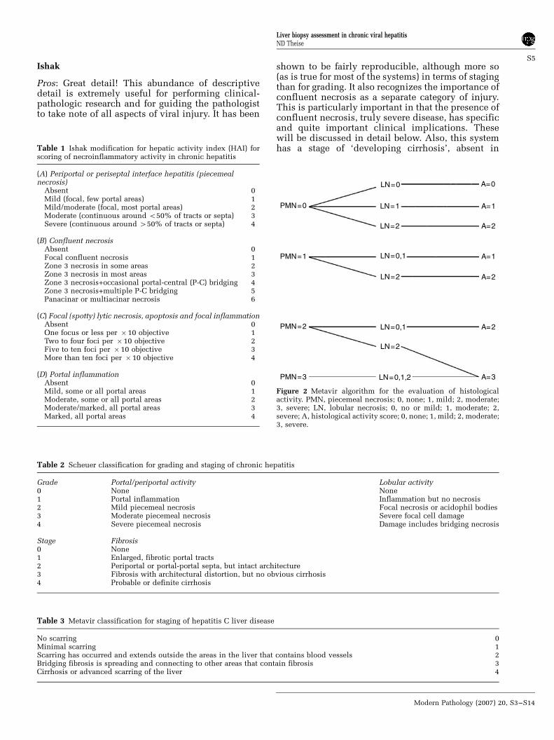
Figure 3 Batts–Ludwig diagrams of necroinflammatory activity. Note that all grades of activity contain portal inflammation; therefore, itis a defining feature of chronic hepatitis and not assessed separately from other necroinflammatory lesions. (a–c) Activity grades 1through 3. Confluent necrosis, in the form of bridging necrosis, is present only in activity grade 4 (d). These versions adapted withpermission from Batts KP, Ludwig J. Chronic hepatitis. An update on terminology and reporting. Am J Surg Pathol 1995;19:1409–1417.15
Liver biopsy assessment in chronic viral hepatitisND Theise
S6
Modern Pathology (2007) 20, S3–S14
categories and scorings were created on the basisof the authors’ knowledge (assumptions) of patho-physiology, which then underwent subsequent test-ing for reproducibility. The Metavir system is alsorelatively simple, yielding scores of mild, moderateor severe for activity and four stages of progression.
Cons: To arrive at those simple scores, thepathologist must evaluate separate criteria and thenfollow a chart to arrive at the final ‘mild, moderate,or severe’. It is not intuitively obvious, at a glance,how to do so, however. Another difficulty is thatthere is a gap between stage 3, ‘frequent fibroussepta’ and stage 4, ‘cirrhosis’. There is no categoryfor a transition to or developing cirrhosis. Thus, ifpart of a specimen is nodular, but other parts are not,then one must suggest something like ‘stage 3–4’,thus creating an implicit fifth stage, which is notexplicitly part of the system.
Scheuer
Pros: This system is also fairly simple, thusimproving its reproducibility. Only two categoriesare presented for evaluation of activity: portal/periportal and lobular. It recognizes that the amountof portal inflammation is not particularly importantas, in the absence of interface hepatitis, it is given agrade of 1 and is explicitly identified as coincidentwith the old ‘chronic persistent hepatitis’.
Cons: While there is a stage of disease progressionthat probably corresponds to transition to cirrhosis,the phrase ‘fibrosis with architectural distortionbut no obvious cirrhosis’ is unclear in its intent.What is ‘architectural distortion’, particularly for ageneralist not steeped in the details of liver anatomyas a focused academic hepatopathologist might be?Does ‘no obvious cirrhosis’ mean no nodularity at
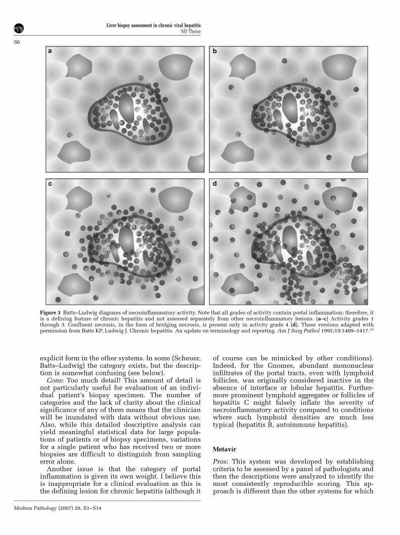
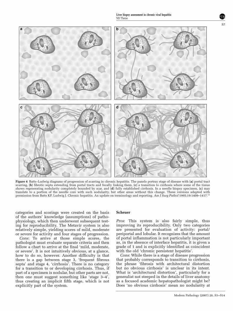
Figure 4 Batts–Ludwig diagrams of progression of scarring in chronic hepatitis. The panels portray stage of disease with (a) portal tractscarring, (b) fibrotic septa extending from portal tracts and focally linking them, (c) a transition to cirrhosis where some of the tissueshows regenerating nodularity completely bounded by scar, and (d) fully established cirrhosis. In a needle biopsy specimen, (c) maytranslate to a portion of the needle core with such nodularity, but other areas without this change. These versions adapted withpermission from Batts KP, Ludwig J. Chronic hepatitis. An update on terminology and reporting. Am J Surg Pathol 1995;19:1409–1417.15
Liver biopsy assessment in chronic viral hepatitisND Theise
S7
Modern Pathology (2007) 20, S3–S14
all? Or that the specimen is only focally nodular sothat one can not confirm fully developed cirrhosis?Knowing how Professor Scheuer discussed biopsyspecimens at the microscope I do believe heintended this to be a category of ‘transition tocirrhosis’, of focal, not total nodularity. But thephrase remains unclear to those who only have thetables to interpret.
Batts–Ludwig
Pros: This system is simple and easily reproducible,like the Metavir and Scheuer systems. In particular,the pictures provided in the original paper (andreproduced more recently elsewhere),5 give a cleargraphic demonstration of what is meant by eachgrade and stage of disease. Within a short time ofusing these figures as reference points, the generalpathologist should be able to confront scoring ofchronic hepatitis specimens with confidence. More-over, the pictures are particularly useful for makingclear to the clinician just what is meant by a verbaldescription or a staging score. The better theclinician can picture in their mind what is beingconveyed by the pathologist report, indeed, imaginethe visuals that the pathologist is converting intotext, the more likely communication among patho-logist, clinician, and patient will be effective.
Cons: As with the Scheuer classification, stage 3 isdescribed as architectural distortion. The drawingmakes clear, however, that the lesion consists offocal nodularity which is a help. However, thedrawing is of a much broader region of liver tissuethan is obtained by needle biopsy, so the applic-ability of the image becomes a matter of how thepathologist regarding the image translates it into afinding in a narrow biopsy specimen. The lack ofclarity of the verbal description then contributes topossible confusion for the less experienced.
So what do I do?
Before I describe my own practice, let me make oneimportant point that is perhaps the most importantpart of this paper and the most useful teaching forthe generalist, so important it deserves to betypographically highlighted:
It does not matter which system you use.‘What? It doesn’t matter?’ so many general
pathologists have responded when I say this. AndI affirm: no, it does not matter. I have eveninformally discussed this with primary authors ofall the different systems and none of them havedisagreed. It is up to the pathologist working withthe clinicians to figure out the needs of the clinicianand to determine what is most comfortable and whatmakes sense. Providing words, not just the numbers,is what seems most important—the words provide apicture to the clinician (and to the patient) so thatthey have a dynamic sense of disease process, rather
than a stack of numbers to play with blithely andoften inappropriately.
The second key point is also worthy of high-lighting:
Name the system clearly in your diagnosis andcommunicate with your clinicians the meaning ofthe different scorings in that system.
If one does not include the name of the systemthen the numbers become meaningless. This isanother reason to be verbally, as well as numerically,clear as to what is seen in the specimen. However,numbers are often more impressive to the clinician(or are perhaps the only thing read by them). Forexample, Metavir stage 3 of 4 is frequent fibroussepta, Scheuer and Batts–Ludwig stage 3 of 4 isextensive fibrosis with architectural distortion,Ishak stage 3 of 6 is widespread portal fibrosis withfocal septa.
This is particularly important to note as patientsoften do not remain with one hepatologist/gastro-enterologist when faced with ambiguous decisionmaking about treatment, or when they move duringthe long course of their disease and have to find newclinicians who may only have the old biopsy reportfor assessment. The clinicians you serve may under-stand your numbers, but the clinicians and/orpathologists that patients are likely to encounterelsewhere, at another time, may not understandyour scores unless you name the system used.
What I Do for Grading of Necroinflammatory Activity
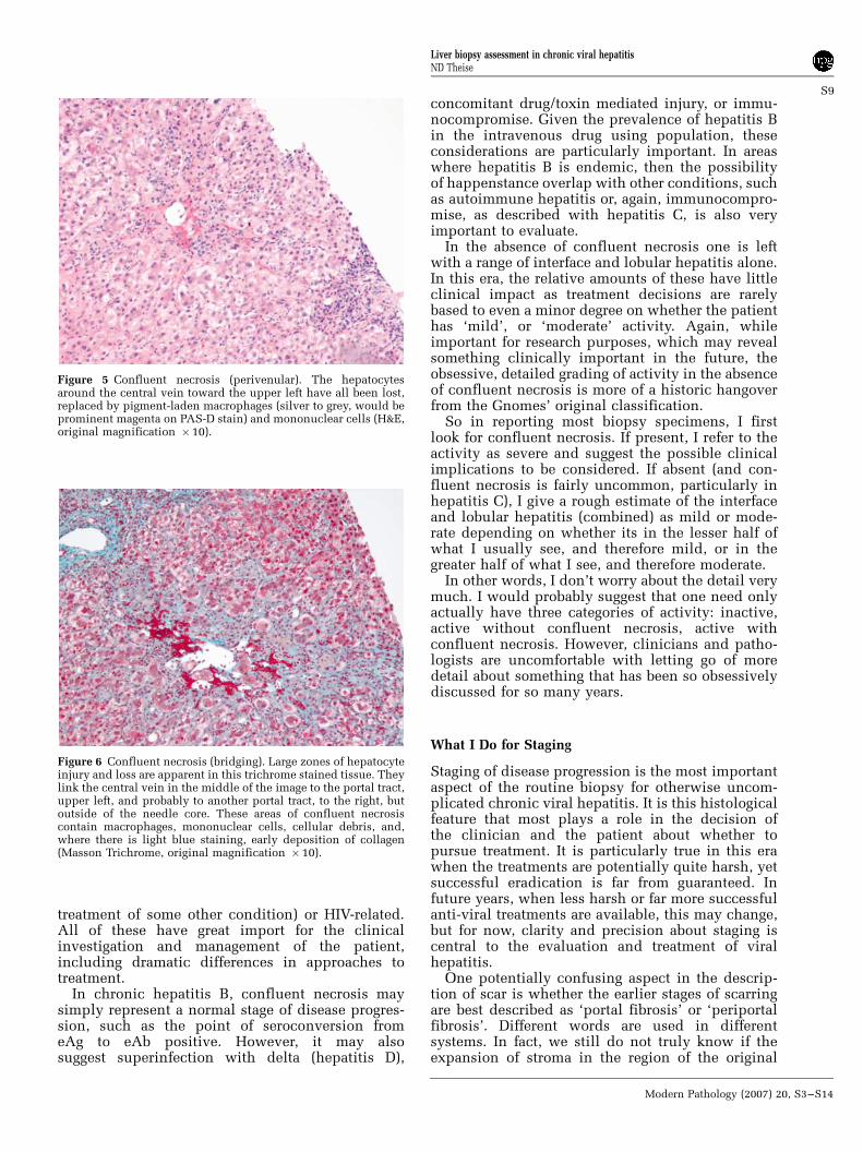
The degree of activity in most specimens is notactually used as much of a guide for treatment.Assessing it in detail for research purposes is ofgreat import and for research purposes I use theIshak system because of its ample detail. But day today, this detail, as I noted, would be more confusingthan helpful and does not, in the ‘real world’ playmuch of a role in treatment decisions except when itis particularly severe. By severe, I mean confluentnecrosis, very much as described in the Ishakstaging: from perivenular hepatocyte dropout(Figure 5) to bridging necrosis (Figure 6) to paren-chymal collapse. Confluent necrosis of any degree ina hepatitis B or hepatitis C biopsy implies specificclinical events or conditions that have possiblyimportant clinical correlates requiring attention.16
In hepatitis C, confluent necrosis may simplyreflect an acute flare of activity in this typicallywaxing and waning disease. However, given theepidemic nature of the disease, some patients withhepatitis C will inevitably also have other concomi-tant conditions requiring evaluation and perhapstreatment. Thus, confluent necrosis may indicateacute coinfection with another hepatotropic virus(acute hepatitis A or B), concomitant autoimmunehepatitis, or superimposed drug/toxin mediatedinjury. There is also the possibility of immunocom-promise in a patient with hepatitis C, iatrogenic (for
Liver biopsy assessment in chronic viral hepatitisND Theise
S8
Modern Pathology (2007) 20, S3–S14
treatment of some other condition) or HIV-related.All of these have great import for the clinicalinvestigation and management of the patient,including dramatic differences in approaches totreatment.
In chronic hepatitis B, confluent necrosis maysimply represent a normal stage of disease progres-sion, such as the point of seroconversion fromeAg to eAb positive. However, it may alsosuggest superinfection with delta (hepatitis D),
concomitant drug/toxin mediated injury, or immu-nocompromise. Given the prevalence of hepatitis Bin the intravenous drug using population, theseconsiderations are particularly important. In areaswhere hepatitis B is endemic, then the possibilityof happenstance overlap with other conditions, suchas autoimmune hepatitis or, again, immunocompro-mise, as described with hepatitis C, is also veryimportant to evaluate.
In the absence of confluent necrosis one is leftwith a range of interface and lobular hepatitis alone.In this era, the relative amounts of these have littleclinical impact as treatment decisions are rarelybased to even a minor degree on whether the patienthas ‘mild’, or ‘moderate’ activity. Again, whileimportant for research purposes, which may revealsomething clinically important in the future, theobsessive, detailed grading of activity in the absenceof confluent necrosis is more of a historic hangoverfrom the Gnomes’ original classification.
So in reporting most biopsy specimens, I firstlook for confluent necrosis. If present, I refer to theactivity as severe and suggest the possible clinicalimplications to be considered. If absent (and con-fluent necrosis is fairly uncommon, particularly inhepatitis C), I give a rough estimate of the interfaceand lobular hepatitis (combined) as mild or mode-rate depending on whether its in the lesser half ofwhat I usually see, and therefore mild, or in thegreater half of what I see, and therefore moderate.
In other words, I don’t worry about the detail verymuch. I would probably suggest that one need onlyactually have three categories of activity: inactive,active without confluent necrosis, active withconfluent necrosis. However, clinicians and patho-logists are uncomfortable with letting go of moredetail about something that has been so obsessivelydiscussed for so many years.
What I Do for Staging
Staging of disease progression is the most importantaspect of the routine biopsy for otherwise uncom-plicated chronic viral hepatitis. It is this histologicalfeature that most plays a role in the decision ofthe clinician and the patient about whether topursue treatment. It is particularly true in this erawhen the treatments are potentially quite harsh, yetsuccessful eradication is far from guaranteed. Infuture years, when less harsh or far more successfulanti-viral treatments are available, this may change,but for now, clarity and precision about staging iscentral to the evaluation and treatment of viralhepatitis.
One potentially confusing aspect in the descrip-tion of scar is whether the earlier stages of scarringare best described as ‘portal fibrosis’ or ‘periportalfibrosis’. Different words are used in differentsystems. In fact, we still do not truly know if theexpansion of stroma in the region of the original
Figure 6 Confluent necrosis (bridging). Large zones of hepatocyteinjury and loss are apparent in this trichrome stained tissue. Theylink the central vein in the middle of the image to the portal tract,upper left, and probably to another portal tract, to the right, butoutside of the needle core. These areas of confluent necrosiscontain macrophages, mononuclear cells, cellular debris, and,where there is light blue staining, early deposition of collagen(Masson Trichrome, original magnification � 10).
Figure 5 Confluent necrosis (perivenular). The hepatocytesaround the central vein toward the upper left have all been lost,replaced by pigment-laden macrophages (silver to grey, would beprominent magenta on PAS-D stain) and mononuclear cells (H&E,original magnification �10).
Liver biopsy assessment in chronic viral hepatitisND Theise
S9
Modern Pathology (2007) 20, S3–S14
portal tract in chronic hepatitis is additionalcollagen deposited within the portal tract as a resultof portal fibroblast activation, around the portal tractresulting from stellate cell activation, or a combina-tion of these two processes. But these issues are tootheoretical at this point to belong in a diagnosticreport. For simplicity’s sake (fewer syllables is betterthan more syllables), I just use the phrase ‘portalfibrosis’.
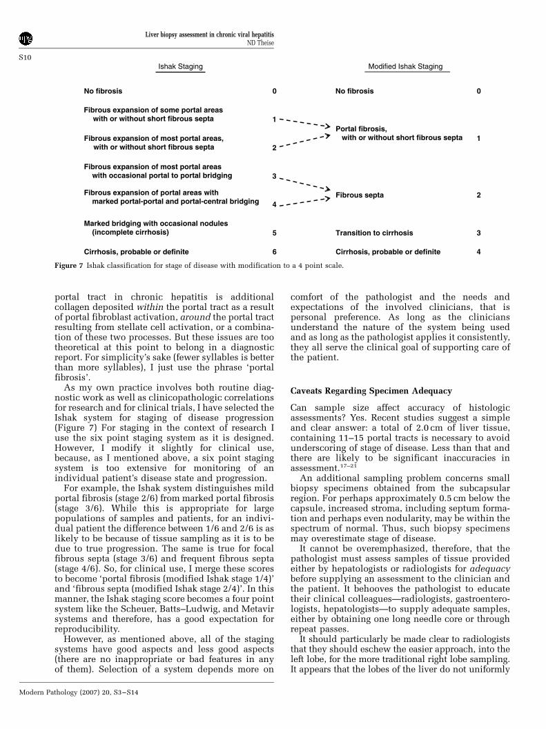
As my own practice involves both routine diag-nostic work as well as clinicopathologic correlationsfor research and for clinical trials, I have selected theIshak system for staging of disease progression(Figure 7) For staging in the context of research Iuse the six point staging system as it is designed.However, I modify it slightly for clinical use,because, as I mentioned above, a six point stagingsystem is too extensive for monitoring of anindividual patient’s disease state and progression.
For example, the Ishak system distinguishes mildportal fibrosis (stage 2/6) from marked portal fibrosis(stage 3/6). While this is appropriate for largepopulations of samples and patients, for an indivi-dual patient the difference between 1/6 and 2/6 is aslikely to be because of tissue sampling as it is to bedue to true progression. The same is true for focalfibrous septa (stage 3/6) and frequent fibrous septa(stage 4/6). So, for clinical use, I merge these scoresto become ‘portal fibrosis (modified Ishak stage 1/4)’and ‘fibrous septa (modified Ishak stage 2/4)’. In thismanner, the Ishak staging score becomes a four pointsystem like the Scheuer, Batts–Ludwig, and Metavirsystems and therefore, has a good expectation forreproducibility.
However, as mentioned above, all of the stagingsystems have good aspects and less good aspects(there are no inappropriate or bad features in anyof them). Selection of a system depends more on
comfort of the pathologist and the needs andexpectations of the involved clinicians, that ispersonal preference. As long as the cliniciansunderstand the nature of the system being usedand as long as the pathologist applies it consistently,they all serve the clinical goal of supporting care ofthe patient.
Caveats Regarding Specimen Adequacy
Can sample size affect accuracy of histologicassessments? Yes. Recent studies suggest a simpleand clear answer: a total of 2.0 cm of liver tissue,containing 11–15 portal tracts is necessary to avoidunderscoring of stage of disease. Less than that andthere are likely to be significant inaccuracies inassessment.17–21
An additional sampling problem concerns smallbiopsy specimens obtained from the subcapsularregion. For perhaps approximately 0.5 cm below thecapsule, increased stroma, including septum forma-tion and perhaps even nodularity, may be within thespectrum of normal. Thus, such biopsy specimensmay overestimate stage of disease.
It cannot be overemphasized, therefore, that thepathologist must assess samples of tissue providedeither by hepatologists or radiologists for adequacybefore supplying an assessment to the clinician andthe patient. It behooves the pathologist to educatetheir clinical colleagues—radiologists, gastroentero-logists, hepatologists—to supply adequate samples,either by obtaining one long needle core or throughrepeat passes.
It should particularly be made clear to radiologiststhat they should eschew the easier approach, into theleft lobe, for the more traditional right lobe sampling.It appears that the lobes of the liver do not uniformly
Ishak Staging Modified Ishak Staging
0No fibrosis No fibrosis
Fibrous expansion of some portal areas with or without short fibrous septa 1
Fibrous expansion of most portal areas, with or without short fibrous septa 2
Fibrous expansion of most portal areas with occasional portal to portal bridging 3
Fibrous expansion of portal areas with marked portal-portal and portal-central bridging 4
Marked bridging with occasional nodules (incomplete cirrhosis) 5
6Cirrhosis, probable or definite
0
Portal fibrosis, with or without short fibrous septa 1
2Fibrous septa
Transition to cirrhosis 3
Cirrhosis, probable or definite 4
Figure 7 Ishak classification for stage of disease with modification to a 4 point scale.
Liver biopsy assessment in chronic viral hepatitisND Theise
S10
Modern Pathology (2007) 20, S3–S14
progress toward cirrhosis and that sampling of the leftlobe may increase the finding of late stage (transitionto or fully developed cirrhosis) in a significantnumber of patients and thus inappropriately influ-ence treatment decisions. Care about not passing toodeeply, and thus through, the left lobe may also leadto more sampling of subcapsular tissue.
Writing a diagnosis
Each written diagnosis for an uncomplicatedchronic viral (or autoimmune) hepatitis biopsyspecimen should contain four pieces of information:
1. The statement that it is, indeed, chronic hepatitis;2. The grade of activity (including the name of the
scoring system used);3. The stage of activity (including the name of the
scoring system used);4. The known or suspected cause of the hepatitis.
For hepatitis B, the identification of ground glasscells or positive immunostaining for B surface andcore antigens allows for definitive statement.Chronic hepatitis C, delta, and autoimmune hepati-tis depend on clinical (serologic) information, sothese should be stated as ‘compatible withy’
The following are examples of how I mightcompose diagnoses using the different scoringsystems. I keep as much histologic detail out of thediagnostic line, keeping it in a descriptive comment,so as to make the important points clear for theclinician and the patient (remember: hepatitispatients, particularly those with hepatitis C, aremore and more acting as their own advocates andwill often be reading their own reports). For clarity,I also put the most important information, that isstaging, into words as well as numbers.
� Chronic hepatitis with Scheuer activity grades2/4 (portal/periportal) and 1/4 (lobular), stage 3/4(septa and focal architectural distortion), compa-tible with hepatitis C.
� Chronic hepatitis B, Metavir grade 1/4 and stage2/4 (fibrous septa).
� Chronic hepatitis, Batts–Ludwig grade 2/4 andstage 4/4 (cirrhosis), compatible with autoimmunehepatitis.
For my own practice, I avoid the numbers forgrading because of the reasons indicated above, somy own diagnostic lines read like this:
� Chronic hepatitis B, mildly active with mildportal fibrosis (modified Ishak stage 1/4).
� Chronic hepatitis, moderately active with transi-tion to cirrhosis (modified Ishak stage 3/4),compatible with hepatitis C.
� Chronic hepatitis, markedly active with confluentnecrosis and fibrous septa (modified Ishak stage2/4), compatible with hepatitis C (see comment).
In this last diagnosis, the ‘see comment’ is todirect the reader to where I describe the confluent
necrosis (perivenular, bridging necrosis, paren-chymal collapse) and make clear that the etiologymay be the suggested one, but that other possibilitiessuggested by the severe activity should be clinicallyassessed.
Another guideline which deserves particularprominence: In the setting of concomitant diseases(frequently and notably fatty liver disease) gradingand, in particular, staging of changes due to theviral hepatitis may be inappropriate. All scoringsystems were created for uncomplicated hepatitis,not for compound disease processes. This caveatbrings us to the final series of points to be maderegarding the assessment of chronic viral hepatitisspecimens.
Additional features to be routinelyassessed
There are, of course, other hepatitis-related featureswhich should be routinely assessed in every chronichepatitis biopsy specimen, these included hepatitisrelated changes (hepatitis C-related fat, increasediron uptake, large cell and small cell change) andparticularly common concomitant diseases, such asalcoholic and nonalcoholic fatty liver disease andhemochromatosis. It behooves the pathologist tokeep these in mind to most completely assess thespecimen and serve patient care.
Steatosis
It is well known that hepatitis C, particularlygenotype 3, can lead to steatotic change in hepato-cytes.22 The trick is in distinguishing this steatosisfrom true fatty liver disease. In hepatitis C-relatedfat, the fat is usually mild (o10%) and it is notzonally distributed, as only some of it will bepericentral, as it is in fatty liver disease, but alsoin the midzone or in periportal locations.
When it is zonal and more extensive, a diagnosisof fatty liver (alcoholic or nonalcoholic) should bemade. In that case, full assessment of fatty liverdisease, including statement of extent of steatosis,evaluation of steatohepatitis (neutrophilic infiltra-tion, hepatocyte ballooning, Mallory bodies) andsteatofibrosis (perivenular, pericellular, and central-portal septa), should be carefully performed. Thedetails of such evaluation are beyond the scope ofthis paper; however, good reviews have been writtenelsewhere.23–25
A key point is that if steatohepatitis or steato-fibrosis is seen, they imply fatty liver disease, ratherthan simple hepatitis C-related steatosis. In parti-cular, it is important to watch for steatofibrosiswhen examining a trichrome stain as even the experteye may not recognize it on H&E stain. Indeed, giventhe epidemic nature of nonalcoholic fatty liverdisease and the significant prevalence of concomi-tant (and synergistic) alcoholic liver disease in
Liver biopsy assessment in chronic viral hepatitisND Theise
S11
Modern Pathology (2007) 20, S3–S14
hepatitis C patients, it is often necessary to write asecond diagnostic line describing this diseaseprocess. In this setting, hepatitis scoring may beinappropriate as distinguishing the contributionsof each disease may not be reliable. Examples ofdiagnoses in such settings might look like this:
� Transition to cirrhosis with mixed features ofchronic hepatitis C (mildly active) and fattyliver disease (alcoholic vs nonalcoholic) (seecomment).
� Cirrhosis with mixed features of chronic hepatitisB (moderately active) and alcoholic liver disease(see comment).
� Frequent fibrous septa with mixed features ofchronic hepatitis C (mildly active) and obesityrelated fatty liver disease (see comment).
Again, the ‘see comment’ is to direct the reader toa diagnostic comment that evaluates to whateverdegree possible, the relationship of each disease tothe changes in the specimen. Thus, the presence of adense mononuclear portal infiltrate, extensive inter-face and lobular hepatitis, marked portal fibrosis,and portal-portal septa would be considered morelikely to be a contribution of the viral hepatitis. Onthe other hand, perivenular and pericellular fibrosis,central-portal septa, and parenchymal neutrophilsmay be taken as evidence of fatty liver disease. Bothdiseases, however, may have some mild degree ofportal fibrosis, mild portal mononuclear infiltrates,mild parenchymal lymphocytosis, and focal apop-tosis, so these features then can belong to eitherdisease.
Hemosiderosis
Histochemical staining should be routine on everyliver biopsy specimen for the screening of hemo-chromatosis, but it is particularly important inchronic viral hepatitis and, more particularly inchronic hepatitis C.23,26 Hepatitis C may itself lead toincreased hepatocyte and/or reticulo-endothelialiron stores. This iron is usually, however, mild.There may also be iron related to prior treatmentwith Ribavirin treatment, which may induce somedegree of hemolysis in some patients.
Hereditary hemochromatosis, however, is nowknown to have quite variable penetrance, so thepresence of any iron, even mild, may be evidence ofthat disease as well.27 Again, how to handle iron-related disease for hemosiderotic liver biopsy speci-mens is more fully covered elsewhere26,28 and laterin this journal, but a simple approach might looksomething like this, in a report:
� Hemosiderosis, grade 1/4, ? hereditary vs second-ary hemochromatosis (see comment).
The comment may then include statements suchas: ‘Prussian blue staining for iron demonstrateshemosiderosis of hepatocytes around some portaltracts. This amount of iron may be related to
hepatitis C infection, however, the small amountdoes not exclude the possibility of hereditaryhemochromatosis due to the variable penetrance ofthis disease. Clinical correlation (genetic testing)may be helpful’. Some clinicians may even prescribevenopuncture for some patients to reduce hepato-cyte iron as it is thought by some to improve thelikelihood of viral clearance with treatment.
Purely reticuloendothelial iron does not suggesthereditary hemochromatosis and is more likely areflection of hemolysis (perhaps Ribavirin-inducedor due to other unrelated causes).29 Also, in patientswith fully established cirrhosis, the cirrhosis itselfmay lead to increased iron uptake from the gut and,then, hemosiderosis of the liver. Thus, when there isclear cirrhosis and iron I make this suggestion firstin the companion comment.
Neoplasia-Related Changes
Both hepatitis C and hepatitis B dramaticallyincrease the risk for hepatocellular carcinoma.Cellular atypia that may suggest early or incipientneoplasia may be recognized in biopsy specimens,though, as with most pathology, if one doesn’tspecifically think to look for it, it may go un-noticed.30 These changes were previously referred toas ‘dysplasia’, but for reasons beyond the scope ofthis paper, are now referred to as ‘change’, as in‘large cell change’ and ‘small cell change’.31
Large cell change is now generally thought to bemalignancy associated, rather than directly pre-malignant. Nonetheless, its presence is thought bymany to indicate a patient with further increasedrisk for development of hepatocellular carcinomawho may benefit from more rigorous cancer surveil-lance, though the need to report it is still not settledand may be dependent on the background liverdisease.32,33 This cellular change is characterized byoften large, atypical nuclei (often reflecting aneu-ploidy), multinucleation, and abundant cytoplasm.(Figure 8). Thus, there is a fairly normal nuclear:cytoplasmic ratio. The cells are not generally innodular array, but instead are intermixed withnormally sized, regenerating hepatocytes, often inperiportal or periseptal locations.
Small cell change, on the other hand, is thought tobe directly premalignant.30,34 It consists of smallhepatocytes, with relatively normal size, buthyperchromatic, often grooved or irregular nuclei(Figure 9). These, therefore, have an increasednuclear:cytoplasmic ratio. Cytoplasm often has abluish appearance on H&E stain. These cells areusually in clustered arrays if not actually formingclear subnodules. Reticulin stain may highlight suchnodularity and also demonstrate slight loss ofreticulin fibers.
It is also becoming increasingly recognized thatthese pre-malignant or malignancy associatedchanges (indeed, even carcinoma itself) may pre-
Liver biopsy assessment in chronic viral hepatitisND Theise
S12
Modern Pathology (2007) 20, S3–S14

cede the development of cirrhosis. In other words,just because the patient does not have cirrhosis doesnot mean that these features are absent or that thepatient may not be at risk for malignancy. Theconcept that cirrhosis itself is premalignant isprobably incorrect; rather, it develops in parallelwith neoplasia in a chronically diseased liver.35,36
Coinfection with HIV
During the early years of hepatitis C recognition,there was also very little that could yet be done forHIV infection. Successful anti-retroviral therapy wasstill more than half a decade away. Hepatitis C inthis setting was generally found to be worse, bothwith greater severity of activity and more rapidlyadvancing fibrosis. Subsequent to successful long-term maintenance of immune competence, thisexpectation of worse or more rapid injury are notnecessarily reflected by the biopsy specimens thatnow come before from patients with coinfection.23,26
Thus, in patients who are stable with competentimmune systems, the findings in liver biopsy speci-mens for hepatitis C or B require no differenttreatment than those from non-HIV infectedpatients. On the other hand, if the patient is notimmune competent, then one may see more severedisease (including confluent necrosis or rapidprogression toward cirrhosis). In addition, in suchpatients, one must also keep an eye open for all the‘old’ lesions: granulomas, infectious organisms(with examination of special stains for fungi andacid fast organisms), and infiltrating neoplasms.
In summary
This era of diagnosis and treatment of chronic viralhepatitis is full of great clinical possibility, rapidlyadvancing past the early decades of the field whennothing but description, watching and waiting wasavailable. But, like any transitional period, theseopportunities come with some measure of confu-sion, even for those with extensive experience, evenexpertise. It is my hope that this text might help cutthrough that ‘scoring jungle’ for the general patho-logists who do not have the time to wade throughdetailed reviews and theoretical speculations. How-ever, my choices and opinions (except, perhaps, thethree statements printed in italics within the text)are not the ‘correct’ ones; we must each make ourown selections from the options available. As Ihopefully have shown, the task is not, in fact,particularly daunting. With the example of theGnomes who came before us, a methodical andrational consideration of histologic findings and therelevant clinical correlates can lead to clear andconsistent descriptions of benefit to our cliniciansand to our patients.
Acknowledgements
There is little in the way of research, teaching, orwriting in my career in hepatopathology that Iwould have accomplished, if it weren’t for thementoring I received from Peter J Scheuer, myteacher, my colleague, my friend, my ‘professionalfather’ as he sometimes said. Indeed, for the manyhepatopathologists for whom he played that role of
Figure 9 Small cell change. Small hepatocytes in the center andleft of the image have slightly smaller nuclei than normalhepatocytes on the right side, but their cytoplasm is considerablyreduced resulting in a markedly increased nuclear to cytoplasmicratio. Their cytoplasm may also be somewhat basophilic.Trabeculae of hepatocytes with small cell change are often thickor difficult to discern yielding a ‘cobblestone’ appearance. Suchcells may form tight subnodules or their clustering may be a littleless well defined, as seen here, though they are not foundscattered individually the way hepatocytes with large cell changeoften are (H&E, original magnification �20).
Figure 8 Large cell change. Large hepatocytes are easily seenscattered among the normal size, regenerating hepatocytes. Theyare large cells with large, often hyperchromatic and atypicalappearing nuclei. Multinucleation may be common. Despite their‘dysplastic’ appearance, they have a relatively normal nuclear:cytoplasmic ratio (H&E, original magnification �20).
Liver biopsy assessment in chronic viral hepatitisND Theise
S13
Modern Pathology (2007) 20, S3–S14
professional father, his influence continues toexpand outward, even after his death earlier thisyear. He will be remembered fondly for his caring,but he will be continually honored by our labors, inthe service of our patients, to correctly and carefullydiagnose our liver biopsy specimens, holding our-selves to the rigorous standards he set for his ownpractice.
Many thanks to Ken Batts for permission toreproduce the figures from his original paper hereand to Linda Ferrell for her patience and guidancein the final organization of this manuscript.
References
1 De Groote J, Desmet VJ, Gedigk P, et al. A classificationof chronic hepatitis. Lancet 1968;2:626–628.
2 Scheuer PJ. The nomenclature of chronic hepatitis:time for a change. J Hepatol 1995;22:112–114.
3 Gerber M. Chronic hepatitis C: the beginning of the endof a time-honored nomenclature? Hepatology 1992;15:733–734.
4 Hunt N, Fleming K. Reproducibility of liver biopsygrading and staging. Liver 1999;19:169–170.
5 Brunt EM. Grading and staging the histopathologicallesions of chronic hepatitis: the Knodell histology activityindex and beyond. Hepatology 2000;31:241–246.
6 Scheuer PJ, Standish RA, Dhillon AP. Scoring ofchronic hepatitis. Clin Liver Dis 2002;6:335–347.
7 Koukoulis G. Chronic hepatitis C: grading, staging, andsearching for reliable predictors of outcome. HumPathol 2001;32:899–903.
8 Desmet VJ. Histological classification of chronichepatitis. Acta Gastroenterol Belg 1997;60:259–267.
9 Ferrell L. Histological classification of chronic hepa-titis. Acta Gastroenterol Belg 1997;60:259–267.
10 Ishak K, Baptista A, Bianchi L, et al. Histologicalgrading and staging of chronic hepatitis. J Hepatol1995;22:696–699.
11 Desmet VJ, Gerber M, Hoofnagle JH, et al. Classifica-tion of chronic hepatitis: diagnosis, grading andstaging. Hepatology 1994;19:1513–1520.
12 Knodell RG, Ishak KG, Black WC, et al. Formulationand application of a numerical scoring system forassessing histological activity in asymptomatic chronicactive hepatitis. Hepatology 1981;1:431–435.
13 Scheuer PJ. Classification of viral hepatitis: a need forreassessment. J Hepatol 1991;13:372–374.
14 Bedossa P, Bioulac-Sage P, Callard P, et al. Intra-observer and interobserver variations in liver biopsyinterpretation in patients with chronic hepatitis C.Hepatology 1994;20:15–20.
15 Batts KP, Ludwig J. Chronic hepatitis. An update onterminology and reporting. Am J Surg Pathol 1995;19:1409–1417.
16 Theise ND, Bodenheimer H, Ferrell L. Viral hepatitis.In: Burt A, Portmann B, Ferrell L (eds). MacSween’sPathology of the Liver, 5th edn. Churchill Livingston/Elsevier: London, UK, 2006 (in press).
17 Guido M, Rugge M. Liver biopsy sampling in chronicviral hepatitis. Semin Liver Dis 2004;24:89–97.
18 Fanning L, Loane J, Kenny-Walsh E, et al. Tissue viralload variability in chronic hepatitis C. Am J Gastro-enterol 2001;96:3384–3389.
19 Regev A, Berho M, Jeffers LJ, et al. Sampling error andintraobserver variation in liver biopsy in patients withchronic HCV infection. Am J Gastroenterol 2002;97:2614–2618.
20 Persico M, Palmentieri B, Vecchione R, et al.Diagnosis of chronic liver disease: reproducibilityand validation of liver biopsy. Am J Gastroenterol2002;97:491–492.
21 Siddique I, El-Naga HA, Madda JP, et al. Samplingvariability on percutaneous liver biopsy in patientswith chronic hepatitis C virus infection. Scand JGastroenterol 2003;38:427–432.
22 Zekry A, McHutchison JG, Diehl AM. Insulin resis-tance and steatosis in hepatitis C virus infection. Gut2005;54:903–906.
23 Lefkowitch JH. Hepatobiliary pathology. Curr OpinGastroenterol 2006;22:198–208.
24 Farrell GC, Larter CZ. Nonalcoholic fatty liverdisease: from steatosis to cirrhosis. Hepatology 2006;43(2 Suppl 1):S99–S112.
25 Brunt EM. Nonalcoholic steatohepatitis. Semin LiverDis 2004;24:3–20.
26 Alla V, Bonkovsky HL. Iron in nonhemochromatoticliver disorders. Semin Liver Dis 2005;25:461–472.
27 Gattoni A, Parlato A, Vangieri B, et al. Role ofhemochromatosis genes in chronic hepatitis C. ClinTher 2006;157:61–68.
28 Brunt EM. Pathology of hepatic iron overload. SeminLiver Dis 2005;25:392–401.
29 Fiel MI, Schiano TD, Guido M, et al. Increased hepaticiron deposition resulting from treatment of chronichepatitis C with ribavirin. Am J Clin Pathol 2000;113:35–39.
30 Libbrecht L, Desmet V, Roskams T. Preneoplasticlesions in human hepatocarcinogenesis. Liver Int2005;25:16–27.
31 Wanless I, Callea R, Craig JR, et al. Terminology ofnodular lesions of the liver: Recommendations of theWorld Congress of Gastroenterology Working Group.Hepatology 1995;22:983–993.
32 Borzio M, Bruno S, Roncalli M, et al. Liver celldysplasia is a major risk factor for hepatocellularcarcinoma in cirrhosis—a prospective study. Gastro-enterology 1995;108:812–817.
33 Ganne-Carrie N, Chastang C, Chapel F, et al. Predictivescore for the development of hepatocellular carcinomaand additional value of liver large cell dysplasia inwestern patients with cirrhosis. Hepatology 1996;23:1112–1118.
34 Marchio A, Terris B, Meddeb M, et al. Chromosomalabnormalities in liver cell dysplasia detected bycomparative genomic hybridisation. J Clin Pathol-MolPathol 2001;54:270–274.
35 Theise ND. Cirrhosis and hepatocellular neoplasia:More like cousins than like parent and child. Gastro-enterology 1996;111:526–528.
36 Vallet-Pichard A, Pol S. Natural history and predictorsof severity of chronic hepatitis C virus (HCV) andhuman immunodeficiency virus (HIV) co-infection.J Hepatol 2006;44(1 Suppl):S28–S34.
Liver biopsy assessment in chronic viral hepatitisND Theise
S14
Modern Pathology (2007) 20, S3–S14


