LITIGATING TRAUMATIC BRAIN INJURY CLAIMS - … · Litigating Traumatic Brain Injury Claims Chapter...
34
LITIGATING TRAUMATIC BRAIN INJURY CLAIMS Presented and Written by: JOHN S. JOSE Slack & Davis, L.L.P. Fort Worth, Texas 817.288.8988 [email protected] Co-written by: KRISTY MAY, RN, BSN Slack & Davis, L.L.P. Austin, Texas State Bar of Texas 30 TH ANNUAL ADVANCED PERSONAL INJURY LAW COURSE July 9 - 11, 2014 Dallas August 6 – 8, 2014 San Antonio August 27 – 29, 2014 Houston CHAPTER 20
Transcript of LITIGATING TRAUMATIC BRAIN INJURY CLAIMS - … · Litigating Traumatic Brain Injury Claims Chapter...
LITIGATING TRAUMATIC BRAIN INJURY CLAIMS
Presented and Written by:
JOHN S. JOSE
Slack & Davis, L.L.P.
Fort Worth, Texas
817.288.8988
Co-written by:
KRISTY MAY, RN, BSN
Slack & Davis, L.L.P.
Austin, Texas
State Bar of Texas
30TH
ANNUAL
ADVANCED PERSONAL INJURY LAW
COURSE
July 9 - 11, 2014 Dallas
August 6 – 8, 2014 San Antonio
August 27 – 29, 2014 Houston
CHAPTER 20
Litigating Traumatic Brain Injury Claims Chapter 20
i
TABLE OF CONTENTS
I. INTRODUCTION. ............................................................................................................................................ 1
II. WHAT IS TRAUMATIC BRAIN INJURY? .................................................................................................... 1 A. Basic Anatomy of the Brain. (Figure 3). .................................................................................................... 1 B. Basic Neuro-Cellular Anatomy. ................................................................................................................. 3 C. Definitions and Classification of Traumatic Brain Injury. ......................................................................... 3
1. The Glasgow Coma Scale. ................................................................................................................. 3 2. ACRM Definition of Mild TBI (mTBI). ............................................................................................ 3 3. Other Definitions of TBI. ................................................................................................................... 4
III. SIGNS AND SYMPTOMS OF MILD TBI. ...................................................................................................... 4 A. When to Look for Signs and Symptoms. ................................................................................................... 4 B. What Signs and Symptoms to Look For. (Figure 9) .................................................................................. 4
1. Loss of Consciousness. ....................................................................................................................... 4 2. Amnesia/Confusion. ........................................................................................................................... 5 3. Post-Trauma Effects. .......................................................................................................................... 5
C. Where to look for Signs and Symptoms. .................................................................................................... 5 1. Client Interviews. ............................................................................................................................... 5 2. Accident Report. ................................................................................................................................. 5 3. Accident Witnesses. ........................................................................................................................... 5 4. Ambulance/EMT Records and Interviews. ........................................................................................ 5 5. Hospital Records. ............................................................................................................................... 5 6. Post-accident Medical/Rehab Records from All Practitioners. (Including Physiotherapists,
Chiropractors, Massage Therapists, Naturopaths, and Counselors). .................................................. 5 7. Prior Medical Records. ....................................................................................................................... 5 8. Scene Photographs. ............................................................................................................................ 5 9. School Records. .................................................................................................................................. 5 10. Employment Records. ........................................................................................................................ 6
IV. MEDICAL EVALUATION OF TBI. ................................................................................................................ 6 A. Neurological Examination.......................................................................................................................... 6 B. Neuropsychological Examination. ............................................................................................................. 6 C. Neuroradiological Evaluation. ................................................................................................................... 6
1. CT Scan. ............................................................................................................................................. 6 2. MRI. ................................................................................................................................................... 6 3. FLAIR. ............................................................................................................................................... 6 4. DTI. .................................................................................................................................................... 6 5. SPECT. ............................................................................................................................................... 6 6. PET. .................................................................................................................................................... 6 7. NeuroQuant® ..................................................................................................................................... 7
V. EXPERT WITNESSES IN TRAUMATIC INJURY CLAIMS. ....................................................................... 7 A. Neurologist. ................................................................................................................................................ 7 B. Neuroradiologist. ........................................................................................................................................ 7 C. Neuropsychologist. ..................................................................................................................................... 7 D. Biomechanical Engineer. ........................................................................................................................... 7 E. Life Care Planner. ...................................................................................................................................... 8 F. Vocational/Economic Experts. ................................................................................................................... 8 G. Economist. .................................................................................................................................................. 9
VI. TRAUMATIC BRAIN INJURY AS A DISEASE PROCESS. ......................................................................... 9 A. TBI Effects on Mortality. ........................................................................................................................... 9 B. TBI Effects on Morbidity. ........................................................................................................................ 10
VII. RESOURCES FOR FURTHER TBI RESEARCH. ........................................................................................ 10

Litigating Traumatic Brain Injury Claims Chapter 20
ii
A. Organizations for Plaintiff Counsel. ......................................................................................................... 10 B. Organizations for Defense Counsel. ......................................................................................................... 10 C. Publications. ............................................................................................................................................. 11
VIII. CONCLUSION. ............................................................................................................................................... 11
APPENDIX (Table of Figures) ..................................................................................................................................... 13
Litigating Traumatic Brain Injury Claims Chapter 20
1
LITIGATING TRAUMATIC BRAIN
INJURY CLAIMS
I. INTRODUCTION.
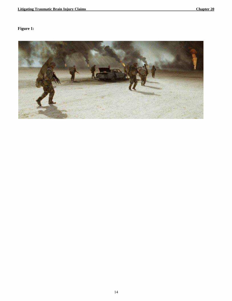
Social forces over the last decade have driven
public awareness (and acceptance) of traumatic brain
injuries. (Figures 1 and 2) More particularly, the Gulf
Wars with their multitude of IED-related blast injuries,
and the NFL players’ claims for sports-related head
injuries have brought publicity to the nature and
devastating long-term effects of insults to the brain.
These same forces have driven scientific research and
medical advances in the diagnosis, treatment, and
understanding of traumatic brain injuries. Public
awareness and scientific means of objective
verification of these injuries have likewise entered the
courtrooms of this country. According to the Centers
for Disease Control and Prevention, traumatic brain
injury (TBI) is a serious health problem in the United
States. Each year, traumatic brain injuries contribute
to a substantial number of deaths and cases of
permanent disability. Studies have estimated that
nearly 1.6 million TBI’s occur in the United States
every year, resulting in over 50,000 deaths and over
70,000 patients with permanent neurological deficits.
This paper is intended to provide a basic framework for
personal injury law practitioners to assist in the
recognition, evaluation, preparation, and presentation
of traumatic brain injury claims. This paper will
concentrate particularly on “mild” traumatic brain
injury (mTBI) claims.
II. WHAT IS TRAUMATIC BRAIN INJURY?
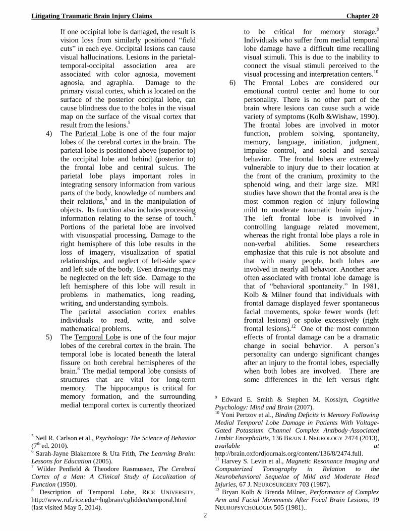
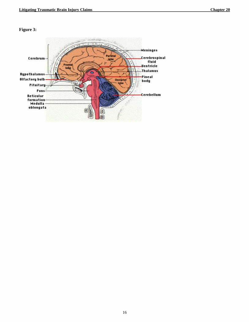
A. Basic Anatomy of the Brain. (Figure 3).
The brain cerebrum is an organ with the
consistency of gelatin that is situated within the skull.
The brain is floating in cerebrospinal fluid within the
hard skull cavity. Portions of the interior of the skull
against which the brain is situated are irregular and
sharp. The cerebrum has an outer layer of tissue
known as the cerebral cortex. The cerebral cortex
surrounds the gray matter. The cerebrum is
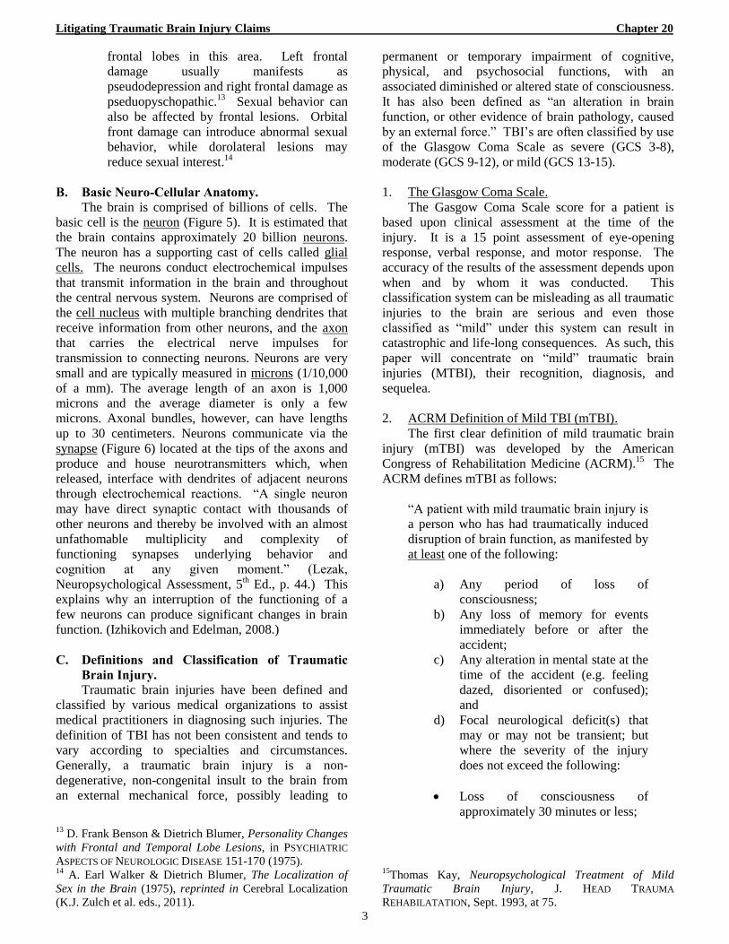
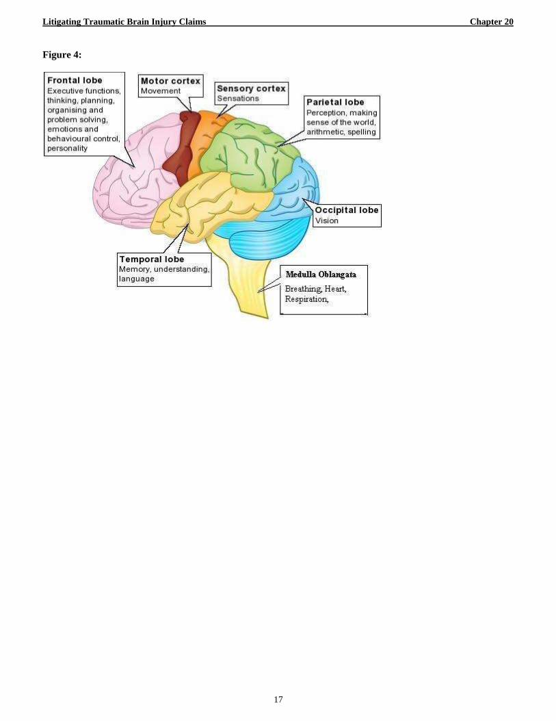
sectionalized into main regions known as lobes.
(Figure 4) The major lobes are the Brain Stem,
Cerebellum, Occipital Lobe, Parietal Lobe, Temporal
Lobe, and Frontal Lobe. Each of these lobes controls
different brain functions. Damage to a particular lobe
can cause impairment of functions controlled by it. It
is also understood that certain of the lobes are within a
network of interconnected structures, and damage to
one such structure can affect brain functions controlled
by other structures within that network. (Figure 4)
1) The Brain Stem is located in the posterior
part of the brain. In humans it is usually
described as including the medulla
oblongata, pons, and midbrain.1 The brain
stem provides the main motor and sensory
innervations to the face and neck via the
cranial nerves. This is an extremely
important part of the brain as the nerve
connections of the motor and sensory
systems from the main part of the brain to the
rest of the body pass through the brainstem.
This includes nerves affecting motor, fine
touch, vibration sensation, pain, temperature,
itch, and crude touch. The brainstem also
plays an important role in the regulation of
cardiac and respiratory function. It also
regulates the central nervous system, and is
pivotal in maintaining consciousness and
regulating the sleep cycle. The brainstem
also controls many basic functions including
heart rate, breathing, sleeping, and eating.
2) The Cerebellum is a region of the brain that
plays an important role in motor control. It
may also be involved in some cognitive
functions such as attention and language, and
in regulating fear and pleasure responses.2 It
contributes to coordination, precision, and
accurate timing. It receives input from
sensory systems of the spinal cord and from
other parts of the brain, and integrates these
inputs to fine tune motor activity.3 Damage
to the cerebella does not cause paralysis, but
instead produces disorders in fine movement,
equilibrium, posture, and motor learning.
Anatomically, the cerebellum has the
appearance of a separate structure attached to
the bottom of the brain, tucked underneath
the cerebral hemispheres.
3) The two Occipital Lobes are the smallest of
four paired lobes in the human cerebral
cortex. Located in the rearmost portion of
the skull, the occipital lobes are part of the
forebrain.4 The occipital lobe is divided into
several functional visual areas. Each visual
area contains a full map of the visual world.
1 Definition of Brainstem, THE FREE DICTIONARY (MEDICAL
DICTIONARY ED.), http://medical-
dictionary.thefreedictionary.com/brainstem (last visited Apr.
30, 2014).
2 Uri Wolf et al., Evaluating the Affective Component of the
Cerebellar Cognitive Affective Syndrome, 21 J.
NEUROPSYCHIATRY & CLINICAL NEUROSCIENCE 245 (2009),
available at
http://neuro.psychiatryonline.org/article.aspx?articleid=1037
81. 3 Edward J. Fine et. al., The History of the Development of
the Cerebellar Examination, 22 SEMINARS NEUROLOGY 375
(2002), available at https://www.thieme-
connect.de/DOI/DOI?10.1055/s-2002-36759. 4 Daniel L. Schacter, Psychology (2
nd ed. 2010).
Litigating Traumatic Brain Injury Claims Chapter 20
2
If one occipital lobe is damaged, the result is
vision loss from similarly positioned “field
cuts” in each eye. Occipital lesions can cause
visual hallucinations. Lesions in the parietal-
temporal-occipital association area are
associated with color agnosia, movement
agnosia, and agraphia. Damage to the
primary visual cortex, which is located on the
surface of the posterior occipital lobe, can
cause blindness due to the holes in the visual
map on the surface of the visual cortex that
result from the lesions.5
4) The Parietal Lobe is one of the four major
lobes of the cerebral cortex in the brain. The
parietal lobe is positioned above (superior to)
the occipital lobe and behind (posterior to)
the frontal lobe and central sulcus. The
parietal lobe plays important roles in
integrating sensory information from various
parts of the body, knowledge of numbers and
their relations,6 and in the manipulation of
objects. Its function also includes processing
information relating to the sense of touch.7
Portions of the parietal lobe are involved
with visuospatial processing. Damage to the
right hemisphere of this lobe results in the
loss of imagery, visualization of spatial
relationships, and neglect of left-side space
and left side of the body. Even drawings may
be neglected on the left side. Damage to the
left hemisphere of this lobe will result in
problems in mathematics, long reading,
writing, and understanding symbols.
The parietal association cortex enables
individuals to read, write, and solve
mathematical problems.
5) The Temporal Lobe is one of the four major
lobes of the cerebral cortex in the brain. The
temporal lobe is located beneath the lateral
fissure on both cerebral hemispheres of the
brain.8 The medial temporal lobe consists of
structures that are vital for long-term
memory. The hippocampus is critical for
memory formation, and the surrounding
medial temporal cortex is currently theorized
5 Neil R. Carlson et al., Psychology: The Science of Behavior
(7th
ed. 2010). 6 Sarah-Jayne Blakemore & Uta Frith, The Learning Brain:
Lessons for Education (2005). 7 Wilder Penfield & Theodore Rasmussen, The Cerebral
Cortex of a Man: A Clinical Study of Localization of
Function (1950). 8 Description of Temporal Lobe, RICE UNIVERSITY,
http://www.ruf.rice.edu/~lngbrain/cglidden/temporal.html
(last visited May 5, 2014).
to be critical for memory storage.9
Individuals who suffer from medial temporal
lobe damage have a difficult time recalling
visual stimuli. This is due to the inability to
connect the visual stimuli perceived to the
visual processing and interpretation centers.10
6) The Frontal Lobes are considered our
emotional control center and home to our
personality. There is no other part of the
brain where lesions can cause such a wide
variety of symptoms (Kolb &Wishaw, 1990).
The frontal lobes are involved in motor
function, problem solving, spontaneity,
memory, language, initiation, judgment,
impulse control, and social and sexual
behavior. The frontal lobes are extremely
vulnerable to injury due to their location at
the front of the cranium, proximity to the
sphenoid wing, and their large size. MRI
studies have shown that the frontal area is the
most common region of injury following
mild to moderate traumatic brain injury.11
The left frontal lobe is involved in
controlling language related movement,
whereas the right frontal lobe plays a role in
non-verbal abilities. Some researchers
emphasize that this rule is not absolute and
that with many people, both lobes are
involved in nearly all behavior. Another area
often associated with frontal lobe damage is
that of “behavioral spontaneity.” In 1981,
Kolb & Milner found that individuals with
frontal damage displayed fewer spontaneous
facial movements, spoke fewer words (left
frontal lesions) or spoke excessively (right
frontal lesions).12
One of the most common
effects of frontal damage can be a dramatic
change in social behavior. A person’s
personality can undergo significant changes
after an injury to the frontal lobes, especially
when both lobes are involved. There are
some differences in the left versus right
9 Edward E. Smith & Stephen M. Kosslyn, Cognitive
Psychology: Mind and Brain (2007). 10
Yoni Pertzov et al., Binding Deficits in Memory Following
Medial Temporal Lobe Damage in Patients With Voltage-
Gated Potassium Channel Complex Antibody-Associated
Limbic Encephalitis, 136 BRAIN J. NEUROLOGY 2474 (2013),
available at
http://brain.oxfordjournals.org/content/136/8/2474.full. 11
Harvey S. Levin et al., Magnetic Resonance Imaging and
Computerized Tomography in Relation to the
Neurobehavioral Sequelae of Mild and Moderate Head
Injuries, 67 J. NEUROSURGERY 703 (1987). 12
Bryan Kolb & Brenda Milner, Performance of Complex
Arm and Facial Movements After Focal Brain Lesions, 19
NEUROPSYCHOLOGIA 505 (1981)..
Litigating Traumatic Brain Injury Claims Chapter 20
3
frontal lobes in this area. Left frontal
damage usually manifests as
pseudodepression and right frontal damage as
pseduopyschopathic.13
Sexual behavior can
also be affected by frontal lesions. Orbital
front damage can introduce abnormal sexual
behavior, while dorolateral lesions may
reduce sexual interest.14
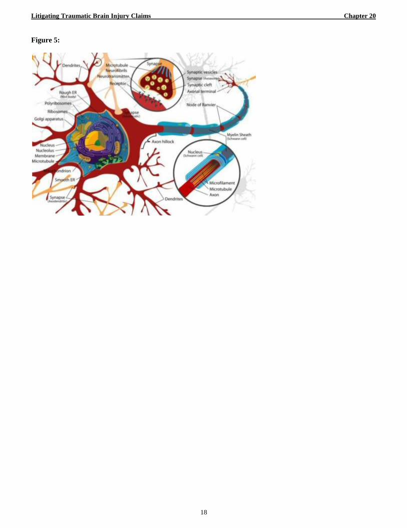
B. Basic Neuro-Cellular Anatomy.
The brain is comprised of billions of cells. The
basic cell is the neuron (Figure 5). It is estimated that
the brain contains approximately 20 billion neurons.
The neuron has a supporting cast of cells called glial
cells. The neurons conduct electrochemical impulses
that transmit information in the brain and throughout
the central nervous system. Neurons are comprised of
the cell nucleus with multiple branching dendrites that
receive information from other neurons, and the axon
that carries the electrical nerve impulses for
transmission to connecting neurons. Neurons are very
small and are typically measured in microns (1/10,000
of a mm). The average length of an axon is 1,000
microns and the average diameter is only a few
microns. Axonal bundles, however, can have lengths
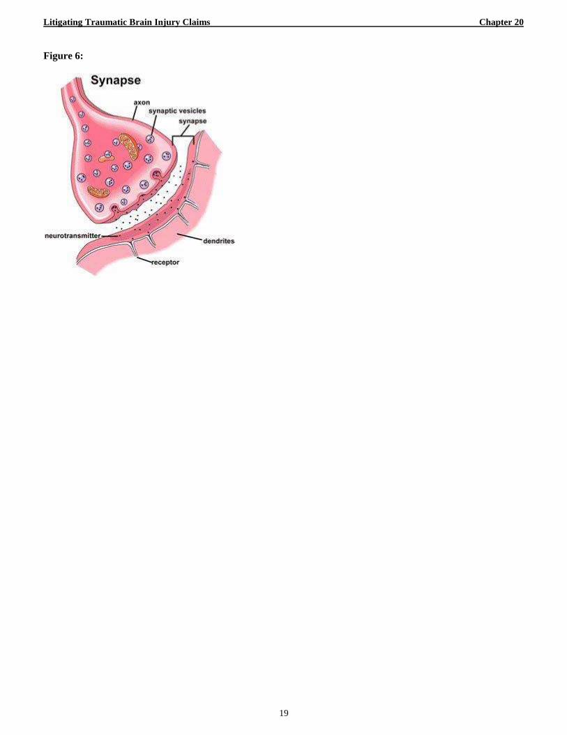
up to 30 centimeters. Neurons communicate via the
synapse (Figure 6) located at the tips of the axons and
produce and house neurotransmitters which, when
released, interface with dendrites of adjacent neurons
through electrochemical reactions. “A single neuron
may have direct synaptic contact with thousands of
other neurons and thereby be involved with an almost
unfathomable multiplicity and complexity of
functioning synapses underlying behavior and
cognition at any given moment.” (Lezak,
Neuropsychological Assessment, 5th Ed., p. 44.) This
explains why an interruption of the functioning of a
few neurons can produce significant changes in brain
function. (Izhikovich and Edelman, 2008.)
C. Definitions and Classification of Traumatic
Brain Injury.
Traumatic brain injuries have been defined and
classified by various medical organizations to assist
medical practitioners in diagnosing such injuries. The
definition of TBI has not been consistent and tends to
vary according to specialties and circumstances.
Generally, a traumatic brain injury is a non-
degenerative, non-congenital insult to the brain from
an external mechanical force, possibly leading to
13
D. Frank Benson & Dietrich Blumer, Personality Changes
with Frontal and Temporal Lobe Lesions, in PSYCHIATRIC
ASPECTS OF NEUROLOGIC DISEASE 151-170 (1975). 14
A. Earl Walker & Dietrich Blumer, The Localization of
Sex in the Brain (1975), reprinted in Cerebral Localization
(K.J. Zulch et al. eds., 2011).
permanent or temporary impairment of cognitive,
physical, and psychosocial functions, with an
associated diminished or altered state of consciousness.
It has also been defined as “an alteration in brain
function, or other evidence of brain pathology, caused
by an external force.” TBI’s are often classified by use
of the Glasgow Coma Scale as severe (GCS 3-8),
moderate (GCS 9-12), or mild (GCS 13-15).
1. The Glasgow Coma Scale.
The Gasgow Coma Scale score for a patient is
based upon clinical assessment at the time of the
injury. It is a 15 point assessment of eye-opening
response, verbal response, and motor response. The
accuracy of the results of the assessment depends upon
when and by whom it was conducted. This
classification system can be misleading as all traumatic
injuries to the brain are serious and even those
classified as “mild” under this system can result in
catastrophic and life-long consequences. As such, this
paper will concentrate on “mild” traumatic brain
injuries (MTBI), their recognition, diagnosis, and
sequelea.
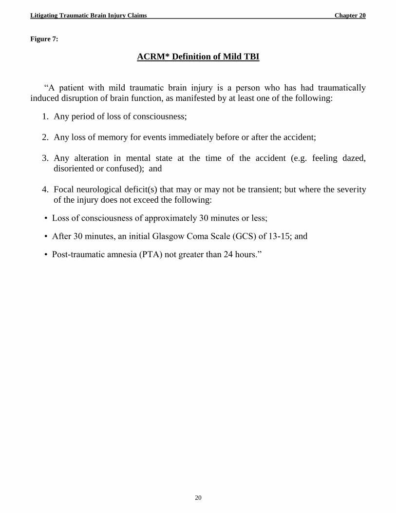
2. ACRM Definition of Mild TBI (mTBI).
The first clear definition of mild traumatic brain
injury (mTBI) was developed by the American
Congress of Rehabilitation Medicine (ACRM).15
The
ACRM defines mTBI as follows:
“A patient with mild traumatic brain injury is
a person who has had traumatically induced
disruption of brain function, as manifested by
at least one of the following:
a) Any period of loss of
consciousness;
b) Any loss of memory for events
immediately before or after the
accident;
c) Any alteration in mental state at the
time of the accident (e.g. feeling
dazed, disoriented or confused);
and
d) Focal neurological deficit(s) that
may or may not be transient; but
where the severity of the injury
does not exceed the following:
Loss of consciousness of
approximately 30 minutes or less;
15
Thomas Kay, Neuropsychological Treatment of Mild
Traumatic Brain Injury, J. HEAD TRAUMA
REHABILATATION, Sept. 1993, at 75.
Litigating Traumatic Brain Injury Claims Chapter 20
4
After 30 minutes, an initial
Glasgow Coma Scale (GCS) of 13-
15; and
Posttraumatic amnesia (PTA) not
greater than 24 hours.” (Figure 7)
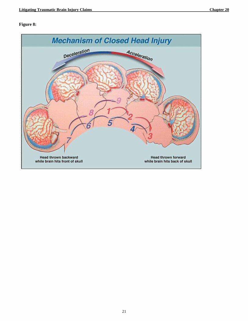
This definition includes the head being struck, the head
striking an object, and the brain undergoing an
acceleration/deceleration movement (i.e. whiplash)
without direct external trauma to the head. (Figure 8)
This definition has gained widespread acceptance and
is recognized by many neurologists, psychiatrists,
physiatrists, and neuropsychologists.
3. Other Definitions of TBI.
Realizing a clear, concise definition of traumatic
brain injury (TBI) was fundamental for reporting,
comparison, and interpretation of studies on TBI. The
Demographics and Clinical Assessment Working
Group of the International and Interagency Initiative
toward Common Data Elements for Research on
Traumatic Brain Injury and Psychological Health
formed an expert group who proposed the following
definition:
TBI is defined as “an alteration in brain
function, or other evidence of brain
pathology, caused by an external force”.16
a) Alteration in brain function is
defined as one of the following
clinical signs:
1) Any period of loss of or
decreased level of
consciousness;
2) Any loss of memory for events
immediately before
(retrograde amnesia) or after
the injury (PTA);
3) Neurologic deficits (weakness,
loss of balance, change in
vision, dyspraxia
paresis/plegia (paralysis),
sensory loss, aphasia, etc); or
4) Any alteration in mental state
at the time of the injury
(confusion, disorientation,
slowed thinking, etc).
b) or other evidence of brain
pathology: Such evidence may
include visual, neuroradiologic or
16
David K. Menon et al., Position Statement: Definition of
Traumatic Brain Injury, 91 ACRHIVES PHYSICAL MED.
REHABILITATION 1637 (2010).
laboratory confirmation of damage
to the brain.
c) Caused by an external force may
include any of the following
events:
1) The head being struck by an
object;
2) The head striking an object;
3) The brain undergoing an
acceleration/deceleration
movement without direct
external trauma to the head;
4) A foreign body penetrating the
brain;
5) Forces generated from events
such as a blast or explosion; or
6) Other force yet to be defined.
Because nomenclature and definitions are more likely
to be a problem in mild TBI, the criteria presented in
this definition under (A) Alteration in brain function is
compatible with the diagnostic criteria presented in the
ACRM definition. This has increased the
understanding that both milder insults and less typical
presentations now fit under the TBI diagnostic
umbrella.
III. SIGNS AND SYMPTOMS OF MILD TBI.
The personal injury practitioner should be
knowledgeable of the symptoms of mild TBI and
should investigate the facts to determine if they are
manifested in the client.
A. When to Look for Signs and Symptoms.
In any case in which the client has sustained a
potential trauma to the brain through external force,
investigation should be made into the presence of
symptoms. The external force can be through a direct
physical impact to the skull or through
acceleration/deceleration (whiplash) of the head.
B. What Signs and Symptoms to Look For.
(Figure 9)
1. Loss of Consciousness.
After establishing that the client sustained an
impact to the head from an external force or an
acceleration/deceleration event, inquiry should be
made into whether the client lost consciousness at the
scene. The loss of consciousness need not be
prolonged in order to meet the criteria for establishing
that a mild TBI occurred. Even a brief loss of
consciousness demonstrates that the impact was
sufficient to have disrupted and damaged structures
and systems in the brain.

Litigating Traumatic Brain Injury Claims Chapter 20
5
2. Amnesia/Confusion.
It is now well recognized that loss of
consciousness is not necessary to confirm the client
suffered mild TBI. Any loss of memory of events
surrounding the incident, any dazed feeling,
disorientation, or confusion by the client is sufficient to
establish that the external force to the brain resulted in
damage to and disruption of functions of the brain.
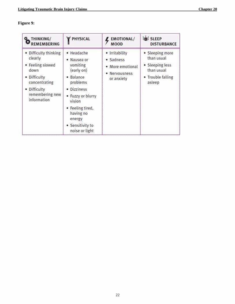
3. Post-Trauma Effects.
Investigation should be made into the presence of
the following symptoms of a TBI:
a) Headaches;
b) Reduced ability to speak clearly;
c) Reduced ability to understand clearly;
d) Reduced ability to focus thoughts or to
concentrate;
e) Reduced ability to read;
f) Unusual fatigue;
g) Changes in sleep patterns;
h) Changes in sex life;
i) Unusual temper;
j) Vision changes;
k) Changes in sense of smell;
l) Changes in sense of taste;
m) Irritability, feelings of hopelessness or
depression;
n) Reduced ability to perform at school or work;
o) Reduced memory;
p) Reduced ability to sequence or perform
complex tasks;
q) Reduced ability to exercise good judgment;
r) Difficulty organizing daily tasks;
s) Problems with balance, dizziness, or vertigo;
t) Reduced ability to perform in a noisy
environment;
u) Development of antisocial behavior.17
C. Where to look for Signs and Symptoms.
The investigation into the potential signs and
symptoms of mild TBI should include interviews and
review of the following:
1. Client Interviews.
The client and family members (and potentially
close friends, co-workers, and accident scene
witnesses) should be interviewed for evidence of any
of the signs and symptoms of mild TBI described
above.
17
Bruce Stern & Dr. Jeffrey Brown, Litigating Brain
Injuries, Vol. 1 §3.2 (2006).
2. Accident Report.
The accident report may have evidence of the
force of impact, the mechanism of injury, loss of
consciousness, confusion/amnesia of events, as well as
identification of scene witnesses and emergency
medical personnel.
3. Accident Witnesses.
Scene witnesses may have evidence of the force
of impact, mechanism of injury, loss of consciousness,
confusion/amnesia, and client complaints (headaches,
dizziness, nausea, etc.).
4. Ambulance/EMT Records and Interviews.
The ambulance crew may have conducted a
Glasgow Coma Scale assessment at the scene and/or
documented loss of consciousness, amnesia or
confusion, bruising, laceration, etc.
5. Hospital Records.
It is important to review not only the initial
history, assessment, and diagnosis but also the nursing
notes which may contain references to cognitive,
emotional, and behavioral symptoms consistent with
TBI.
6. Post-accident Medical/Rehab Records from All
Practitioners. (Including Physiotherapists,
Chiropractors, Massage Therapists, Naturopaths,
and Counselors).
These records may contain references to
complaints of headaches, dizziness, nausea,
memory/concentration problems, and other symptoms
consistent with TBI.
7. Prior Medical Records.
These records may contain evidence of a prior
brain injury or other medical conditions which would
increase the vulnerability to TBI to help explain why
the client is one of the 10-20% of victims of TBI that
never recover.
8. Scene Photographs.
These photographs depicting damage to vehicles,
equipment, etc., may assist in demonstrating the force
of impact and mechanism of injury consistent with
TBI.
9. School Records.
These records, including standardized testing and
performance scores both before and after injury, can
demonstrate decreased scholastic aptitude and/or
behavioral problems consistent with TBI. These
records should be made available to the
neuropsychologist for consideration.
Litigating Traumatic Brain Injury Claims Chapter 20
6
10. Employment Records.
Job performance, attendance, pay raises, etc., can
be contrasted pre- and post-accident to indicate
changes consistent with TBI.
IV. MEDICAL EVALUATION OF TBI.
Once the investigation shows that the client
probably sustained a TBI, the practitioner should
assure that the client receives a thorough medical
evaluation of the nature and extent of the client’s
injury. Often emergency medical treatment of the
acute injury will not include such an evaluation.
Typically, emergent care of a head trauma will be
overseen by a trauma physician and be limited to x-ray,
CT scan, and/or MRI which, in many cases, are not
effective to detect and define the injury to the brain.
A. Neurological Examination.
The client should be thoroughly assessed by a
qualified neurologist in order to evaluate and document
the loss of consciousness, amnesia/confusion, and any
and all symptoms of the TBI. If warranted, the
neurologist can make the referral for
neuropsychological and neuroradiological examination
of the client to augment the diagnosis and quantify the
resulting deficits in brain function.
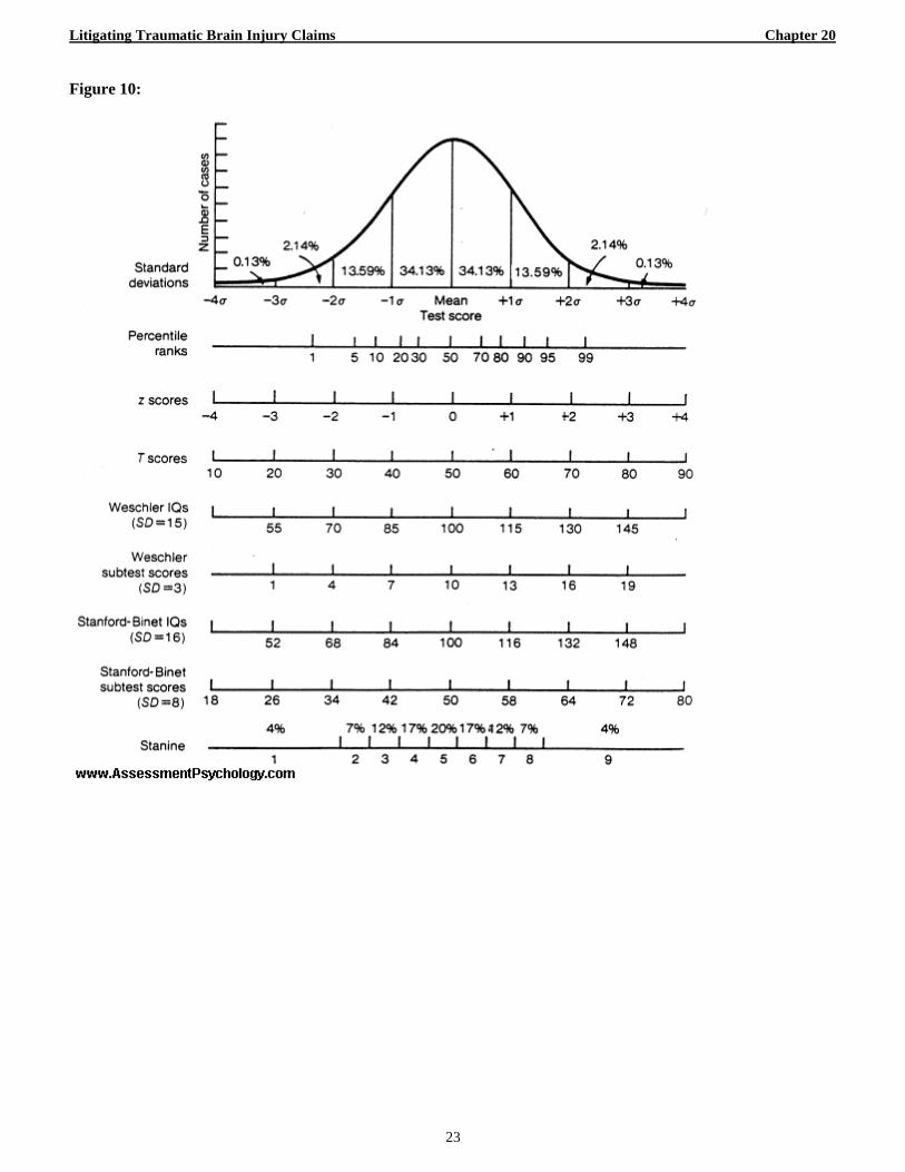
B. Neuropsychological Examination.
The client should be examined by a qualified
neuropsychologist to evaluate the nature and extent of
any neurological dysfunction. This should include
taking of a history, review of medical records including
neuroimaging studies, and the administration and
interpretation of a battery of standardized tests.
Cognitive deficits and abnormal behavior should be
correlated to areas of the brain that control related
functions and any clinical abnormalities noted there.
These standardized tests measure memory, complex or
sequenced tasks, I.Q., reasoning, emotional response,
vision, and other brain functions. Where possible,
post-morbid levels of function should be compared to
pre-morbid levels in order to evaluate the reduction in
brain function attributable to the TBI. (Figure 10)
C. Neuroradiological Evaluation.
Various neuroimaging techniques play an
imperative role in TBI diagnosis. Certain of these
techniques are capable of outlining neuroanatomical
abnormalities, as well as cellular and metabolic
dysfunction on the microscopic level. Therefore, it is
possible to correlate neurophysiological damage
caused by TBI with neuropsychological deficits
incurred by the client.18
18
Benjamin J. Hayempour et al., The Role of Neuroimaging
in Assessing Neuropsychological Deficits Following
Traumatic Brain Injury, 39 J. PSYCHIATRY LAW 537 (2011).
1. CT Scan.
Computed Tomography is capable of detecting
skull fracture and subarachnoid hemorrhage, and can
differentiate acute hemorrhage of the parenchyma from
edema or swelling. However, it is not reliable to show
specific deficits related to regional damage to the brain.
(Figure 11)
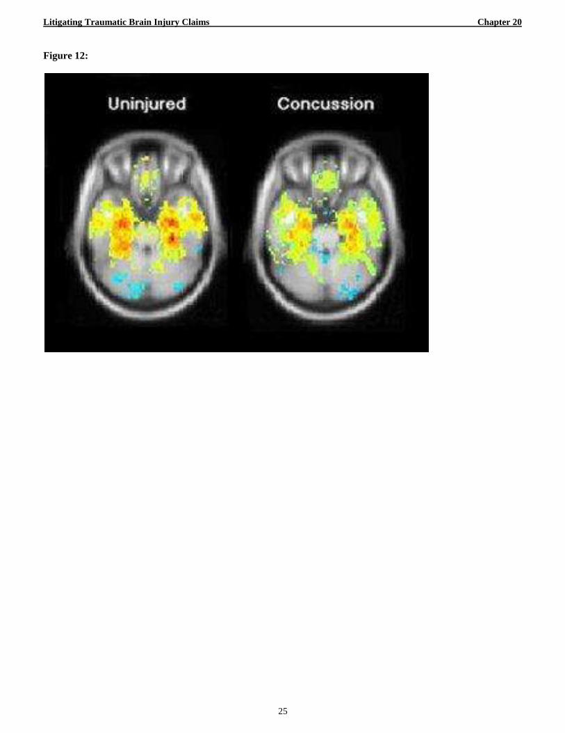
2. MRI.
Magnetic Resonance Imaging is the preferred
imaging technique for detecting sub-acute and chronic
TBI; however, both traditional MRI and CT are not
reliable to detect mild TBI microscopic shear injury or
metabolic dysfunction on the microscopic level.
(Figure 12)
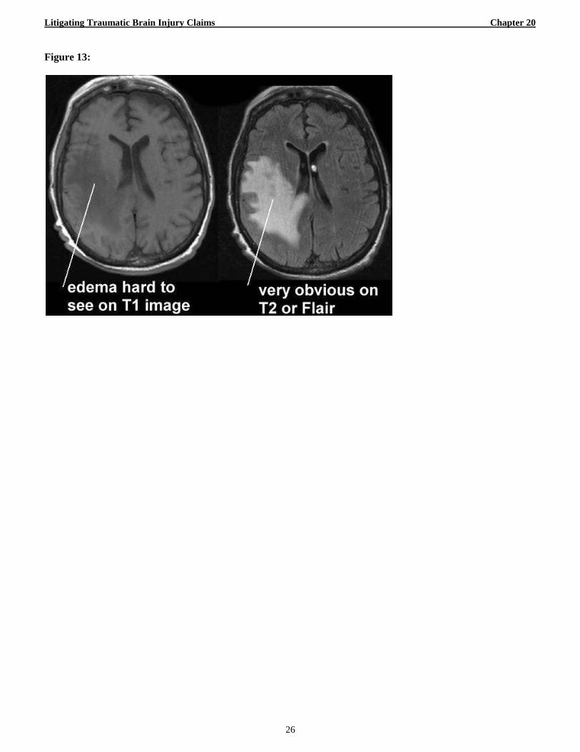
3. FLAIR.
Fluid Attenuated Inversion Recovery uses a pulse
to selectively reduce signal from cerebrospinal fluid
(CSF). (Figure 13) FLAIR imaging increases the
detection of contusions, white matter injuries, and
subarachnoid hemorrhages. It also improves the
detection of diffuse axonal injuries.19
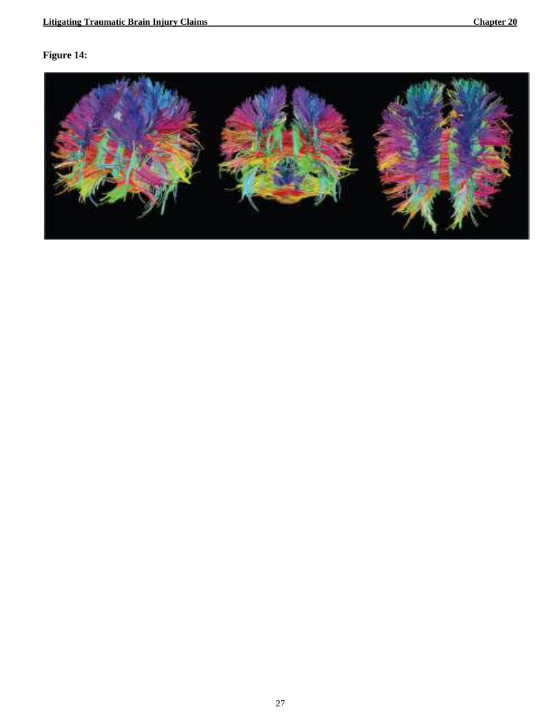
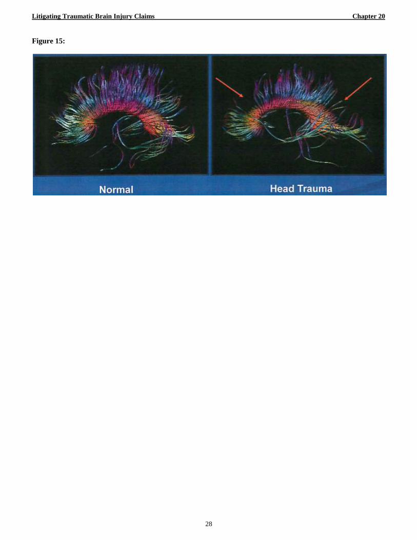
4. DTI.
Diffusion Tensor Imaging measures the random
motion of water molecules in brain tissue. (Figures 14
and 15) The white matter tracts are clearly shown by
DTI. It also shows disruption in those tracts and is an
excellent technique for showing diffuse axonal injury.
DTI can reveal pathology where a conventional MRI is
negative or normal in appearance.20
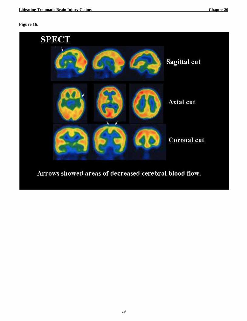
5. SPECT.
Single Photon Emission Tomography (Figure 16)
measures cerebral blood flow in brain tissue.21
Measuring blood flow is an indirect measurement of
brain metabolism. SPECT is highly sensitive for
detecting regional blood flow disturbances in patients
with TBI. It is particularly more effective than CT or
conventional MRI in cases of mild TBI.22
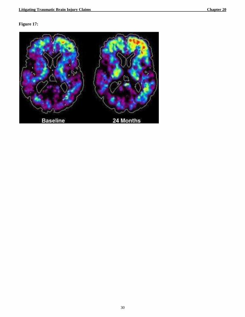
6. PET.
Positron Emission Tomography (Figure 17) can
evaluate glucose metabolism in various regions of the
19
Tuong H. Le & Alisa D. Gean, Neuroimaging of
Traumatic Brain Injury, 76 MOUNT SINIAI J. MED. 145
(2009). 20
Thierry A.G.M. Huisman et al., Diffusion Tensor Imaging
as Potential Biomarker of White Matter Injury in Diffuse
Axonal Injury, 25 AM. J. NEURORADIOLOGY 370 (2004). 21
Bruce G. Gray et al., Technetium-99m-HMPAO SPECT in
the Evaluation of Patients with a Remote History of
Traumatic Brain Injury: A Comparison with X-Ray
Computed Tomography, 33 J. NUCLEAR MED. 52 (1992). 22
Daniel K. Kido et al., Traumatic Brain Injuries: Predictive
Usefulness of CT, 182 RADIOLOGY 777 (1992).
Litigating Traumatic Brain Injury Claims Chapter 20
7
brain. Slowed glucose metabolism indicates neuronal
dysfunction in that region of the brain. Thus, PET has a
significant advantage for illustrating regional brain
dysfunction.
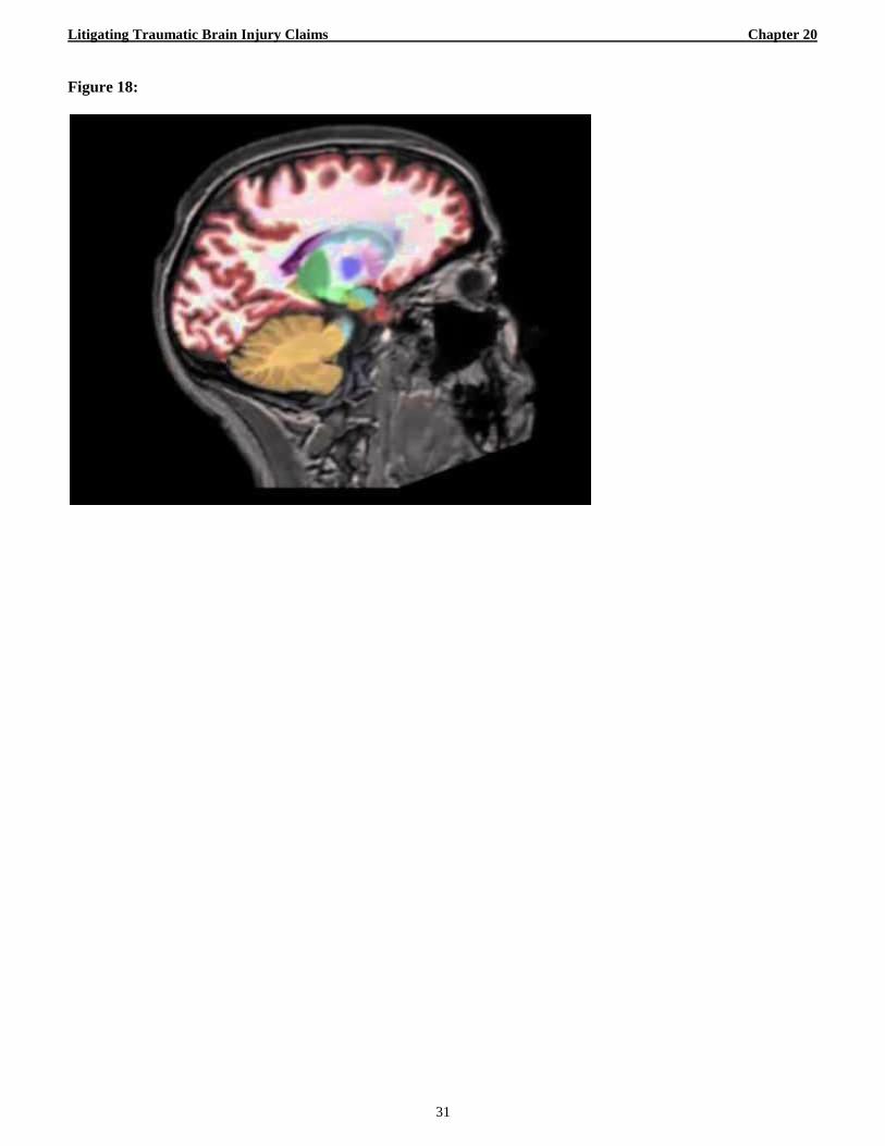
7. NeuroQuant®
(Figure 18) is an FDA-approved method of
analyzing MRI data in measuring brain volume of a
patient and comparing it to normal controls. Brain
atrophy or shrinkage is associated with damage to the
brain. NeuroQuant® can measure atrophy to various
areas of the patient’s brain which can then be
correlated to the patient’s TBI symptoms.23
V. EXPERT WITNESSES IN TRAUMATIC
INJURY CLAIMS.
The litigation of traumatic brain injury claims can
be expert-intensive. The fact of injury, its cause, the
nature and extent of the injury, and the past and future
damages from the injury can each require expert
testimony in order to effectively persuade the trier of
fact. As such, expert witness selection, qualification,
and presentation are often the battleground in these
cases. The following is to assist the practitioner in the
selection, preparation, and presentation of these
important witnesses.
A. Neurologist.
In any case in which a traumatic brain injury is
suspected, it is important to assure that the plaintiff is
evaluated by a qualified neurologist. A neurologist is a
medical doctor or osteopath who is trained in the
diagnosis and treatment of nervous system disorders,
including injuries to and diseases of the brain, spinal
cord, nerves, and muscles. Neurologists perform
neurological examinations of the nerves of the head
and neck, movement, balance, sensation, memory,
speech, language, and other cognitive abilities. A
neurologist qualified to examine and treat a TBI victim
should also be familiar with advanced neuroimaging
techniques such as CT, MRI, DTI, SPECT, FLAIR,
PET, and NeuroQuant®. She should be capable of
interpreting the results of such imaging methods and
correlation of positive findings with any neurological
deficits reported by history and/or neuropsychological
testing. The neurologist should be familiar with
documented long-term sequelea of traumatic brain
injury. That is, the neurologist should understand that
TBI is a “disease process” and not a one-time injury.
The neurologist should also be capable of diagnosing
the TBI and formulating a treatment plan including
medications, therapy, and other recommendations for
23
David E. Ross et al., NeuroQuant® Revealed
Hippocampal Atrophy in a Patient with Traumatic Brain
Injury, 24 J. NEUROPSYCHIATRY & CLINICAL
NEUROSCIENCES E33 (2012).
future medical and custodial care. The neurologist
should opine as to the causation of the injury, the
nature and extent of the injury, its probable duration,
and the resultant limitations. The neurologist should
interface with the neuroradiologist, neuropsychologist,
vocational expert, rehabilitation expert, life care
planner, and biomechanical expert.
B. Neuroradiologist.
A neuroradiologist is a medical doctor or
osteopath that practices a sub-specialty of radiology
focusing on the diagnosis and characterization of
abnormalities of the central and peripheral nervous
systems, spine, and brain by the use of neuroimaging
techniques. The neuroradiologist should be familiar
with not only traditional imaging modalities, CT and
MRI, but also advanced modalities for the diagnosis of
TBI such as DTI, FLAIR, SPECT, PET, and
NeuroQuant®. In addition to coordination with the
neurologist, the neuroradiologist should be able and
willing to coordinate with medical illustrators and
video production professionals to assist in the
formulation and authentication of exhibits to illustrate
the positive neuroradiological findings.
C. Neuropsychologist.
A neuropsychologist is educated and trained to
understand the relationship between the brain and
emotions, behavior, memory, perception, motivation,
and motor skills. The neuropsychologist should be
trained in the interpretation of neuroimaging studies
and the administration and interpretation of the results
of standardized testing of the patient so as to correlate
structural abnormalities with resultant brain
dysfunctions. The clinician should be able to relate the
brain trauma to the abnormality and resulting
impairment. The neuropsychologist should also assist
in the development of treatment modalities and
estimations of the probable effect of brain dysfunction
on the patient’s future family, social, and work
performance. The neuropsychologist should interface
with the neurologist, neuroradiologist, biomechanical
engineer, and vocational experts.
D. Biomechanical Engineer.
The forensic biomechanical engineer is educated
and trained in the analysis of mechanical forces
generated from a given impact and the probable
disruption caused to anatomical regions of the human
body. In cases where it is disputed that a given event
would cause a diagnosed traumatic brain injury, a
biomechanical engineer should be employed to
conduct such an analysis. Typically, in a motor vehicle
collision, this analysis will involve a three-step
approach. In the first step, the type and severity of the
collision is determined, including the change in
velocity of the vehicle (delta-v), and the associated
Litigating Traumatic Brain Injury Claims Chapter 20
8
acceleration time-history of the occupant compartment.
Step two is the occupant dynamics analysis which
models the kinematics of the occupant when subjected
to the forces determined in step one. Step three, the
injury biomechanics analysis, consists of comparing
the occupant loading states determined in step two with
experimentally determined injury tolerance thresholds
of the human body.24
From this analysis, the
biomechanical engineer can opine as to whether or not
the collision probably caused the traumatic brain injury
that has been diagnosed.
E. Life Care Planner.
In cases where TBI is likely to result in substantial
future care needs, a life care planner should be
considered for the plaintiff. “The Life Care Plan is a
dynamic document based upon published standards of
practice, comprehensive assessment, data analysis and
research, which provides an organized, concise plan for
current and future needs with associated costs for
individuals who have experienced catastrophic injury
or have chronic health needs.”25
The life care planner
should be experienced in planning life care for the
traumatic brain injured and have an understanding that
TBI is a disease process and not a finite injury with
finite sequalea. The life care plan may address the
following areas:
Projected patient evaluations
Projected therapeutic modalities
Medication
Diagnostic testing and educational assessments
Supply needs
Wheelchair needs (including accessories and
maintenance)
Home care or facility-based care needs
Transportation needs
Projected surgical or aggressive medical needs
Home furnishings and accessories
Architectural renovations
Aides for independent function
The life care planner may be from diverse fields of
practice, including rehabilitative medicine,
rehabilitative nursing, rehabilitative counseling,
psychiatry, case management, and other areas. The
planner may also be board certified in life care
planning (CLP), which is granted by the Commission
on Health Care Certification. The life care planner
24
Wilson C. Hayes et al., Forensic Injury Biomechanics, 9
ANN. REV. BIOMEDICAL ENGINEERING 55 (2007). 25
International Association of Rehabilitation Professionals,
Standards of Practice §1,
http://www.rehabpro.org/sections/ialcp/focus/standards/secti
on-i-introduction (last visited May 6, 2014).
should interface with the neurologist,
neuropsychologist, vocational expert, and other related
experts and medical providers for the preparation of
the life care plan. The plan, its projected care needs,
and associated costs should also be approved and
agreed to by the testifying physician. The
methodology for preparation of the life care plan may
include the following:
Review of medical records and supporting
documents
Clinical interview with the patient and/or family
members for comparison of pre-morbid and post-
morbid conditions
Consultation with medical treatment team
Research to develop clinical practice guidelines
and costs of care, supplies, equipment, etc.
The life care services and supply costs, as
reflected in the Life Care Plan, should then be
totaled and reduced to their present value by a
qualified economic expert.
F. Vocational/Economic Experts.
Persons that suffer even “mild” traumatic brain
injuries can suffer deficits affecting behavior and
performance that last for varying periods of time and
often last for life. The deficits often result in difficulty
comprehending what is read, problems with attention
to tasks, and not hearing or understanding what is said
by others. These patients cannot process multiple
commands or new information. They have memory
problems, especially when there are two people talking
or there are interruptions. They also complain of
dizziness, irritability, anti-social behavior, anxiety, and
depression. All of these common complaints can be
devastating to affect employment even though the
patients, at first, appear normal to the casual observer.
These patients often can gain or return to employment
following injury, but cannot sustain over time.26
27
On
average, the brain injured person works less over a life
time than the non-injured person and, when they do
work, the brain injured person earns less.28
A vocational economic analysis compares the
injured person’s pre-accident earning capacity (based
on education, work experience, and earning levels) to
26
Laurence M. Binder, Persisting Symptoms After Mild
Head Injury; a Review of Past Concussion Syndrome, 8 J.
CLINICAL & EXPERIMENTAL NEUROPSYCHOLOGY 323
(1986). 27
Deborah Abrams et al., The Economies of Return to Work
for Survivors of Traumatic Brain Injuries: Vocational
Services are Worth the Investment, J. HEAD TRAUMA
REHABILITATION, Dec. 1993, at 59. 28
U.S. Bureau of the Census, Series P-23, Labor Force
Status and Other Characteristics of Persons with a Work
Disability: 1981-1988, No. 160, Table 9 (1989).
Litigating Traumatic Brain Injury Claims Chapter 20
9
their post-injury earning capacity (based on medical,
neuropsychological, and vocational information) and
makes a two-step determination:
1) can the injured person return to competitive
employment and, if so, what are the types of
employment to which they can realistically
return and retain; and
2) if partially or completely disabled, what is
the projected loss of earning capacity over
their lifetime in terms of “present value”.
In determining the foregoing, five key elements are
addressed:
Pre-Injury Earning Capacity: The vocational
expert will consider the person’s education,
training, experience, and earning history to
estimate his “capacity” to earn before the brain
injury.
Pre-Injury Work Life Expectancy: The expert will
estimate pre-injury with life expectancy based
upon the person’s education, health status,
profession, population averages, and expression of
intentions by the person.
Post-Injury Earning Capacity: The expert will
consider the person’s post-injury disability and his
residual capacity to earn. Statistically, disabled
persons earn about 35% less than non-disabled
persons.
Post-Injury Work Life Expectancy: Work life
expectancy varies greatly based upon disability
status. The expert should consider that brain
injuries are progressing in nature and are likely to
shorten the person’s work life. Studies show that a
45 year old nondisabled worker with 12 years of
education has a work life expectancy of 16.5 years
while a disabled person with similar age and
education has a work life expectancy of 4.2
years.29
Present Value Calculation: The economic expert,
whether the same or separate from the vocational
expert, should then calculate the “present value”
of the pre-injury earning capacity per year
multiplied by the pre-injury work life expectancy
minus the post-injury earning capacity multiplied
by the post-injury work life expectancy.
The vocational expert should be qualified by his
education, training, and experience to testify as to the
five key elements. The expert typically will have at
least a bachelor’s degree but preferably a master’s or
doctoral level degree in economics, accounting,
29
Anthony M. Gamboa Jr., The New Work Life Expectancy
Table, Tables 1 & 4 (1990).
vocational rehabilitation, counseling, or psychology.
Board certifications are available through the
American Board of Vocational Experts, International
Association of Rehabilitation, and the American
Rehabilitation Economics Association.
G. Economist.
In cases in which the Plaintiff has suffered a TBI
resulting in lost future earning capacity, lost profits
from business activities, and/or substantial medical
and/or custodial expenses, a qualified economist
should be retained. An economist typically has a
degree in economics, finance, or accounting and has
sufficient education, training, and experience to offer
opinions concerning the “present value” of future lost
income earning capacity, lost profits, and medical
expenses. The economist should review the life care
plan and its projected future costs so as to reduce those
costs to their present value.
VI. TRAUMATIC BRAIN INJURY AS A
DISEASE PROCESS.
Too often, traumatic injury to the brain is seen as
“an event”. It is often mistakenly treated as a broken
bone, the final outcome to an injury of an isolated body
system. That is, once it is “fixed” and given some
therapy, no further treatments would be needed in the
future and, certainly, there will be no affect on other
organs of the body. However, brain injury is not an
event. It is a disease. It never, ever, ever goes away.30
A. TBI Effects on Mortality.
Those suffering a TBI are more likely to die or
have a reduced life span.
1) Those with moderate to severe TBI have a 7
year reduction in life expectancy. Those with
mild TBI have a small but statistically
significant reduction in long term survival.31
2) Of 767 subjects with mild-moderate-severe
TBI followed for 13 years, those surviving
over a year, death rate was 2.5 times higher
than controls and 2 times higher than controls
for mTBIs.32
3) As to the causes of death of TBI victims,
they are 37 times more likely to die from
30
Brent Masel, Brain Injury as a Disease, Address at North
American Brain Injury Society (NABIS) (Mar. 19-21,
2014). 31
Cindy Harrison-Felix et al., Mortality Following
Rehabilitation in the Traumatic Brain Injury Model Systems
of
Care, 19 NEUROREHABILITATION 45 (2004). 32
Thomas McMillan et al., Death After Head Injury: The 13
Year Outcome of a Case Control Study, 82 J.
NEUROLOGY NEUROSURGERY & PSYCHIATRY 931 (2011).
Litigating Traumatic Brain Injury Claims Chapter 20
10
seizure, 12 times more likely to die from
septicemia, 4 times more likely to die from
pneumonia, and 3 times more likely to die
from other respiratory conditions as were the
non-TBI controls.33
B. TBI Effects on Morbidity.
Those suffering TBIs are more likely to develop
other diseases of the body.
1) TBI Victims have an increased risk of stroke,
even higher risk than those with
hypertension.34
2) TBI is the leading cause of epilepsy in young
adults. TBI victims are 1.5 to 17 times more
likely to develop seizures than the general
population.35
3) 70% of chronic TBI out-patients have
subjective complaints of sleep disturbances.36
4) Patients with TBI are 5 times more likely to
develop malignant brain tumors.37
5) There is an increased incidence of
neuroendocrine dysfunction in the TBI
victims. 30% of severe TBIs have
hypopituitarism after 1 year.38
6) 20% of moderate to severe TBI victims after
1 year have Growth Hormone Dysfunction
with increased osteoporosis, high cholesterol,
increased abdominal fat, cardiovascular
disease, and decreased cognitive
functioning.39
33
Cindy Harrison-Felix et al., Causes of Death Following 1
Year Post Injury Among Individuals with Traumatic
Brain Injury, 21 J. HEAD TRAUMA & REHABILITATION 22
(2006). 34
James F. Burke, et al., Traumatic Brain Injury May be an
Independent Risk Factor for Stroke, 81 NEUROLOGY 33
(2013). 35
John F. Annegars et al., A Population Based Study of
Seizures After Traumatic Brain Injuries, 338 N. ENG. J.
MED.
20 (1998). 36
Brent Masel et al., Excessive Day Time Sleepiness in
Adults with Brain Injuries, 821 ARCHIVES PHYSICAL MED. &
REHABILITATION 1526 (2001). 37
Yi-Hua Chen et al., Association Between Traumatic Brain
Injury and the Risk of Brain Cancer, 29 J.
NEUROTRAUMA 1328 (2012). 38
Harald Jorn Schneider et al., Hypothalamopituitary
Dysfuction Following Traumatic Brain Injury and
Aneurysmal
Subarachnoid Hemmorage: A Systematic Review, 298 J. AM.
MED. ASS’N 1429 (2007).
39 Gianluca Aimaretti, Traumatic Brain Injury and
Hypopituitarism, 5 SCI. WORLD J. 777 (2005).
7) Sexual Dysfunction is reported by 40-
50% of individuals post-TBI.40
8) There is a higher incidence of psychiatric
disease in patients with chronic TBI; 20%
report psychosis, 18-61% report
depression, 1-22% have mania, 3-59%
PTSD, and 20-40% have aggression. TBI
is associated with
9) higher rates of suicidal ideation, suicide
attempts, and completed suicides.41
10) Moderate to severe TBIs in adults result
in 2 times the risk of developing
Alzheimer’s and other forms of dementia
later in life.42
VII. RESOURCES FOR FURTHER TBI
RESEARCH.
This paper is merely a “primer” on the subject
of litigation of traumatic brain injuries. Organizations
for plaintiff and defense counsel exist that provide in-
depth programs and materials on the subject as well as
excellent publications for the interested practitioner.
A. Organizations for Plaintiff Counsel.
1) The American Association for Justice
(AAJ) has a Traumatic Brain Injury
Group that sponsors conferences at least
two times a year with excellent topics and
speakers. www.justice.org.
2) The North American Brain Injury Society
(NABIS) is a society of professionals
involved in the issues surrounding brain
injury. It provides education programs,
scientific updates, and a platform for
communication and professional
exchange. www.nabis.org.
B. Organizations for Defense Counsel.
The Defense Research Institute (DRI) is the
leading organization of defense attorneys and in-house
counsel. It sponsors seminars that regularly feature
speakers and papers that treat the issues surrounding
traumatic brain injury litigation from the defense
perspective. www.dri.org.
40
Nathan Zasler et al., Brain Injury Medicine (2006). 41
Edward Kim et al., Neuropsychiatric Complications of
Traumatic Brain Injury: A Critical Review of the Literature
(A report by the ANPA Committee on Research), 19 J.
NEUROPSYCHIATRY & CLINICAL NEUROSCIENCES 106
(2007). 42
Scott Gottlieb, Head Injury Doubles the Risk of
Alzheimer’s Disease, BRIT. MED. J., Nov. 4, 2000, at 1100.
Litigating Traumatic Brain Injury Claims Chapter 20
11
C. Publications.
1) Litigating Brain Injuries, Vols. 1 and 2,
Stern, Bruce. Thomson-West Publishing.
2006.
2) Neuropsychological Assessment, Lezak,
et al., 5th Edition, Oxford University
Press. 2012.
3) The Handbook of Clinical
Neuropsychology, Gurd, et al. Oxford
University Press. 2012.
4) The Handbook of Functional
Neuroimaging of Cognition. Cabeza, et
al, Second Edition.
VIII. CONCLUSION.
Recent events have brought public awareness
to traumatic brain injuries and their devastating effects
on the lives of victims, including war heroes and
athletic legends. Scientific advances in neuroimaging
have enabled objective verification of many of the
injuries. Even so called “mild” traumatic brain injuries
(which may go undetected during emergent care of the
acute injury) can have life-long effects. The prudent
practitioner must be vigilant in identifying these
injuries and thorough in the assessment and
presentation of them. It is imperative that personal
injury lawyers understand the symptoms and science
related to TBI and the proper modalities for verifying
and explaining such injuries.
Litigating Traumatic Brain Injury Claims Chapter 20
13
APPENDIX
Table of Figures
1. Gulf War Photo ................................................................................................................................................... 14
2. D Magazine Cover with Tony Dorsett ................................................................................................................ 15
3. Basic Anatomy of the Brain ................................................................................................................................ 16
4. Lobes v. Function ................................................................................................................................................ 17
5. Anatomy of the Neuron ....................................................................................................................................... 18
6. Synapse ............................................................................................................................................................... 19
7. ACRM Definition of mTBI ................................................................................................................................. 20
8. Acceleration/Deceleration of the Brain ............................................................................................................... 21
9. Signs and Symptoms of mTBI ............................................................................................................................ 22
10. Neuropsychological “Bell Curve” ....................................................................................................................... 23
11. CT Scan ............................................................................................................................................................... 24
12. MRI ..................................................................................................................................................................... 25
13. FLAIR ................................................................................................................................................................. 26
14. DTI ...................................................................................................................................................................... 27
15. DTOI – Normal vs. Head Trauma ....................................................................................................................... 28
16. SPECT ................................................................................................................................................................. 29
17. PET ...................................................................................................................................................................... 30
18. NeuroQuant® ...................................................................................................................................................... 31
19. Road Sign of the Brain Injured............................................................................................................................ 32
Litigating Traumatic Brain Injury Claims Chapter 20
14
Figure 1:

Litigating Traumatic Brain Injury Claims Chapter 20
15
Figure 2:
Litigating Traumatic Brain Injury Claims Chapter 20
16
Figure 3:
Litigating Traumatic Brain Injury Claims Chapter 20
17
Figure 4:
Litigating Traumatic Brain Injury Claims Chapter 20
18
Figure 5:
Litigating Traumatic Brain Injury Claims Chapter 20
19
Figure 6:
Litigating Traumatic Brain Injury Claims Chapter 20
20
Figure 7:
ACRM* Definition of Mild TBI
“A patient with mild traumatic brain injury is a person who has had traumatically
induced disruption of brain function, as manifested by at least one of the following:
1. Any period of loss of consciousness;
2. Any loss of memory for events immediately before or after the accident;
3. Any alteration in mental state at the time of the accident (e.g. feeling dazed,
disoriented or confused); and
4. Focal neurological deficit(s) that may or may not be transient; but where the severity
of the injury does not exceed the following:
• Loss of consciousness of approximately 30 minutes or less;
• After 30 minutes, an initial Glasgow Coma Scale (GCS) of 13-15; and
• Post-traumatic amnesia (PTA) not greater than 24 hours.”
Litigating Traumatic Brain Injury Claims Chapter 20
21
Figure 8:
Litigating Traumatic Brain Injury Claims Chapter 20
22
Figure 9:
Litigating Traumatic Brain Injury Claims Chapter 20
23
Figure 10:

Litigating Traumatic Brain Injury Claims Chapter 20
24
Figure 11:
Litigating Traumatic Brain Injury Claims Chapter 20
25
Figure 12:
Litigating Traumatic Brain Injury Claims Chapter 20
26
Figure 13:
Litigating Traumatic Brain Injury Claims Chapter 20
27
Figure 14:
Litigating Traumatic Brain Injury Claims Chapter 20
28
Figure 15:
Litigating Traumatic Brain Injury Claims Chapter 20
29
Figure 16:
Litigating Traumatic Brain Injury Claims Chapter 20
30
Figure 17:
Litigating Traumatic Brain Injury Claims Chapter 20
31
Figure 18:
Litigating Traumatic Brain Injury Claims Chapter 20
32
Figure 19: