Literature review of modelling muscle mechanics, damage and adaptation · Literature review of...
26
Literature review of modelling muscle mechanics, damage and adaptation Gielen, A.W.J. Published: 01/01/1995 Document Version Publisher’s PDF, also known as Version of Record (includes final page, issue and volume numbers) Please check the document version of this publication: • A submitted manuscript is the author's version of the article upon submission and before peer-review. There can be important differences between the submitted version and the official published version of record. People interested in the research are advised to contact the author for the final version of the publication, or visit the DOI to the publisher's website. • The final author version and the galley proof are versions of the publication after peer review. • The final published version features the final layout of the paper including the volume, issue and page numbers. Link to publication Citation for published version (APA): Gielen, A. W. J. (1995). Literature review of modelling muscle mechanics, damage and adaptation. (DCT rapporten; Vol. 1995.018). Eindhoven: Technische Universiteit Eindhoven. General rights Copyright and moral rights for the publications made accessible in the public portal are retained by the authors and/or other copyright owners and it is a condition of accessing publications that users recognise and abide by the legal requirements associated with these rights. • Users may download and print one copy of any publication from the public portal for the purpose of private study or research. • You may not further distribute the material or use it for any profit-making activity or commercial gain • You may freely distribute the URL identifying the publication in the public portal ? Take down policy If you believe that this document breaches copyright please contact us providing details, and we will remove access to the work immediately and investigate your claim. Download date: 07. Sep. 2018
-
Upload
vuongquynh -
Category
Documents
-
view
217 -
download
0
Transcript of Literature review of modelling muscle mechanics, damage and adaptation · Literature review of...

Literature review of modelling muscle mechanics,damage and adaptationGielen, A.W.J.
Published: 01/01/1995
Document VersionPublisher’s PDF, also known as Version of Record (includes final page, issue and volume numbers)
Please check the document version of this publication:
• A submitted manuscript is the author's version of the article upon submission and before peer-review. There can be important differencesbetween the submitted version and the official published version of record. People interested in the research are advised to contact theauthor for the final version of the publication, or visit the DOI to the publisher's website.• The final author version and the galley proof are versions of the publication after peer review.• The final published version features the final layout of the paper including the volume, issue and page numbers.
Link to publication
Citation for published version (APA):Gielen, A. W. J. (1995). Literature review of modelling muscle mechanics, damage and adaptation. (DCTrapporten; Vol. 1995.018). Eindhoven: Technische Universiteit Eindhoven.
General rightsCopyright and moral rights for the publications made accessible in the public portal are retained by the authors and/or other copyright ownersand it is a condition of accessing publications that users recognise and abide by the legal requirements associated with these rights.
• Users may download and print one copy of any publication from the public portal for the purpose of private study or research. • You may not further distribute the material or use it for any profit-making activity or commercial gain • You may freely distribute the URL identifying the publication in the public portal ?
Take down policyIf you believe that this document breaches copyright please contact us providing details, and we will remove access to the work immediatelyand investigate your claim.
Download date: 07. Sep. 2018

Literature review of modelling mus- cle mechanics, damage and adapta- tion
WFW-report: 95.018
A.W.J. Gielen

Contents
2 1 Introduction
2 3 Physiology of skeletal muscles 2.1 Morphology 3
2.1.1 Connective tissue 4 2.1.2 Musclefibre 5 2.1.3 Myofibril 5 2.1.4 Sarcomere 7 2.1.5 Sarcotubular system . . . . . . . . . . . . . . . . . . . . . . . . . . . . . 8
2.2 Muscle function: the contraction mechanism . . . . . . . . . . . . . . . . . . . . 8 2.2.1 Exitation or the electrical phenomena . . . . . . . . . . . . . . . . . . . 8 2.2.2 Single twitch and wave summation . . . . . . . . . . . . . . . . . . . . . 9
2.2.4 Length and speed dependence of the contraction . . . . . . . . . . . . . 9
. . . . . . . . . . . . . . . . . . . . . . . . . . . . . . . . . . . . . . . . . . . . . . . . . . . . . . . . . . . . . . . . . . . . .
. . . . . . . . . . . . . . . . . . . . . . . . . . . . . . . . . . . . . . . . . . . . . . . . . . . . . . . . . . . . . . . . . . . . . . . . . . . . . . . . . . . . . . . . . . . . . . . . . . . . . . . .
. . . . . . . . . . . . . . . . . . . . . . . . . . . . . . . . . . . 2.2.3 Mechanics 9
2.2.5 Metabolics 10 . . . . . . . . . . . . . . . . . . . . . . . . . . . . . . . . . .
11
3.1.1 Hill model 11 12
3 Modelling skeletal muscles 3.1 Models for the contracting fibers . . . . . . . . . . . . . . . . . . . . . . . . . . 11
3.1.2 Huxley model . . . . . . . . . . . . . . . . . . . . . . . . . . . . . . . . . 3.1.3 Distributed moments model . . . . . . . . . . . . . . . . . . . . . . . . . 14
3.2 Models for the connective tissue and tendons . . . . . . . . . . . . . . . . . . . 15 3.2.1 Construction of constitutive laws . . . . . . . . . . . . . . . . . . . . . . 15
. . . . . . . . . . . . . . . . . . . . . . . . . . . . . . . . . . .
17 4 Damage and Adaptation 4.1 Morphological aspects of damage . . . . . . . . . . . . . . . . . . . . . . . . . . 17
4.1.1 Direct myofiber damage . . . . . . . . . . . . . . . . . . . . . . . . . . . 17 4.1.2 Delayed myofiber damage . . . . . . . . . . . . . . . . . . . . . . . . . . 18 4.1.3 Connective tissue damage . . . . . . . . . . . . . . . . . . . . . . . . . . 18
4.2 Causes of muscle damage . . . . . . . . . . . . . . . . . . . . . . . . . . . . . . 18 4.2.1 Mechanical component . . . . . . . . . . . . . . . . . . . . . . . . . . . . 18 4.2.2 Metabolic component . . . . . . . . . . . . . . . . . . . . . . . . . . . . 18
4.3.1 Morphological aspects . . . . . . . . . . . . . . . . . . . . . . . . . . . . 19 4.3.2 Adaptation control . . . . . . . . . . . . . . . . . . . . . . . . . . . . . . 19
. . . . . . . . . . . . . . . . . . . . . . . . . . . . . . . . . . . . . . 4.3 Adaptation 19
20 20
A Mathematics of the DM model A.l From Huxley to DM . . . . . . . . . . . . . . . . . . . . . . . . . . . . . . . . .
1

Chapter 1
Introduction
The mechanical properties of skeletal muscle change as a result of all kinds of internal and external influences, like blood perfusion and mechanically applied loads. These changes can be split into damage and adaptation. When a muscle is subjected to a long term change of mechanically loads, it will alter properties and compostion, like fibre type, connective tissue and fibre directions, in order to accommodate to the new circumstances. When the adaptive ability is unable to follow the loads with respect to magnitude and/or time, then muscle damage will OCCUT. Damage may vary from very small localised micro defects upto massive structural defects. Muscle damage will result in a change of chemical compostion of extracellular fluids in the muscle, which leads to swelling and/or all kind of inflammatory reactions. The border between adaptation and damage is still very unclear.
Adaptation and damage of skeletal muscles can be applied in several terrains: What’s the difference between use and mis/over-use of muscles in training or working conditions. How do muscles react on inactivity for ill and elderly persons. Can a muscle from the back be used to support the pumping function of the heart.
Since the 1980’s muscle damage and adaption are topics in experimental research at the Rijksuniversiteit Limburg. A mathematical model of the experiments, which predicts the response of muscles, could add an inportant ingredient in the interpretation of the experiments. Development of the mathematical model is subject of research in the project damage and adaptation of skeletal muscles at Eindhoven University of Technology.
Although lots of mathematical models of muscles exist, they give a limited description of the geometrical and mechanical properties. These models are essentially one-dimensional models of whole muscles (i.e. muscle-tendon complex) or single fibres. There have been attempts to include the complex geometrical properties of the muscle (e.g. [21, 39]), but none of them paid attention to the 3-dimensional behaviour of the intramuscular tissue. Finite element models could be a solution to this problem. Both the 3D geometry and 3D material properties can be taken into accound, as well as non-homogeneous properties like nonuniform sarcomere distributions (e.g. [i, 28]), damage (e.g. [9, 22, 37, 381 and adaptation (e.g. [lo, 14, 341).
This report is a literature survey about skeletal muscles, damage and adaptation. It gives an introduction into the physiology of skeletal muscle, models of skeletal muscle and the major topics of damage and adaptation of skeletal muscles.
The chapter about damage and adaptation must be considered as a first attempt to sum- marise the most important issues in damage and adaptation of skeletal muscles. As I have a different scope with respect to the subject than the authors of most the literature, it’s possi- ble that I emphasized subjects that are considered minor by biologists/physiologists and vice versa.
Sander Gielen, February 6, 1995
2

Chapter 2
Physiology of skeletal muscles
Muscles can be divided into three types: skeletal, cardiac, and smooth muscle. The skeletal muscles comprise the great mass of the somatic musculature. They are striated and controlled by the voluntary nerve system. Cardiac muscle is also striated but is functionally different. It contracts rhythmically in absence of external innervation. Pacemaker cells in the myocardium, that may discharge spontaneously, innervate the cardiac muscle. Smooth muscles are different in structure and are controled by the vegetative nerve system.
Only skeletal muscles will be considered here.
2.1 Morphology A skeletal muscle has several components:
e muscle fibres e connective tissue e blood and lymphe vessels o motor and sensor nerves.
sarmlemma
Figure 2.1: Location of the fascia, vessels and fibres.
The muscle is connected to the skeleton by tendons and shielded by a sheath of connective tissue, the epi-mysium. Vessels and nerves are important €or the function o€ the muscle, but only muscle fibres and connective tissue will be considered.
3

2.1.1 Connective tissue
The muscle is protected by a fascia, the epimysium. In the muscle the perimysium connects the fibre bundles to one another and to the epimysium. The perimysium is also the 'carrier' for the bloodvessels, lymphevessels and nerves into the muscle. The endomysium, which is actually a part of the perimysium, is the fine network of connective tissue at muscle-fibre level. It also supports the vessels and nerves to the individual muscle fibres.
The peri- and endomysium allow individual fibres and bundles to move independently within a certain range'.
The main function of the connective tissue is transmission of force through/allong the muscle onto the skeleton. The endomysium transmits the force of the muscle-fibres to the perimysium, which in its turn transmits it to the epimysium and the tendons. They are in fact not seperate structures, but the endo-, peri- and epimysium form together with the tendons a big continuous network of tissue. When a muscle is suddenly overloaded it is more likely that rupture will occure at the connection bone-tendon or in 2the tendon, but seldom at the t endon-muscle transition.
In a tendon the collagen molecules will have a more parrallel arrangement, while in muscle perimysium it will have a more 'woven' structure.
The main 'construction' material of tendons and fascia is collagen. The space in the fascia not occupied by vessels or collagen is filled with a hydrophilic (=water attracting) gel called ground substance. A small part of the tendons and fascia is formed by elastin.
Collagen
Collagen strands, also called a-chains, are formed from threesomes of one glycine and two amino acids, arranged in a left-hand helix. Three a-chains are right-hand twisted and form the collagen-molecule. (See figure 2.2). There are 12 types of collagen identified. The differences are variations in the a-chains and variations in spatial arrangement. The differences are related to the function of the collegen in the tissue.
860nrn
1 87nrn 1 r7
Glycine Aminoaads
Figure 2.2: Schematic drawing of collagen triple helix. The individual a-chains are ieft-handed helices with approximately three residues per turn. The chains are, in turn, coiled around each other following a right-hand twist. [15]
Ground substance
Dense connective tissues contain little ground substance, while loose connective tissues contain lost of ground substance. The composition of the subs? Lnce varies with the tissue, but it contains muco-poly-saccharides and water with all kinds of ions and minerals. The fluid in
'Does this imply that for wide range of shear strains the shear stress is low, then the shear stress rises quickly to limit the shear strain?
4

this substance complicates the mechanical behaviour in two ways. The fluid can move trough the tissue as well as it can bind to collagen (hydration).
Elastin
Elastin is the most 'linear' biosolid material known. From the stress-strain curve (figure 2.3)
i.g. Nudiae denaiurated
I 20
% Strain = p L L O ) W O
Figure 2.3: Stress-strain curve of elastin. 1151
one can see that elastin is slightly visco-elastic. However the energy dissipation is small. The elastic properties shown remain at least up to a/& = 1.6.
2.1.2 Muscle fibre
In the muscle the fibres are arranged in bundles containing a few to over a hundred fibres. Each bundle is shielded by a sheath of connective tissue, the peri-mysium. Within the bundle the fibres are connected to one another by the endo-mysium. At this level blood and lymphe vessels are still visible (see figure 2.5). There are two types of fibres: Slow-twitch (S) and fast- twitch (F). The latter can be divided into oxidative glycolytic (FOG) or glycolytic (FG). The slow-twitch fibres are always oxidative (SO). The muscle fibres have a diameter of 10 - 95pm [14, 151 and usually a length of several millimeters to several centimeters. This means that a muscle fibre has a length-diameter ratio of 100 upto 1O.QOO. According to [14] for rats the SO fibres are 70 - 80pm, the FOG fibres 55 - 60pm and the F fibres 80pm. For humans these are: SO 80pm and F 90 - 95pm. Sometimes the length can reach 30cm in long muscles. The fibres can span the entire muscle, from end to end, or only a part, ending in tendons or other connective tissue intersections.
The muscle fibre is a special kind of cell. It has more than one nucleus, because in the embryonal fase the cell is formed by fusion of many myoblasts. Besides, it has a sareo-tubular system and long threads or myofibrils . The tubuli are important in the stimulation, activation of the muscle, while the myofibrils are the drivers of the contraction mechanism.
2.1.3 Myofibril
Each muscle fibre (muscle cell) contains l o 2 . . . lo3 myofibrils. A myofibril is a thin thread ( d = 1.. .2pum) with the same length as the muscle fibre. The fibrils can be divided into individual filaments. Each filament is made up of contractile proteins. These proteins are: myosin, actin and troponin. The troponin is made of 3 subunits, called troponin I, T and C.
5

Figure 2.4: The muscle belly constist of bundle of muscle fibres. Each bundle is protected by a sheath of connective tissue. A bundle of fibres is surrounded by all kind of vessels, while each fibre consists of many myofibrils. The fibrils are repeating units of actin and myosin filaments. One unit is called a sarcomere. [15]
3 2 Triad 4 4 5
Figure 2.5: Construction of the muscle fibre. 1: myofibrils. 2: sarcoplasmatic reticulum. 3: terminal cistern. 4: T-system of the tubuli. 5: basal lamina. 6: mitochondria. A: A-band. I: I-band. [16]
6

Myosin i
Figure 2.6: top: actin filament, bot- tom: myosin filament. [5]
Figure 2.7: Cross-bridge and troponin configuration on actin. [16]
The myosin molecules are asymmetric with enlarged globular heads. The head contains an actin-binding site and a catalytic site that hydrolyzes ATP (adenosine tri-phosphate).
The actin and troponin are linked on two supporting tropomyosin molecules, which form a double helix. (See figure 2.7). The actin is spaced regulary on the tropomyosin on small intervals and the troponin is spaced on slightly bigger intervals. The troponin-?' binds the other troponin parts to the tropomyosin. The troponin-I inhibits the interaction of myosin with actin and troponin-C contains sites for Cu2+ -ions that initiate contraction.
Actin
A single actin filament has a diameter of approximately 7nm. The tensile force of the filament is 108 f 5pN (measured on 61 filaments by Kishino and Yanagida in 1988. See fl51). This force is comparable with the force on a single actin element in a muscle cell during isometric contraction. The tensile strength of the actin filament is, on assuming a force of 108pN sustained by a fiber of diameter 7 n m at least 2.2MPu.
2.1.4 Sarcomere
In the muscle fibre there are repeating units of actin and myosin filaments. Such a unit is called a sarcomere. The striation of the skeletal muscle is due to the differences in the
Mand H-band Mand
A-band
Figure 2.8: The repetitive unit sarcomere. In the sarcomere various bands and lines are seen, due to differences in refractive indices. [5]
refractive indexes of the various parts of the sarcomeres. The different parts are identified by letters. The light I band is divided by the dark Z line. The dark A band has a lighter H band
7
in the middel, while the M line divides this band. The unit between two adjacent Z lines is called the sarcomere and has a length of 2 . . .3,3m. See figure 2.8.
The actin-troponin filaments are attached at one end to the Z line, also called Z disk, and free at the other end to interdigitate with the myosin filaments. The myosin filaments are believed to be connected to the Z-disk by means of a connecting filament [33].
2.1.5 Sarcotubular system
The muscle fibrils are surrounded by the sarcotubular system. This system is made up of a T-tubular system and a sarcoplasmatic reticulum (SR). The SR forms an irregular curtain around each of the fibrils Setween its contacts with the T-system. In mammalian skeletal muscle these contacts are the junction of the A and I bands. The arrangement of a T-system tubulus with a SR on either side is called the triad. This triad is situated at the junction of the A and I band. The contact of SR with the T-system is also referred to as the terminal cisterns [5].
As stated before the sarcotubular system is important for the exitation, innervation mech- anism. The T-system transmits rapidly the action potential from the cell membrane to all the fibrils and the SR is concerned with Cu2+ movement and muscle metabolism.
2.2 Muscle function: the contraction mechanism When a muscle is activated there are two major processes: the exitation, or the electrical phenomena and the mechanics. The electrical phenomena are responsable for the transport of the chemicals needed for the contraction and the mechanics occur due to the chemical processes in the muscle fibrils.
The sequence of events in the contraction process are as follows [16, 461:
1. discharge of motoneurons (not discussed). 2. release of transmitter at motor end-plate (not discussed). 3. generation of end-plate potential (not discussed). 4. generation of muscle action potential. 5. inward spread and depolarization of T-tubuli and SR. 6. release of Cu2+ out of the SR. 7. binding of Cu2+ to troponin C, uncovering the troponin I site. 8. formation of cross-linkages between actin and myosin and sliding of the filaments. 9. Cu2+ pumped back into SR.
10. release of Cu2+ from troponin C, covering the troponin I site. 11. end of interaction between actin and myosin.
The items 4-7 are discussed in the next section. Item 8 will be discussed in section 2.2.3.
2.2.1 Exitation or the electrical phenomena
The resting membrane potential of skeletal muscle is -90mV. The action potential of at least 15mV lasts 2 - 4ms and is conducted along the muscle fibre at about lm/s [5] to 5 m / s [16, 451 and triggers a Nu+ influx into the muscle cells. The Nu+ triggers the SR and its terminal cisterns to become permeabel. The action potential is followed by a refractory period of 0.5 - 3ms. This is the time needed for the terminal cisterns to release their Ca2+ ions into the muscle fibrils. As long as there is enough Cu2+ the actin can bind the myosin. The Cu2+ is actively being pumped back into the SR. This implies that for a long contraction a train of action potentials is required.
8

2.2.2
A single action potential causes a brief contraction followed by a relaxation. This action is called muscle twitch. The duration of the twitch depends on the type of muscle. Muscles concerned with fine, rapid precise movement have twitch duration of 7.5ms [16]. Muscles involving strong, gross sustained movement have twitch duration up to 100ms. In order to obtain a stronger and longer contraction, more impulses must be given. When an impuls arrives before the contraction is over, contraction will last. The individual contractions will sum up. This tendency is stronger when the twitches come at a higher frequency. (See figure 2.9). At a critical frequency, the successive contractions fuse together and the individual twitches can not be distinguished one from the other. This state is called tetanized state 01
tetanic contraction.
Single twitch and wave summation
external eye-muscle ~astiocnemius medialis I
soleus
O 20 40 60 80 1;;
Figure 2.9: Left: Single twitch for different muscles. Right: Wave summation
2.2.3 Mechanics
The contraction is often described by a sliding filament model. This model describes how the muscle works according to most researchers. When the sarcomere is activated (sufficient Ca2+ and ATP is present) acto-myosin cross-bridges can be formed. The heads of the myosin molecules link to actin at an angle, produce movement of myosin on actin by swiveling (rotating the heads), and then disconnect and reconnect at the next linking site, repeating the process in serial fashion. Each cycle of attaching, swiveling and detaching shortens the sarcomere by 1%. Recently, more evidence is found that support this hypothesis [13, 351 in great detail.
2.2.4
Besides the availability of Ca2+ and energy (ATB) the tensioc developped in the milscle fibres depends on the overlap of the actin and myosin filaments, which relate to the actual muscle length. In a unloaded muscle the sarcomere length will be about 1.9-2.1pum7 the slack length. The overlap of the actin and myosin filaments is optimal. An extension of the muscle will result in a reduction of overlap area, less cross-bridges, less tension, while contraction of the actin filaments will hinder the actin filaments at the other half of the sarcomere. This is called steric hindering. See figure 2.10.
The speed dependence is due to the speed of cross-bridge attachment/detachment. When the shortening speed is low, the cross-bridges will drive the contraction. At high shortening speeds the cross-bridges can just keep pace, producing little tension, or even produce tension opposing the movement when attachment/detachment of the cross-bridges are too slow.
Length and speed dependence of the contraction
9

Force (F/FO) 1 .o
O. 5
O I I I I
0.5 1 .c 1.5 2.0 ULO
I I I
- . . i 1.90 + . I 2.20
Figure 2.10: Length dependence of contraction.
2.2.5 Metabolics
The energy needed for the contraction process is obtained by hydrolysis of ATP into ADP (adenosine di-phosphate). This process takes place in the head of the myosin molecules. When there is not enough Cu2+ , the muscle is in the resting state, the troponin I is tightly bound to actin, and tropomyosin covers the sites where myosin heads bind to actin.
L / ar Activation
Substrate ,
Figure 2.11: Metabolic process in the muscle [27]
The force generated in the muscle fibre depends on the number of active fibrils, the number of available sites for cross-bridges (Cu2+ dependent and length dependent) and the available ATP. The ADP is converted into ATP in the muscle cells using oxigen and pyruvat by the mitochondria.
10

Chapter 3
Modelling skelet al muscles
The modelling of skeletal muscle will be split into two basic components: models for the active, contracting behaviour of the muscle, and the passive behaviour. The latter can be split into passive behaviour of the muscle belly and tendons. However, as these models resemble, they will be discussed in a single section.
3.1 Models for the contracting fibers Two types of models exist for contracting fibers. They are Hill type models and Huxley type model. The Distributed Moments model (DM model), which also will be discussed here, is a mathematical simplification of the Huxley model. It has nice mathematical properties and the same physical background as the Huxley model and seems very promising for FEM modelling [32]. For this reason only a brief introduction of the Hill type model will be given here, while the Huxley type model will be discussed in more detail.
3.1.1 Hill model
The Hill model is a phenomenological model, which is almost exclusively used by bioengineers and movement scientists. It is a simple model which has excellent properties for the analysis of whole muscle (i.e. muscle including tendons) and multiple muscle systems [20, 451.
The model has its origins in the 1920's and '30, when several investigators careful measured the mechanical behaviour of isolated maximally stimulated muscles. The transient respons of the active muscle resembled that of a passive viscoelastic system. Later the measurements concentrated on heat production in muscle, relating shortening to work production. This resulted in a model for the mechanical behaviour of the muscle, which can be summarized as [15]:
(w t b)(P + u ) = b(Po + u )
where P represents the tension in the muscle, o the velocity of the contraction, and a, b and Po are constants.
The main advantage of the model is its great simplicity, familiarity, and a direct connection with macroscopic muscle experiments. The contractile dynamics are represented by one simple nonlinear first-order ordinary differential equation. Inclusion of electrical stimulation/calcium activation is possible adding an extra differential equation. The parameters can be determined using simple standard tests. Over the years the parameters of different muscles are determined (e.g. [i, 3, 4, 11, 191 etc.).
The disadvantage lies if the fact that the model is a viscoelastic analogy which has no connection with the underlying physiological mechanisms of muscle contraction. Moreover the "series elastic element" and the "active state" (see for example [15]) are purely conceptual constructs that have no independent physical existence: the cannot be defined and measured in
11

general without invoking the Hill model. The "active state" has been defined for the Hill model by several authors (e.g. [20]). The model is not able to describe all experiments (e.g. [li]) and implementation of damage and adaptation will be 'unnatural' due to the phenomenological character.
Depending on the application, the Hill-type model is modified to give the desired be- haviour [2, 7, 8, 12, ll, 20, 36, 411.
3.1.2 Huxley model
The Huxley cross-bridge models are mostly used by biophysicists and biochemists. The models describe the mechanics of contraction at the molecular level. The mechanism of contraction can be verified from results of mechanical, thermodynamical and biochemical experiments on muscle [17]. This type of model is very elegant, but might be to complex to serve as mathematical representation of the mechanics of the muscle in multiple muscle or whole muscle modelling [45]. In the following subsection the most simple Huxley model is derived. Since the introduction of this model in 1957 the model has been extended in order to incorporate detailed features of the contraction process, such as activation, length-dependence and metabolics.
Derivation of the basic Huxley model
In the activated muscle, force and motion are produced by the formation of cross-bridges between the actin and myosin filaments. From time to time the cross-bridges will bind to an actin site and the link will be stretched or compressed, generating a force. The force generated in a sarcomere will be the sum of the forces of all cross-bridges.
Myosin
Bonding k x >
0 Actin site k) D
i+> , Figure 3.1: Basic Huxley model
The basis of the Huxley theory focusses on an ensemble of M myosin heads or crossbridges. The following hypotheses are used to derive the theory [15, 461:
The lengths of the actin and myosin filaments are unaltered by stretch or contraction of the muscle. The cross-bridges are independent force generators. At any instant of time, with significant probability each crossbridge has access to only one actin bonding site. The distance between successive actin bonding sites is 1. The nearest accessible actin site thus lies within the interval [-l/2,1/2]. It is assumed that the probability density of the distance to the nearest actin site over this interval is constant. The model assumes that only a specified number of biochemical states are possible. The most simple model assumes only two states possible for the myosin cross-bridge: (1) attached to an actin site, or (2) detached from actin. In the attached state, the cross- bridge is elastically connected to the myosin filament backbone and generates a force
12
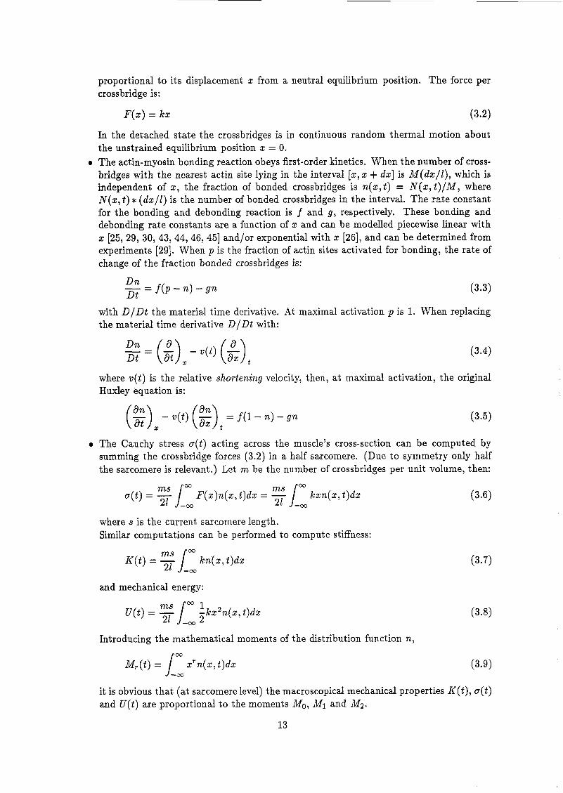
proportional to its displacement z from a neutral equilibrium position. The force per crossbridge is:
F ( z ) = ka: (3.2)
In the detached state the crossbridges is in continuous random thermal motion about the unstrained equilibrium position z = O.
o The actin-myosin bonding reaction obeys first-order kinetics. When the number of cross- bridges with the nearest actin site lying in the interval [a:,a: + dz] is M(dz/Z), which is independent of 2, the fraction of bonded crossbridges is n ( z , t ) = N ( z , t ) / M , where N i z , t ) >i< ( d s / l ) is the nxmbeï of bonded crossbridges in the interval. The rate constaat for the bonding and debonding reaction is f and g , respectively. These bonding and debonding rate constants are a function of a: and can be modelled piecewise Linear with z [25, 29, 30, 43 ,44 ,46 ,45] and/or exponential with z [26], and can be determined from experiments [29]. When p is the fraction of actin sites activated for bonding, the rate of change of the fraction bonded crossbridges is:
with D / D t the material time derivative. At maximal activation p is 1. When replacing the material time derivative D/Dt with:
D n Dt t
(3.4)
where v(t) is the relative shortening velocity, then, at maximal activation, the original Huxley equation is:
(:)z- u ( t ) (a:>, - = f ( i - n ) - g n (3.5)
o The Cauchy stress o(t) acting across the muscle's cross-section can be computed by summing the crossbridge forces (3.2) in a half sarcomere. (Due to symmetry only half the sarcomere is relevant.) Let m be the number of crossbridges per unit volume, then:
F(z )n ( z , t )d z =
where s is the current sarcomere length. Similar computations can be performed to compute stiffness:
and mechanical energy:
ms O 3 1 -k22n(z , t)dz
= 21 J_, 2
Introducing the mathematical moments of the distribution function n, 03
~ , ( t ) = zrn(a:,t)dz J -03
it is obvious that (at sarcomere level) the macroscopical mechanical properties K ( t ) , a(t) and ü(t) are proportional to the moments Mo, Ml and M2.
13

This model is a concise mathematical description of the way the muscle is believed to work by the great majority of biologists. Recent researchs [13, 351 support the hypothesis of the biologists. The model links the mechanics, chemistry, and structure of muscle into an integrated whole. Moreover, the model focusses on the contractile part of a muscle and not the muscle as a whole [45].
Main drawback of the model is its complexity. One searches for a bond distribution which can’t be measured directly. The model even can describe mechanical behaviour, observed in experiments in which the muscle is loaded in a complex way (which is probably ’unnatural’).
3.ï.3 Distributed moments model
In the Distributed Moments (DM) model [26, 25, 30, 43, 45, 461, the objective of the theory is generating a macroscopic model, which has the same simplicity as the Hill model, while retaining the presumed microscopic crossbridge behaviour of the Huxley model.
The DM approximation is introduced in assuming a functional form of the bond distri- bution function n. This is allowed as we are not interested in the details of the distribution function, but in the macroscopic mechanical behaviour, which is proportional t o the math- ematical moments of the distribution function (3.9). In the present DM models a Gaussian form is used for the bond distribution function [43].
where
(3.10)
and a( t ) = - (m) Ml( t ) P O ) = Mo(t) (3.11)
in which case the distribution function n(z , t ) can be expressed as n(Mo(t) , MI( t ) , Mz(t)) . Thus n(2, t ) is characterized by its first three mathematical moments. This means that equation (3.5) can be rewritten as [43] (see appendix A for details):
The main advantage lies in the fact that the bond distribution doesn’t need to be determined, as in the Huxley model, but only the three moments. These mathematical moments are proportional to the mechanical properties stiffness, force and energy.
Simulations with DM models show that it reproduces many features of the complex me- chanical behaviour of the muscle observed in experiments. It even simulates experiments which can not be reproduced with the traditional Hill model [li, 321.
The DM model is a mathematical approximation of the Huxley model. It has the same physical background as the Huxley model. With the right parameters it offers reasonable predictions of most measured behaviours of the muscle, both in shortening and stretch, both of mechanics and energetics. It doesn’t include internal variables like ”active state”. The model can be extended to describe effects, such as: length-dependence [19], electrical stimulation and calcium-activation dynamics [25], as other relevant chemical/metabolical processes [30].
The DM model is more complex then the Hill model, but computationally less expensive than the Huxley model. As most parameters enter the model in a nonlinear manner, estimation
14

of these parameters is difficult. However, most parameters have a physical background, which implies that sometimes they can be estimated independently. For other parameters reasonable estimates have been obtained for only a few muscles, which are documented weli [25].
3.2 Models for the connective tissue and tendons The muscle fibers are held together by connective tissue. Two basic tissues can be distin- guished. The fascia (epimysium) and the interconnecting tissue (or peri- and endomysium). The tendons and the fascia/connective tissue are constructed from the same family of mate- rids? actin, elastin and collagen as solid materia! and ñuid (i.e. water with ail kinds of ions) See also chapter 2.
3.2.1 Construction of constitutive laws
For the construction of constitutive laws, there are two ways.
Phenomenological models
The general approach in phenomenological models is to find a mathematical description of a piece of material, describing as close as possible the observed stress-strain behaviour. For bio-material this is often done by choosing a strain-energy function of the desired form. Strain- energy functions are useful as they are able to describe the stress-strain behaviour for large deformations.
In order to get a description of the three-dimensional behaviour of the material, it is necessary to do a complete three-dimensional measurement. This is very difficult for biological tissues.
Microstuctural models
The basic assumption for microstructural models is that the mechanical response of a tissue is the sum of the responses of its constituents. So, if the structure of the constituents, their mechanics and interactions are known, the overall tissue’s response can be evaluated.
For tendons and connective tissue, the three major components are elastin and collagen fibres and a fluid. The properties of these materials are known (see e.g. [15] and chapter 2). The structure can be defined as the orientation of the fibres in the tissue. Lanir [24] assumes that:
o Each fibre is thin and perfectly flexible. It has no compressive strength: It will buckle
o The effect of the fluid during deformation is that of a hydrostatic pressure. o Each fibre is subjected to a uniaxial strain. o Curved fibres will be stretched before loaded.
under zero load.
The structural approach has several important advantages:
e It is physical in the sense that all material functions are physical quantities. o The resulting model facilitates the understanding of the tissue’s function and provides
o The structural models offer a convenient way to determine and follow up pathological an insight into the tissue’s response to a given deformation.
alterations in the structure and mechanics of various tissue components.
15

o Structural model have a distict advantage in tissue characterisation since the structure is often known or can be investigated accuratly by one of several available morphometric techniques. Mechanical properties of tissue components can be determined independently by isolating them from the tissue.
Disadvantages associate with this approach:
o Computation of the tissue response is expensive. o It is not always possible to measure the response of the constituents. e The interaction ofthe constituents is ofter more complex than can be modelled /measured
/determined (or: lots of assumptions!).
16

Chapter 4
Damage
Biological tissues, like
and AdaDtation A
the muscle, possess the ability to adjust to changes in the activity pattern. Muscle fibres can change the metabolic potetial without changing fibre type [27]. Muscle fibres can change from type, by changing the isoforms of myosin [18]. Muscle fibres can change in length by adding or deleting sarcomeres at the ends of the myofibrils, in order to maintain an optimum working range.
Unaccustomed exercise or over-use of muscles, mostly results in temporary repairable dam- age. Evidence of muscle damage includes morphological changes (structure), delayed-onset muscle soreness, pain, performance decrements and elevation of muscle protein levels in the blood, especially creatine kinase (CK). As muscle damage and adaptation are closely related [9, 22, 37, 341, both will be discussed in this chapter.
Both processes are difficult to understand as the mechanics and the chemics in the muscle interact. Damage can be induced through mechanics and chemics, while small mechanical damage can result in major chemical damage and vice versa. Moreover, damage can initiate adapt at ion.
Key questions for damage and adaptation are: (i) Where does it occur, (2) when does it occur, (3) what is the cause and (4) what is the result.
In the following part, damage as a result of exercise will we discussed and the adaptation process is considered to be caused by damage and/or training.
4.1 Morphological aspects of damage
The morphological aspects of muscle damage can be divided into temporal and spatial aspects. For the present it useful to split the aspects to muscle fibres and connective tissue (spatial). The fibre damage will also be split in time. So, the three aspects discussed here are: direct myofiber damage, delayed myofiber damage and connective tissue or fascial damage [34]. The damage often restricts to small areas (localisation), which seem to be distributed uniformly through the muscle [38].
4.1.1 Direct myofiber damage
Direct myofiber damage can be defined as an alteration in muscle fiber structure or function which can be observed immediately after the exercise has been completed. The structural abnormalities might include sarcomere derangement (broadened and disrupted 2-disks), my- ofibrillar disruption, swollen mitochondria, fragmented or swollen sarcoplasmatic reticular el- ements, dilated T-tubules, and lesions in the plasma membrane. Or: All micro components can be damaged, but according to [37] the structural components in the A-band and 2-line are more susceptible for failure in eccentric exercise experiments.
17

4.1.2 Delayed myofiber damage
Delayed myofiber damage is probably the best documented injury, as the signs of this type of damage start at the end of the exercise and continue for a period of days. Often more damage can be found a few days post-exercise than directly after the exercise [37], which is caused by the process where the injured sites are degraded by the proteolytic and lipolytic systems (i.e. garbage cleaning). At the same time elevated levels of creatine kinase can be found in the blood. As creatine kinase is found almost exclusively in muscle tissue, it is considered an indicator of muscle damage [9]. Swelling of the muscle fibres (hypertrophy) also occurs more pronounced a few days post-exercise.
4.1.3 Connective tissue damage
The connective tissue of skeletal muscle form sheathes around the bundles of muscles fibres and include the endomysium, perimysium and epimysium. The endomysium is perhaps the most important for this discussion, since it forms a sheath around each myofiber and may interconnect adjacent myofibers. The surface of a myofiber is composed of an endomysial fibrous network, a basal lamina, and the sarcolemma (plasma membrane). The endomysium and basal lamina are so intimately related physically that they probably should be considered, from a mechanical viewpoint, as a single elastic structure [34].
As there are distinct differences in the content and composition of the endomysium in different fiber types of animals and man, the damage found in the different fiber types are related [34]. Fast-twitch fibers, who have a less developed endomysium compared to slow- twitch fibers, are believed to be more susceptibel to stretch induced injury [9].
4.2 Causes of muscle damage
Muscle damage can be elicited with short intense eccentric exercise as well as long exhaustive endurance exercise at medium intensity [37, 381. The damage can be cause by mechanical overload and metabolic defects (ischaemia).
4.2.1 Mechanical component
Although damage can occur during concentric and isometric contractions, it is most effectively induced with eccentric contractions (i.e. lengthening of the muscle during activation). When subjected to tension and stretched, different muscle components could break: the connective tissue between adjacent myofibers, the basal lamina, the plasma membrane. When the con- tractile proteins can not relax at the appropriate speed, locally very high tensile stresses can occur E221 or fibres could get a rigor-type state. If this happens very locally (one fibre or one myofibril), shear stresses will develop between the adjacent structures, easily resulting in rupture or myofilament damage [34, 371.
High local temperatures, due to high energy consumption, could also result in damage of the muscle.
4.2.2 Metabolic component
When muscle suffers a temporary oxygen deficit, ATP is generated by anaerobic glycolysis, which results in the production of lactate. This decreases the intracellular pH, which may compromise normal muscle function. Another factor is the combination of glycogen depletion and ischaemia, which may lead to an increased vulnerability of the muscle to encounter damage. Further it has been proved that free radicals can initiate muscle damage.
18

4.3 Adaptation A muscle is able to adapt to the changes in the activity pattern. Due to the complexity of the muscle as a organ, there are several components of the muscle which can alter. According to [9, 10, 14,18, 27,34,40] the following properties must be considered: Fibre size, optimal work- ing range of the sarcomere, number of cross-bridges per cross-sectional area, force development (fast and slow twitch), force-Ca2+ relationship, force-stimulation frequency relationship, maxi- mum shortening velocity, the force-velocity relationship, muscle metabolics, contractile protein prcducticn, elevated Ca2+ and creatine kinase levels and changes in connective tissues. Sevrral of these aspects depend up one another. Some aspects of muscle adaptation are unclear as it is difficult to distinguish muscle repair and actual adaptation.
4.3.1 Morphological aspects
Although myofibres have the ability to be repaired and remain functional after damage, there is no reason to believe that a special injury-resistant muscle fibre is manufactured in response to myofiber damage. Neither would a hypertrophic muscle be developped from a single exercise session. As long as the innervation is intact, a repaired muscle will not be physiological different from its injured precursor[9].
When a muscle is increased in length (constant stretch) sarcomeres seem to be added at the ends of the myofibrils [34] in order to obtain optimal overlap of actin and myosin filaments for the given working range.
Training increases the number and size of mitochondria supplying ATP (energy) to the muscle, which reduces fatigue effects [lo, 271 and muscle fibres can change from type by generating different myosin proteins [Nl. However, rapid adaptation is more likely to be explained by muscle membrane or connective tissue responses than muscle fibre respons.
4.3.2 Adaptation control
According to [9, 18, 311 muscle adaptation is more likely a local than a central response to loading. Stretching of tissue (i.e. tissue strain) and damage seem to be controlling factors in adaptation.
19

Appendix A
Mathematics Q€ the DM model
A.l From Huxley to DM Starting from Huxley's original equation:
and the assumed distribution function:
where
where MT(t) is defined by the definition of the mathematical moments of a distribution func- tion:
o3
M,(t) = Lo3 xTn(x , t )dx
derivation of the DM-model is as follows.
Multiply (-4.1) with x' and integrate from minus infinity to infinity:
with T = O, 1 ,2 , . . .. This gives
The integral of the second term of the left-hand-side needs some attention: co o3
= [xTn]Fo3 - T xT-lndx = -T Lo3 xT-'ndx
This step is allowed, as the limit of x T n at rtco will go to zero because of the choice of the distribution function (A.2). For the first right-hand-side term of (AA) the moments of the bonding rate function will be introduced:
o3
bT = Lo3 x T f ( x ) d x
20

while the second RHS term will be symbolised by:
-03
Substituting (A.8) and (A.9) in (8.6) and limiting r to 0 , l and 2, we get the final form of the DM-model, as used by Zahalak [43]:
- - - bo - F o p 0 7 Ml, J 4 2 ) (A.lO) dM0 dt
d t - - - b2 - FZ(M0, Ml, J 4 2 ) - 2 W J 4 1 (A.12) dM2
d t
- - dM1 - b; - F1(&,iW;,M2) - .(t)Ma (All)
21

Bibliography
T. Allinger and W. Herzog. Nonuniform sarcomere dynamics during shortening and stretch-shortening contractions with whole muscle. In Bouisset et al. [6], pages 84-85. T. Arts, P. Veenstra, and R. Reneman. Epicardial deformation and left ventricular wail mechanics during ejection in the dog. American Journal of Physiology, 243:H379-H3907 1982. R. Baratta, M. Solomonow, R. Best, and R. D’Ambrosia. Force-velocity realtionship models of nine skeletal muscles. In Bouisset et al. [6], pages 140-141. R. Baratta, M. Solomonow, T. Vance, R. Best, and R. D’Ambrosia. Models of length-force relationships in nine skeletal muscles. In Bouisset et al. [6], pages 138-141. J. Bernards and E. Bouman. Fysiologie van de mens. Bohn, Scheltema & Holkema, Emmalaan 27, 3581 HN Utrecht (NL), 5th edition, 1988. S. Bouisset, S. Métral, and H. Monod, editors. International Society of Biornechan- ics X I V h congress Paris, 4-S July 1993, 58500 Clamecy, Paris, 1993. Société de Bioméchanique, l’imprimerie Laballery. P. Bovendeerd. The mechanics of the normal and ischemic left ventricle during the cardiac cycle a numerical and experimental analysis. PhD thesis, Rijksuniversiteit Limburg, 1990. P. Bovendeerd, T. Arts, J. Huyghe, D. van Campen, and R. Reneman. Dependence of local left ventricular wall mechanics on myocardial fiber orientation: a model study. Journal of Biomechanics, 25:1129-1140,1992. C. B. Ebbeling and P. M. Clarkson. Exercise-induced muscle damage and adaptation. Sports Medicine, pages 207-234, 1989. V. R. Edgerton and R. Roy. Regulation of skeletal muscle fiber size, shape and function. Journal of biomechanics, 24 suppl. 1:123-133,1991. G. Ettema and P. Huijing. Frequency response of rat gastrocnemius medialis in small amplitude vibrations. Journal of Biomechanics, 27:1015-1022,1994. G. J. Ettema and P. A. Huijing. Architecture and elastic properties of the series elastic element of muscle-tendon complex, chapter 4, pages 57-68. In Winters and Woo [42], 1990. J. T. Finer, R. M. Simmons, and J. A. Spudich. Single myosin molecule mechanics: piconewton forces and nanometre steps. Nature, 368:113-i19,1994. R. Fitts, E(. McDonald, and J. Schluter. The determinants of skeletal muscle force and power: their adaptability with changes in activity pattern. Journal of Biomechanics,
Y . Fung. Biomechanics, mechanical properties of living tissues. 2nd edition. Springer- Verlag, 1993. W. Ganong. review of Medical Physiology. LANGE Medical Publications, Los Altos, California 94022, 10th edition, 1981. Y. Goldman and B. Brenner. Molecular mechanism of muscle contraction. Annual Review
24: 111-122, 1991.
of Ph~siolûgy, 49:629-636, 1987.
22

[18] G. Goldspink, A. Scutt, P. T. Loughna, D. J. Wells, T. Jaenicke, and G. F. Gerlach. Gene expression in skeletal muscle in response to stretch and force generation. American Journal of Physiology, 262:R356-R363, 1992.
[19] H. Granzier, D. Burns, and G. Pollack. Sarcomere length dependence of the force-velocity relation in single frog muscle fibers. Biophysical Journal, 55:499-507, 1989.
[20] H. Hatze. Myocybernetic control models of skeletal muscle: Characteristics and applica- tions. University of South Africa, Muckleneuk, Pretoria, 1981.
[ai] K. Kaufman, K. An, and E. Chao. Incorporation of muscle architecture into the muscle length-tension relationship. Journal of biomechanics, 22:943-948,1989.
i221 H. Kuipers. Exercise-induced muscle damage. Knternationab Journal o,’ Sports Medicine,
[23] B. Landjerit , editor. Ivh international symposium on computer simulation in biomechan-
[24] Y. Lanir. Constitutive equations for fibrous connective tissues. Journal of Biomechanics,
[25] S. Ma and G. I. Zahalak. A distibution-moment model of energetics in skeletal muscle. Journal of biomechanics, Vol. 24:21-35, 1991.
[26] S.-P. Ma and G. I. Zahalak. A simple self-consistent distribution-moment model for muscle: chemical energy and heat rates. Mathematical Biosciences, 84:211-230, 1987.
[27] K. McCully, H. Kakihira, K. Vandenborne, and i. Kent-Braun. Nonivasive measurements of activity-induced changes in muscle metabolism. Journal of biomechanics, 24:153-161, 1991.
[28] D. Morgan. Modeling of lengthening muscle: the role of inter-sarcomere dynamics, chap- ter 3, pages 46-56. In Winters and Woo [42], 1990.
[29] R. Podolsky, A. Nolan, and S. Zaveler. Cross-bridge properties derived from muscle isotonic velocity transients. Proc. Nat. Acad. Sei. U.S.A., 64:504-511, 1969.
[30] E. Rouhaud and G. Zahalak. Interaction between contraction and metabolism in skeletal muscle: a distribution-moment model. In Landjerit [23], pages BMTC 18-21.
[31] J.-I. Sadochima and S. Izumo. Mechanotransduction in stretch-induced hypertrophy of cardio myocytes. Journal of receptor research, 13:777-794,1993.
[32] C. Schaap. Evaluatie van het distributed momentsmodel voor spiercontractie. stager- apport wfw 94.154. (in dutch). Technical report, University of Technology Eindhoven, 1994.
[33] L. Skubiszak. Mechanism of muscle contraction. Technology and health care, 1:133-142, 1993.
[34] W. T. Stauber. Eccentric action of muscles: Physiology, injury and adaptation. Ezercize and Sportsscience review, pages 157-185, 1989.
[35] K. Svoboda, C. F. Schmidt, B. J. Schnapp, and S. M. Block. Direct observation of kinesin stepping by optical trapping interferometry. Nature, 365: 721-727,1993.
[36] H. ter Keurs, W. Rijnsburger, R. van Heuningen, and M. Nagelsmit. Tension development and sarcomere length in rat cardia trabeculae evidence of length-dependent activation. Circulation research, 46:703-714,1980.
[37] J. van der Meulen. Execise-induced muscle damage: morphological, biochemical and func- tional aspects. PhD thesis, Rijksuniversiteit Limburg (NL), 1991.
[38] J. van der Meulen, H. Kuipers, J. van der Wal, and J. Drukker. Quatitative and spatial aspects of degenerative changes in rat soleus muscle after exercise of different durations. Journal of Anatomy, pages 349-353,1993.
15~132-135, 1994.
ics., Paris, 1993.
16~1-12, 1983.
23

[39] J. van Leeuwen and C. Spoor. Modelling mechanically stable muscle architectures. Phil. Trans. R. Soc. Lond. B, 336:275-292,1992.
[40] H. Vandenburgh, S. Hatfaludy, P. Karlisch, and J. Shansky. Mechanically induced alter- ations in cultured skeletal muscle growth. Journal of biomechanics, 24 suppl. 1:91-99, 1991.
[41] J. M. Winters. Hill-based muscle models: a system engineering perspective, chapter 5 , pages 69-93. In Winters and Woo [42], 1990.
[42] J. M. Winters and S. L.-Y. Woo, editors. Multiple muscle systems, biomechanics and movement organization. Springer-Verlag, New York, 1990.
[43] G. I. Zahalak. A dlstribution-moment approximation for kinetic theories of muscular contraction. Mathematical Biosciences, 55239-114, 1981.
[44] G. I. Zahalak. A comparison of the mechanical behavior of the cat soleus muscle with a distribution-moment model. Journal of biomechanica2 engeneering, 108:131-140, 1986.
[45] G. I. Zahalak. Modeling muscle mechanics (and energetics), chapter 1, pages 1-23. In Winters and Woo [42], 1990.
[46] G. I. Zahalak and S.-P. Ma. Muscle activation and contraction: constitutive relations based directly on cross-bridge kinetics. Journal of biomechanical engineering, 112:52-62, 1990.
24