Listeria monocytogenes Multidrug Resistance Transporters...
12
Listeria monocytogenes Multidrug Resistance Transporters and Cyclic Di-AMP, Which Contribute to Type I Interferon Induction, Play a Role in Cell Wall Stress Millie Kaplan Zeevi, a Nirit S. Shafir, a Shira Shaham, a Sivan Friedman, a Nadejda Sigal, a Ran Nir Paz, b Ivo G. Boneca, c,d Anat A. Herskovits a Department of Molecular Microbiology and Biotechnology, The George S. Wise Life Sciences Faculty, Tel Aviv University, Tel Aviv, Israel a ; Hadassah-Hebrew University Medical Center, Jerusalem, Israel b ; Institut Pasteur, Group Biology and Genetics of the Bacterial Cell Wall, Paris, France c ; INSERM, Group Avenir, France d The intracellular bacterial pathogen Listeria monocytogenes activates a robust type I interferon response upon infection. This response is partially dependent on the multidrug resistance (MDR) transporter MdrM and relies on cyclic-di-AMP (c-di-AMP) secretion, yet the functions of MdrM and cyclic-di-AMP that lead to this response are unknown. Here we report that it is not MdrM alone but a cohort of MDR transporters that together contribute to type I interferon induction during infection. In a search for a physiological function of these transporters, we revealed that they play a role in cell wall stress responses. A mutant with deletion of four transporter genes (mdrMTAC) was found to be sensitive to sublethal concentrations of vancomycin due to an inability to produce and shed peptidoglycan under this stress. Remarkably, c-di-AMP is involved in this phenotype, as overexpression of the c-di-AMP phosphodiesterase (PdeA) resulted in increased susceptibility of the mdrMTAC mutant to van- comycin, whereas overexpression of the c-di-AMP diadenylate cyclase (DacA) reduced susceptibility to this drug. These observa- tions suggest a physiological association between c-di-AMP and the MDR transporters and support the model that MDR trans- porters mediate c-di-AMP secretion to regulate peptidoglycan synthesis in response to cell wall stress. L isteria monocytogenes is a Gram-positive, facultative, intracel- lular pathogen that invades a wide range of mammalian cells (1). Following internalization, the bacteria escape to the cell cyto- sol by secreting several virulence factors, primarily the pore-form- ing hemolysin, listeriolysin O (LLO). Once in the cytosol, L. monocytogenes replicates and spreads from cell to cell by recruiting host actin filaments (1). During infection, L. monocytogenes trig- gers a robust type I interferon response, as manifested by en- hanced expression and secretion of the cytokine beta interferon (IFN-)(2). This response was shown to be independent of Toll- like receptors but reliant on several innate immune signaling mol- ecules (i.e., STING, TBK-1, and IRF3) (3–6). Remarkably, the bacteria must be replicating in the macrophages’ cytosol to elicit a type I interferon response, as phagosomally trapped bacteria, such as an LLO-negative mutant, do not induce this response (2). A previous study aimed at identifying L. monocytogenes determi- nants involved in IFN- activation identified multidrug resistance (MDR) transporters as modulators of the type I interferon re- sponse in vivo (7). Specifically, overexpression in bacteria of two closely related MDR transporters, MdrM and MdrT, was found to trigger enhanced induction of IFN- by infected macrophages. However, only deletion of the mdrM gene resulted in reduced levels of IFN- secreted by infected macrophages (7). This obser- vation indicated that MdrM plays an active role during bacterial cytosolic growth that leads to induction of the type I interferon response. MdrM and MdrT belong to the major facilitator superfamily (MFS) of MDR transporters and are closely related to the well- characterized MDR transporter, QacA, of Staphylococcus aureus (8). MDR transporters, such as QacA, are notorious for their abil- ity to confer resistance to a wide variety of toxic compounds and drugs, including antibiotics, by utilizing proton motive force to actively extrude these compounds outside the cell (9). Accord- ingly, MdrM and MdrT were shown to be transcriptionally in- duced upon bacterial exposure to rhodamine 6G (R6G) and tet- raphenylphosphonium (TPP), both well-known substrates of MDRs, and to confer resistance to cholic acid (7, 10). Neverthe- less, none of these classical MDR functions could explain the ob- served role of these proteins in activating the innate immune sys- tem, implying they might possess distinct physiological roles during infection. It was recently proposed that MdrM and MdrT transporters extrude cyclic-di-AMP (c-di-AMP) during L. monocytogenes in- tracellular growth, which in turn activates infected macrophages to elicit the IFN- response (11, 12). Indeed, c-di-AMP activates a robust type I interferon response when added exogenously; how- ever, a physiological association between c-di-AMP and the MDR transporters was not established. Notably, several reports had in- dicated that c-di-AMP serves as a second messenger molecule that influences central cellular processes of bacteria: e.g., genome sur- veillance, response to cell wall stresses, and, more recently, pepti- doglycan homeostasis (13–17). In bacteria, c-di-AMP is synthe- sized by diadenylate cyclase (DAC) using ATP as a substrate and, conversely, linearized to 5=-pApA by a specific c-di-AMP phos- phodiesterase (PDE) (15). While it was shown that the level of c-di-AMP is largely dependent on the expression levels of DAC Received 9 July 2013 Accepted 13 September 2013 Published ahead of print 20 September 2013 Address correspondence to Anat A. Herskovits, [email protected]. Supplemental material for this article may be found at http://dx.doi.org/10.1128 /JB.00794-13. Copyright © 2013, American Society for Microbiology. All Rights Reserved. doi:10.1128/JB.00794-13 5250 jb.asm.org Journal of Bacteriology p. 5250 –5261 December 2013 Volume 195 Number 23 on July 7, 2018 by guest http://jb.asm.org/ Downloaded from
Transcript of Listeria monocytogenes Multidrug Resistance Transporters...
Listeria monocytogenes Multidrug Resistance Transporters and CyclicDi-AMP, Which Contribute to Type I Interferon Induction, Play aRole in Cell Wall Stress
Millie Kaplan Zeevi,a Nirit S. Shafir,a Shira Shaham,a Sivan Friedman,a Nadejda Sigal,a Ran Nir Paz,b Ivo G. Boneca,c,d
Anat A. Herskovitsa
Department of Molecular Microbiology and Biotechnology, The George S. Wise Life Sciences Faculty, Tel Aviv University, Tel Aviv, Israela; Hadassah-Hebrew UniversityMedical Center, Jerusalem, Israelb; Institut Pasteur, Group Biology and Genetics of the Bacterial Cell Wall, Paris, Francec; INSERM, Group Avenir, Franced
The intracellular bacterial pathogen Listeria monocytogenes activates a robust type I interferon response upon infection. Thisresponse is partially dependent on the multidrug resistance (MDR) transporter MdrM and relies on cyclic-di-AMP (c-di-AMP)secretion, yet the functions of MdrM and cyclic-di-AMP that lead to this response are unknown. Here we report that it is notMdrM alone but a cohort of MDR transporters that together contribute to type I interferon induction during infection. In asearch for a physiological function of these transporters, we revealed that they play a role in cell wall stress responses. A mutantwith deletion of four transporter genes (�mdrMTAC) was found to be sensitive to sublethal concentrations of vancomycin dueto an inability to produce and shed peptidoglycan under this stress. Remarkably, c-di-AMP is involved in this phenotype, asoverexpression of the c-di-AMP phosphodiesterase (PdeA) resulted in increased susceptibility of the �mdrMTAC mutant to van-comycin, whereas overexpression of the c-di-AMP diadenylate cyclase (DacA) reduced susceptibility to this drug. These observa-tions suggest a physiological association between c-di-AMP and the MDR transporters and support the model that MDR trans-porters mediate c-di-AMP secretion to regulate peptidoglycan synthesis in response to cell wall stress.
Listeria monocytogenes is a Gram-positive, facultative, intracel-lular pathogen that invades a wide range of mammalian cells
(1). Following internalization, the bacteria escape to the cell cyto-sol by secreting several virulence factors, primarily the pore-form-ing hemolysin, listeriolysin O (LLO). Once in the cytosol, L.monocytogenes replicates and spreads from cell to cell by recruitinghost actin filaments (1). During infection, L. monocytogenes trig-gers a robust type I interferon response, as manifested by en-hanced expression and secretion of the cytokine beta interferon(IFN-�) (2). This response was shown to be independent of Toll-like receptors but reliant on several innate immune signaling mol-ecules (i.e., STING, TBK-1, and IRF3) (3–6). Remarkably, thebacteria must be replicating in the macrophages’ cytosol to elicit atype I interferon response, as phagosomally trapped bacteria, suchas an LLO-negative mutant, do not induce this response (2). Aprevious study aimed at identifying L. monocytogenes determi-nants involved in IFN-� activation identified multidrug resistance(MDR) transporters as modulators of the type I interferon re-sponse in vivo (7). Specifically, overexpression in bacteria of twoclosely related MDR transporters, MdrM and MdrT, was found totrigger enhanced induction of IFN-� by infected macrophages.However, only deletion of the mdrM gene resulted in reducedlevels of IFN-� secreted by infected macrophages (7). This obser-vation indicated that MdrM plays an active role during bacterialcytosolic growth that leads to induction of the type I interferonresponse.
MdrM and MdrT belong to the major facilitator superfamily(MFS) of MDR transporters and are closely related to the well-characterized MDR transporter, QacA, of Staphylococcus aureus(8). MDR transporters, such as QacA, are notorious for their abil-ity to confer resistance to a wide variety of toxic compounds anddrugs, including antibiotics, by utilizing proton motive force toactively extrude these compounds outside the cell (9). Accord-
ingly, MdrM and MdrT were shown to be transcriptionally in-duced upon bacterial exposure to rhodamine 6G (R6G) and tet-raphenylphosphonium (TPP), both well-known substrates ofMDRs, and to confer resistance to cholic acid (7, 10). Neverthe-less, none of these classical MDR functions could explain the ob-served role of these proteins in activating the innate immune sys-tem, implying they might possess distinct physiological rolesduring infection.
It was recently proposed that MdrM and MdrT transportersextrude cyclic-di-AMP (c-di-AMP) during L. monocytogenes in-tracellular growth, which in turn activates infected macrophagesto elicit the IFN-� response (11, 12). Indeed, c-di-AMP activates arobust type I interferon response when added exogenously; how-ever, a physiological association between c-di-AMP and the MDRtransporters was not established. Notably, several reports had in-dicated that c-di-AMP serves as a second messenger molecule thatinfluences central cellular processes of bacteria: e.g., genome sur-veillance, response to cell wall stresses, and, more recently, pepti-doglycan homeostasis (13–17). In bacteria, c-di-AMP is synthe-sized by diadenylate cyclase (DAC) using ATP as a substrate and,conversely, linearized to 5=-pApA by a specific c-di-AMP phos-phodiesterase (PDE) (15). While it was shown that the level ofc-di-AMP is largely dependent on the expression levels of DAC
Received 9 July 2013 Accepted 13 September 2013
Published ahead of print 20 September 2013
Address correspondence to Anat A. Herskovits, [email protected].
Supplemental material for this article may be found at http://dx.doi.org/10.1128/JB.00794-13.
Copyright © 2013, American Society for Microbiology. All Rights Reserved.
doi:10.1128/JB.00794-13
5250 jb.asm.org Journal of Bacteriology p. 5250 –5261 December 2013 Volume 195 Number 23
on July 7, 2018 by guesthttp://jb.asm
.org/D
ownloaded from
and PDE enzymes (15, 18), the mechanism coordinating the ac-tivity of these enzymes is not known. The prevalence of DACdomains among bacteria and archaea strengthens the premise thatthis c-di-AMP is fundamentally involved in microbial physiology(13). The L. monocytogenes genome contains both c-di-AMP dacand pde genes (dacA [lmo2120] and pdeA [lmo0052]). The dacAgene was shown to be essential for growth and to be the generesponsible for c-di-AMP production, while pdeA was shown todegrade c-di-AMP (11, 18).
In the present study, we aimed to identify a physiological asso-ciation between L. monocytogenes MDR transporters and c-di-AMP. We discovered that MDR transporters play a role in the L.monocytogenes response to cell wall stress and found that thisMDR function was linked to c-di-AMP production. More gener-ally, this report furthers the understanding of the molecularmechanism whereby intracellular L. monocytogenes cells triggertype I interferon responses during infection.
MATERIALS AND METHODSBacterial strains, cells, growth media, and reagents. L. monocytogenesstrain 10403S was used as the wild-type (WT) strain and as the parentalstrain for all mutants generated in this work (Table 1). The Escherichia coliXL-1 Blue (Stratagene) and DH12 strains were used for vector propaga-tion. E. coli strain SM-10 (19) was used for conjugative plasmid delivery toL. monocytogenes. L. monocytogenes strains were grown in brain heart in-fusion (BHI) (BD) medium or minimal medium (20) at 37°C, and E. colistrains were grown in Luria-Bertani (LB) medium (BD) at 37°C. For in-fection experiments, L. monocytogenes bacteria were grown overnight inBHI at 30°C without agitation. IPTG (isopropyl-�-D-1-thiogalactopyra-noside) was purchased from Bio-Lab, Ltd. (Israel), penicillin G, vanco-mycin hydrochloride, and mutanolysin were purchased from Sigma, andc-di-AMP and c-di-GMP were purchased from the Biolog Institute (Ger-many). Primary bone marrow-derived (BMD) macrophages were isolatedfrom 6- to 8-week-old female C57BL/6 mice (Harlan Laboratories, Ltd.,Israel) and cultured as described previously (21). RAW264 macrophageswere grown and maintained in Dulbecco’s modified Eagle’s medium(DMEM)-based media.
Generation of L. monocytogenes in-frame deletion mutants. Dele-tion mutants were generated by standard techniques using the pKSV7oriTvector, as described in reference 7. Plasmid pLIV2-mdrM was used forgeneration of 6�His-tagged MdrM and the F58V mutant (for primers,see Table S1C in the supplemental material).
Protein analysis by Western blotting. Overnight cultures were di-luted 1:100 and grown to an optical density at 600 nm (OD600) of 1 U andthen supplemented with 0.25 mM IPTG when indicated. Bacteria wereharvested, treated with 50 U of mutanolysin for 1 h, and sonicated in a
mixture of 20 mM Tris-HCl (pH 8), 0.5 M NaCl, 1 mM EDTA, and 1 mMphenylmethylsulfonyl fluoride (PMSF). After removal of cell debris,membranes were collected by ultracentrifugation. Membrane fractions(50 �g of protein) were then subjected to 12.5% SDS-PAGE and blottedfor His tag detection using India HisProbe-horseradish peroxidase (HRP)(Pierce) and the ECL enhanced chemiluminescence reagent.
Bacterial growth curves. Overnight cultures were adjusted to anOD600 of 0.03 in 20 ml fresh BHI broth, supplemented when indicatedwith 0.25 mM IPTG, 0.08 �g ml�1 penicillin G, 1 �g ml�1 vancomycin, or3 �g ml�1 c-di-AMP. For bacterial RNA extraction, bacteria were grownat 37°C to an OD600 of 0.4 and then supplemented with 3 �g ml�1 linco-mycin, 50 �M rhodamine 6 G (R6G), 1 �g ml�1 vancomycin, and 0.08 �gml�1 penicillin G for 2 h or centrifuged and resuspended in: minimalmedium (pH 5) (lactic acid), minimal medium with 10 mM H2O2, ordefined minimal medium (pH 5) with 10 mM H2O2 for 30 min. Formicroscopy, bacteria were grown similarly and supplemented with 1 �gml�1 vancomycin for 2 h. Growth curves in the presence of drugs wereperformed in a Synergy HT Biotek plate reader at 37°C with continuousshaking and monitoring of the OD600 every 15 min for 24 h. Of note,bacterial growth in the plate reader is different from that in flasks withrespect to the OD levels that are measured. In each experiment, growthconditions are indicated.
L. monocytogenes intracellular growth in cells. Intracellular growthcurves were performed as described previously (22). Briefly, 2 � 106 cellswere seeded on a petri dish with glass coverslips and infected with 8 � 106
bacteria. At 0.5 h postinfection (p.i.), cells were washed, and at 1 h p.i.,gentamicin was added. At each time point, cells from 3 coverslips werelysed and CFU were counted. For bacterial gene expression of intracellu-larly grown L. monocytogenes cells, 25 � 106 BMD macrophages wereinfected with 1 � 108 bacteria and lysed in 20 ml of ice-cold water at 6 hp.i., and the released bacteria were collected on 0.45-�m-pore hydroxy-apatite (HA) filters (Millipore, catalog no. HAWP04700).
Gene expression analysis. RNA was purified from bacteria in mid-log-phase growth in BHI or from infected cells using standard phenol-chloroform extraction methods. RNA from intracellularly grown bacteriawas amplified using the MessageAmp II (Ambion) bacterial RNA ampli-fication kit according to the manufacturer’s instructions. RNA of infectedmacrophages was extracted using TRIzol reagent according to standardprotocols. In all cases, 1 �g of RNA was reverse transcribed to cDNA usingan Applied Biosystems high-capacity reverse transcription kit. Real-timequantitative PCR (RT-qPCR) was performed on 10 ng of cDNA usingSYBR green with the Step-One Plus RT-PCR system (Applied Biosys-tems). The transcription of bacterial genes was normalized using 16SrRNA or the rpoB gene, and that of macrophage cytokines was normalizedusing the glyceraldehyde 3-phosphate dehydrogenase gene (gpdh). Statis-tical analysis was performed using the StepOne V2.1 software. Error barsrepresent 95% confidence intervals; in a case where the error bars of twosamples do not overlap, the P value is ��0.01. Primer sequences are de-scribed in Table S1A and B in the supplemental material. The completeintracellular expression profile of L. monocytogenes 10403S was publishedseparately (23).
�-Galactosidase MUG assay for mdrC transcription. Overnight cul-tures of WT L. monocytogenes pPL2-PmdrClacZ and �mdrMTA pPL2-PmdrClacZ cells were adjusted to an OD600 of 0.05. Cultures were grown in96-well black plates (200 �l) with a clear bottom to an OD600 of �0.4 at37°C. Next, the plates were centrifuged for 10 min at 3,800 rpm, superna-tants were aspirated, and the cells were washed twice with phosphate-buffered saline (PBS). Two hundred microliters of ABT buffer (60 mMK2HPO4, 40 mM KH2PO4, 100 mM NaCl, 0.1% Triton X-100 [pH 7]),supplemented with 80 �g ml�1 of MUG substrate (4-methylumbelliferyl-�-D-galactopyranoside) (Sigma), was added to each well. Plates wereshaken for 30 s and incubated at room temperature for 1 h in the dark.Following incubation, the optical density (600 nm) and the fluorescenceintensity (excitation, 360 nm; emission, 460 nm) were measured using aSynergy HT Biotek plate reader. �-Galactosidase activity was normalized
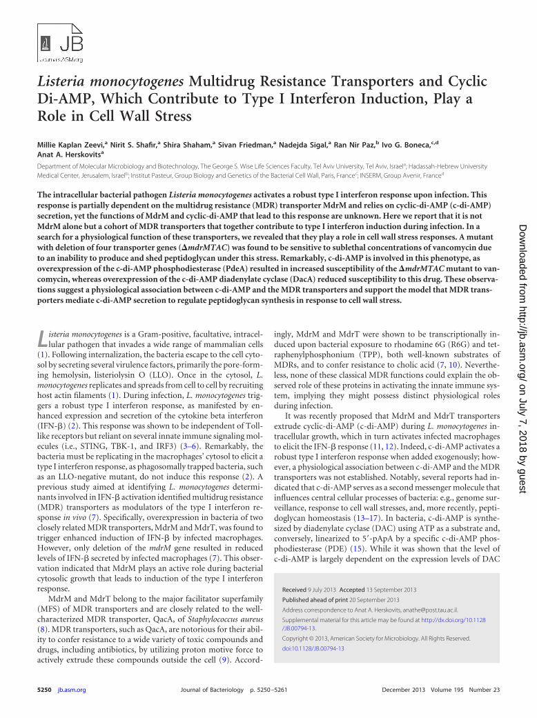
TABLE 1 Genes similar to mdrM in L. monocytogenes strain 10403Sbased on protein sequence
L. monocytogenes10403S gene no.
L. monocytogenesEGDe geneidentifier no.
% identity, %similarity ofamino acidsequence
Intracellularinductiona
Genename
LMRG_02976.6 lmo1617 Yes mdrMLMRG_02679.6 lmo2588 45, 65 Yes mdrTLMRG_00200.6 lmo0519 35, 60 Yes mdrALMRG_02080.6 lmo0981 25, 46 NoLMRG_01853.6 lmo2845 23, 44 Yes mdrBLMRG_01880.6 lmo2818 22, 41 Yes mdrCLMRG_02296.6 lmo0872 20, 38 Yes mdrDLMRG_01872.6 lmo2826 16, 33 Yes mdrEa Based on microarray analysis (23).
MDRs and Cyclic Di-AMP Regulate Cell Wall Stress
December 2013 Volume 195 Number 23 jb.asm.org 5251
on July 7, 2018 by guesthttp://jb.asm
.org/D
ownloaded from
to the sample’s OD (24, 25). The experiment was performed in triplicateand was repeated three times independently.
TEM. Bacteria were grown as described above with and without van-comycin treatment. For negative staining, PBS-washed bacteria were ad-sorbed on Formvar carbon-coated grids and stained with 2% aqueousuranyl acetate. For transmission electron microscopy (TEM) sections, abacterial pellet from 20 ml of culture was fixed in 2.5% glutaraldehyde inPBS at 4°C for 20 h, washed three times with PBS, and postfixed in 1%OsO4 in PBS at 4°C for 2 h. Dehydration was carried out in graded ethanoland embedding in glycid ether. Thin sections were mounted on Formvarcarbon-coated grids and stained with uranyl acetate and lead citrate. Allimages were acquired using a Jeol 1200 EX transmission electron micro-scope (Jeol, Japan). Measurements of cell wall thickness were performedfrom three independent biological repeats; a total of 35 frames were takenfor each strain and condition.
Mouse infection. L. monocytogenes bacteria were grown in BHI me-dium at 30°C overnight. Six- to 8-week-old C57BL/6 female mice (HarlanLaboratories, Ltd., Israel) were infected via tail vein injections with 4 �104 washed bacteria (5 mice in each group). Spleens and livers were har-vested 72 h p.i. and homogenized in 0.2% saponin, and bacterial CFUwere determined by plating. The experiment was repeated twice.
Measurement of peptidoglycan synthesis rate. Overnight bacterialculture was diluted 1:100 into 10 ml of BHI, grown to an OD600 of 0.4, andsupplemented with 20 �M N-acetylglucosamine and 10 �l of 1 �Ci �l�1
of 14C-N-acetylglucosamine (American Radiolabeled Chemicals). Theculture then was divided into two parts, and 0.8 �g ml�1 of vancomycinwas added to one of them. One-hundred-microliter aliquots from cul-tures incubated without agitation at 37°C were withdrawn in triplicateevery 30 min and added to 100 �l of boiling 8% SDS, and the mixture wasincubated for 5 min at 95°C. Cell wall was collected on 0.45-�m-pore-sizemembrane filters (Millipore, catalog no. HAWP02500), washed with 15ml of water, and counted using 5 ml of EcoLite() liquid scintillationcocktail with a PerkinElmer TriCarb 3110TR �-counter.
Peptidoglycan extraction and muropeptide analysis. Cell wall andpeptidoglycan were purified as described previously (26). Muropeptideswere generated from highly purified cell wall and peptidoglycan samplesby mutanolysin and then reduced using sodium borohydride. Muropep-tide separation was performed by high-performance liquid chromatogra-phy (HPLC) as previously described for L. monocytogenes (27, 28). Foractivation of cytokines by cell wall samples, lyophilized cell wall extractswere resuspended at a concentration of 1.5 mg ml�1, and then the pH wasadjusted to 7.5 with NaOH, and 20 �l was added to 2 � 106 BMD mac-rophages in 2.5 ml medium. After 6 h, macrophage RNA was harvestedand analyzed for cytokine induction.
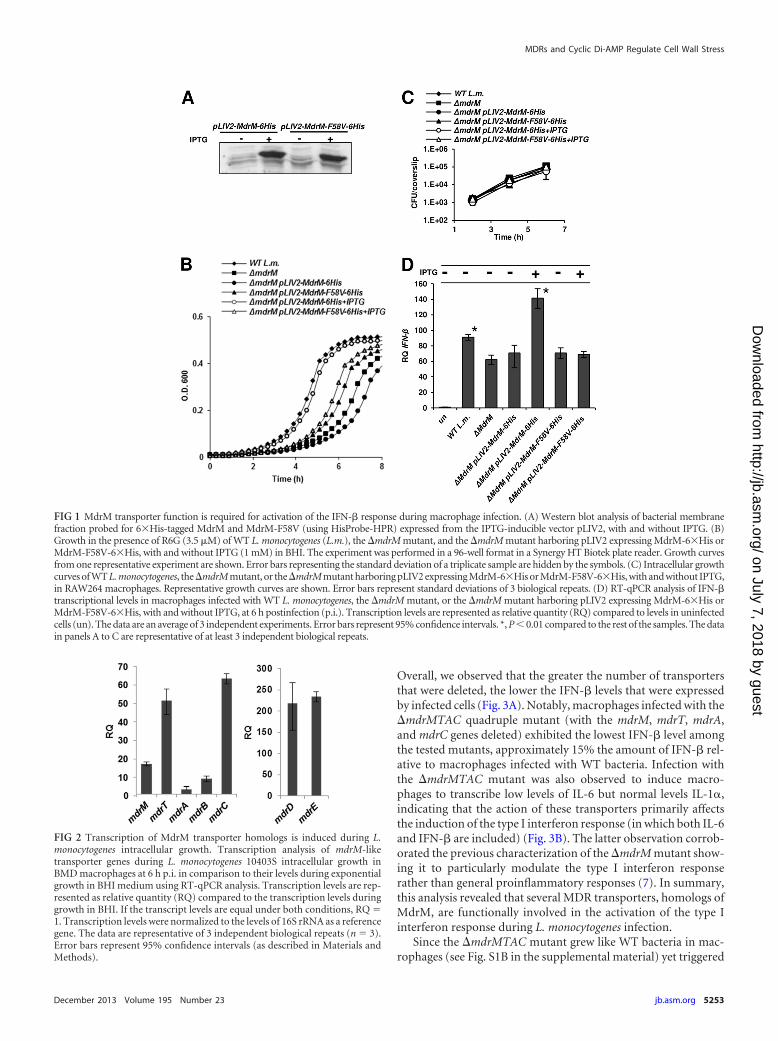
RESULTSA functional MdrM transporter is required to trigger macro-phages to elicit the IFN-� response. As mentioned, L. monocyto-genes bacteria overexpressing the MdrM transporter have beenshown to trigger infected macrophages to express enhancedIFN-� levels (7). Initially, we wanted to validate that this enhance-ment of the IFN-� response requires MdrM to be functional. Tothis end, an mdrM mutant was generated that harbored a muta-tion that inactivates function but preserves expression. In the S.aureus QacA transporter, substitution for tyrosine at position 63with valine resulted in a nonfunctional transporter and enhancedsensitivity of the bacteria to a wide variety of drugs (29). There-fore, using site-directed mutagenesis, the corresponding phenyl-alanine in MdrM at position 58, F58, was substituted with valine.The resulting mdrM-F58V gene construct was tagged with histi-dine at the 3= end and cloned into the integrative pLIV2 vectorunder an IPTG-inducible promoter to generate pLIV2-mdrM-F58V-6His. This plasmid or a control plasmid containing the His-tagged native mdrM gene (pLIV2-mdrM-6His) was conjugated to
a �mdrM mutant, and the expression levels of the native andmutated MdrM were compared by Western blotting. Indeed, boththe native MdrM and MdrM-F58V proteins were expressed andfound in the membrane fraction at similar levels upon IPTG ad-dition (Fig. 1A). Next, the ability of the MdrM and MdrM-F58Vproteins to confer resistance to R6G was tested. As expected, the�mdrM mutant was more sensitive to R6G than the wild-type(WT) bacteria were, and introduction of the native mdrM gene(via pLIV2-mdrM-6His with IPTG induction) rescued the sensi-tivity. However, introduction of MdrM-F58V did not restore fullgrowth, providing support that the F58V mutation does interferewith MdrM’s transport function (Fig. 1B). Next, the capacity ofthis mutant to enhance the IFN-� response was tested. To thisend, macrophages were infected with the �mdrM mutant harbor-ing the pLIV2 plasmid expressing the native or the mutatedMdrM. As shown in Fig. 1C, all strains grew to a similar extentintracellularly (Fig. 1C). Notably, only bacteria overexpressing thenative MdrM induced an enhanced IFN-� response, while bacte-ria overexpressing the mutated MdrM did not (Fig. 1D). Theseresults indicate that MdrM’s function is required for induction ofan IFN-� response during infection.
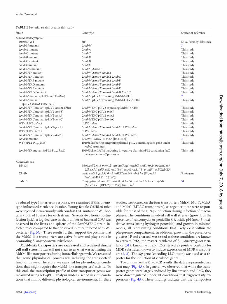
MdrM transporter and several MDR homologs are transcrip-tionally induced during intracellular growth. MDR transportersare known to exhibit functional redundancy due to overlappingsubstrate specificity (9, 30). Since MdrM was shown to be respon-sible for a third of the IFN-� induction by infected macrophages(7), we examined if additional transporters are involved in medi-ation of the IFN-� response. A search of the L. monocytogenesstrain 10403S genome for mdrM homologs revealed several genesencoding putative MDR transporters, among them the previouslyidentified gene mdrT (Table 1). On this list, the LMRG_00200.6gene (an ortholog of lmo0519 in EGD-e), named here mdrA, washighly similar to mdrM and mdrT, with 60% similarity and 45%identity in protein sequence, whereas the other protein genes ex-hibited only 33 to 44% sequence similarity (Table 1). To gaininsight into the potential requirement for these transporters dur-ing L. monocytogenes infection, we analyzed their transcriptionlevels during intracellular growth in macrophages. Our previoustranscriptomic data from intracellularly grown bacteria indicatedthat all of these transporters are induced during infection of mac-rophages, except for LMRG_02080.6 (using microarray analysis[23]) (Table 1). We thus further compared the transcription levelsof the induced MDR transporters by real-time quantitative PCR(RT-qPCR) analysis. As shown in Fig. 2, all of the transporterswere transcriptionally upregulated during intracellular growth inmacrophages, at least 4-fold, over their levels in BHI. These resultssuggest that each of the transporters might play an active roleduring L. monocytogenes infection.
A set of MdrM-like transporters control most of the type Iinterferon response to L. monocytogenes infection and viru-lence. To examine whether MdrM homologs contribute to IFN-�induction during infection, we generated a series of in-frame de-letion mutants harboring single or multiple (double, triple, qua-druple, and quintuple) MDR gene deletions (Table 2). All of theMDR mutants grew similarly to WT bacteria both in BHI brothand intracellularly in macrophages, except for the mdrMTAD mu-tant, which exhibited a moderate intracellular growth defect (seeFig. S1A and B in the supplemental material). The IFN-� responseelicited by macrophages after infection with each of the mutantswas evaluated using RT-qPCR analysis of IFN-� transcript levels.
Kaplan Zeevi et al.
5252 jb.asm.org Journal of Bacteriology
on July 7, 2018 by guesthttp://jb.asm
.org/D
ownloaded from
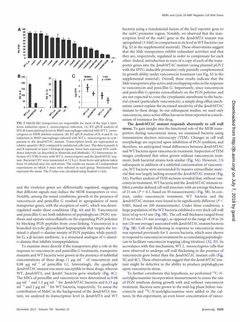
Overall, we observed that the greater the number of transportersthat were deleted, the lower the IFN-� levels that were expressedby infected cells (Fig. 3A). Notably, macrophages infected with the�mdrMTAC quadruple mutant (with the mdrM, mdrT, mdrA,and mdrC genes deleted) exhibited the lowest IFN-� level amongthe tested mutants, approximately 15% the amount of IFN-� rel-ative to macrophages infected with WT bacteria. Infection withthe �mdrMTAC mutant was also observed to induce macro-phages to transcribe low levels of IL-6 but normal levels IL-1,indicating that the action of these transporters primarily affectsthe induction of the type I interferon response (in which both IL-6and IFN-� are included) (Fig. 3B). The latter observation corrob-orated the previous characterization of the �mdrM mutant show-ing it to particularly modulate the type I interferon responserather than general proinflammatory responses (7). In summary,this analysis revealed that several MDR transporters, homologs ofMdrM, are functionally involved in the activation of the type Iinterferon response during L. monocytogenes infection.
Since the �mdrMTAC mutant grew like WT bacteria in mac-rophages (see Fig. S1B in the supplemental material) yet triggered
FIG 2 Transcription of MdrM transporter homologs is induced during L.monocytogenes intracellular growth. Transcription analysis of mdrM-liketransporter genes during L. monocytogenes 10403S intracellular growth inBMD macrophages at 6 h p.i. in comparison to their levels during exponentialgrowth in BHI medium using RT-qPCR analysis. Transcription levels are rep-resented as relative quantity (RQ) compared to the transcription levels duringgrowth in BHI. If the transcript levels are equal under both conditions, RQ �1. Transcription levels were normalized to the levels of 16S rRNA as a referencegene. The data are representative of 3 independent biological repeats (n � 3).Error bars represent 95% confidence intervals (as described in Materials andMethods).
FIG 1 MdrM transporter function is required for activation of the IFN-� response during macrophage infection. (A) Western blot analysis of bacterial membranefraction probed for 6�His-tagged MdrM and MdrM-F58V (using HisProbe-HPR) expressed from the IPTG-inducible vector pLIV2, with and without IPTG. (B)Growth in the presence of R6G (3.5 �M) of WT L. monocytogenes (L.m.), the �mdrM mutant, and the �mdrM mutant harboring pLIV2 expressing MdrM-6�His orMdrM-F58V-6�His, with and without IPTG (1 mM) in BHI. The experiment was performed in a 96-well format in a Synergy HT Biotek plate reader. Growth curvesfrom one representative experiment are shown. Error bars representing the standard deviation of a triplicate sample are hidden by the symbols. (C) Intracellular growthcurves of WT L. monocytogenes, the �mdrM mutant, or the �mdrM mutant harboring pLIV2 expressing MdrM-6�His or MdrM-F58V-6�His, with and without IPTG,in RAW264 macrophages. Representative growth curves are shown. Error bars represent standard deviations of 3 biological repeats. (D) RT-qPCR analysis of IFN-�transcriptional levels in macrophages infected with WT L. monocytogenes, the �mdrM mutant, or the �mdrM mutant harboring pLIV2 expressing MdrM-6�His orMdrM-F58V-6�His, with and without IPTG, at 6 h postinfection (p.i.). Transcription levels are represented as relative quantity (RQ) compared to levels in uninfectedcells (un). The data are an average of 3 independent experiments. Error bars represent 95% confidence intervals. *, P � 0.01 compared to the rest of the samples. The datain panels A to C are representative of at least 3 independent biological repeats.
MDRs and Cyclic Di-AMP Regulate Cell Wall Stress
December 2013 Volume 195 Number 23 jb.asm.org 5253
on July 7, 2018 by guesthttp://jb.asm
.org/D
ownloaded from
a reduced type I interferon response, we examined if this pheno-type influenced virulence in mice. Young female C57BL/6 micewere injected intravenously with �mdrMTAC mutant or WT bac-teria (total of 10 mice for each strain). Seventy-two hours postin-fection (p.i.), a log decrease in the number of bacterial CFU wasobserved in the livers and spleens of the �mdrMTAC strain-in-fected mice compared to that observed in mice infected with WTbacteria (Fig. 3C). These results further support the premise thatthe MdrM-like transporters are active in vivo and play a role inpromoting L. monocytogenes virulence.
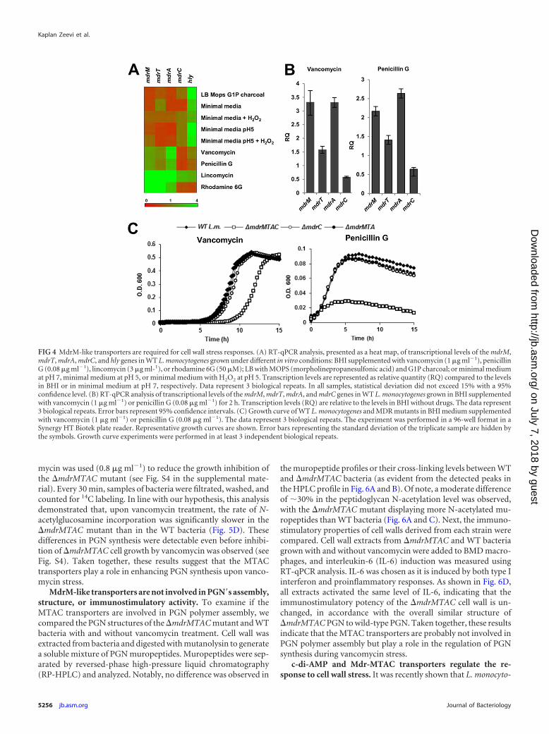
MdrM-like transporters are expressed and required duringcell wall stress. It was still not clear to us what was activating theMdrM-like transporters during intracellular growth. We reasonedthat some physiological process was inducing the transporters’function in vivo. Therefore, we searched for physiological condi-tions that might require the MdrM-like transporters’ activity. Tothis end, the transcription profile of four transporter genes wasmeasured using RT-qPCR analysis under a set of in vitro condi-tions that mimic different physiological environments. In these
studies, we focused on the four transporters MdrM, MdrT, MdrA,and MdrC (MTAC transporters), as together these were respon-sible for most of the IFN-� induction during infection of macro-phages. The conditions involved cell wall stresses (growth in thepresence of vancomycin or penicillin G), acidic pH (near 5), oxi-dative stress (using hydrogen peroxide), and growth in minimalmedia, all representing conditions that likely exist within thephagosome compartment. In addition, growth in the presence ofglucose-1P and charcoal was tested as these conditions are knownto activate PrfA, the master regulator of L. monocytogenes viru-lence (31). Lincomycin and R6G served as positive controls forMDR substrates known to induce expression of MDR transport-ers (7, 8). The hly gene (encoding LLO toxin) was used as a re-porter for the induction of virulence genes.
To summarize the RT-qPCR results, the data are presented as aheat map (Fig. 4A). In general, we observed that while the trans-porter genes were largely induced by lincomycin and R6G, theywere downregulated under all conditions that triggered hly ex-pression (Fig. 4A). These findings indicate that the transporters
TABLE 2 Bacterial strains used in this study
Strain Genotype Source or reference
Listeria monocytogenes10403S (WT) Strr D. A. Portnoy, lab stock�mdrM mutant �mdrM 7�mdrA mutant �mdrA This study�mdrC mutant �mdrC This study�mdrB mutant �mdrB This study�mdrD mutant �mdrD This study�mdrE mutant �mdrE This study�mdrMC mutant �mdrM �mdrC This study�mdrMTA mutant �mdrM �mdrT �mdrA This study�mdrMTAC mutant �mdrM �mdrT �mdrA �mdrC This study�mdrMTAB mutant �mdrM �mdrT �mdrA �mdrB This study�mdrMTAD mutant �mdrM �mdrT �mdrA �mdrD This study�mdrMTAE mutant �mdrM �mdrT �mdrA �mdrE This study�mdrMTABC mutant �mdrM �mdrT �mdrA �mdrB �mdrC This study�mdrM mutant (pLIV2-mdrM-6His) �mdrM pLIV2 expressing MdrM-6�His 7�mdrM mutant
(pLIV2-mdrM-F58V-6His)�mdrM pLIV2 expressing MdrM-F58V-6�His This study
�mdrMTAC mutant (pLIV2-mdrM-6His) �mdrMTAC pLIV2 expressing MdrM-6�His This study�mdrMTAC mutant (pLIV2-mdrT) �mdrMTAC pLIV2-mdrT This study�mdrMTAC mutant (pLIV2-mdrA) �mdrMTAC pLIV2-mdrA This study�mdrMTAC mutant (pLIV2-mdrC) �mdrMTAC pLIV2-mdrC This studyWT (pLIV2-pdeA) pLIV2-pdeA This study�mdrMTAC mutant (pLIV2-pdeA) �mdrM �mdrT �mdrA �mdrC pLIV2-pdeA This studyWT (pLIV2-dacA) pLIV2-dacA This study�mdrMTAC mutant (pLIV2-dacA) �mdrM �mdrT �mdrA �mdrC pLIV2-dacA This study�marR mutant �marR (LMRG_01348.6 [lmo1618]) 7WT (pPL2-PmdrClacZ) 10403S harboring integrative plasmid pPL2 containing lacZ gene under
mdrC promoterThis study
�mdrMTA mutant (pPL2-PmdrClacZ) 10403S �mdrMTA harboring integrative plasmid pPL2 containing lacZgene under mdrC promoter
This study
Escherichia coliDH12s �80dlacZ�M15 mcrA �(mrr-hsdRMS-mcrBC) araD139 �(ara leu)7697
�(lacX74) galU galK rpsL (Strr) nupG recA1/[F= proAB lacIqZ�M15]XL-1b recA1 endA1 gyrA96 thi-1 hsdR17 supE44 relA1 lac [F= proAB
lacIqZ�M15 Tn10 (Tetr)]Stratagene
SM-10 Conjugation donor; F� thi-1 thr-1 leuB6 recA tonA21 lacY1 supE44(Muc) � [RP4-2(Tc::Mu)] Kmr Tra
54
Kaplan Zeevi et al.
5254 jb.asm.org Journal of Bacteriology
on July 7, 2018 by guesthttp://jb.asm
.org/D
ownloaded from
and the virulence genes are differentially regulated, suggestingthat different signals may induce the MDR transporters in vivo.Notably, among the tested conditions, growth in the presence ofvancomycin and penicillin G resulted in upregulation of mosttransporter genes, with the exception of mdrC, which was down-regulated under these conditions (Fig. 4A and B). Vancomycinand penicillin G are both inhibitors of peptidoglycan (PGN) syn-thesis and operate extracellularly on the expanding PGN polymerby blocking PGN peptides from cross-linking. Vancomycin is abranched tricyclic glycosylated heptapeptide that targets the ter-minal D-alanyl-D-alanine moiety of PGN peptides, while penicil-lin G, a �-lactam antibiotic, is a structural analogue of D-alanyl-D-alanine that inhibits transpeptidation.
To examine more directly if the transporters play a role in theresponse to vancomycin and penicillin G treatments, transportermutants and WT bacteria were grown in the presence of sublethalconcentrations of these drugs (1 �g ml�1 of vancomycin and0.08 �g ml�1 of penicillin G). Interestingly, the quadruple�mdrMTAC mutant was more susceptible to these drugs, whereasWT, �mdrMTA, and �mdrC bacteria grew similarly (Fig. 4C).The MICs of penicillin and vancomycin were determined as 0.08�g ml�1 and 1.5 �g ml�1 for �mdrMTAC bacteria and 0.15 �gml�1 and 2 �g ml�1 for WT bacteria, respectively. To assess thecontribution of MdrC in the background of the �mdrMTA mu-tant, we analyzed its transcription level in �mdrMTA and WT
bacteria using a translational fusion of the lacZ reporter gene tothe mdrC promoter region. Notably, we observed that the tran-scription level of the mdrC gene in the �mdrMTA mutant wasupregulated (3-fold) in comparison to its level in WT bacteria (seeFig. S2 in the supplemental material). These observations suggestthat the Mdr transporters exhibit redundant activities and thatthey are, respectively, regulated in order to compensate for eachother. Indeed, introduction in trans of a copy of each of the trans-porter genes into the �mdrMTAC mutant (using plasmid pLIV2with the IPTG-inducible promoter) only partially complementedits growth ability under vancomycin treatment (see Fig. S3 in thesupplemental material). Overall, these results indicate that theMdr transporters play active and overlapping roles in the responseto vancomycin and penicillin G. Importantly, since vancomycinand penicillin G operate extracellularly on the PGN polymer andare not expected to cross the cytoplasmic membrane to the bacte-rial cytosol (particularly vancomycin), a simple drug efflux mech-anism cannot explain the increased sensitivity of the �mdrMTACmutant to these drugs. In our subsequent studies, we used onlyvancomycin, since active efflux has never been reported as a mech-anism of resistance for this drug.
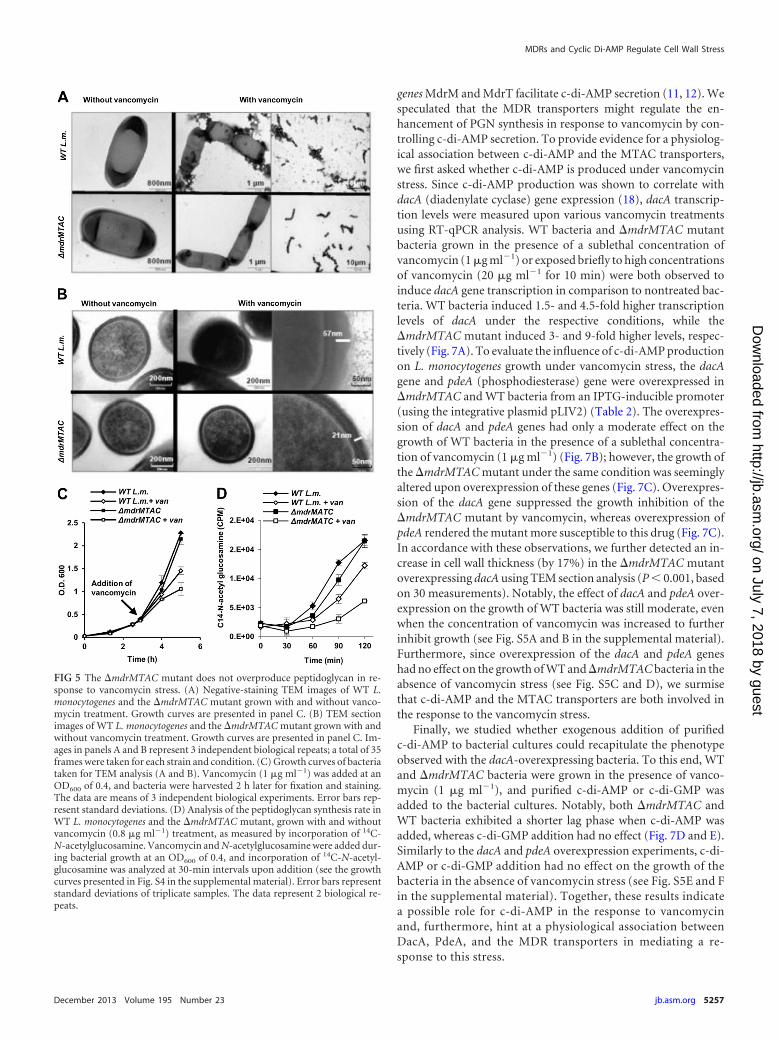
The �mdrMTAC mutant responds aberrantly to cell wallstress. To gain insight into the functional role of the MDR trans-porters during vancomycin stress, we examined bacteria usingtransmission electron microscopy (TEM). Changes in cell wallmorphology are expected upon inhibition of PGN synthesis, andtherefore, we anticipated visual differences between �mdrMTACand WT bacteria upon vancomycin treatment. Inspection of TEMimages confirmed that when grown without vancomycin treat-ment, both bacterial strains look similar (Fig. 5A). However, 2 hsubsequent to addition of a sublethal concentration of vancomy-cin, WT bacteria were surrounded by massive extracellular mate-rial that was largely lacking around the �mdrMTAC mutant (Fig.5A). Further analysis of TEM sections revealed that, without van-comycin treatment, WT bacteria and the �mdrMTAC mutant ex-hibit a similar defined cell wall structure with an average thicknessof 21 nm (P � 0.1, based on 50 measurements) (Fig. 5B). In con-trast, under vancomycin treatment, WT bacteria and the�mdrMTAC mutant were found to be significantly different (P �0.001, based on 100 measurements). Under these conditions, alarge population of the WT bacteria exhibited a very thick cell walllayer of up to 63 nm (Fig. 5B). The cell wall thickness ranged from25 to 63 nm (35-nm average), as opposed to the range of 18 to 26nm (24-nm average) associated with �mdrMTAC mutant bacteria(Fig. 5B). Cell wall thickening in response to vancomycin stresswas reported previously for S. aureus bacteria, which were shownto respond to vancomycin treatment by accumulating peptidogly-can to facilitate vancomycin trapping (drug titration) (32, 33). Inaccordance with this mechanism, WT L. monocytogenes cells thatwere observed to undergo cell wall thickening in the presence ofvancomycin grew better than the �mdrMTAC mutant cells (Fig.5C and 4C). These observations suggest that the �mdrMTAC mu-tant might be defective in the ability to produce peptidoglycanupon vancomycin stress.
To further corroborate this hypothesis, we performed 14C-N-acetylglucosamine incorporation measurements to assess the rateof PGN synthesis during growth with and without vancomycintreatment. Bacteria were grown to the mid-log phase before van-comycin and 14C-N-acetylglucosamine were added to the cul-tures. In this experiment, an even lower concentration of vanco-
FIG 3 MdrM-like transporters are responsible for most of the type I inter-feron induction upon L. monocytogenes infection. (A) RT-qPCR analysis ofIFN-� transcriptional levels in BMD macrophages infected with WT L. mono-cytogenes or MDR deletion mutants. (B) RT-qPCR analysis of IL-6 and IL-1induction in BMD macrophages infected with WT L. monocytogenes in com-parison to the �mdrMTAC mutant. Transcription levels are represented asrelative quantity (RQ) compared to uninfected cells (un). The data in panels Aand B represent at least 3 biological repeats. Error bars represent 95% confi-dence intervals (as described in Materials and Methods). (C) Intravenous in-fection of C57BL/6 mice with WT L. monocytogenes and the �mdrMTAC mu-tant. Bacterial CFU were numerated at 72 h p.i. from livers and spleens takenfrom 10 infected mice for each strain. The results are means of 2 independentexperiments in which 5 mice were infected in each group. Horizontal barsrepresent the mean. The P value was calculated using Student’s t test.
MDRs and Cyclic Di-AMP Regulate Cell Wall Stress
December 2013 Volume 195 Number 23 jb.asm.org 5255
on July 7, 2018 by guesthttp://jb.asm
.org/D
ownloaded from
mycin was used (0.8 �g ml�1) to reduce the growth inhibition ofthe �mdrMTAC mutant (see Fig. S4 in the supplemental mate-rial). Every 30 min, samples of bacteria were filtrated, washed, andcounted for 14C labeling. In line with our hypothesis, this analysisdemonstrated that, upon vancomycin treatment, the rate of N-acetylglucosamine incorporation was significantly slower in the�mdrMTAC mutant than in the WT bacteria (Fig. 5D). Thesedifferences in PGN synthesis were detectable even before inhibi-tion of �mdrMTAC cell growth by vancomycin was observed (seeFig. S4). Taken together, these results suggest that the MTACtransporters play a role in enhancing PGN synthesis upon vanco-mycin stress.
MdrM-like transporters are not involved in PGN=s assembly,structure, or immunostimulatory activity. To examine if theMTAC transporters are involved in PGN polymer assembly, wecompared the PGN structures of the �mdrMTAC mutant and WTbacteria with and without vancomycin treatment. Cell wall wasextracted from bacteria and digested with mutanolysin to generatea soluble mixture of PGN muropeptides. Muropeptides were sep-arated by reversed-phase high-pressure liquid chromatography(RP-HPLC) and analyzed. Notably, no difference was observed in
the muropeptide profiles or their cross-linking levels between WTand �mdrMTAC bacteria (as evident from the detected peaks inthe HPLC profile in Fig. 6A and B). Of note, a moderate differenceof �30% in the peptidoglycan N-acetylation level was observed,with the �mdrMTAC mutant displaying more N-acetylated mu-ropeptides than WT bacteria (Fig. 6A and C). Next, the immuno-stimulatory properties of cell walls derived from each strain werecompared. Cell wall extracts from �mdrMTAC and WT bacteriagrown with and without vancomycin were added to BMD macro-phages, and interleukin-6 (IL-6) induction was measured usingRT-qPCR analysis. IL-6 was chosen as it is induced by both type Iinterferon and proinflammatory responses. As shown in Fig. 6D,all extracts activated the same level of IL-6, indicating that theimmunostimulatory potency of the �mdrMTAC cell wall is un-changed, in accordance with the overall similar structure of�mdrMTAC PGN to wild-type PGN. Taken together, these resultsindicate that the MTAC transporters are probably not involved inPGN polymer assembly but play a role in the regulation of PGNsynthesis during vancomycin stress.
c-di-AMP and Mdr-MTAC transporters regulate the re-sponse to cell wall stress. It was recently shown that L. monocyto-
FIG 4 MdrM-like transporters are required for cell wall stress responses. (A) RT-qPCR analysis, presented as a heat map, of transcriptional levels of the mdrM,mdrT, mdrA, mdrC, and hly genes in WT L. monocytogenes grown under different in vitro conditions: BHI supplemented with vancomycin (1 �g ml�1), penicillinG (0.08 �g ml�1), lincomycin (3 �g ml-1), or rhodamine 6G (50 �M); LB with MOPS (morpholinepropanesulfonic acid) and G1P charcoal; or minimal mediumat pH 7, minimal medium at pH 5, or minimal medium with H2O2 at pH 5. Transcription levels are represented as relative quantity (RQ) compared to the levelsin BHI or in minimal medium at pH 7, respectively. Data represent 3 biological repeats. In all samples, statistical deviation did not exceed 15% with a 95%confidence level. (B) RT-qPCR analysis of transcriptional levels of the mdrM, mdrT, mdrA, and mdrC genes in WT L. monocytogenes grown in BHI supplementedwith vancomycin (1 �g ml�1) or penicillin G (0.08 �g ml�1) for 2 h. Transcription levels (RQ) are relative to the levels in BHI without drugs. The data represent3 biological repeats. Error bars represent 95% confidence intervals. (C) Growth curve of WT L. monocytogenes and MDR mutants in BHI medium supplementedwith vancomycin (1 �g ml�1) or penicillin G (0.08 �g ml�1). The data represent 3 biological repeats. The experiment was performed in a 96-well format in aSynergy HT Biotek plate reader. Representative growth curves are shown. Error bars representing the standard deviation of the triplicate sample are hidden bythe symbols. Growth curve experiments were performed in at least 3 independent biological repeats.
Kaplan Zeevi et al.
5256 jb.asm.org Journal of Bacteriology
on July 7, 2018 by guesthttp://jb.asm
.org/D
ownloaded from
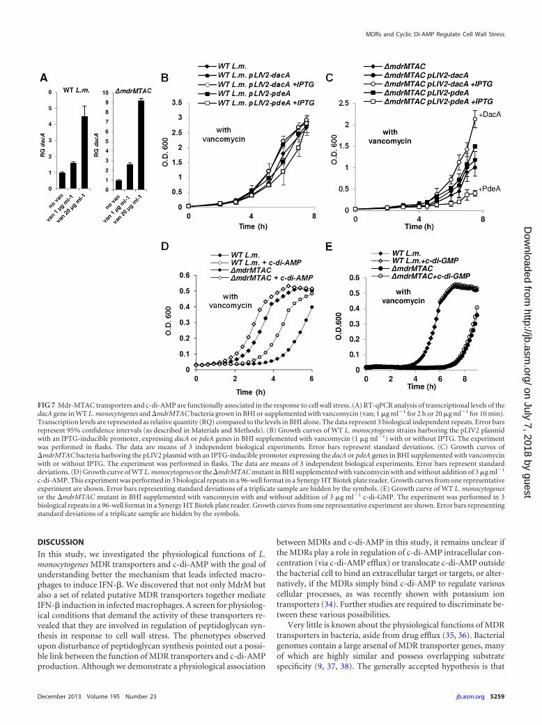
genes MdrM and MdrT facilitate c-di-AMP secretion (11, 12). Wespeculated that the MDR transporters might regulate the en-hancement of PGN synthesis in response to vancomycin by con-trolling c-di-AMP secretion. To provide evidence for a physiolog-ical association between c-di-AMP and the MTAC transporters,we first asked whether c-di-AMP is produced under vancomycinstress. Since c-di-AMP production was shown to correlate withdacA (diadenylate cyclase) gene expression (18), dacA transcrip-tion levels were measured upon various vancomycin treatmentsusing RT-qPCR analysis. WT bacteria and �mdrMTAC mutantbacteria grown in the presence of a sublethal concentration ofvancomycin (1 �g ml�1) or exposed briefly to high concentrationsof vancomycin (20 �g ml�1 for 10 min) were both observed toinduce dacA gene transcription in comparison to nontreated bac-teria. WT bacteria induced 1.5- and 4.5-fold higher transcriptionlevels of dacA under the respective conditions, while the�mdrMTAC mutant induced 3- and 9-fold higher levels, respec-tively (Fig. 7A). To evaluate the influence of c-di-AMP productionon L. monocytogenes growth under vancomycin stress, the dacAgene and pdeA (phosphodiesterase) gene were overexpressed in�mdrMTAC and WT bacteria from an IPTG-inducible promoter(using the integrative plasmid pLIV2) (Table 2). The overexpres-sion of dacA and pdeA genes had only a moderate effect on thegrowth of WT bacteria in the presence of a sublethal concentra-tion of vancomycin (1 �g ml�1) (Fig. 7B); however, the growth ofthe �mdrMTAC mutant under the same condition was seeminglyaltered upon overexpression of these genes (Fig. 7C). Overexpres-sion of the dacA gene suppressed the growth inhibition of the�mdrMTAC mutant by vancomycin, whereas overexpression ofpdeA rendered the mutant more susceptible to this drug (Fig. 7C).In accordance with these observations, we further detected an in-crease in cell wall thickness (by 17%) in the �mdrMTAC mutantoverexpressing dacA using TEM section analysis (P � 0.001, basedon 30 measurements). Notably, the effect of dacA and pdeA over-expression on the growth of WT bacteria was still moderate, evenwhen the concentration of vancomycin was increased to furtherinhibit growth (see Fig. S5A and B in the supplemental material).Furthermore, since overexpression of the dacA and pdeA geneshad no effect on the growth of WT and �mdrMTAC bacteria in theabsence of vancomycin stress (see Fig. S5C and D), we surmisethat c-di-AMP and the MTAC transporters are both involved inthe response to the vancomycin stress.
Finally, we studied whether exogenous addition of purifiedc-di-AMP to bacterial cultures could recapitulate the phenotypeobserved with the dacA-overexpressing bacteria. To this end, WTand �mdrMTAC bacteria were grown in the presence of vanco-mycin (1 �g ml�1), and purified c-di-AMP or c-di-GMP wasadded to the bacterial cultures. Notably, both �mdrMTAC andWT bacteria exhibited a shorter lag phase when c-di-AMP wasadded, whereas c-di-GMP addition had no effect (Fig. 7D and E).Similarly to the dacA and pdeA overexpression experiments, c-di-AMP or c-di-GMP addition had no effect on the growth of thebacteria in the absence of vancomycin stress (see Fig. S5E and Fin the supplemental material). Together, these results indicatea possible role for c-di-AMP in the response to vancomycinand, furthermore, hint at a physiological association betweenDacA, PdeA, and the MDR transporters in mediating a re-sponse to this stress.
FIG 5 The �mdrMTAC mutant does not overproduce peptidoglycan in re-sponse to vancomycin stress. (A) Negative-staining TEM images of WT L.monocytogenes and the �mdrMTAC mutant grown with and without vanco-mycin treatment. Growth curves are presented in panel C. (B) TEM sectionimages of WT L. monocytogenes and the �mdrMTAC mutant grown with andwithout vancomycin treatment. Growth curves are presented in panel C. Im-ages in panels A and B represent 3 independent biological repeats; a total of 35frames were taken for each strain and condition. (C) Growth curves of bacteriataken for TEM analysis (A and B). Vancomycin (1 �g ml�1) was added at anOD600 of 0.4, and bacteria were harvested 2 h later for fixation and staining.The data are means of 3 independent biological experiments. Error bars rep-resent standard deviations. (D) Analysis of the peptidoglycan synthesis rate inWT L. monocytogenes and the �mdrMTAC mutant, grown with and withoutvancomycin (0.8 �g ml�1) treatment, as measured by incorporation of 14C-N-acetylglucosamine. Vancomycin and N-acetylglucosamine were added dur-ing bacterial growth at an OD600 of 0.4, and incorporation of 14C-N-acetyl-glucosamine was analyzed at 30-min intervals upon addition (see the growthcurves presented in Fig. S4 in the supplemental material). Error bars representstandard deviations of triplicate samples. The data represent 2 biological re-peats.
MDRs and Cyclic Di-AMP Regulate Cell Wall Stress
December 2013 Volume 195 Number 23 jb.asm.org 5257
on July 7, 2018 by guesthttp://jb.asm
.org/D
ownloaded from
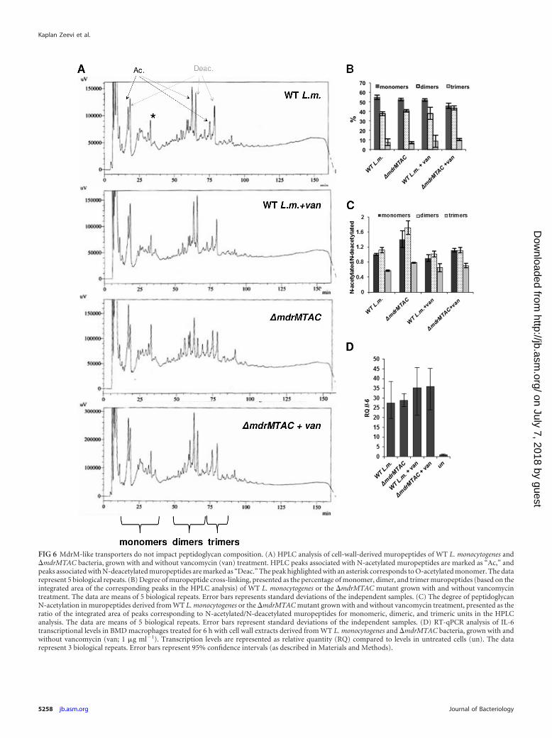
FIG 6 MdrM-like transporters do not impact peptidoglycan composition. (A) HPLC analysis of cell-wall-derived muropeptides of WT L. monocytogenes and�mdrMTAC bacteria, grown with and without vancomycin (van) treatment. HPLC peaks associated with N-acetylated muropeptides are marked as “Ac,” andpeaks associated with N-deacetylated muropeptides are marked as “Deac.” The peak highlighted with an asterisk corresponds to O-acetylated monomer. The datarepresent 5 biological repeats. (B) Degree of muropeptide cross-linking, presented as the percentage of monomer, dimer, and trimer muropeptides (based on theintegrated area of the corresponding peaks in the HPLC analysis) of WT L. monocytogenes or the �mdrMTAC mutant grown with and without vancomycintreatment. The data are means of 5 biological repeats. Error bars represents standard deviations of the independent samples. (C) The degree of peptidoglycanN-acetylation in muropeptides derived from WT L. monocytogenes or the �mdrMTAC mutant grown with and without vancomycin treatment, presented as theratio of the integrated area of peaks corresponding to N-acetylated/N-deacetylated muropeptides for monomeric, dimeric, and trimeric units in the HPLCanalysis. The data are means of 5 biological repeats. Error bars represent standard deviations of the independent samples. (D) RT-qPCR analysis of IL-6transcriptional levels in BMD macrophages treated for 6 h with cell wall extracts derived from WT L. monocytogenes and �mdrMTAC bacteria, grown with andwithout vancomycin (van; 1 �g ml�1). Transcription levels are represented as relative quantity (RQ) compared to levels in untreated cells (un). The datarepresent 3 biological repeats. Error bars represent 95% confidence intervals (as described in Materials and Methods).
Kaplan Zeevi et al.
5258 jb.asm.org Journal of Bacteriology
on July 7, 2018 by guesthttp://jb.asm
.org/D
ownloaded from
DISCUSSION
In this study, we investigated the physiological functions of L.monocytogenes MDR transporters and c-di-AMP with the goal ofunderstanding better the mechanism that leads infected macro-phages to induce IFN-�. We discovered that not only MdrM butalso a set of related putative MDR transporters together mediateIFN-� induction in infected macrophages. A screen for physiolog-ical conditions that demand the activity of these transporters re-vealed that they are involved in regulation of peptidoglycan syn-thesis in response to cell wall stress. The phenotypes observedupon disturbance of peptidoglycan synthesis pointed out a possi-ble link between the function of MDR transporters and c-di-AMPproduction. Although we demonstrate a physiological association
between MDRs and c-di-AMP in this study, it remains unclear ifthe MDRs play a role in regulation of c-di-AMP intracellular con-centration (via c-di-AMP efflux) or translocate c-di-AMP outsidethe bacterial cell to bind an extracellular target or targets, or alter-natively, if the MDRs simply bind c-di-AMP to regulate variouscellular processes, as was recently shown with potassium iontransporters (34). Further studies are required to discriminate be-tween these various possibilities.
Very little is known about the physiological functions of MDRtransporters in bacteria, aside from drug efflux (35, 36). Bacterialgenomes contain a large arsenal of MDR transporter genes, manyof which are highly similar and possess overlapping substratespecificity (9, 37, 38). The generally accepted hypothesis is that
FIG 7 Mdr-MTAC transporters and c-di-AMP are functionally associated in the response to cell wall stress. (A) RT-qPCR analysis of transcriptional levels of thedacA gene in WT L. monocytogenes and �mdrMTAC bacteria grown in BHI or supplemented with vancomycin (van; 1 �g ml�1 for 2 h or 20 �g ml�1 for 10 min).Transcription levels are represented as relative quantity (RQ) compared to the levels in BHI alone. The data represent 3 biological independent repeats. Error barsrepresent 95% confidence intervals (as described in Materials and Methods). (B) Growth curves of WT L. monocytogenes strains harboring the pLIV2 plasmidwith an IPTG-inducible promoter, expressing dacA or pdeA genes in BHI supplemented with vancomycin (1 �g ml�1) with or without IPTG. The experimentwas performed in flasks. The data are means of 3 independent biological experiments. Error bars represent standard deviations. (C) Growth curves of�mdrMTAC bacteria harboring the pLIV2 plasmid with an IPTG-inducible promoter expressing the dacA or pdeA genes in BHI supplemented with vancomycinwith or without IPTG. The experiment was performed in flasks. The data are means of 3 independent biological experiments. Error bars represent standarddeviations. (D) Growth curve of WT L. monocytogenes or the �mdrMTAC mutant in BHI supplemented with vancomycin with and without addition of 3 �g ml�1
c-di-AMP. This experiment was performed in 3 biological repeats in a 96-well format in a Synergy HT Biotek plate reader. Growth curves from one representativeexperiment are shown. Error bars representing standard deviations of a triplicate sample are hidden by the symbols. (E) Growth curve of WT L. monocytogenesor the �mdrMTAC mutant in BHI supplemented with vancomycin with and without addition of 3 �g ml�1 c-di-GMP. The experiment was performed in 3biological repeats in a 96-well format in a Synergy HT Biotek plate reader. Growth curves from one representative experiment are shown. Error bars representingstandard deviations of a triplicate sample are hidden by the symbols.
MDRs and Cyclic Di-AMP Regulate Cell Wall Stress
December 2013 Volume 195 Number 23 jb.asm.org 5259
on July 7, 2018 by guesthttp://jb.asm
.org/D
ownloaded from
MDR transporters evolved independently during evolution tocope with a wide array of physiological substrates, allowing bacte-ria to survive diverse ecological niches (39). Studies in the lastdecade have revealed diverse functions of MDR transporters thatare not related to drug efflux (36, 40). For example, involvementof MDR transporters in lipid transport, pH homeostasis (41), vir-ulence (42, 43), and quorum sensing (44) has been documented,with fatty acids, ions, bile salts, antibacterial peptides, and precur-sors of quorum-sensing molecules suggested as natural substrates(45–51). Despite the advance in our understanding of MDR trans-porters, in silico prediction of MDR transporter functions remainschallenging. The initial discovery that the L. monocytogenes MdrMtransporter modulates the type I interferon response raised manyquestions regarding the mechanism of this function. Primarily, itwas not clear whether this function evolved specifically to subvertthe host immune system or, alternatively, represents the inadver-tent consequence of a more basic bacterial physiological function.Later the report that c-di-AMP is secreted by MdrM and leads toIFN-� induction (11) further highlighted the need to better un-derstand the natural biological process that involves MdrM andc-di-AMP. Here we show that four homologous L. monocytogenesMDR transporters not only are triggering IFN-� induction duringinfection but also are novel players in the response to cell wallstress. Remarkably, the �mdrMTAC mutant lacking the fourtransporters failed to trigger enhanced production of PGN in re-sponse to vancomycin stress, a mechanism known to facilitatevancomycin resistance (32). In light of the observation that thegrowth defect of the �mdrMTAC mutant under vancomycinstress was not accompanied by production of aberrant PGN, wepropose that the MDR transporters play a regulatory role in PGNsynthesis rather than a direct role in PGN biogenesis. The findingsthat increased production of c-di-AMP (via dacA overexpression)or addition of purified c-di-AMP rescued the growth inhibition ofthe �mdrMTAC mutant under vancomycin stress support thatc-di-AMP is involved downstream of the transporters’ function.However, the possibility that c-di-AMP affects the observed phe-notypes indirectly cannot be excluded (14, 34, 52, 53).
Cyclic di-AMP has been recently demonstrated to play a role inthe response to cell wall stresses in both S. aureus and Bacillus subtilis.Two independent genetic screens highlighted c-di-AMP as a key reg-ulator of lipoteichoic acid (LTA)- and PGN-related stress. In S. au-reus, a genetic screen designed to identify suppressor mutations thatrestore the ability of LTA-deficient mutants to grow identified muta-tions in pde gene and revealed a global role for c-di-AMP in cell wallregulation (15). In B. subtilis, a search for genes that facilitate intrinsicresistance to �-lactam antibiotics also identified a pde gene and c-di-AMP as key players (16). Notably, increased production of c-di-AMPresulted in enhanced resistance of B. subtilis bacteria to �-lactams(16), a phenotype that is similar to our observation that overexpres-sion of the dacA gene suppressed the growth defect of the�mdrMTAC mutant under vancomycin stress. Interestingly, a con-nection between c-di-AMP, MDRs, and cell wall stress was pointedout previously in a study that examined global genomic changes in a�-lactam-resistant methicillin-resistant S. aureus (MRSA) strain. Inthis study, mutations were identified in three genes encoding a peni-cillin binding protein, PDE, and an MDR transporter (54). While itwas not clear to the authors how these genes associate, we now sur-mise that c-di-AMP and MDR transporters function together to reg-ulate cell wall synthesis. Notably, this transporter’s function might bepart of an intrinsic mechanism of bacteria to overcome cell wall stress.
Such a function is most likely required during infection of mamma-lian cells, where bacteria are subjected to various host antimicrobialmechanisms targeting the bacterial cell wall. In this regard, the in vivovirulence defect of the �mdrMTAC mutant may be linked to thetransporters’ function in resistance to cell wall stress; however, sincethe transporter’s function poses additional effects on the innate im-mune system, the observed in vivo phenotype is complex to decipher.
Finally, in light of the growing notion that bacteria use diversecyclic nucleotide (or dinucleotide) messengers (such as c-di-GMPand c-di-AMP) to regulate basic processes (e.g., adhesion, biofilm,virulence, DNA damage, and cell wall stress) (55, 56), it is not surpris-ing that the innate immune system developed mechanisms to detectthese vital molecules. The study presented here further supports thepremise that c-di-AMP is a vital molecule that exhibits multiple func-tions during L. monocytogenes infection of mammalian cells.
ACKNOWLEDGMENTS
We thank Eitan Bibi, Ilya Borovok, and the Herskovits lab members forcritical review of this work. We thank Lev Rabinovich for help with ex-periments.
This work was partially supported by ERA-NET Pathogenomics(Israel Ministry of Health [MOA]), IRG-FP7 and Israel Science Founda-tion grants to A.A.H., by the Legacy Heritage Grant 1640/08 of the IsraeliScience Foundation to R.N.-P., and by ERC starting grant 202283 (PGNfrom SHAPE to VIR) to I.G.B. The Rina and Yoel Saraf Family fundedM.K.Z.’s scholarship.
REFERENCES1. Dussurget O, Pizarro-Cerda J, Cossart P. 2004. Molecular determinants
of Listeria monocytogenes virulence. Annu. Rev. Microbiol. 58:587– 610.2. O’Riordan M, Yi CH, Gonzales R, Lee KD, Portnoy DA. 2002. Innate
recognition of bacteria by a macrophage cytosolic surveillance pathway.Proc. Natl. Acad. Sci. U. S. A. 99:13861–13866.
3. Ishikawa H, Ma Z, Barber GN. 2009. STING regulates intracellularDNA-mediated, type I interferon-dependent innate immunity. Nature461:788 –792.
4. Perry AK, Chen G, Zheng D, Tang H, Cheng G. 2005. The host type Iinterferon response to viral and bacterial infections. Cell Res. 15:407– 422.
5. Stockinger S, Reutterer B, Schaljo B, Schellack C, Brunner S, MaternaT, Yamamoto M, Akira S, Taniguchi T, Murray PJ, Muller M, DeckerT. 2004. IFN regulatory factor 3-dependent induction of type I IFNs byintracellular bacteria is mediated by a TLR- and Nod2-independent mech-anism. J. Immunol. 173:7416 –7425.
6. O’Connell RM, Vaidya SA, Perry AK, Saha SK, Dempsey PW, Cheng G.2005. Immune activation of type I IFNs by Listeria monocytogenes occursindependently of TLR4, TLR2, and receptor interacting protein 2 but in-volves TNFR-associated NF kappa B kinase-binding kinase 1. J. Immunol.174:1602–1607.
7. Crimmins GT, Herskovits AA, Rehder K, Sivick KE, Lauer P, DubenskyTW, Jr, Portnoy DA. 2008. Listeria monocytogenes multidrug resistancetransporters activate a cytosolic surveillance pathway of innate immunity.Proc. Natl. Acad. Sci. U. S. A. 105:10191–10196.
8. Brown MH, Skurray RA. 2001. Staphylococcal multidrug efflux proteinQacA. J. Mol. Microbiol. Biotechnol. 3:163–170.
9. Nikaido H. 2009. Multidrug resistance in bacteria. Annu. Rev. Biochem.78:119 –146.
10. Quillin SJ, Schwartz KT, Leber JH. 2011. The novel Listeria monocyto-genes bile sensor BrtA controls expression of the cholic acid efflux pumpMdrT. Mol. Microbiol. 81:129 –142.
11. Woodward JJ, Iavarone AT, Portnoy DA. 2010. c-di-AMP secreted byintracellular Listeria monocytogenes activates a host type I interferon re-sponse. Science 328:1703–1705.
12. Yamamoto T, Hara H, Tsuchiya K, Sakai S, Fang R, Matsuura M,Nomura T, Sato F, Mitsuyama M, Kawamura I. 2012. Listeria mono-cytogenes strain-specific impairment of the TetR regulator underlies thedrastic increase in cyclic di-AMP secretion and beta interferon-inducingability. Infect. Immun. 80:2323–2332.
13. Romling U. 2008. Great times for small molecules: c-di-AMP, a second
Kaplan Zeevi et al.
5260 jb.asm.org Journal of Bacteriology
on July 7, 2018 by guesthttp://jb.asm
.org/D
ownloaded from
messenger candidate in Bacteria and Archaea. Sci. Signal. 1:pe39. doi:10.1126/scisignal.133pe39.
14. Oppenheimer-Shaanan Y, Wexselblatt E, Katzhendler J, Yavin E, Ben-Yehuda S. 2011. c-di-AMP reports DNA integrity during sporulation inBacillus subtilis. EMBO Rep. 12:594 – 601.
15. Corrigan RM, Abbott JC, Burhenne H, Kaever V, Grundling A. 2011.c-di-AMP is a new second messenger in Staphylococcus aureus with a rolein controlling cell size and envelope stress. PLoS Pathog. 7:e1002217. doi:10.1371/journal.ppat.1002217.
16. Luo Y, Helmann JD. 2012. Analysis of the role of Bacillus subtilis sig-ma(M) in beta-lactam resistance reveals an essential role for c-di-AMP inpeptidoglycan homeostasis. Mol. Microbiol. 83:623– 639.
17. Witte G, Hartung S, Buttner K, Hopfner KP. 2008. Structural biochem-istry of a bacterial checkpoint protein reveals diadenylate cyclase activityregulated by DNA recombination intermediates. Mol. Cell 30:167–178.
18. Witte CE, Whiteley AT, Burke TP, Sauer JD, Portnoy DA, WoodwardJJ. 2013. Cyclic di-AMP is critical for Listeria monocytogenes growth, cellwall homeostasis, and establishment of infection. mBio 4(3):e00282–13.doi:10.1128/mBio.00282-13.
19. Simon R, Priefer U, Puhler A. 1983. A broad host range mobilizationsystem for in vivo genetic engineering: transposon mutagenesis in Gramnegative bacteria. Nat. Biotechnol. 1:784 –791.
20. Phan-Thanh L, Gormon T. 1997. A chemically defined minimal mediumfor the optimal culture of Listeria. Int. J. Food Microbiol. 35:91–95.
21. Portnoy DA, Schreiber RD, Connelly P, Tilney LG. 1989. Gammainterferon limits access of Listeria monocytogenes to the macrophage cy-toplasm. J. Exp. Med. 170:2141–2146.
22. Herskovits AA, Auerbuch V, Portnoy DA. 2007. Bacterial ligands gen-erated in a phagosome are targets of the cytosolic innate immune system.PLoS Pathog. 3:e51. doi:10.1371/journal.ppat.0030051.
23. Lobel L, Sigal N, Borovok I, Ruppin E, Herskovits AA. 2012. Integrativegenomic analysis identifies isoleucine and CodY as regulators of Listeriamonocytogenes virulence. PLoS Genet. 8:e1002887. doi:10.1371/journal.pgen.1002887.
24. Vidal-Aroca F, Giannattasio M, Brunelli E, Vezzoli A, Plevani P, Muzi-Falconi M, Bertoni G. 2006. One-step high-throughput assay for quantitativedetection of beta-galactosidase activity in intact gram-negative bacteria, yeast,and mammalian cells. Biotechniques 40:433–434, 436, 438.
25. Chanda PK, Ganguly T, Das M, Lee CY, Luong TT, Sau S. 2007.Detection of antistaphylococcal and toxic compounds by biological assaysystems developed with a reporter Staphylococcus aureus strain harboringa heat inducible promoter-lacZ transcriptional fusion. J. Biochem. Mol.Biol. 40:936 –943.
26. Girardin SE, Boneca IG, Viala J, Chamaillard M, Labigne A, Thomas G,Philpott DJ, Sansonetti PJ. 2003. Nod2 is a general sensor of peptidogly-can through muramyl dipeptide (MDP) detection. J. Biol. Chem. 278:8869 – 8872.
27. Boneca IG, Dussurget O, Cabanes D, Nahori MA, Sousa S, Lecuit M,Psylinakis E, Bouriotis V, Hugot JP, Giovannini M, Coyle A, Bertin J,Namane A, Rousselle JC, Cayet N, Prevost MC, Balloy V, Chignard M,Philpott DJ, Cossart P, Girardin SE. 2007. A critical role for peptidogly-can N-deacetylation in Listeria evasion from the host innate immune sys-tem. Proc. Natl. Acad. Sci. U. S. A. 104:997–1002.
28. Aubry C, Goulard C, Nahori MA, Cayet N, Decalf J, Sachse M, BonecaIG, Cossart P, Dussurget O. 2011. OatA, a peptidoglycan O-acetyltransferase involved in Listeria monocytogenes immune escape, iscritical for virulence. J. Infect. Dis. 204:731–740.
29. Wu J, Hassan KA, Skurray RA, Brown MH. 2008. Functional analysesreveal an important role for tyrosine residues in the staphylococcal mul-tidrug efflux protein QacA. BMC Microbiol. 8:147. doi:10.1186/1471-2180-8-147.
30. Nishino K, Yamaguchi A. 2001. Analysis of a complete library of putativedrug transporter genes in Escherichia coli. J. Bacteriol. 183:5803–5812.
31. Ripio MT, Brehm K, Lara M, Suarez M, Vazquez-Boland JA. 1997.Glucose-1-phosphate utilization by Listeria monocytogenes is PrfA de-pendent and coordinately expressed with virulence factors. J. Bacteriol.179:7174 –7180.
32. Cui L, Ma X, Sato K, Okuma K, Tenover FC, Mamizuka EM, GemmellCG, Kim MN, Ploy MC, El-Solh N, Ferraz V, Hiramatsu K. 2003. Cellwall thickening is a common feature of vancomycin resistance in Staphy-lococcus aureus. J. Clin. Microbiol. 41:5–14.
33. Kawai M, Yamada S, Ishidoshiro A, Oyamada Y, Ito H, Yamagishi J.2009. Cell-wall thickness: possible mechanism of acriflavine resistance in
meticillin-resistant Staphylococcus aureus. J. Med. Microbiol. 58:331–336.
34. Corrigan RM, Campeotto I, Jeganathan T, Roelofs KG, Lee VT, Grun-dling A. 2013. Systematic identification of conserved bacterial c-di-AMPreceptor proteins. Proc. Natl. Acad. Sci. U. S. A. 110:9084 –9089.
35. Neyfakh AA. 1997. Natural functions of bacterial multidrug transporters.Trends Microbiol. 5:309 –313.
36. Piddock LJ. 2006. Multidrug-resistance efflux pumps—not just for resis-tance. Nat. Rev. Microbiol. 4:629 – 636.
37. Ren Q, Paulsen IT. 2007. Large-scale comparative genomic analyses ofcytoplasmic membrane transport systems in prokaryotes. J. Mol. Micro-biol. Biotechnol. 12:165–179.
38. Lewinson O, Adler J, Sigal N, Bibi E. 2006. Promiscuity in multidrugrecognition and transport: the bacterial MFS Mdr transporters. Mol. Mi-crobiol. 61:277–284.
39. Paulsen IT. 2003. Multidrug efflux pumps and resistance: regulation andevolution. Curr. Opin. Microbiol. 6:446 – 451.
40. Martinez JL, Sanchez MB, Martinez-Solano L, Hernandez A, Garmen-dia L, Fajardo A, Alvarez-Ortega C. 2009. Functional role of bacterialmultidrug efflux pumps in microbial natural ecosystems. FEMS Micro-biol. Rev. 33:430 – 449.
41. Krulwich TA, Lewinson O, Padan E, Bibi E. 2005. Do physiological rolesfoster persistence of drug/multidrug-efflux transporters? A case study.Nat. Rev. Microbiol. 3:566 –572.
42. Hirakata Y, Srikumar R, Poole K, Gotoh N, Suematsu T, Kohno S,Kamihira S, Hancock RE, Speert DP. 2002. Multidrug efflux systems playan important role in the invasiveness of Pseudomonas aeruginosa. J. Exp.Med. 196:109 –118.
43. Nishino K, Latifi T, Groisman EA. 2006. Virulence and drug resistanceroles of multidrug efflux systems of Salmonella enterica serovar Typhimu-rium. Mol. Microbiol. 59:126 –141.
44. Evans K, Passador L, Srikumar R, Tsang E, Nezezon J, Poole K. 1998.Influence of the MexAB-OprM multidrug efflux system on quorum sens-ing in Pseudomonas aeruginosa. J. Bacteriol. 180:5443–5447.
45. Lee EH, Shafer WM. 1999. The farAB-encoded efflux pump mediatesresistance of gonococci to long-chained antibacterial fatty acids. Mol. Mi-crobiol. 33:839 – 845.
46. Lewinson O, Padan E, Bibi E. 2004. Alkalitolerance: a biological functionfor a multidrug transporter in pH homeostasis. Proc. Natl. Acad. Sci.U. S. A. 101:14073–14078.
47. Lacroix FJ, Cloeckaert A, Grepinet O, Pinault C, Popoff MY, Waxin H,Pardon P. 1996. Salmonella typhimurium acrB-like gene: identificationand role in resistance to biliary salts and detergents and in murine infec-tion. FEMS Microbiol. Lett. 135:161–167.
48. Thanassi DG, Cheng LW, Nikaido H. 1997. Active efflux of bile salts byEscherichia coli. J. Bacteriol. 179:2512–2518.
49. Bengoechea JA, Skurnik M. 2000. Temperature-regulated efflux pump/potassium antiporter system mediates resistance to cationic antimicrobialpeptides in Yersinia. Mol. Microbiol. 37:67– 80.
50. Pearson JP, Van Delden C, Iglewski BH. 1999. Active efflux and diffu-sion are involved in transport of Pseudomonas aeruginosa cell-to-cell sig-nals. J. Bacteriol. 181:1203–1210.
51. Aendekerk S, Diggle SP, Song Z, Hoiby N, Cornelis P, Williams P,Camara M. 2005. The MexGHI-OpmD multidrug efflux pump controlsgrowth, antibiotic susceptibility and virulence in Pseudomonas aerugi-nosa via 4-quinolone-dependent cell-to-cell communication. Microbiol-ogy 151:1113–1125.
52. Rao F, See RY, Zhang D, Toh DC, Ji Q, Liang ZX. 2010. YybT is asignaling protein that contains a cyclic dinucleotide phosphodiesterasedomain and a GGDEF domain with ATPase activity. J. Biol. Chem. 285:473– 482.
53. Zhang L, Li W, He ZG. 2013. DarR, a TetR-like transcriptional factor, isa cyclic di-AMP-responsive repressor in Mycobacterium smegmatis. J.Biol. Chem. 288:3085–3096.
54. Banerjee R, Gretes M, Harlem C, Basuino L, Chambers HF. 2010. AmecA-negative strain of methicillin-resistant Staphylococcus aureus withhigh-level beta-lactam resistance contains mutations in three genes. Anti-microb. Agents Chemother. 54:4900 – 4902.
55. Gomelsky M. 2011. cAMP, c-di-GMP, c-di-AMP and now cGMP: bacte-ria use them all! Mol. Microbiol. 79:562–565.
56. Hengge R. 2009. Principles of c-di-GMP signalling in bacteria. Nat. Rev.Microbiol. 7:263–273.
MDRs and Cyclic Di-AMP Regulate Cell Wall Stress
December 2013 Volume 195 Number 23 jb.asm.org 5261
on July 7, 2018 by guesthttp://jb.asm
.org/D
ownloaded from