CD14 and Toll-like Receptors Regulate Lipopolysaccharide ...
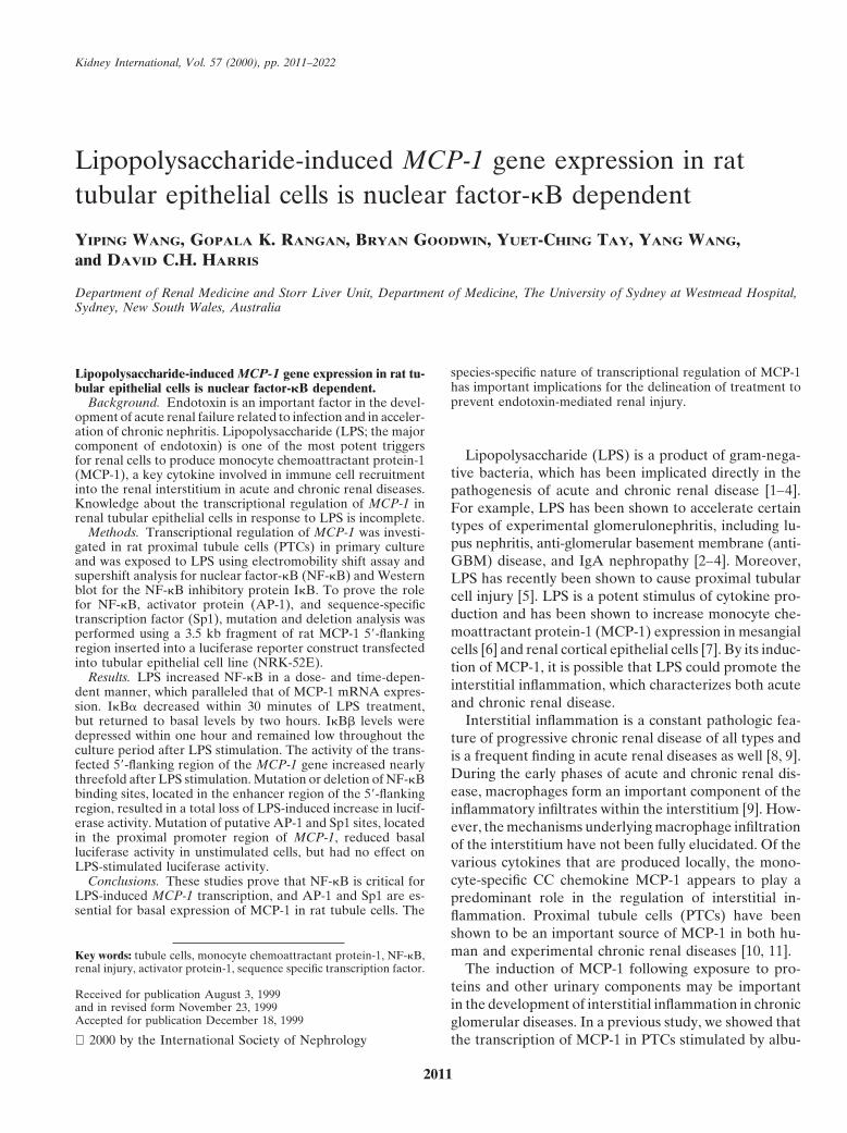
Kidney International, Vol. 57 (2000), pp. 2011–2022
Lipopolysaccharide-induced MCP-1 gene expression in rattubular epithelial cells is nuclear factor-kB dependent
YIPING WANG, GOPALA K. RANGAN, BRYAN GOODWIN, YUET-CHING TAY, YANG WANG,and DAVID C.H. HARRIS
Department of Renal Medicine and Storr Liver Unit, Department of Medicine, The University of Sydney at Westmead Hospital,Sydney, New South Wales, Australia
species-specific nature of transcriptional regulation of MCP-1Lipopolysaccharide-induced MCP-1 gene expression in rat tu-has important implications for the delineation of treatment tobular epithelial cells is nuclear factor-kB dependent.prevent endotoxin-mediated renal injury.Background. Endotoxin is an important factor in the devel-
opment of acute renal failure related to infection and in acceler-ation of chronic nephritis. Lipopolysaccharide (LPS; the majorcomponent of endotoxin) is one of the most potent triggers
Lipopolysaccharide (LPS) is a product of gram-nega-for renal cells to produce monocyte chemoattractant protein-1tive bacteria, which has been implicated directly in the(MCP-1), a key cytokine involved in immune cell recruitment
into the renal interstitium in acute and chronic renal diseases. pathogenesis of acute and chronic renal disease [1–4].Knowledge about the transcriptional regulation of MCP-1 in For example, LPS has been shown to accelerate certainrenal tubular epithelial cells in response to LPS is incomplete. types of experimental glomerulonephritis, including lu-Methods. Transcriptional regulation of MCP-1 was investi-
pus nephritis, anti-glomerular basement membrane (anti-gated in rat proximal tubule cells (PTCs) in primary cultureand was exposed to LPS using electromobility shift assay and GBM) disease, and IgA nephropathy [2–4]. Moreover,supershift analysis for nuclear factor-kB (NF-kB) and Western LPS has recently been shown to cause proximal tubularblot for the NF-kB inhibitory protein IkB. To prove the role cell injury [5]. LPS is a potent stimulus of cytokine pro-for NF-kB, activator protein (AP-1), and sequence-specific
duction and has been shown to increase monocyte che-transcription factor (Sp1), mutation and deletion analysis wasmoattractant protein-1 (MCP-1) expression in mesangialperformed using a 3.5 kb fragment of rat MCP-1 59-flanking
region inserted into a luciferase reporter construct transfected cells [6] and renal cortical epithelial cells [7]. By its induc-into tubular epithelial cell line (NRK-52E). tion of MCP-1, it is possible that LPS could promote the
Results. LPS increased NF-kB in a dose- and time-depen- interstitial inflammation, which characterizes both acutedent manner, which paralleled that of MCP-1 mRNA expres-and chronic renal disease.sion. IkBa decreased within 30 minutes of LPS treatment,
Interstitial inflammation is a constant pathologic fea-but returned to basal levels by two hours. IkBb levels weredepressed within one hour and remained low throughout the ture of progressive chronic renal disease of all types andculture period after LPS stimulation. The activity of the trans- is a frequent finding in acute renal diseases as well [8, 9].fected 59-flanking region of the MCP-1 gene increased nearly
During the early phases of acute and chronic renal dis-threefold after LPS stimulation. Mutation or deletion of NF-kBease, macrophages form an important component of thebinding sites, located in the enhancer region of the 59-flanking
region, resulted in a total loss of LPS-induced increase in lucif- inflammatory infiltrates within the interstitium [9]. How-erase activity. Mutation of putative AP-1 and Sp1 sites, located ever, the mechanisms underlying macrophage infiltrationin the proximal promoter region of MCP-1, reduced basal of the interstitium have not been fully elucidated. Of theluciferase activity in unstimulated cells, but had no effect on
various cytokines that are produced locally, the mono-LPS-stimulated luciferase activity.cyte-specific CC chemokine MCP-1 appears to play aConclusions. These studies prove that NF-kB is critical for
LPS-induced MCP-1 transcription, and AP-1 and Sp1 are es- predominant role in the regulation of interstitial in-sential for basal expression of MCP-1 in rat tubule cells. The flammation. Proximal tubule cells (PTCs) have been
shown to be an important source of MCP-1 in both hu-man and experimental chronic renal diseases [10, 11].Key words: tubule cells, monocyte chemoattractant protein-1, NF-kB,
renal injury, activator protein-1, sequence specific transcription factor. The induction of MCP-1 following exposure to pro-teins and other urinary components may be important
Received for publication August 3, 1999in the development of interstitial inflammation in chronicand in revised form November 23, 1999
Accepted for publication December 18, 1999 glomerular diseases. In a previous study, we showed thatthe transcription of MCP-1 in PTCs stimulated by albu- 2000 by the International Society of Nephrology
2011

Wang et al: LPS, MCP-1, and NF-kB2012
min correlated with activation of the transcription factor experiments [16]. Experiments were commenced aftercells had reached confluence, which was usually betweennuclear factor-kB (NF-kB). We further demonstrated
that the 59-flanking region of rat MCP-1 gene (which five and six days after the isolation procedure. TheNRK52E cell line was obtained originally from normalexhibited a 64% homology with the human MCP-1 59-
flanking region) contained binding sites for NF-kB, acti- rat kidney at passage 15 and showed epithelial morphol-ogy. The cells were cultured in DMEM containing 5%vator protein (AP-1), and sequence-specific transcription
factor (Sp1) [12]. fetal calf serum and were maintained at 378C in 5% CO2/95% O2. The cells from primary culture or cell line wereA number of studies have focused on the regulators
of murine and human MCP-1 genes [13, 14]. However, treated with or without LPS (0 to 10 ng/mL) for 0 to 16hours in serum-free medium in various experiments.much less is known about the regulation of MCP-1 in
rats, the species frequently used in in vivo studies ofAnalysis of monocyte chemoattractantacute and chronic renal disease. As LPS is likely to beprotein-1 transcriptiona clinically important stimulus for MCP-1 production in
a variety of animal and human renal diseases, this study Total RNA was isolated using RNAZOL B (Teltest,TX, USA). The transcription of MCP-1 mRNA in PTCsought to investigate the transcriptional regulation of rat
MCP-1 in PTCs in response to stimulation with LPS. We was determined using semiquantitative reverse tran-scriptase-polymerase chain reaction (RT-PCR). The de-hypothesized that the signal transduction pathways of
MCP-1 gene in PTCs require the activation of NF-kB, sign of primers and conditions for RT-PCR have beendescribed previously [12]. Ten percent of the PCR prod-AP-1, and Sp1. To investigate this hypothesis, the role
of these factors in MCP-1 regulation was examined by uct was loaded into a 1.2% agarose gel stained by ethid-ium bromide (0.5 mg/mL) and was photographed (Polar-transient transfection of chimeric MCP-1-luciferase re-
porter gene constructs containing MCP-1–regulatory re- oid 665 film) over ultraviolet light. The bands on thenegative film were scanned by Densitometry (Personalgions into a rat tubular epithelial cell line.Densitometer; Molecular Dynamics, Sunnyvale, CA,USA), and the signal intensity for MCP-1 was expressed
METHODSrelative to the housekeeping gene glyceraldehyde-3-
Reagents phosphate dehydrogenase (GAPDH).Dulbecco’s modified Eagle’s medium (DMEM), LPS
Extraction of nuclear protein and electrophoretic(Escherichia coli, serotype 026:B6), dexamethasone,mobility shift assayN-tosyl-phe-chloro-methyl-ketone (TPCK), and all other
cell culture reagents were purchased from Sigma-Aldrich Nuclear extracts from PTCs were prepared usingmethods described previously [15, 17]. The nuclear pro-(Sydney, Australia). Plasmid pGL3-basic and luciferase
assay system, gel shift assay system, and oligonucleotide teins were diluted to a standard concentration (3 mg/mL)and were stored in aliquots at 2708C. Double-strandedlabeling kit were from Promega (Sydney, Australia). The
rabbit polyclonal antibodies against p50, p65, c-Rel, oligonucleotides, containing NF-kB consensus bindingsites, were radiolabeled using T4 polynucleotide kinaseIkBa, and IkBb were from Santa Cruz Biotechnology
(Santa Cruz, CA, USA). P32-gATP, Hybond-C extra ny- (Promega) and [32P] ATP and were purified by centrifu-gation over a G-50 sephadex spin column. Specific oli-lon, peroxidase-conjugated sheep antirabbit IgG, and
enhanced chemiluminescence (ECL) detection reagent gonucleotide probes of NF-kB rat MCP-1-probe A(59-GTCTGGGAACTTCCAATGC-39, base 22287 towere from Amersham (Sydney, Australia). A transformer
site-directed mutagenesis kit was from Clontech (Palo 22278) and MCP-1-probe B (59-GAATGGGAATTTCCC-39, base 22261 to 22252) carrying rat MCP-1Alto, CA, USA), and transfection reagent lipofect AMIN-
plus was from Life Technologies (Sydney, Australia). NF-kB consensus sequences and their mutations [muta-tion A (GTCTGGGAACTcggAATGC) and mutation BDNA-modifying enzymes, unless otherwise stated, were
from Boehringer Mannheim (Sydney, Australia). The (GGAATGGccATTTCCACCAC)] were radiolabeledand employed to examine the specificity of NF-kB bind-rat tubular cell line (NRK52E) was purchased from
American Type culture Collection (Rockville, MD, USA). ing to the rat MCP-1 gene. Five micrograms of nuclearprotein were incubated with 50,000 cpm 32P-labeled
Cells probes for 20 minutes at room temperature according tothe manufacturer’s instructions (Promega). The samplesFor primary culture, PTCs were isolated and cultured
from normal male Wistar rats using isopycnic centrifuga- were loaded onto a 7% polyacrylamide gel with 1 3Tris-acetate/EDTA buffer, and electrophoresis was per-tion. Cells were grown in DMEM supplemented with
epidermal growth factor (10 ng/mL) and insulin (5 mg/mL) formed at 100 V for one hour. The gel was vacuum driedand exposed to x-ray film for two to four hours. Thein a 5% CO2/95% O2 environment [15, 16]. The proximal
tubular cell origin of these cells was verified in previous specificity of the reaction was determined by competition

Wang et al: LPS, MCP-1, and NF-kB 2013
reactions in which a 100-fold molar excess of unlabeled 59-CTCAAAGGTGCTGCAGAGTTACTT-39, and anti-NF-kB probe was added to the binding reaction 10 min- sense primer, 59-TGCATAGTGGTGGAGGAAGA-39.utes before the addition of a radiolabeled probe. The resultant amplicon was gel purified (Qiagen Pty Ltd.,
The composition of activated NF-kB was analyzed Victoria, Australia), digested with Bgl II, and insertedusing a supershift assay. In these studies, 1 mL of antibod- into Bgl II-linearized pGL3-basic to form pGL3-MCP-1.ies reactive to the rat p50, p65, or cRel protein was The 215 bp rMCP-1 promoter region between 2107 andincubated with the reaction mixture for 30 minutes and 174 was amplified by standard PCR using the sense primerwas then added to radiolabeled NF-kB probes (A and B) 59-AGCAGATTCAAACTTCCA-39 and antisensefor 15 minutes. Electrophoresis was performed as de- primer 59-GGAGGAAGAGAGATTCGAAGG-39 andscribed, and the autoradiographs were analyzed for re- was digested with Hind III and Xho I and inserted intoduction in signal intensity and the presence of a super- Hind III-Xho I–linearized pGL3-basic to form pGL3-shifted band. MCP (2107/174). The 343 bp rMCP-1 enhancer region
between 22407 and 22065 was amplified using a senseWestern blot analysis primer 59-GAGCTCAGACTATGCCTTTGT-39 and an-
Nuclear factor-kB activity is primarily regulated by tisense primer 59-GAGCCTGGGAGGTCACCATT-39.the turnover of IkB proteins in the cytosol. Therefore, The resultant fragment was digested with KpnI andWestern immunoblotting was performed to analyze the Xho I and inserted into KpnI-Xho I-linearized pGL3-kinetics of IkB proteins. Cells were lyzed with buffer TK, which contains bases 2105 to 153 of the herpes sim-[0.1 mol/L Tris, pH 6.8, 0.1% Nonidet P-40, 10% plex virus thymidine kinase (TK) promoter linked to ab-mercaptoethanol, 4 mmol/L ethylenediaminetetraace- luciferase reporter gene to form pGL3-MCP (22407/tic acid (EDTA), and 1 mmol/L orthovanadate]. Protein 22065).content of the samples was determined. Samples con-taining 10 mg of protein were loaded into each lane Mutagenesisand electrophoresed on a 10% sodium dodecyl sulfate- Site-directed mutagenesis was performed on the puta-polyacrylamide gel (SDS-PAGE) under reducing condi- tive NF-kB binding sites in the distal enhancer regiontions. The proteins were transferred to a nitrocellulose using the Transformer Site-Directed Mutagenesis kitfilter for electrophoresis performed for one hour at 100 V. according to the manufacturer’s instructions. The oligo-To block nonspecific binding sites, the membrane was nucleotides used were 59-GGAATGGccATTTCCACCincubated sequentially with 5% bovine serum albumin AC-39 and 59-GTCTGGGAACTcggAATGC-39 to obtainand then primary polyclonal antibodies (rabbit anti-
mutated NF-kB construct pGL3-kB-MA and pGL3-kB-IkBa and anti-IkBb, 1:1000) for one hour each at room
MB. To delete the NF-kB binding sites A and B, thetemperature. Blots were washed with Tris buffer andpGL3-MCP (22219/174) was prepared by excising aincubated with peroxidase-conjugated sheep antirabbitfragment from 22219 upstream on pGL3-MCP-1. TheIgG (1:1000) for one hour at room temperature. Reactiv-putative Sp1 and AP-1 binding sites in the proximality was detected with ECL detection reagent. The molec-promoter region were revealed by nucleotide sequenceular weights of the immunoreactive bands were esti-analysis and mutated using the oligonucleotides 59-mated by comparison with known molecular standards.GCCTGACTCCAagCTCTGGCTTAC-39 and 59-CACCCTGCCTGgtaCCACCCTCTG-39 to form the mu-Construction of plasmidstated Sp1 and AP-1 construct pGL3-SP-M and pGL3-Previously, the rat MCP-1 (rMCP-1) gene was clonedAP-M.and sequenced up to 2940 bp upstream, which does not
encompass all of the regulatory regions delineated in Cell transfection and luciferase assaythis study [18]. To elucidate the mechanism of rMCP-1
In six-well cell culture plates, NRK-52E cells weretranscriptional regulation, the 59-flanking region of theseeded at a density of 3 3 105 cells per well in 1 mLrMCP-1 from base 23580 to 180 was cloned and se-medium. After 20 hours of incubation, the cells were 60quenced [15]. The relevant sequence of the 59-flankingto 80% confluent. The NRK52E cells were cotransfectedregion of the rMCP-1 gene is illustrated in Figure 1. Twowith luciferase expression vector (6 mg/mL) and b-galNF-kB binding sites were found in the counterpart ofvector (3 mg/mL as control) by lipofectAMIN-plus fol-human MCP-1 enhancer region: A, 2287/22278, and B,lowing the instructions of the kit. After 24 hours of trans-2261/22252, relative to the major transcriptional startfection, the cells were incubated with or without LPS forsite. In the proximal promoter region of rMCP-1 gene,eight hours and then harvested. The luciferase activity inan AP-1 binding site at base 254 to 244 and Sp1 bindingcell extracts was determined by an a-counter followingsite at base 251 to 239 were identified. The 59-flankingthe instructions of luciferase assay kit, and b-gal activityregion of rMCP-1 from base 23555 to 174 was amplified
from rat genomic DNA by PCR using the sense primer, was assayed by spectrophotometer.

Wang et al: LPS, MCP-1, and NF-kB2014
Fig. 1. Sequence of the enhancer and pro-moter region of rat monocyte chemoattractantprotein-1 (rMCP-1) gene. The kB bindingsites A (22287/22278) and B (22261/22252)in the enhancer region are underlined. Theactivated protein-1 (AP-1) binding site (254/244) is underlined, and the sequence specifictranscription factor (Sp1) binding site (251/239) is highlighted in promoter region.
Statistics but had no effect on the density of complex II. Incubationwith labeled NF-kB and unlabeled mutant NF-kB probeData are presented as mean 6 SD of three to five(1 to 103) only reduced the density of complex II (dataseparate experiments in which values were determinednot shown). Therefore, complex II is likely to representin triplicate. Analysis of variance (ANOVA) and Fisher’sthe binding by a non–NF-kB transcription factor,least significant method were used for comparisonswhereas complex I is specific for NF-kB binding. In un-among multiple means, and the Student’s unpaired t-teststimulated PTCs, there was weak binding activity ofwas used for comparison between two means. A P valueNF-kB. LPS stimulation (1 to 10 mg/mL) increasedless than 0.05 was considered significant.NF-kB binding activity dose dependently; maximal bind-ing was observed at 5 mg/mL LPS (Fig. 2). NF-kB binding
RESULTS activity was strongly induced at 2 hours, peaked at 8Nuclear factor-kB activation and monocyte hours, and was maintained a high level at least 16 hourschemoattractant protein-1 mRNA expression following stimulation with LPS. The addition of excess
unlabeled NF-kB oligonucleotide completely abrogatedThe activation of NF-kB in response to LPS was exam-complex formation, thereby confirming the specificityined using an NF-kB consensus binding oligonucleotide.of the reaction (Fig. 3). The time- and dose-dependentTwo bands, upper band (complex I) and lower bandactivation of NF-kB paralleled that of MCP-1 mRNA(complex II), appeared on the electrophoretic mobilityaccumulation. LPS (5 mg/mL) produce a fivefold increaseshift assay (EMSA; Fig. 2). Nuclear protein incubatedin MCP-1 expression, with a peak at 4 to 8 hours and awith labeled NF-kB oligonucleotide and 1 to 10 timessustained increase for up to 24 hours with continuedthe molar excess of unlabeled oligonucleotide reduced
the density of complex I in a dose-dependent manner, exposure (Fig. 4). Pretreatment of cells with NF-kB in-

Wang et al: LPS, MCP-1, and NF-kB 2015
Fig. 2. Activation of nuclear factor-kB(NF-kB) by lipopolysaccharide (LPS) in prox-imal tubule cells (PTC). The arrow indicatesthe upper band (complex I), which is anNF-kB specific binding complex. The arrow-head indicates the lower band (complex II),which is a result of nonspecific binding. Elec-tromobility shift assay (EMSA) shows thedose–response pattern for NF-kB binding innuclear extracts from proximal tubule cellsstimulated by LPS (0 to 10 mg/mL) and thetime course for NF-kB binding activity afterexposure to LPS (5 mg/mL) for 0 to 16 hours.
hibitor TPCK resulted in an attenuated expression of B contained the NF-kB components p50, p65, and c-Rel(Fig. 7).NF-kB after exposure to LPS. In contrast, pretreatment
of cells with dexamethasone (0.1 mmol/L) resulted in a To elucidate the mechanism of rMCP-1 transcriptionalregulation in tubular epithelial cells by LPS stimulation,partial suppression of NF-kB activity (Fig. 3). The dose
response (0.01 to 10 mmol/L) was tested for dexametha- the 3567 bp 59-flanking region (base 23580 to 174) wascloned into a luciferase reporter plasmid. rMCP-1 genesone; NF-kB activity was partially suppressed with as
little as 0.03 mmol/L, and no extra suppression was seen contains two NF-kB binding sites in an enhancer regionthat are identical to human MCP-1 (hMCP-1). However,for doses beyond 0.1 mmol/L. In parallel, LPS-induced
expression of MCP-1 was inhibited by both TPCK and unlike hMCP-1, there is no NF-kB binding site in theproximal promoter region of rMCP-1. As seen in Figuredexamethasone, as previously described [15].8, tubule cells (NRK-52E cells) transfected with pGL3-
Degradation of cytoplasm IkB MCP-1 demonstrated a significant increase in luciferaseactivity when exposed to LPS. The induced levels ofTo further understand the mechanism of NF-kB ex-
pression in PTC stimulated with LPS, the levels of IkB-a luciferase activity were twofold to threefold greater thanin unstimulated cells. To investigate which regions areand IkB-b were determined with Western blots of cyto-
plasmic extracts (Fig. 5). Stimulation of cells with LPS involved in the transcriptional activity of rMCP-1 byLPS, tubule cells were transfected with pGL3-MCPled to a rapid, but transient decrease in IkB-a. In cells
stimulated by LPS, IkB-a was reduced within 30 minutes (22407/22065), which contains the enhancer region ofMCP-1 linked to a minimal TK promoter, and exhibitedand returned to control levels within 4 hours, despite
continued exposure. Levels of IkB-b also decreased, but an fivefold increase of luciferase activity in response toLPS. However, tubular epithelial cells transfected withunlike with IkBa, this was not seen until after a one-
hour stimulation with LPS. Also unlike IkB-a, lower pGL3MCP (2107/174), which contains the MCP-1 pro-moter region alone, showed no change in luciferase activ-levels of IkB-b persisted under continuing stimulation
with LPS. ity when incubated with LPS. This indicated that thesequence of promoter region was not essential for induc-
Functional analysis of MCP-1 gene regulation tion of MCP-1 by LPS.To examine the role of NF-kB, AP-1, and Sp1, tubularAn EMSA was performed to test whether NF-kB
binds to the kB consensus sequences identified in the epithelial cells were transfected with plasmids containingmutations in these binding sites. Mutants of the naturalenhancer region of MCP-1 gene in response to LPS. LPS
increased the specific binding activity of NF-kB to probes NF-kB binding sites were generated and analyzed bytransfection in tubule cells (Fig. 9). Mutagenesis ofcontaining the kB consensus sequences (probe A and
probe B), but not to mutated probes (mutation A or NF-kB binding site A elicited a significant reduction inluciferase activity in both unstimulated controls and cellsmutation B; Fig. 6). Supershift analysis using specific
probes that bind to kB consensus sites of rMCP-1 showed exposed to LPS, although luciferase activity was stillinduced twofold in LPS-stimulated versus control cells.that the complexes that formed on probe A and probe

Wang et al: LPS, MCP-1, and NF-kB2016
Fig. 3. Inhibition of NF-kB. EMSA shows that NF-kB binding activityafter stimulation by LPS (5 mg/mL) was completely inhibited by TPCK(100 mmol/L) and excess unlabeled NF-kB oligonucleotide (Comp)and partially inhibited by dexamethasone (Dex. 0.1 mmol/L). This isrepresentative of five separate experiments.
In contrast, mutation of NF-kB binding site B resulted Fig. 4. Expression of MCP-1 mRNA relative to GAPDH by reversetranscriptase polymerase chain reaction (RT-PCR) in PTC stimulatedin complete loss of LPS-induced luciferase activity. Thisby LPS. (A) PTCs were exposed to LPS in concentrations up to 10indicated that NF-kB binding site B is critical to the mg/mL for eight hours. (B) PTCs were exposed to 5 mg/mL LPS for 0,
regulation of rMCP-1 transcription by LPS. Following 2, 4, 8, 16, and 24 hours. *P , 0.01.the deletion of both NF-kB binding sites A and B in theenhancer region, the basal reporter gene activity wasdramatically reduced, and LPS effects were abolished.
of wild-type expression. These results suggest that AP-1This demonstrated that the NF-kB binding sites in theand Sp1 play an important role in maintaining the basaldistal enhancer region were necessary for LPS to inducetranscriptional expression of rMCP-1. Neither AP-1 norrMCP-1 transcription.Sp1 in the promoter region increased luciferasis activityWe next examined the effect of mutation of AP-1 andunder stimulation of LPS. Moreover, LPS stimulationSp1 sites in the promoter region of rMCP-1 (Fig. 10). Acaused no effect on luciferase activity induced by eithermutation of the AP-1 site substantially reduced reporterAP-1 or Sp1 mutants. This indicated that LPS-inducedgene expression to approximately 60% of the wild-typerMCP-1 transcription is not dependent on either AP-1activity. Likewise, mutation of the putative Sp1 binding
motif was shown to reduce reporter gene activity to 30% or Sp1 binding sites in the promoter region.

Wang et al: LPS, MCP-1, and NF-kB 2017
Fig. 7. Supershift analysis using specific probes that bind to kB consen-sus sites of rat MCP-1 revealed that activated NF-kB bands are com-posed of p50, p65, and c-Rel. The lower arrowhead indicates NF-kBbinding bands. The upper arrow indicates the supershifted band. Theis representative of three separate experiments.
DISCUSSIONFig. 5. Expression of IkBa (s) and IkBb (n) in PTCs stimulated byLPS (5 mg/mL). (A) IkBa levels decreased rapidly and returned to Despite intensive investigation of the MCP-1 gene,baseline after four hours of stimulation, whereas IkBb levels decreased
there is little information known about the molecularafter one hour and did not recover by eight hours (N 5 3). *P , 0.001vs. baseline. (B) Representative Western blots showing the changes in mechanisms involved in transcriptional region of MCP-1IkBa (upper panel) and IkBb (lower panel) levels in cytoplasmic ex- in the rat species, as the 59-flanking region of rat MCP-1tracts of PTCs exposed to LPS.
gene was isolated and sequenced only recently. To ourknowledge, the present study is the first direct demon-stration of transcription regulation of MCP-1 in rat renaltissue, using functional analysis of MCP-1 transcriptionalregulatory region. In this study, we have shown that LPSactivated NF-kB in a time- and dose-dependent manner,which correlated with MCP-1 expression. Furthermore,inhibition of NF-kB with TPCK or dexamethasone fullyor partially reduced MCP-1 expression. Moreover, muta-tion or deletion of NF-kB binding sites in the enhancerregion attenuated LPS-induced transcription activity andreduced basal transcription activity of reporter gene con-structs. However, mutation of AP-1 or Sp1 binding sitesin the promoter region only reduced basal transcriptionactivity of reporter gene. These results demonstrate thatthe induction of MCP-1 transcription in PTCs by LPSdepends on the activation of NF-kB, but not transcrip-tion factors AP-1 or Sp1.
Three functional NF-kB binding sites have been dem-onstrated to be important for the transcription of bothhuman and murine MCP-1 gene: one in the proximalpromoter region and two in the distal enhancer regionFig. 6. NF-kB binding to kB sites of rat MCP-1 gene. Exposure to
LPS (5 mg/mL) for eight hours increased NF-kB binding activity to [19]. Interestingly, unlike the hMCP-1 gene, rMCP-1 hasprobe A and probe B containing NF-kB consensus sequences, but not no NF-kB binding site in its promoter region. However,their mutations, mutation A and B. This is representative of three
two NF-kB binding sites exist in the enhancer regionseparate experiments.of rMCP-1 gene, and these are identical to hMCP-1.According to studies on hMCP-1 gene, the kB site B(59-GGGAATTTCC) sequence is identical to inter-

Wang et al: LPS, MCP-1, and NF-kB2018
Fig. 8. Transactivity of different fragments of 59-flanking region of rMCP-1 gene in response to LPS stimulation. (A) Schematic structure ofdifferent regions of rMCP-1 gene. (B) Different fragments of 59-flanking region linked to the luciferase gene were transfected into NRK-52E cells.The cells were stimulated with LPS (5 mg/mL) for eight hours, and the luciferase activity was measured. Values are means 6 SD for at least threeseparate experiments and are normalized by cotransfected b-galactosidase activity. Symbols are: ( ) unstimulated; ( ) LPS. The 59-flankingregion and enhancer region increased luciferase activity, whereas the promoter region had no effect on luciferase activity when incubated withLPS. *P , 0.01 vs. unstimulated cells.
feron-beta (INF-b), tumor necrosis factor-a (TNF-a) site B caused a complete loss of LPS-induced transcrip-tion. This demonstrated that NF-kB binding site B playsand multiple histocompatibility complex (MHC) class II
associate invariant chain kB sequence [13, 20]. The kB the dominant role in LPS-mediated transcription ofMCP-1 expression in rat tubule cells.site A (59-GGGAACTCC) with sequence similarity to
site B has a high affinity for both p65 and c-Rel, as A LPS-induced increase of hMCP-1 gene transcriptiondepends on the activity of its enhancer and/or promoterdistinct from the NF-kB binding site on IL-8 gene, which
has a high affinity for only p65, and on Ig j-chain gene, region [14]. In the present studies, distal enhancer re-gions were capable of conferring LPS responsiveness onwhich has a high affinity to only p50 [21, 22]. An interest-
ing finding in this study is that both NF-kB binding sites luciferase reporter gene, whereas the proximal promoterregion of rMCP-1 was not LPS responsive. This suggestshave a high affinity not only for p65 and c-Rel, but also
for p50, suggesting these binding sites in the rMCP-1 that in rat, unlike humans, the enhancer region has thepredominant role in LPS-induced MCP-1 transcription.gene have broader binding affinities for different NF-
kB/Rel proteins than do the sites in human MCP-1 gene. Monocyte chemoattractant protein-1 gene expressionis stimulus and cell specific [24]. This observation is ex-Of more importance to gene regulation than binding is
the transactivating potential of p50, p60 and cRel, which plained mainly by specific differences in the process ofNF-kB activation. The mechanism by which LPS acti-is not clear. For example, Sheppard et al have shown
that although NF-kB sites within the vascular cellular vates NF-kB in PTCs is not clear, but is probably medi-ated by phosphorylation of IkB. IkB is a family of inhibi-adhesion molecule-1 promoter region bind p50/p50 and
p65/p65 homodimers and p50/p65 heterodimers, only tors that complex with NF-kB in the cytoplasm andtightly control the nuclear translocation of NF-kB.p50/p65 heterodimers lead to gene activation during neu-
ral differentiation [23]. Among the different IkB, IkBa and IkBb play a majorrole in regulation of NF-kB [13, 25]. Studies have con-To further confirm the role of NF-kB in MCP-1 pro-
duction, the kB binding sites (sites A and B) in the cluded that TNF-a or phorbol 12-myristate 13-acetate(PMA) induce degradation of IkBa but not IkBb, sug-enhancer region were mutated, resulting in a loss of
NF-kB binding ability and a reduction of gene transcrip- gesting that these IkB proteins are regulated separatelyin a stimulus-dependent manner [26, 27]. In the currenttion. Mutation of site A resulted in a partial loss of gene
transcription in response to LPS, whereas mutation of study, increased nuclear binding of NF-kB was accompa-

Wang et al: LPS, MCP-1, and NF-kB 2019
Fig. 9. Mutation and deletion analysis of the distal kB sites in enhancer region regulating MCP-1 induction. (A) Schematic structure of 59-flankingregion of the MCP-1 gene, indicating the two kB binding sites A and B. (B) kB sites in the enhancer region were mutated or deleted, and theresultant gene fragment was linked to the luciferase gene and transfected into NRK-52E cells. The cells were stimulated with LPS (5 mg/mL) foreight hours, and the luciferase activity was measured. Values are means 6 SD for at least three separate experiments and are normalized bycotransfected b-galactosidase activity. kB sites A and B are shown by squares and mutated sites are marked with an x. Symbols are: ( )unstimulated; (j) LPS. #P , 0.01 vs. unstimulated cells transfected with unmutated 59-flanking region. *P , 0.01 vs. stimulated cells transfectedwith unmutated 59-flanking region.
nied by a decline in IkB concentration. LPS led to a line with observations made with hMCP-1, is a functionalcis-acting element.rapid and transient reduction in IkBa levels, but a later
and persistent decrease in IkBb. The reappearance of Activated protein-1 is another important regulatoryelement involved in the regulation of many cytokineIkBa has been proposed to be an internal counter-regu-
latory mechanism to prevent excessive activation of genes, including MCP-1. It has been reported that muta-tion of either AP-1 or NF-kB binding sites in the pro-NF-kB [28]. From this study, it appears that up-regula-
tion of MCP-1 by LPS is caused by activation of NF-kB moter region suppressed cytokine-induced MCP-1 ex-pression in human endothelial cells, suggesting that thesevia temporary degradation of IkBa and persistent degra-
dation of IkBb. The rapid degradation of IkBa may two elements act in a cooperative manner to regulateMCP-1 production [29]. However, in this study, mutationcontribute to early activation of NF-kB, and persistent
degradation of IkBb contributes to more prolonged acti- of the AP-1 site reduced only the basal transcription levelof rMCP-1, but had no effect on LPS-induced MCP-1vation of NF-kB.
Several studies have shown that pathways of MCP-1 expression. The explanation for this observation may berelated to the lack of NF-kB binding sites in the promotergene activation involve multiple transcription factors,
including Sp1 and AP-1. Nucleotide sequence analysis region of rat MCP-1 gene or the possibility that AP-1does not play a major role in rMCP-1 induction by LPS.of rMCP-1 gene revealed a potential Sp1 binding motif.
Previously, Ueda et al demonstrated that this site was It has been proposed that inhibition of NF-kB might beuseful therapeutically to alter the progression of chronicimportant for the basal expression of hMCP-1 gene [14].
However, the Sp1 binding site in rMCP-1 is not identical inflammation. Although glucocorticoids are capable ofinhibiting NF-kB binding activity by LPS, interleukin-1to that of hMCP-1, and it was unclear whether this site
in the rat gene was involved in basal expression. Indeed, (IL-1), and TNF-a in lymphocytes and endothelial cells,a recent study showed that they were unable to do this inin the present study, a mutation of the Sp1 site in
rMCP-1 substantially reduced the transcriptional activity mesangial cells [30]. In our study, activation of NF-kBand induction of MCP-1 by LPS were completely blockedof reporter gene constructs, demonstrating that this, in

Wang et al: LPS, MCP-1, and NF-kB2020
Fig. 10. Mutation analysis of AP-1 and Sp1 sites in the promoter region of the rMCP-1 gene. (A) Schematic structure of promoter region of theMCP-1 gene, showing the AP-1 and Sp1 binding sites. (B) AP-1 or Sp1 sites in promoter region were mutated, and resultant fragments werelinked to the luciferase gene and transfected into NRK-52E cells. The cells were stimulated with LPS (5 mg/mL) for eight hours, and luciferaseactivity was measured. Values are means 6 SD for at least three separate experiments and are normalized by cotransfected b-galactosidase activity.The AP-1 site is represented by a square and the Sp1 site by a circle, and mutated sites are marked with an x. Symbols are: ( ) unstimulated;( ) LPS. *P , 0.01 vs. unstimulated cells transfected with intact promoter region.
by TPCK (an inhibitor of cytoplasmic IkB proteolysis) of B cells to produce large amounts of immunoglobinbut only partially suppressed by dexamethasone. The and immune complexes [34]. However, Cavallo andreason for partial efficacy of steroids in this situation Granholm found that activation of proliferative lupusremains uncertain, but may be explained by cell specific- nephritis by LPS could not be attributed to altered con-ity of response. It has been demonstrated that up-regula- centration of complement nor to delayed removal oftion of IkBa by glucocorticoids is not sufficient to block circulating pathogenic immune complexes [35]. Such ob-NF-kB activation in endothelial cells and epithelial cells servations suggest the presence of alternative explana-[31, 32]. tions for promotion of nephritis by LPS. Endothelial
Lipopolysaccharide is a constituent of various gram- cells are generally thought to be the primary target ofnegative bacteria that are present in the gut in substantial LPS, but recently, tubular epithelial cells have also beenamounts and a common resource of subclinical and clini-
demonstrated to be important early targets of LPS [5].cal infection. The mechanism of action of LPS is relatedTrachtman et al have shown that interactions betweento CD14, a membrane protein that is considered to berenal tubular epithelial cells and E. coli can stimulatea receptor for the LPS complex [33]. In animal models,nitric oxide production in renal cells and suggested thatLPS has been shown to exacerbate the severity of renalnitric oxide production could responsible for tubuloin-damage in lupus nephritis, anti-GBM disease and IgAterstitial inflammation [36]. In the present studies, LPSnephropathy [2–4]. Furthermore, subclinical and clinicaldirectly activated NF-kB and induced MCP-1 transcrip-infections could accelerate chronic renal disease. Thetion in PTCs. It is possible that such a mechanism mightmechanisms by which LPS accelerate renal disease are
unknown and have been proposed to involve activation underlie the acceleration of nephritis by LPS, that is,

Wang et al: LPS, MCP-1, and NF-kB 2021
Induction of monocyte chemoattractant protein-1 in proximal tu-induction of MCP-1 leading to infiltration of leukocytesbule cells by urinary protein. J Am Soc Nephrol 8:1537–1545, 1997
into the interstitium. 13. Brown K, Park S, Kanno T, Franzoso G, Siebenlis U: Mutualregulation of the transcriptional activator NF-kappa B and its inhib-In summary, LPS induced MCP-1 expression in a time-itor, I kappa B-alpha. Proc Natl Acad Sci USA 90:2532–2536, 1993and dose-dependent manner, which correlated with
14. Ueda A, Okuda K, Ohno S, Shirai A, Igarashi T, MatsunagaNF-kB activation and was inhibited fully or partially with K, Fukushima J, Kawanoto S, Ishigatsubo Y, Okubo T: NF-
kappa B and Sp1 regulate transcription of the human monocyteNF-kB inhibitors TPCK or dexamethasone. Deletion orchemoattractant protein-1 gene. J Immunol 153:2052–2063, 1994mutation of the NF-kB binding sites in the enhancer
15. Wang Y, Rangan G, Tay YC, Wang YA, Harris DCH: Inductionregion of rat MCP-1 gene dramatically reduced tran- of monocyte chemoattractant protein-1 by albumin is mediated by
nuclear factor kB in proximal tubule cells. J Am Soc Nephrolscription activity in response to LPS, whereas mutation10:1204–1213, 1999of AP-1 and Sp1 binding sites in the promoter region 16. Chen L, Boadle RA, Harris DC: Toxicity of holotransferrin but
resulted in reduced basal transcription level of rat MCP- not albumin in proximal tubule cells in primary culture. J Am SocNephrol 9:77–84, 19981 gene. In conclusion, these studies prove that the up-
17. Dignam JD, Lebovitz RM, Roeder RG: Accurate transcriptionregulation of MCP-1 transcription in rat tubule cells by initiation by RNA polymerase II in a soluble extract from isolatedLPS depends on NF-kB activation. This information mammalian nuclei. Nucleic Acids Res 11:1475–1489, 1983
18. Timmers HT, Pronk GJ, Bos JL, Van Der AJ: Analysis of thecould be useful in the design of anti-inflammatory strate-rat JE gene promoter identifies an AP-1 binding site essential for
gies to specifically suppress transcriptional activation of basal expression but not for TPA induction. Nucleic Acids Res18:23–34, 1990MCP-1 in rat models of renal disease.
19. Ping D, Boekhoudt GH, Togers EM, Boss JM: Nuclear factor-kappa B p65 mediates the assembly and activation of the TNF-responsive element of the murine monocyte chemoattractant-1gene. J Immunol 162:727–734, 1999ACKNOWLEDGMENT
20. Ito CY, Kazantsev AG, Baldwin AS: Three NF-kappa B sitesThis study was supported by Grant 970721 from the National Health in the I kappa B-alpha promoter are required for induction of
and Medical Research Council, Australia. gene expression by TNF alpha. Nucleic Acids Res 22:3787–3792,1994
Reprint requests to David C.H. Harris, M.D., Department of Renal 21. Kunsch C, Rosen CA: NF-kappa B subunit-specific regulation ofMedicine, Westmead Hospital, Westmead NSW 2145, Australia. the interleukin-8 promoter. Mol Cell Biol 13:6137–6146, 1993E-mail: [email protected] 22. Ueda A, Okuda K, Ohno S, Shirai A, Igarashi T, Matsunaga
K, Fukushima J, Kawanoto S, Ishigatsubo Y, Okubo T, Yoshi-mura T: Transcriptional regulation of the human monocyte chemo-attractant protein-1 gene. Cooperation of two NF-kappaB sites and
REFERENCES NF-kappaB/Rel subunit specificity. J Biol Chem 272:31092–31099,19971. Haas C, Car B, Ryffel BLE, Hir M: Lipopolysaccharide-induced
23. Sheppard AM, McQuillan JJ, Iademarco MF, Dean DC: Controlglomerulonephritis develops in the absence of interferon-gamma of vascular cell adhesion molecule-1 gene promoter activity duringsignaling. Exp Nephrol 4:222–230, 1996 neural differentiation. J Biol Chem 270:3710–3719, 19952. Granholm NA, Cavallo T: Long-lasting effects of bacterial lipo- 24. Brown AM, Linhoff MW, Stein B, Wright KL, Baldwin AS,
polysaccharide promote progression of lupus nephritis in NZB/W Basta PV, Ting JP: Function of NF-kappa B/Rel binding sitesmice. Lupus 3:507–514, 1994 in the major histocompatibility complex class II invariant chain
3. Karkar AM, Rees AJ: Influence of endotoxin contamination on promoter is dependent on cell-specific binding of different NF-anti-GBM antibody induced glomerular injury in rats. Kidney Int kappa B/Rel subunits. Mol Cell Biol 14:2926–2935, 199452:1579–1583, 1997 25. Thompson JE, Phillips RJ, Erdjument-Bromage H, Tempst P,
4. Endo Y, Kanbayashi H, Hara M: Experimental immunoglobulin Ghosh S: I kappa B-beta regulates the persistent response in aA nephropathy induced by gram-negative bacteria. Nephron biphasic activation of NF-kappa B. Cell 80:573–582, 199565:196–205, 1993 26. Beg AA, Finco TS, Nantermet PV, Baldwin AS: Tumor necrosis
5. Kang YH, Falk MC, Bentley TB, Lee CH: Distribution and role factor and interleukin-1 lead to phosphorylation and loss of I kappaof lipopolysaccharide in the pathogenesis of acute renal proximal B alpha: A mechanism for NF-kappa B activation. Mol Cell Bioltubule injury. Shock 4:441–449, 1995 13:3301–3310, 1993
6. Rovin BH, Dickerson JA, Tan LC, Hebert CA: Activation of 27. Tran K, Merika M, Thanos D: Distinct functional properties ofnuclear factor-kappa B correlates with MCP-1 expression by hu- IkappaB alpha and IkappaB beta. Mol Cell Biol 17:5386–5399,man mesangial cells. Kidney Int 48:1263–1271, 1995 1997
7. Schmouder RL, Strieter RM, Kunkel SL: Interferon-gamma reg- 28. Chiao PJ, Miyamoto S, Verma IM: Autoregulation of I kappa Bulation of human renal cortical epithelial cell-derived monocyte alpha activity. Proc Natl Acad Sci USA 91:28–32, 1994chemotactic peptide-1. Kidney Int 44:43–49, 1993 29. Martin T, Cardarelli PM, Parry GC, Fells KA, Cobb RR:
8. Jones CL, Eddy AA: Tubulointerstitial nephritis. Pediatr Nephrol Cytokine induction of monocyte chemoattractant protein-1 expres-1992:572–586, 1992 sion in human endothelial cells depends on the cooperative action
9. Boucher A, Droz D, Adafer E, Noel LH: Characterization of of NF-kB and AP-1. Eur J Immunol 27:1091–1097, 1997mononuclear cell subsets in renal cellular interstitial infiltrates. 30. Auwardt RB, Mudge SJ, Che CG, Power DA: Regulation ofKidney Int 29:1043–1049, 1986 nuclear factor kappaB by corticosteroids in rat mesangial cells.
10. Prodjosudjadi W, Gerritsma JS, van Es LA, Daha MR, Bruijn J Am Soc Nephrol 9:1620–1628, 1998JA: Monocyte chemoattractant protein-1 in normal and diseased 31. de Bosscher K, Schmitz ML, Vanden BW, Plaisance S, Fiershuman kidneys: An immunohistochemical analysis. Clin Nephrol W, Haegeman G: Glucocorticoid-mediated repression of NF-kB-44:148–155, 1995 dependent transcription involves direct interference with transacti-
11. Eddy AA, Giachelli CM: Renal expression of genes that promote vation. Proc Natl Acad Sci USA 94:13504–13509, 1997interstitial inflammation and fibrosis in rats with protein-overload 32. Wissink S, Van Heerde EC, Vand Der Burg B, Van Der Saagproteinuria. Kidney Int 47:1546–1557, 1995 PT: A dual mechanism mediates repression of NF-kB activity by
glucocorticoids. Mol Endocrinol 12:355–363, 199812. Wang Y, Chen J, Chen L, Tay YC, Rangan GK, Harris DCH:

Wang et al: LPS, MCP-1, and NF-kB2022
33. Yang T, Sun D, Huang YG, Smart A, Briggs JP, Schnermann ing with processing of pathogenic immune complexes in NZB/Wmice. Am J Pathol 137:971–978, 1990JB: Differential regulation of COX-2 expression in the kidney by
lipopolysaccharide: Role of CD14. Am J Physiol 276:F10–F16, 1999 36. Trachtman H, Futterweit S, Singhal PC, Sankaran R, Franki N:Renal tubular epithelial cell-E. coli interaction products stimulate34. Klinmam DM, Steinberg AD: Systemic autoimmune disease arises
from polyclonal B cell activation. J Exp Med 165:1755–1760, 1987 nitric oxide production in cultured rat renal medullary interstitialand mesangial cells. Res Commun Mol Pathol Pharmacol 94:227–35. Cavallo T, Granholm NA: Bacterial lipopolysaccharide trans-
forms mesangial into proliferative lupus nephritis without interfer- 238, 1996