Lipophilic components and evaluation of the cytotoxic and ...
11
GRASAS Y ACEITES 69 (3) July–September 2018, e270 ISSN-L: 0017-3495 https://doi.org/10.3989/gya.0234181 Lipophilic components and evaluation of the cytotoxic and antioxidant activities of Impatiens glandulifera Royle and Impatiens noli – tangere L. (Balsaminaceae) K. Szewczyk a,* , R. Bonikowski b , A. Maciąg-Krajewska b , J. Abramek c and A. Bogucka-Kocka c a Department of Pharmaceutical Botany, Medical University of Lublin, Chodźki 1, 20-093 Lublin, Poland b Łódź University of Technology, Faculty of Biotechnology and Food Sciences, Institute of General Food Chemistry, Stefanowskiego 4/10, 90-924 Lodz, Poland c Department of Biology and Genetics, Medical University of Lublin, Chodźki 4a,20-093 Lublin, Poland * Corresponding author: [email protected] Submitted: 27 February 2018; Accepted: 10 May 2018 SUMMARY: The chemical composition of the lipophilic fractions of Impatiens glandulifera Royle and I. noli- tangere L. were analyzed by gas chromatography-mass spectrometry (GC-MS)., The study focused on the fatty acids, triterpenoids and sterols in the leaves, roots and seeds. Most of the identified compounds are new for these species. a-linolenic, oleic and palmitic acids were the most abundant in the fatty acid fractions, β-amyrin and 5a-lup-20(29)-en-3β-ol in the triterpenoid fractions, and β-sitosterol, spinasterol and chondrillasterol in the sterol fractions. The fatty acid and triterpenoid fractions showed strong antioxidant activity, similar to positive controls. Moreover, the triterpenoid fraction from I. noli-tangere seeds significantly inhibited HL-60 human leukemia cells. Other fractions showed moderate cytotoxicity. The present study suggests that I. glandulifera and I. noli-tangere are good source of omega-3 fatty acids, and they might be considered as antioxidant and chemopreventive agents. KEYWORDS: Antioxidant; Cytotoxicity; Fatty acids; Impatiens; Phytosterols; Triterpenoids RESUMEN: Componentes lipofílicos y evaluación de las actividades citotóxicas y antioxidantes de Impatiens glandulifera Royle e Impantiet noli-tangere L. (Balsaminaceae). La composición química de las fracciones lipofí- licas, centrada en los ácidos grasos, triterpenoides y esteroles de las partes aéreas, raíces y semillas de Impatiens glandulifera Royle e Impatient. noli-tangere L. se analizaron por cromatografía de gases-espectrometría de masas (GC-MS). La mayoría de los compuestos identificados son nuevos para estas especies. Los ácidos a-linolé- nico, oleico y palmítico fueron los más abundantes en las fracciones de ácidos grasos, β-amirina y 5a-lup-20 (29)-en-3β-ol en las fracciones triterpenoides, y β-sitosterol, espinasterol y condriplasterol en las fracciones de esteroles. Las fracciones de ácidos grasos y triterpenos mostraron una fuerte actividad antioxidante, similar a los controles positivos. Además, la fracción triterpenoidea de las semillas de I. noli-tangere inhibió significativa- mente las células de leucemia humana HL-60. Otras fracciones mostraron citotoxicidad moderada. El presente estudio sugiere que I. glandulifera e I. noli-tangere son la buena fuente de ácidos grasos omega-3, y podrían considerarse antioxidantes y agentes quimiopreventivos. PALABRAS CLAVE: Ácidos grasos; Antioxidante; Cytotoxicidad; Fitoesteroless; Impatiens; Triterpenoides ORCID ID: Szewczyk K https://orcid.org/0000-0003-4884-9110, Bonikowski R https://orcid.org/0000-0003-3327- 8984, Maciąg-Krajewska A https://orcid.org/0000-0001-9892-1219, Abramek J https://orcid.org/0000-0001-7925-4350, Bogucka-Kocka A https://orcid.org/0000-0001-8473-7429 Citation/Cómo citar este artículo: Szewczyk K, Bonikowski R, Maciąg-Krajewska A, Abramek J, Bogucka-Kocka A. 2018. Lipophilic components and evaluation of the cytotoxic and antioxidant activities of Impatiens glandulifera Royle and Impatiens noli – tangere L. (Balsaminaceae). Grasas Aceites 69 (3), e270. https://doi.org/10.3989/gya.0234181 Copyright: ©2018 CSIC. This is an open-access article distributed under the terms of the Creative Commons Attribution 4.0 International (CC BY 4.0) License.
Transcript of Lipophilic components and evaluation of the cytotoxic and ...

GRASAS Y ACEITES 69 (3)July–September 2018, e270
ISSN-L: 0017-3495https://doi.org/10.3989/gya.0234181
Lipophilic components and evaluation of the cytotoxic and antioxidant activities of Impatiens glandulifera Royle and
Impatiens noli – tangere L. (Balsaminaceae)
K. Szewczyka,*, R. Bonikowskib, A. Maciąg-Krajewskab, J. Abramekc and A. Bogucka-Kockac
aDepartment of Pharmaceutical Botany, Medical University of Lublin, Chodźki 1, 20-093 Lublin, PolandbŁódź University of Technology, Faculty of Biotechnology and Food Sciences,
Institute of General Food Chemistry, Stefanowskiego 4/10, 90-924 Lodz, PolandcDepartment of Biology and Genetics, Medical University of Lublin, Chodźki 4a,20-093 Lublin, Poland
*Corresponding author: [email protected]
Submitted: 27 February 2018; Accepted: 10 May 2018
SUMMARY: The chemical composition of the lipophilic fractions of Impatiens glandulifera Royle and I. noli-tangere L. were analyzed by gas chromatography-mass spectrometry (GC-MS)., The study focused on the fatty acids, triterpenoids and sterols in the leaves, roots and seeds. Most of the identified compounds are new for these species. a-linolenic, oleic and palmitic acids were the most abundant in the fatty acid fractions, β-amyrin and 5a-lup-20(29)-en-3β-ol in the triterpenoid fractions, and β-sitosterol, spinasterol and chondrillasterol in the sterol fractions. The fatty acid and triterpenoid fractions showed strong antioxidant activity, similar to positive controls. Moreover, the triterpenoid fraction from I. noli-tangere seeds significantly inhibited HL-60 human leukemia cells. Other fractions showed moderate cytotoxicity. The present study suggests that I. glandulifera and I. noli-tangere are good source of omega-3 fatty acids, and they might be considered as antioxidant and chemopreventive agents.
KEYWORDS: Antioxidant; Cytotoxicity; Fatty acids; Impatiens; Phytosterols; Triterpenoids
RESUMEN: Componentes lipofílicos y evaluación de las actividades citotóxicas y antioxidantes de Impatiens glandulifera Royle e Impantiet noli-tangere L. (Balsaminaceae). La composición química de las fracciones lipofí-licas, centrada en los ácidos grasos, triterpenoides y esteroles de las partes aéreas, raíces y semillas de Impatiens glandulifera Royle e Impatient. noli-tangere L. se analizaron por cromatografía de gases-espectrometría de masas (GC-MS). La mayoría de los compuestos identificados son nuevos para estas especies. Los ácidos a-linolé-nico, oleico y palmítico fueron los más abundantes en las fracciones de ácidos grasos, β-amirina y 5a-lup-20 (29)-en-3β-ol en las fracciones triterpenoides, y β-sitosterol, espinasterol y condriplasterol en las fracciones de esteroles. Las fracciones de ácidos grasos y triterpenos mostraron una fuerte actividad antioxidante, similar a los controles positivos. Además, la fracción triterpenoidea de las semillas de I. noli-tangere inhibió significativa-mente las células de leucemia humana HL-60. Otras fracciones mostraron citotoxicidad moderada. El presente estudio sugiere que I. glandulifera e I. noli-tangere son la buena fuente de ácidos grasos omega-3, y podrían considerarse antioxidantes y agentes quimiopreventivos.
PALABRAS CLAVE: Ácidos grasos; Antioxidante; Cytotoxicidad; Fitoesteroless; Impatiens; Triterpenoides
ORCID ID: Szewczyk K https://orcid.org/0000-0003-4884-9110, Bonikowski R https://orcid.org/0000-0003-3327-8984, Maciąg-Krajewska A https://orcid.org/0000-0001-9892-1219, Abramek J https://orcid.org/0000-0001-7925-4350, Bogucka-Kocka A https://orcid.org/0000-0001-8473-7429
Citation/Cómo citar este artículo: Szewczyk K, Bonikowski R, Maciąg-Krajewska A, Abramek J, Bogucka-Kocka A. 2018. Lipophilic components and evaluation of the cytotoxic and antioxidant activities of Impatiens glandulifera Royle and Impatiens noli – tangere L. (Balsaminaceae). Grasas Aceites 69 (3), e270. https://doi.org/10.3989/gya.0234181
Copyright: ©2018 CSIC. This is an open-access article distributed under the terms of the Creative Commons Attribution 4.0 International (CC BY 4.0) License.

2 • K. Szewczyk, R. Bonikowski, A. Maciąg-Krajewska, J. Abramek and A. Bogucka-Kocka
Grasas Aceites 69 (3), July–September 2018, e270. ISSN-L: 0017–3495 https://doi.org/10.3989/gya.0234181
1. INTRODUCTION
Impatiens glandulifera Royle and Impatiens noli-tangere L. are an annual herbaceous species which belongs to the Balsaminaceae family. These plants are native to North-Western, Central Europe and Asia (Tříska et al., 2013). In Poland, I. noli-tangere and I. glandulifera are two of the top 25 invasive alien plants (Tokarska-Guzik et al., 2010).
From a chemical point of view, I. glandulifera and I. noli-tangere have been the subject of few studies which have reported on the isolation or iden-tification of flavonoids (Szewczyk et al., 2016a), phenolic acids (Szewczyk and Olech, 2017), essen-tial oils (Szewczyk et al., 2016b), triterpenoid sapo-nins (Grabowska et al., 2017), glucosylated steroids (Cimmino et al., 2016), and naphthoquinones (Lobstein et al., 2001; Tříska et al., 2013).
It has been reported that several Impatiens species have valuable biological properties. For example, the triterpenoid saponins isolated from the leaves of I. parviflora exhibited cytotoxic activ-ity against human prostate and melanoma cancer cells (Grabowska et al., 2017), and the glucosylated steroids from I. glandulifera had cytostatic activity against U373 glioblastoma cells (Cimmino et al., 2016). Grabowska et al. (2016) noticed that fresh leaves of I. parviflora may be beneficial in inflam-matory conditions. Moreover, I. balsamina has been used for a very long time in traditional Asian and American medicine. Depending on the type of ail-ment, it was applied by compression directly on the skin, or as a tea prepared by pouring hot water on the dried plant (Yang et al., 2001). The plant is also utilized in Chinese medicine for rheumatism ther-apy; in treating fractures, swellings and contusions, as well as beriberi disease and as a plant with anti-cancer properties (Fukumoto et al., 1996).
Due to the biological importance of Impatiens species and the fact that the current knowledge about the lipophilic components in these plants is negligible, the aim of the present study is to determine and to compare the fatty acids, triterpenoids and sterol con-tents in the leaves, roots and seeds of I. glandulifera and I. noli-tangere. Moreover, their antioxidant and cytotoxic potential were investigated.
2. MATERIALS AND METHODS
2.1. Plant material
The leaves, roots and seeds of two Impatiens species were collected in August 2015. I. glandu-lifera Royle (no. IG-0815) were collected in Józefów, near Biłgoraj (Poland) at an altitude of 240 m a.m.s.l. (coordinates N 50°29’06; E 23°02’12’’) and I. noli-tangere L. (no. INT-0815) were gathered in Zalesie Górne near Warsaw (Poland) at an alti-tude of 115 m a.m.s.l. (coordinates N 52°2’16’’;
E 21°1’55’’). Voucher specimens were deposited in the Department of Pharmaceutical Botany, Faculty of Pharmacy, Medical University of Lublin. The plants were identified by Prof. Tadeusz Krzaczek.
2.2. Chemicals and reagents
All chemical reagents used in the experiment were purchased from various commercial suppliers and were of the highest purity available. 2,2-diphenyl-1-picrylhydrazyl (DPPH), ferrozine (3-(2-pyridyl)-5,6-bis (4-phenyl-sulfonic acid)-1,2,4-triazine), a-tocopherol, 2,6-di-tert-butyl-4-methylphenol (BHT), were purchased from Sigma-Aldrich (St. Louis, MO, USA). Phosphate-buffered saline (PBS) was purchased from Gibco (Carlsbad, CA, USA). Other chemicals used for the preparation of the extracts were of analytical grade, and were obtained from Polish Reagents (POCH, Gliwice, Poland).
2.3. Extraction and isolation procedure
The air-dried, ground leaves (100.0 g), roots (100.0 g) and seeds (20.0 g) of two plants were extracted with petroleum ether (bp 45–60 °C) in a Soxhlet apparatus for over 50 h. The obtained lipid extracts were evaporated under vacuum. Subsequently, yellowish oil residues were saponi-fied with a 10% ethanol solution of potassium hydroxide for 9 h, and the solutions were reduced to about half of their volume under vacuum, diluted with water, and extracted with diethyl ether. The ether phases were washed with distilled water until neutral, dried, and concentrated to yield crude unsaponifiable fractions. The sterols were first sep-arated from these fractions in the typical manner by precipitation with digitonine (Jerzmanowska, 1967). The obtained digitonides were filtered and washed with ethanol, and acetylation was per-formed (acetic acid anhydrous, with anhydrous sodium acetate, 2 h, 100 °C). Sterol acetates were crystallized from 50% ethanol (Jerzmanowska, 1967; Harrabi et al., 2016). Saponification of these mixtures yielded the free sterols which were iden-tified by GC-MS. The total amount of the phy-tosterols was determined by means of the weight method (Jerzmanowska, 1967).
Triterpenoids were then isolated from the fil-trates remaining after the crystallization of sterol digitonides. The eluates were reduced to about half of their volume under vacuum, and methanol was added for the crystallization of triterpenoids. The obtained precipitates were again crystallized from ethanol, acetylated (acetic acid anhydride, anhy-drous pyridine, 20 min), and again crystallized from ethanol. Hydrolysis of the triterpenoid acetate mix-tures (5% KOH in anhydrous ethanol, benzene, 7 h) yielded free triterpenoids (Jerzmanowska, 1967). Both free and triterpenoid acetates were analyzed

Lipophilic components and evaluation of the cytotoxic and antioxidant activities • 3
Grasas Aceites 69 (3), July–September 2018, e270. ISSN-L: 0017–3495 https://doi.org/10.3989/gya.0234181
using GC-MS. A schematic diagram of the isola-tion sterol and triterpenoid fractions is presented in Scheme 1.
In the next stage, powdered plant materials were sonicated with n-hexane (3 × 30 min) at a controlled temperature (40 ± 2 °C). The supernatants were con-centrated to dryness under vacuum at a controlled temperature. Then, 5 mL of methyl-tert-butyl ether
were added to 0.1 g oil. The fatty acid methyl esters (FAMEs) were obtained by adding a trimethylsul-fonium hydroxide solution (TMSH). The mixtures were then incubated (60 min, 60 °C), and analyzed using GC-MS.
For antioxidant and cytotoxic assays, 0.1 g of tri-terpenoid and fatty acid fractions were diluted in 80 and 95% ethanol (10 mL), respectively.
Scheme 1. Schematic diagram of the isolation sterol and triterpenoid fractions from Impatiens species.
Raw material
Lipid extract
Extraction with petroleum ether in Soxhlet (over 50 h)
Oil residue
Evaporation under vacuum
Water-alkaline extract
Alkaline hydrolysis (10% KOH, 9 h), solution reduction, water
Etheral phase Water phase
Extraction with diethyl ether
Sterols and triterpenoids
Evaporation under vacuum
eliminated
Filtrate Precipitate
1% ethanol solution of digitonine, filtration, washed with ethanol and diethyl ether
Triterpene alcohols
Evaporation, methanol crystallization, filtration
FiltrateSterol
digitonides
Washed with chloroform and acetone
Sterol acetate
Acetylation (anhydrous natrium acetate, 2h, 100°C)
GC-MS Free sterols
GC-MS
Triterpenoid acetate
Ethanol crystallization, acetylation (aceticacid anhydride, anhydrous pyridine, 20 min)
Free triterpenoid
GC MS-
Alkaline hydrolysis with 5% KOH

4 • K. Szewczyk, R. Bonikowski, A. Maciąg-Krajewska, J. Abramek and A. Bogucka-Kocka
Grasas Aceites 69 (3), July–September 2018, e270. ISSN-L: 0017–3495 https://doi.org/10.3989/gya.0234181
2.4. Chromatographic analysis
The analyses were performed on a Trace GC Ultra coupled with a DSQII mass spectrometer (Thermo Electron Corporation). GC-FID and MS analyses was performed using a MS-FID splitter (SGE Analytical Science). Mass range: 33-550 amu, ion source-heating: 200 °C, EI: 70eV, He (p: 300 kPa for phytosterols; 91 kPa for FAMEs). Operating conditions for derivatives of phytosterols: BPX5 (30 m×0.25 mm i.d., film thickness 0.25 μm), tem-perature program 100 °C (1 min, 10 °C/min) – 250 °C (15 min, 4 °C/min) – 300 °C (30 min). Injector temperature: 310 °C, and detector: 300 °C. Operating conditions for FAMEs: Stabilwax-DA, Restek (30m x 0.25 mm i.d., film thickness 0.25 µm), 50 °C (3 min) – 250 °C (40 min), 4 °C/min. Injector: 250 °C, detector 260 °C.
2.5. Identification of compounds
Phytosterols were analyzed as TMS (trimethyl-silyl ethers; Thanh et al., 2006) and fatty acids as FAME derivatives along with their mass spectra, compared with the MS of the NIST/EPA/NIH Mass Spectral Library 2009 and Wiley Registry of Mass Spectral Data, 8th edition. The retention times (Rt) were compared with a standard mixture Rt.
Area percent was obtained electronically from the GC-FID response without the use of an internal standard or correction factors. Quantitative deter-minations corresponded to the means of three rep-licates ± SD.
2.6. Cell lines and cell cultures
Human leukemia cell lines HL-60 and HL-60/MX2 were cell lines of AML (acute myeloid leu-kemia) origin and were obtained from the ECACC (the European Collection of Cell Cultures).
The HL-60 and HL-60/MX2 cells were main-tained in RPMI 1640 medium (Biomed Lublin) sup-plemented with 20% and 10% of fetal bovine serum (FBS; PAA Laboratories, Linz, Austria), respec-tively and the antibiotics were 100 U/mL penicillin, 100 µg/mL streptomycin, and 2.5 µg/mL amphoteri-cin B (Gibco, Carlsbad, USA), incubated at 37 ºC in a humidified atmosphere of 5% CO2.
2.7. In Vitro cytotoxicity assay
The effect of the obtained triterpenoid and fatty acid fractions on HL-60 and HL-60/MX2 cell lines was measured using the trypan blue assay. These cells were seeded on 12-well plates (Sarstedt, Wiener, Austria) at a density of 2×10⁵ and 3×10⁵ cells per well, respectively. After 24 hours, the cell suspen-sions were treated with plant samples at concentra-tions ranging from 10 to 5000 µg/mL for triterpenoid
and from 10 to 2500 µg/mL for fatty acid fractions, and incubated for 24 hours. Then, cell suspensions were centrifugated (800 rpm, 5 min), washed with PBS and centrifugated again. The cells were stained with a 0.4% solution of trypan blue (Bio-Rad) and counted with a TC10TM Automated Cell Counter (Bio-Rad). Each experiment was repeated three times. Dose response curves were made and IC50 values were found.
2.8. Antioxidant activity
To determine the antioxidant activity of the obtained triterpenoid and fatty acid fractions, two methods were used. DPPH (2.2-diphenyl-1-picryl-hydrazyl) free radical scavenging activity was mea-sured according to the Brand–Williams et al., (1995) method. The changes in color from deep-violet to light-yellow were measured at 515 nm in a UV/visible light spectrophotometer. The metal chelating power was determined using the Guo et al., (2001) method. Absorbance was measured at 562 nm and the per-centage of inhibition of ferrozine- Fe2+ complex for-mation was calculated according to the formula:
% inhibition = [1-(As/Ab)] x100
where Ab - absorbance of the blank, As – absorbance in the presence of the test sample
Both antioxidant activities were expressed as an efficient concentration EC50, the extract solu-tion concentration provided 50% of the activity in a dose-dependent manner. a-Tocopherol and BHT were used as a positive control.
2.9. Statistical analysis
All extractions and determinations were done in triplicate. The results are presented as experimen-tal means and ± SD. The obtained data were sub-jected to statistical analysis using Statistica 10.0. (StatSoft, Cracow).
3. RESULTS AND DISCUSSION
In continuation of our research on the genus Impatiens L., the present study deals with the deter-mination of phytosterols, triterpenoids and fatty acids from the leaves, roots and seeds of I. glandu-lifera and I. noli-tangere, as well as the examination of their cytotoxicity and antioxidant activity.
3.1. Sterol composition
The mixtures of sterol acetates and free sterols were obtained in typical way, and then analyzed using the GC-MS method. The total amount of sterols was determined using the weight method and the results are given in Table 1. On the basis of GC-MS analysis,

Lipophilic components and evaluation of the cytotoxic and antioxidant activities • 5
Grasas Aceites 69 (3), July–September 2018, e270. ISSN-L: 0017–3495 https://doi.org/10.3989/gya.0234181
in the sample from the roots of I. Glandulifera (IGR) fifteen phytosterols were identified. In the leaves of both species (IGL; INL) in the seeds of I. noli-tangere (INS), in the seeds of I. Glandulifera (IGS), and the roots of I. noli-tangere (INR),eleven and twelve com-pounds were observed, respectively.
The major components of the total sterol frac-tions obtained from the leaves of I. glandulifera were spinasterol + chondrillasterol and Δ7-sitosterol. The co-eluted compounds were separated in the GCxGC analysis (Figure 1). Similarly, spinasterol was observed in the root cultures of I. balsamina (Panichayupakaranant et al., 1995). This compound is known as an antimutagen agent that was tested using the mouse skin tumor assay (Villaseñor and Domingo, 2000). a-Spinasterol, together with glan-duliferins A and B (belonging to a cholestane sub-group), were isolated from I. glandulifera (Cimmino et al., 2016). In the rhizomes of I. pritzellii, some sterols, such as a-spinasterol, spinasteryl-3-one, a-spinasteryl-3-O-β-ᴅ-glucopyranoside, and 3-O-[6’-O-palmitylo-β-ᴅ-glucosyl]-spinasterol were noticed previously (Zhou et al., 2007). Moreover, a-spinas-terol was isolated from the seeds, roots, leaves and fruits of I. balsamina (Wang et al., 2011).
In other samples in our research, the most impor-tant sterol was β-sitosterol (ranging from 23.25 to 59.00% of total sterols). High levels of campes-terol and sitostanol were also observed in these frac-tions. Other compounds such as ergost-8(14)-en-3-ol,
campestenol, Δ7-stigmasterol, Δ5- avenasterol, and cholesterol occurred in low concentrations.
3.2. Triterpenoid composition
The appropriate GC-MS procedure allowed for the identification of the triterpenoid acetate fraction from the leaves of I. glandulifera (IGLt).Thirteen compounds such as 5a-ergost-7-en-3β-ol, taraxasteryl, a-spinasterol, 13,27-cyclour-san-3-ol, (3β,13β,14β)-, β-amyrin, cycloeucalenyl, β-simiarenol, stigmast-7-en-3-ol, (3β,5a)-, 5a-lup-20(29)-en3β-ol, 9,19-cyclolanostan-3-ol, 24-meth-ylene-, (3β)-, Ψ-taraxasteryl, lupan-3-ol, and olean-12-ene-3,28-diol, (3β)- were detected. The occurrence of eleven compounds was observed in the fraction from the roots of I. glandulifera (IGRt). In the roots of I. noli-tangere (INRt) and the seeds of I. glandulifera (IGSt), eight compounds were observed. In the leaves of I. noli-tangere (INLt), ten compounds were detected; and in the seeds of I. noli-tangere (INSt), seven compounds were found. As can be seen in Table 2, phytosterols were also present in these fractions, with the exception of triterpenoids. An exemplary GC-MS chromato-gram of the triterpenoid acetates from the leaves of I. Glandulifera is shown in Figure 1.
In the GC-MS analysis of the triterpenoid acetate fraction from the leaves of I. glandulifera, peak 1 had an identical retention time to 5a-ergost-7-en-3β-ol
Table 1. Sterol content (% of total fraction; mass %, GC) of I. glandulifera and I. noli-tangere.
Compound IGL IGR IGS INL INR INS
cholesterol nd 2.00±0.15 1.04±0.02 1.21±0.01 1.55±0.08 3.79±0.13
campesterol 0.26±0.01 11.63±0.38 3.54±0.11 13.78±0.21 14.05±0.35 6.17±0.19
campestanol nd 6.50±0.10 5.21±0.08 4.70±0.05 5.06±0.03 3.18±0.15
ergosta-7,22-dien-3-ol 0.10±0.01 0.10±0.01 nd 0.10±0.01 nd nd
ergost-8(14)-en-3-ol nd 0.70±0.02 0.85±0.05 0.81±0.01 1.00±0.03 0.59±0.01
stigmasterol nd 0.21±0.03 0.42±0.03 0.19±0.01 0.36±0.04 nd
β-sitosterol 2.43±0.11 44.36±0.25 53.61±0.69 59.00±0.38 54.18±0.43 23.25±0.50
sitostanol 0.88±0.04 18.65±0.42 13.57±0.32 17.26±0.19 19.05±0.35 5.91±0.13
∆5-avenasterol nd 0.21±0.01 tr 0.91±0.10 1.72±0.05 0.68±0.08
∆7-stigmastenol nd 0.29±0.02 2.61±0.06 1.70±0.11 2.26±0.04 1.46±0.01
ergosterol tr 0.18±0.02 nd nd nd nd
3 β-hydroxy-5 `-cholestane-6-one tr 0.19±0.01 nd nd nd nd
5.Xi.-Ergost-7-ene-3β-ol 1.75±0.13 tr nd nd 0.08±0.02 1.12±0.02
Spinasterol + chondrillasterol 68.86±0.75 9.31±0.25 11.05±0.31 0.34±0.08 0.23±0.01 4.86±0.06
∆7-sitosterol 23.01±0.38 1.72±0.02 7.30±0.15 nd nd 1.25±0.06
stigmasta-7,24(28)-dien-3β-ol (∆7-avenasterol) 2.20±0.02 nd nd nd tr 0.37±0.01
Sum of identified [%] 99.49±0.01 96.05±0.05 99.20±0.03 100.00±0.03 99.54±0.01 52.63±0.01
Total amount of sterol [%] 0.14 0.28 0.30 0.23 0.16 0.07
Values are mean ± SD of three samples; nd – not detected; tr – traces; IGL, I. glandulifera leaves; IGR, I. glandulifera roots; IGS, I. glandulifera seeds; INL, I. noli-tangere leaves; INR, I. noli-tangere roots; INS, I. noli-tangere seeds.

6 • K. Szewczyk, R. Bonikowski, A. Maciąg-Krajewska, J. Abramek and A. Bogucka-Kocka
Grasas Aceites 69 (3), July–September 2018, e270. ISSN-L: 0017–3495 https://doi.org/10.3989/gya.0234181
Figure 1. The GC x GC chromatogram (Rxi-5 x BPX50) of spinasterol and chondrillasterol (around 40 min) separation which were co-eluted (Rt=85.98 min) in GC analysis.
41.798836.7988
6.76
5
Masses: TIC
9.76
58.
765
7.76
5
38.4655 40.1322
Figure 2. The GC-MS chromatogram of triterpenoid acetates from the leaves of I. Glandulifera with marked compounds. 1-5a-ergost-7-en-3β-ol; 2- taraxasteryl; 3- a-spinasterol; 4- 13,27-cycloursan-3-ol, (3β,13β,14β)-; 5- β-amyrin; 6- cycloeucalenyl; 7- β-simiarenol; 8- stigmast-7-en-3-ol, (3β,5a)-; 9- 5a-lup-20(29)-en3β-ol; 10- 9,19-cyclolanostan-3-ol,- 24-methylene-, (3β)-; 11-
Ψ-taraxasteryl; 12- lupan-3-ol; 13 -olean-12-ene-3,28-diol, (3β)-.
84
RT:49.78–94.99
74
76
62
48
42
40
50 52 54˚ 58˚ 60 62 68 74˚70˚ 84˚ 86 88 90 92 94˚82˚8078˚76˚72˚
Time (min)
666458
38
44
46
50
52
1
2
4
6
8
7
9
5
3
10
11
12
13
54
56
58
60
64
66
68
70
70
72
78
80
82
94.6
394
.10
93.0
1
92.9
692
.41
91.9
391
.40
91.2
090
.81
90.1
989
.94
89.2
488
.86
88.6
488
.09
87.3
986
.95
86.0
986
.01
85.7
584
.98
84.6
084
.32
84.1
083
.16
82.9
782
.17
81.9
6
80.5
9
80.0
079
.19
78.6
377
.97
77.7
977
.33
76.8
676
.47
75.7
975
.65
75.1
6
74.4
474
.44
74.0
273
.45
73.1
272
.77
71.8
971
.56
71.0
070
.31
70.0
6
89.3
465
.56
66.2
6
67.9
067
.41
66.9
166
.52
66.2
465
.85
64.7
764
.40
63.5
6
63.0
2 62
.76
62.6
561
.58
61.3
2
60.5
060
.22
59.9
559
.2358
.78
57.1
256.5
4
56.7
1
Rel
ativ
e In
tens
ity
58.2
2
54.8
5
56.6
6
55.5
1
54.3
5
63.6
353
.38
52.2
751
.09
51.3
851
.08
50.8
050
.45
81.3
2
93.2
0

Lipophilic components and evaluation of the cytotoxic and antioxidant activities • 7
Grasas Aceites 69 (3), July–September 2018, e270. ISSN-L: 0017–3495 https://doi.org/10.3989/gya.0234181
acetate. The mass spectrum was similar to that obtained for authentic 5a-ergost-7-en-3β-ol and showed a molecular ion at m/e 426 and other impor-tant ions at m/e 411, 393, 355, 341, 302, 287, 269, 257, 218, and 204. This compound was only present in this sample. 13,27-cycloursan-3-ol, (3β,13β,14β)- was observed in the leaves of both species, and cycloeucalenyl was detected only in the leaves and roots of I. glandulifera. Ψ-Taraxasteryl acetate was absent in the analyzed seeds. The taraxasteryl, a-spinasterol, β-amyrin, β-simiarenol, stigmast-7-en-3-ol, (3β,5a)-, and 5a-lup-20(29)-en3β-ol ace-tates were identified in all the examined fractions from both Impatiens species.
The GC-MS method permitted an estimation of the contents of triterpenoid acetates determined in the leaves, roots and seeds of I. glandulifera and I. noli-tangere, based on the total fraction (Table 2). The quantitative analysis was done in triplicate. Amongst triterpenoid acetate fractions, the most abundant compounds in all samples were a-spinas-terol (from 2.42 to 33.91%), 5a-lup-20(29)-en3β-ol (5.73 - 22.91%) and β-amyrin (6.34 – 17.08%).
3.3. Fatty acid composition
In the hexane extracts from the leaves, roots and seeds of I. glandulifera and I. noli-tangere ten fatty acids were identified (Table 3). The saturated fatty acids comprised from 12.2 (roots of I. noli-tangere; INRf) to 27.2% (leaves of I. glandulifera; IGLf), monounsaturated – 16.9 (IGLf) to 34.1% (leaves of
I. noli-tangere; INLf), and polyunsaturated fatty acids comprised 40.3 (INRf) to 55.8% (IGLf). The unsaturated fatty acids a-linolenic and oleic acids were dominant compounds in all the examined samples. In I. noli-tangere γ-linolenic acid was also found at a high level, from 5.8% for the roots to 7.9% for the leaves. Trace fatty acids such as capric acid (C10:0) was detected only in the roots and seeds of I. noli-tangere (INSf). Small amounts of arachi-donic acid (C20:4) were noticed in the leaves and seeds of I. glandulifera (IGLf, IGSf). The presence of azelaic acid in the studied plants is interesting, as this acid has antibacterial, anti-inflammatory, keratolytic and sebostatic and tyrosinase-inhibiting properties, which is quite rare among plants (Reszke and Szepietowski, 2016).
It was observed that the amounts of the saturated fatty acids from the leaves and seeds of both exam-ined species were higher than in the roots (13.8% in I. glandulifera and 12.2% in I. noli – tangere). Differences in the values of the unsaturated / satu-rated ratios were observed from 2.7 for the leaves and seeds of I. glandulifera to 5.1 for the roots of I. noli-tangere. The polyunsaturated fatty acids were the most abundant of the unsaturated fatty acids in all organs of both species. The polyunsaturated (PUFAs) / monounsaturated (MUFAs) fatty acids ratios were from 1.2 for the leaves of I. glandulifera to 3.3 for the leaves of I. noli-tangere.
All the examined samples contained both ω-3 and ω-6 fatty acids. It has been reported that the ideal intake ratio of ω-6 to ω-3 fatty acids is
Table 2. Composition of triterpene acetate fractions (% of total fraction; mass%, GC) of I. glandulifera and I. noli-tangere.
Compound IGLt IGRt IGSt INLt INRt INSt
5`-ergost-7-en-3β-ol acetate 0.96±0.05 tr nd nd nd nd
taraxasteryl acetate 1.90±0.17 0.20±0.01 2.64±0.16 0.93±0.06 tr 1.13±0.02
`-spinasterol acetate 33.91±0.20 29.06±0.51 21.63±0.19 14.89±0.09 18.02±0.39 2.42±0.11
13,27-cycloursan-3-ol, acetate, (3β,13β,14β)- 2.24±0.10 nd nd 4.75±0.13 nd nd
β-amyrin acetate 14.00±0.40 6.34±0.19 10.02±0.31 17.08±0.43 12.97±0.24 8.73±0.08
cycloeucalenyl acetate 2.45±0.18 0.91±0.12 nd nd nd nd
β-simiarenol acetate 6.06±0.20 5.01±0.14 1.85±0.01 4.19±0.13 1.58±0.02 3.42±0.05
stigmast-7-en-3-ol, acetate, (3β,5`)- 4.78±0.08 3.96±0.02 tr 7.34±0.12 6.01±0.06 2.51±0.07
5` -lup-20(29)-en-3β-ol, acetate 20.17±0.56 16.04±0.30 5.73±0.10 22.91±0.38 9.75±0.29 13.08±0.69
9,19-cyclolanostan-3-ol, 24-methylene-, acetate, (3β)-
4.32±0.09 nd 2.71±0.02 1.04±0.10 nd nd
Ψ-taraxasteryl acetate 1.12±0.01 1.79±0.02 nd 4.53±0.21 2.80±0.08 nd
lupan-3-ol acetate tr 2.13±0.04 0.58±0.01 nd nd nd
olean-12-ene-3,28-diol, diacetate, (3β)- 1.85±0.03 0.70±0.01 nd 3.51±0.14 5.04±0.23 0.73±0.01
Sum of identified [%] 93.76±0.01 66.14±0.01 45.16±0.05 81.17±0.01 56.17±0.02 32.02±0.07
Total triterpene content[%] 0.61 0.37 0.59 0.42 0.48 0.29
Values are mean ± SD of three samples; nd – not detected; tr – traces; IGLt, I. glandulifera leaves; IGRt, I. glandulifera roots; IGSt, I. glandulifera seeds; INLt, I. noli-tangere leaves; INRt, I. noli-tangere roots; INSt, I. noli-tangere seeds.

8 • K. Szewczyk, R. Bonikowski, A. Maciąg-Krajewska, J. Abramek and A. Bogucka-Kocka
Grasas Aceites 69 (3), July–September 2018, e270. ISSN-L: 0017–3495 https://doi.org/10.3989/gya.0234181
between 1:1 and 4:1 (Simopoulos, 2006). According to Simopoulos (2006), increased levels of ω-3 fatty acids exert suppressive effects on many diseases such as cancer, cardiovascular disease, and inflammatory and autoimmune diseases. In our study, the ratio of ω-6 to ω-3 fatty acids is from 1/2.5 (roots of I. glan-dulifera; IGRf) to 1/12.8 (roots of I. noli-tangere; INRf). The obtained lower ratio of omega-6 to omega-3 fatty acids suggests that I. glandulifera and I. noli–tangere may be considered for the prevention and management of chronic diseases. The richer source of omega-3 fatty acids is I. glandulifera. A comparison of the present results with those of other authors has revealed that some of the iden-tified compounds in the leaves, roots and seeds of I. glandulifera and I. noli-tangere have been reported in I. glandulifera. Ortin and Evans, (2013) noticed that the hydrophobic extracts from flower stalks with seed pods of I. glandulifera contained linole-nic, palmitic, stearic, arachidic and trans-tetradec-2-enoic acids. According to Kaufmann and Keller, (1948), the Impatiens species contained oils with acetic and parinaric acid glycerides. They found that the seeds of I. glandulifera contain 50% and I. noli-tangere 55% of these types of oils. Saponification provided 13% glycerol, 10% acetic acid, 40% pari-naric acid, about 3% palmitic acid, about 3% stearic acid and about 20% mixture of oleic, linoleic and
linolenic acid. Nisar et al., (2012) analyzed a n-hex-ane extract of I. bicolor. Their study showed that the fatty acid esters, such as trans-methyl 13-octa-decenoate, methyl heptadecanoate, methyl octadec-anoate, methyl docosanoate, methyl tetracosanoate, and methyl eicosanoate are the major compounds of this extract.
3.4. In vitro cytotoxicity assay
Because plant triterpenoids and fatty acids have been reported to exhibit a variety of antioxi-dant, anti-inflammatory, antimicrobial, and antitu-mor promoting biological activities (Topçu, 2006; Dzubak et al., 2006), the antioxidant and cytotoxic properties of fractions obtained from the leaves, roots and seeds of I. glandulifera and I. noli-tangere were also evaluated.
In this study, the effect of the triterpenoid and fatty acid fractions in increasing concentrations on two types of cancer cell lines, HL-60 and HL-60/MX2, were investigated.
The cytotoxicity was estimated using trypan blue vital staining. The experiment was performed in triplicate and the mean values were calculated from the given values. The IC50 (Half Maximal Inhibitory Concentration, the inhibitor concentation when cell viability is 50%) values of the examined samples
Table 3. Fatty acid composition (mass % of total fatty acids) in the hexane extracts of I. glandulifera and I. noli-tangere.
Fatty acid
Fatty acid composition (% of total fatty acids)
IGLf IGRf IGSf INLf INRf INSf
Caprylic (C8:0) 0.3 ± 0.01 0.3 ± 0.00 0.4 ± 0.03 tr 0.3 ± 0.02 0.2 ± 0.10
Capric (C10:0) nd nd nd nd 0.1 ± 0.00 0.2 ± 0.01
Azelaic (C9:0) 2.2 ± 0.02 nd 2.0 ± 0.10 0.8 ± 0.02 nd 0.1 ± 0.10
Palmitic (C16:0) 21.1 ± 0.14 12.6 ± 0.19 21.0 ± 0.44 14.0 ± 0.05 7.2 ± 0.01 11.5 ± 0.13
Stearic (C18:0) 3.6 ± 0.02 0.9 ± 0.06 3.6 ± 0.11 9.4 ± 0.33 4.6 ± 0.18 7.6 ± 0.08
Oleic (C18:1) 16.9 ± 0.19 17.1 ± 0.11 18.3 ± 0.05 34.1 ± 0.66 21.4 ± 0.25 30.0 ± 0.50
Linoleic (C18:2) ω-6 12.9 ± 0.13 14.2 ± 0.10 12.3 ± 0.04 nd 2.5 ± 0.02 12.0 ± 0.03
`-Linolenic (C18:3) ω-3 40.5 ± 1.05 35.6 ± 0.66 40.3 ± 0.25 33.8 ± 0.14 32.0 ± 1.2 31.6 ± 0.90
γ-Linolenic (C18:3) 1.1 ± 0.10 tr 1.0 ± 0.01 7.9 ± 0.13 5.8 ± 0.08 6.6 ± 0.33
Arachidonic (C20:4) 1.3 ± 0.02 nd 1.1 ± 0.05 nd nd nd
Total [%] 99.9±0.01 80.7±0.03 100.0±0.01 100.0±0.00 73.9±0.02 99.8±0.01
Σ SAFAs 27.2±0.03 13.8±0.01 27.0±0.00 24.2±0.01 12.2±0.03 19.6±0.01
Σ UNSAFAs 72.7±0.01 66.9±0.05 73.0±0.02 75.8±0.01 61.7±0.06 80.2±0.02
UNSAFAs/SAFAs ratio 2.7 4.9 2.7 3.1 5.1 4.1
Σ MUFAs 16.9±0.01 17.1±0.02 18.3±0.01 34.1±0.03 21.4±0.01 30.0±0.01
Σ PUFAs 55.8±0.05 49.8±0.03 54.5±0.02 41.7±0.01 40.3±0.02 50.2±0.03
PUFAs/ MUFAs ratio 3.3 2.9 3.0 1.2 1.9 1.7
ω-6/ ω-3 ratio 1/3.1 1/2.5 1/3.3 - 1/12.8 1/2.6
Values are mean ± SD of three samples; nd – not detected; tr – traces; IGLf, I. glandulifera leaves; IGRf, I. glandulifera roots; IGSf, I. glandulifera seeds; INLf, I. noli-tangere leaves; INRf, I. noli-tangere roots; INSf, I. noli-tangere seeds; SAFAs, saturated fatty acids; UNSAFAs, unsaturated fatty acids; MUFAs, monounsaturated fatty acids; PUFAs, polyunsaturated fatty acids.

Lipophilic components and evaluation of the cytotoxic and antioxidant activities • 9
Grasas Aceites 69 (3), July–September 2018, e270. ISSN-L: 0017–3495 https://doi.org/10.3989/gya.0234181
were determined using MS Excel. The cells of both cancer lines exposed to these fractions presented diverse cytotoxicity depending on the dose of IC50.
Based on the obtained results, it was found that the analyzed fractions from I. glandulifera and I. noli-tangere induce apoptosis of the cells of the both tested cell lines. The results, which are given in Table 4, showed that the triterpenoid fraction from I. noli-tangere seeds significantly inhibited HL-60 human leukemia cells, and was the most potent fraction with IC50 values of 11.69 µg/mL, followed by fatty acid fractions from the roots and leaves of I. glandulifera with IC50 of 41.54 and 61.81 µg/mL, respectively. Moreover, the fatty acids from the roots of I. noli-tangere and triterpenoids from the roots of I. glandulifera showed a moderate cytotox-icity against HL-60 with IC50 values of 65.37 and 65.56 µg/mL.
On the other hand, the triterpenoid fractions from the seeds of I. glandulifera and I. noli-tangere showed high inhibition activity against the HL-60/MX2 cell line with IC50 of 33.92 and 43.35 µg/mL, respectively. Relatively high cytotoxicity against HL-60/MX2, fatty acid fractions from the leaves of I. noli-tangere (IC50 = 50.82 µg/mL) and roots of I. glandulifera (IC50 = 52.81 µg/mL) was shown.
The weakest cytotoxic activity was found in the triterpenoid fraction from the leaves of I. glandulifera. The lowest IC50 doses against both cell lines used in the research was determined.
Based on the IC50 values, it can be concluded that the HL-60 cell line is more sensitive to the Impatien fractions studied. The highest IC50 value was observed after the exposure of HL-60 cells to leaf extract (IC50 = 963.69 µg/mL).
The presence of 5a-lup-20 (29)-en3β-ol and a-spinasterol may be responsible for the cytotoxic activity of the studied fractions. It has been reported that 5a-lup-20(29)-en3β-ol inhibits skin cancer in CD-1, and induces apoptosis in HL-60 human leu-kemia cells (Saleem et al., 2004; Zhang et al., 2009). Spinasterol has shown antitumor effect against ovar-ian, skin and breast cancer cells (Jeon et al.,2006).
Previous studies report the cytotoxic and antitu-mor activities of extracts and compounds isolated from I. balsamina. For example, the ethanol extract of I. balsamina was investigated for in vitro cytotoxic and in vivo antitumor activities against transplantable
tumors and human cell lines. The obtained results showed significant antitumor and cytotoxic effects against Dalton’s ascites lymphoma and human can-cer cell lines (Baskar et al., 2012). Moreover, bal-saminone C (dinaphthofuran-7,12- dione derivative) isolated from the seeds of I. balsamina exhibited cytotoxicity against A549, Bel-7402 and Hela cancer cell lines (Pei et al., 2012).
3.5. Antioxidant activity
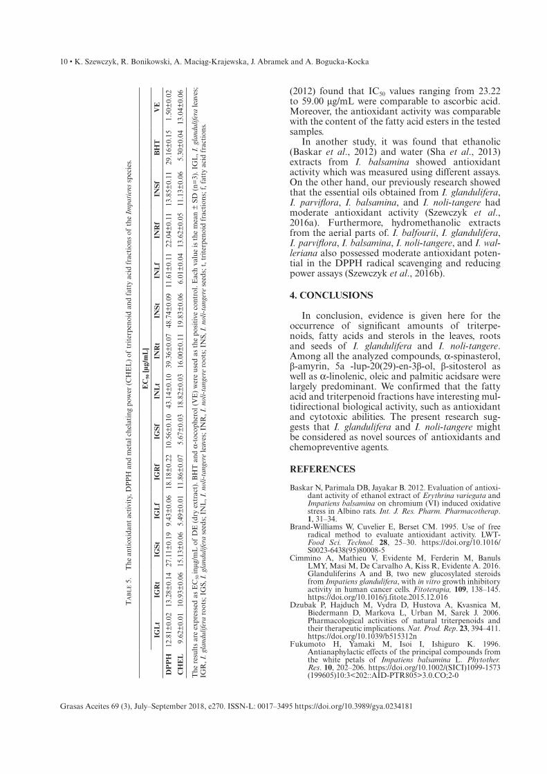
The antioxidant activities of triterpenoid and fatty acid samples were evaluated using the DPPH radical-scavenging test and metal chelating power. As seen in Table 5, EC50 values for the analyzed fractions and standards on the DPPH radical were found in the range of 12.81 to 27.11 µg/mL and from 39.36 to 48.74 µg/mL for triterpenoid fractions from I. glandulifera and I. noli-tangere, respectively, from 9.43 to 18.18 µg/mL and from 11.61 to 22.04 µg/mL for fatty acid fractions from I. glandulifera and I. noli-tangere, respectively, 29.16 µg/mL for BHT and 1.50 µg/mL for a-tocopherol. Most of the samples tested showed better antioxidant activity than BHT (the lower EC50 values) but weaker than a-tocopherol. The fatty acid fraction from the leaves of I. glandulifera showed the strongest DPPH radi-cal scavenging activity with EC50 at 9.43 µg/mL, fol-lowed by the fatty acid fraction from the seeds of I. glandulifera and the leaves of I. noli-tangere with EC50 of 10.56 and 11.61µg/mL, respectively. The strong antioxidant activity may be attributed to the presence of high amounts of omega-3 fatty acids in these fractions.
EC50 values for the Fe2+ chelating capacities of the analyzed fractions were found in the range from 9.62 to 15.13 µg/mL and from 16.00 to 19.83 µg/mL for triterpenoid fractions from I. glandulifera and I. noli-tangere, respectively. For fatty acid fractions the values of EC50 ranged from 5.49 to 11.86 µg/mL and from 6.01 to 13.62 µg/mL for I. glandulifera and I. noli-tangere, respectively. The positive controls BHT and a-tocopherol showed EC50 values of 5.30 and 13.04 µg/mL, respectively. The results obtained in this test showed that metal chelating power was similar to positive controls.
Similar findings were obtained for the n-hexane extract of I. bicolor in the DPPH assay. Nisar et al.,
Table 4. The IC50 values for HL-60 and HL-60/MX2 line cells.
IC50 [µg/mL]
IGLt IGRt IGSt IGLf IGRf IGSf INLt INRt INSt INLf INRf INSf
HL-60 963.69 65.56 88.07 61.81 41.54 246.54 145.69 92.21 11.69 74.46 65.37 71.20
HL-60/MX2 875.91 60.56 33.92 169.36 52.81 288.47 243.99 105.71 43.35 50.82 95.37 157.91
IGL, I. glandulifera leaves; IGR, I. glandulifera roots; IGS, I. glandulifera seeds; INL, I. noli-tangere leaves; INR, I. noli-tangere roots; INS, I. noli-tangere seeds; t, triterpenoid fractions; f, fatty acid fractions.
10 • K. Szewczyk, R. Bonikowski, A. Maciąg-Krajewska, J. Abramek and A. Bogucka-Kocka
Grasas Aceites 69 (3), July–September 2018, e270. ISSN-L: 0017–3495 https://doi.org/10.3989/gya.0234181
(2012) found that IC50 values ranging from 23.22 to 59.00 µg/mL were comparable to ascorbic acid. Moreover, the antioxidant activity was comparable with the content of the fatty acid esters in the tested samples.
In another study, it was found that ethanolic (Baskar et al., 2012) and water (Sha et al., 2013) extracts from I. balsamina showed antioxidant activity which was measured using different assays. On the other hand, our previously research showed that the essential oils obtained from I. glandulifera, I. parviflora, I. balsamina, and I. noli-tangere had moderate antioxidant activity (Szewczyk et al., 2016a). Furthermore, hydromethanolic extracts from the aerial parts of. I. balfourii, I. glandulifera, I. parviflora, I. balsamina, I. noli-tangere, and I. wal-leriana also possessed moderate antioxidant poten-tial in the DPPH radical scavenging and reducing power assays (Szewczyk et al., 2016b).
4. CONCLUSIONS
In conclusion, evidence is given here for the occurrence of significant amounts of triterpe-noids, fatty acids and sterols in the leaves, roots and seeds of I. glandulifera and I. noli-tangere. Among all the analyzed compounds, a-spinasterol, β-amyrin, 5a - lup-20(29)-en-3β-ol, β-sitosterol as well as a-linolenic, oleic and palmitic acidsare were largely predominant. We confirmed that the fatty acid and triterpenoid fractions have interesting mul-tidirectional biological activity, such as antioxidant and cytotoxic abilities. The present research sug-gests that I. glandulifera and I. noli-tangere might be considered as novel sources of antioxidants and chemopreventive agents.
REFERENCES
Baskar N, Parimala DB, Jayakar B. 2012. Evaluation of antioxi-dant activity of ethanol extract of Erythrina variegata and Impatiens balsamina on chromium (VI) induced oxidative stress in Albino rats. Int. J. Res. Pharm. Pharmacotherap. 1, 31–34.
Brand-Williams W, Cuvelier E, Berset CM. 1995. Use of free radical method to evaluate antioxidant activity. LWT-Food Sci. Technol. 28, 25–30. https://doi.org/10.1016/S0023-6438(95)80008-5
Cimmino A, Mathieu V, Evidente M, Ferderin M, Banuls LMY, Masi M, De Carvalho A, Kiss R, Evidente A. 2016. Glanduliferins A and B, two new glucosylated steroids from Impatiens glandulifera, with in vitro growth inhibitory activity in human cancer cells. Fitoterapia, 109, 138–145. https://doi.org/10.1016/j.fitote.2015.12.016
Dzubak P, Hajduch M, Vydra D, Hustova A, Kvasnica M, Biedermann D, Markova L, Urban M, Sarek J. 2006. Pharmacological activities of natural triterpenoids and their therapeutic implications. Nat. Prod. Rep. 23, 394–411. https://doi.org/10.1039/b515312n
Fukumoto H, Yamaki M, Isoi I, Ishiguro K. 1996. Antianaphylactic effects of the principal compounds from the white petals of Impatiens balsamina L. Phytother. Res. 10, 202–206. https://doi.org/10.1002/(SICI)1099-1573 (199605)10:3<202::AID-PTR805>3.0.CO;2-0
Ta
bl
e 5
. T
he a
ntio
xida
nt a
ctiv
ity,
DP
PH
and
met
al c
hela
ting
pow
er (
CH
EL
) of
tri
terp
enoi
d an
d fa
tty
acid
fra
ctio
ns o
f th
e Im
pati
ens
spec
ies.
EC
50 [µ
g/m
L]
IGL
tIG
Rt
IGS
tIG
Lf
IGR
fIG
Sf
INL
tIN
Rt
INS
tIN
Lf
INR
fIN
Sf
BH
TV
E
DP
PH
12.8
1±0.
0213
.28±
0.14
27.1
1±0.
199.
43±
0.06
18.1
8±0.
2210
.56±
0.10
43.1
4±0.
1039
.36±
0.07
48.7
4±0.
0911
.61±
0.11
22.0
4±0.
1113
.85±
0.11
29.1
6±0.
151.
50±
0.02
CH
EL
9.62
±0.
0110
.93±
0.06
15.1
3±0.
065.
49±
0.01
11.8
6±0.
075.
67±
0.03
18.8
2±0.
0316
.00±
0.11
19.8
3±0.
066.
01±
0.04
13.6
2±0.
0511
.13±
0.06
5.30
±0.
0413
.04±
0.06
The
res
ults
are
exp
ress
ed a
s E
C50
inµg
/mL
of
DE
(dr
y ex
trac
t). B
HT
and
a-t
ocop
hero
l (V
E)
wer
e us
ed a
s th
e po
siti
ve c
ontr
ol. E
ach
valu
e is
the
mea
n ±
SD
(n=
3). I
GL
, I. g
land
ulife
ra le
aves
; IG
R, I
. gla
ndul
ifera
roo
ts; I
GS,
I. g
land
ulife
ra s
eeds
; IN
L, I
. nol
i-ta
nger
e le
aves
; IN
R, I
. nol
i-ta
nger
e ro
ots;
IN
S, I
. nol
i-ta
nger
e se
eds;
t, t
rite
rpen
oid
frac
tion
s; f,
fat
ty a
cid
frac
tion
s.

Lipophilic components and evaluation of the cytotoxic and antioxidant activities • 11
Grasas Aceites 69 (3), July–September 2018, e270. ISSN-L: 0017–3495 https://doi.org/10.3989/gya.0234181
Grabowska K, Podolak I, Galanty A, Załuski D, Makowska-Wąs J, Sobolewska D, Janeczko Z, Żmudzki P. 2016. In vitro anti-denaturation and anti-hyaluronidase activities of extracts and galactolipids from leaves of Impatiens parvi-flora DC. Nat. Prod. Res. 30, 1219–1223. https://doi.org/ 10.1080/14786419.2015.1049175
Grabowska K, Podolak I, Galanty A, Żmudzki P, Koczurkiewicz P, Piska K, Pękala E, Janeczko Z. 2017. Two new triterpe-noid saponins from the leaves of Impatiens parviflora DC. and their cytotoxic activity. Ind. Crops Prod. 96, 71–79. https://doi.org/10.1016/j.indcrop.2016.11.022
Guo JT, Lee HL, Chiang SH, Lin HI, Chang CY. 2001. Antioxidant properties of the extracts from different parts of broccoli in Taiwan. J. Food Drug Anal. 9, 96–101.
Harrabi S, Curtis S, Hayet F, Mayer PM. 2016. Changes in the sterol compositions of milk thistle oil (Silybium marianum L.) during seed maturation. Grasas Aceites 67, https://doi.org/10.3989/gya.0495151
Jeon GC, Park MS, Yoon DY, Shin CH, Sin HS, Um SJ. 2006. Antitumor activity of spinasterol isolated from Pueraria roots. Exp. Mol. Med. 37, 111–120. https://doi.org/10.1038/emm.2005.15
Jerzmanowska Z. 1967. Substancje roślinne. Metody wyodrębniania. PWN, Warszawa.
Kaufman HP, Keller M. 1948. Über das Vorkommen von Parinarsäure und Essigsäure in den Samenfetten der Balsaminaceae. Chem. Ber. 81, 152–158. https://doi.org/ 10.1002/cber.19480810212
Lobstein A, Brenne X, Feist E, Metz N, Weniger B, Anton R. 2001. Quantitative determination of naphthoquinones of Impatiens species. Phytochem. Anal. 12, 202–205. https://doi.org/10.1002/pca.574
Nisar M, Qayum M, Shah M, Zia-Ul-Haq M, Khan I, Ahmad K, Qayum Z. 2012. Chemical constituents and antioxi-dant activity of n-hexane extract of Impatiens bicolor. Chem. Nat. Compd. 48, 143–146. https://doi.org/10.1007/s10600-012-0184-6
Ortin Y, Evans, P. 2013. trans-Tetradec-2-enoic acid in Impatiens glandulifera. Synthetic Communications 43, 1404–1412. https://doi.org/10.1080/00397911.2011.635395
Panichayupakaranant P, Noguchi H, De-Eknamkul W, Sankawa U. 1995. Naphthoquinones and coumarins from Impatiens balsamina root cultures. Phytochem. 40, 1141–1143. https://doi.org/10.1016/0031-9422(95)00418-7
Pei H, Lei J, Qian S H. 2012. A new cytotoxic dinaphthofuran-7,12dione derivatives from the seeds of Impatiens balsam-ina. Zhong. Yao. Cai. 35, 407–410.
Reszke R, Szepietowski J. 2016. Azelaic acid in dermatological treatment – current state of knowledge. Przegl. Dermatol. 103, 337–343. https://doi.org/10.5114/dr.2016.61785
Saleem M, Afaq F, Adhami VM, Mukhtar H. 2004. Lupeol modulates NF-κB and PI3K/Akt pathways and inhibits
skin cancer in CD-1 mice. Oncogene 23, 5203–5214. https://doi.org/10.1038/sj.onc.1207641
Simopoulos AP. 2006. Evolutionary aspects of diet, the omega-6/omega-3 ratio and genetic variation: nutritional implications for chronic diseases. Biomed. Pharmacother. 60, 502–507. https://doi.org/10.1016/j.biopha.2006.07.080
Szewczyk K, Kalemba D, Komsta Ł, Nowak R. 2016a. Comparison of the essential oil composition of selected Impatiens species and its antioxidant activities. Molecules 21, https://doi.org/10.3390/molecules21091162
Szewczyk K, Olech M. 2017. Optimization of extraction method for LC-MS based determination of phenolic acid profiles in different Impatiens species. Phytochem. Lett. 20, 322–330. https://doi.org/10.1016/j.phytol.2017.02.005
Szewczyk K, Zidorn C, Biernasiuk A, Komsta Ł, Granica S. 2016b. Polyphenols from Impatiens (Balsaminaceae) and their anti-oxidant and antimicrobial activities. Ind. Crop. Prod. 86, 262–272. https://doi.org/10.1016/j.indcrop.2016.03.053
Thanh T, Vergenes MF, Kaloustian J, El-Moselhy T, Amiot-Carlin M, Portugal H. 2006. Effect of storage and heating on phytosterol concentrations in vegetables oils determined by GC/MS. J. Sci. Food Agric. 86, 220–225. https://doi.org/10.1002/jsfa.2322
Tokarska-Guzik B, Węgrzynek B, Urbisz A, Urbisz A, Nowak T, Bzdęga K. 2010. Alien vascular plants in the Silesian Upland of Poland: distribution, patterns, impacts and threats. Biodivers. Res. Conserv. 19, 33–54. https://doi.org/10.2478/v10119-010-0019-x
Topçu G. Bioactive triterpenoids from Salvia species. 2006. J. Nat. Prod. 69, 482–487. https://doi.org/10.1021/np0600402
Tříska J, Vrchotová N, Sýkora J, Moos M. 2013. Separation and identification of 1,2,4-trihydroxynaphthalene1-O-glucoside in Impatiens glandulifera Royle. Molecules 18, 8429–8439. https://doi.org/10.3390/molecules18078429
Villaseñor IM, Domingo AP. 2000. Anticarcinogenicity poten-tial of spinasterol isolated from squash flowers. Teratog. Carcinog. Mutagen. 20, 99–105. https://doi.org/10.1002/(SICI)1520-6866(2000)20:3<99::AID-TCM1>3.0.CO;2-7
Yang X, Summerhurst DK, Koval SF, Ficker C, Smith ML, Bernards MA. 2001. Isolation of an antimicrobial com-pound from Impatiens balsamina using bioassay–guided fractionation. Phytother. Res. 15, 676–680. https://doi.org/ 10.1002/ptr.906
Zhang L, Zhang Y, Zhang L, Yang X, Lv Z. 2009. Lupeol, a dietary triterpene, inhibited growth, and induced apoptosis through down-regulation of DR3 in SMMC7721 cells. Cancer Invest. 27, 163–170. https://doi.org/10.1080/07357900802210745
Zhou XF, Zhao XY, Tang L, Ruan HL, Zhang YH, Pi HF, Xiao WL, Sun HD, Wu JZ. 2007. Three new triterpenoid saponins from the rhizomes of Impatienspritzellii var. hupehensis. J. Asian Prod. Res. 9, 379–385. https://doi.org/10.1080/10286020600781019