Oxidant Damage of Lipids and Proteins Membranes Unstable ...

Lipids, membranes and chemiosmosis
The figure below illustrates the interior of a eucaryotic cell such as found in all higher
animals.
Its outer limit is the plasma membrane and it contains many different types of structures each of
which is also highly membranous. The nucleus is surrounded by a membranous structure with
large holes depicted by large dots through which messenger RNAs (ribonucleic acids),
transcribed from genes in the nucleus, are transported to the rough endoplasmic reticulum where
they are translated into proteins. This is depicted by strings of small dots that represent
ribosomes sequentially reading the messenger RNAs and translating them into proteins. These
proteins are subsequently released from the ribosomes and are transported into the Golgi
apparatus for post-translational processing. They may subsequently be sequestered in secretion
granules, that are small intracellular membranous compartments that can fuse with the plasma
membrane in a process called exocytosis whereby their contents are released to the exterior of the
cell. Endocytosis is the reverse of this process and produces endosomes that contain molecules
from outside the cell. These may be digested in lysosomes with which the endosomes fuse. All of
these structures are dynamic, membranous structures. The membranes are lipid bilayers with a
thickness of 6 to 10 nm (nanometers).
Obviously, the picture in the figure is not a photo-micrograph made with a light
microscope because such photo-micrographs are restricted to larger than micron scale, and
nanometer scale structures are depicted in the picture. The picture is a deduced representation

made from scores of electron-micrographs made with the electron-microscope. The revolution in
electron microscopy techniques in the 1940s through the 1960s made this picture possible. This
picture is a static picture as well. In a living cell the membranous structures are in motion and
undergo fusions and fissions that constantly change their number, size and shape. Of special
interest for us in this text are the mitochondria, two of which are shown in the picture (a typical
cell may contain hundreds of mitochondria). These are the power plants of the cell in which a
variety of processes take place. Before a detailed account of their structure and processes is
given, however, a digression into membrane structure is required.
Lipids and membranes
Membranes self-assemble from lipid molecules and proteins. Membranes are highly
selective permeability barriers. Except for the processes of exocytosis and endocytosis
mentioned above (in fact, both of these processes require the aid of a structural protein, clathrin),
transport across membranes is mediated by embedded proteins. These proteins confer on the
membrane its distinctive features. They serve as pumps, gates, transporters, receptors, energy
tranducers, structural elements and enzymes. The ratio of protein to lipid by weight in a
membrane can range from 3 to 1 to 1 to 5. In myelin, a nerve fiber insulator, the membrane is
18% protein, the plasma membrane is 50% protein and the inner mitochondrial membrane is
75% protein.
The three major kinds of membrane lipids are phospholipids, glycolipids and cholesterol.
Phospholipids are derived from the three carbon alcohol gycerol
and glycolipids are derived from the more complex alcohol sphingosine

Those derived from glycerol were called phosphoglycerides but are now called
glycerophospholipids and contain two fatty acid side chains and a phosphorylated alcohol such
as choline, ethanolamine, inositol or serine. Typically, the fatty acid chains in phospholipids and
glycolipids contain between 14 and 24 carbon atoms. Most common are 16 (palmitate)
and 18 (oleate)

Oleate contains one unsaturated carbon-carbon double bond in the middle of the chain. It is in
the cis conformation and this is almost always the case with unsaturated biological fatty acid
chains. The bend this causes in the fatty acids creates a disruption of regular order in the
membrane resulting in a more fluid membrane interior.
In glycerophospholipids, the carboxyl groups of the fatty acids are esterified to the C-1
and C-2 hydroxyl groups of glycerol. In the simplest glycerophospholipid, the C-3 hydroxyl of
glycerol is esterified to phosphate, forming diacylglycerol-3-phosphate

Membranes contain only a small amount of this compound. The major glycerophospholipids are
derivatives of this compound in which the phosphate group is esterified to the hydroxly group of
one of several alcohols
Glycolipids are based on sphingosine. This molecule contains an amino group and
already has one fatty acid chain. A second fatty acid carboxly group is linked by an amide bond
to this amino group. In sphingomyelin, phosphoryl choline is esterified to the hydroxly group of
sphingosine. In glycolipids generally, the unit linked to the hydroxly group of sphingosine is a
simple sugar or a small sugar polymer. If glucose or galactose is used then the result is a
cerebroside and if a branched sugar chain with up to seven units is used then the result is a
ganglioside. Another important neutral lipid in some membranes is cholesterol a sterol
It is found in most eucaryotes but not in procaryotes. The single O that it contains derives from
atmospheric O2. Cholesterol must have evolved after the earth’s atmosphere accumulated O2.
Eucaryotic plasma membranes are often rich in cholesterol but the membranes of sub-cellular
organelles have less. The evolutionary theory that mitochondria and cholorplasts in eucaryotes
arose from captured procaryotic ancestors suggests that their membranes should contain no
cholesterol.
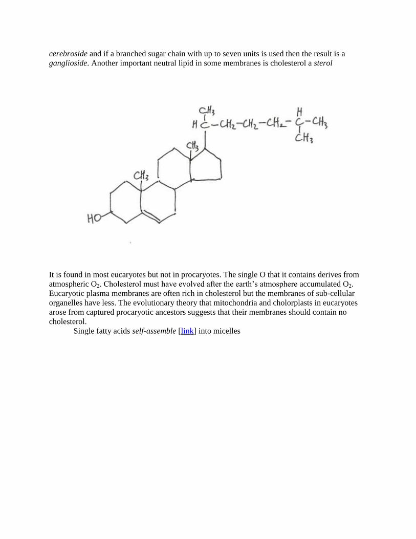
Single fatty acids self-assemble [link] into micelles
Phospholipids and glycolipids, on the other hand generally form lipid bilayers (also called
bimolecular sheets)
These structures are the result of the amphipathic nature of these molecules. That is, they contain
both a hydrophobic region, the fatty acid chains, and a hydrophilic region, the charged phosphate
groups and in some cases charged amine groups. The essence of self-assembly has to do with the
way water molecules interact with the two regions of the lipid molecules. Water molecules are
polar and can hydrogen bond with other molecules. The hydrophobic fatty acid regions of lipids
interact more favorably with other hydrophobic molecules then with water and the hydrophilic

regions interact strongly with water. To accommodate both tendencies, fatty acids form micelles
in which the hydrophilic carboxyl groups are on the outside in direct contact with water and the
hydrophobic fatty acid chains are on the inside interacting with each other. Micelles are usually
less than 20 nm in diameter. Phospholipids and glycolipids have two fatty acid side chains and
are, therefore, too bulky to readily form micelles without exposing the fatty acids chains to water
from the outside. Instead, a bilayer forms in which all of the fatty acids are inside the bilayer,
sequestered away from water while the charged hydrophobic ends are on the outside of the
bilayer
The bilayers tend to close up making a closed compartment that can have a diameter as large as
mm’s (millimeter). This is a much more versatile structure than a micelle. Bilayers tend to be
highly impermeable to ions and polar molecules, with the conspicuous exception of water
molecules. Permeability coefficients are measured in cm/s (centimeters per second). The table
below shows the great range of values observed

For an ion such as Na+ (sodium) the incredibly low permeability compared to H2O results from
the solvation shell of water molecules around Na+ in the aqueous milieu. This shell would have
to be removed in order for Na+ to permeate the bilayer. This is highly unfavorable energetically.
The permeability of H2O is a result of its very small size and its high thermal velocity at
physiological temperatures, about 4 x 104 cm/s ( this speed is equivalent to 1440 km/hr
(kilometer per hour).
When the bilayer closes into a compartment so that there are no edges exposing fatty acid
side chains to water, the resulting structure is asymmetric. Sphingomyelin and phosphatidyl
choline are preferentially located in the outer layer while phosphatidyl ethanolamine and
phosphatidyl serine are preferentially located in the inner layer in the case of red blood cells.
This brings up an important fact regarding the nature of the association of lipid molecules in the
bilayer. It is not a covalent arrangement. The fatty acid side chains of different lipid molecules
interact by hydrophobic forces and van der Waals forces, whereas the charged, hydrophilic ends
interact with water molecules by ionic forces and hydrogen bonds.
The individual lipid molecules are free to move laterally within the bilayer with a
diffusion constant of about 10-8
cm2/s. Thus, a lipid molecule can diffuse, on the average, a
distance of one mm (micron) in one second. This is roughly the size scale of the mitochondria.
Nevertheless, a lipid molecule is very unlikely to flip-flop from one layer of the bilayer to the
other. It takes a phospholipid 109 times longer to flip-flop across the membrane than it does to
laterally diffuse 5 nm (which takes 6 microseconds). Thus, the asymmetry of location of lipid
species in the bilayer is well preserved in time.

Proteins are associated with lipids in many different ways. Some are peripherally
associated with the hydrophilic exterior/interior of the bilayer compartment. These proteins tend
to have lots of charged amino acid residues.

The free energy barrier for a protein to cross the bilayer is even greater than for a lipid molecule
to flip-flop across. Other proteins are deeply embedded in the bilayer because of extensive
stretches of hydrophobic amino acid residues that make these stretches hydrophobic and lipid-
like as well. Some proteins strands traverse back and forth across the bilayer many times.
Proteins can laterally diffuse in the membrane almost as well as lipids can but they are generally
bigger than lipids and do so more slowly. Other proteins are linked to other molecules by groups
that are external to the lipid phase of the membrane and this can greatly reduce their laterally
mobility.
Complexes of proteins that are associated with the membrane occur and possess well
maintained positions relative to the membrane’s interior/exterior orientation, i.e. some of the
proteins may be peripheral on the inside or the outside while others are embedded, either
crossing the bilayer altogether or situated closer to the exterior or the interior side of the
membrane compartment. These elaborate arrangements self-assemble from the proteins that are
synthesized elsewhere in the cell (endoplasmic reticulum and Golgi apparatus) and find their
way to the correct associations in the correct membrane by diffusion and specific identification
labels that may be enzymatically removed once the proteins are in the correct position. Many
proteins are facilitated in their association with the membrane by chaperone proteins that require
the energy of ATP (adenosine triphosphate) to do their jobs.
Cholesterol associates with the fatty acid side chains of the lipids in a bilayer. By
disrupting the interactions of saturated fatty acids chains with each other, cholesterol actually
promotes fluidity of the lipid interior of the bilayer. However, too much cholesterol can have the
reverse effect.
Electron transport chains
Let’s look more closely at the mitochondria

They are the power plants of the cell. They have the shape of a cylinder with a size of roughly
1.0 mm diameter and 2.0 mm length. They have two membranes, a smooth outer membrane and
an extensively invaginated inner membrane. The invaginations are called cristae. The proteins
mediating electron transport and ATP synthesis are bound to the inner membrane. The outer
membrane contains the protein porin, a non-specific pore that permits the diffusive passage
through the membrane of molecules up to 10 kd (kilo-daltons). This includes coenzymes, small
peptides and small oligonucleotides. The inner membrane contains ~ 75% proteins and is freely
permeable to only O2, CO2 and H2O. It contains proteins for electron transport, ATP synthesis
and metabolite transport. These proteins are embedded in the inner membrane lipid interior.
Contained within the compartment surrounded by the inner membrane is a gel-like matrix
rich in proteins for pyruvate dehydrogenase, the citric acid cycle, fatty acid oxidation and for the
mitochondion’s genetic machinery, as well as mitochondrial DNA (deoxyribonucleic acid), RNA
and ribosomes. While the cytoplasm of the cell is an aqueous solution of many substances, the
matrix of the mitochondria is so jam packed with proteins and other molecules that there is room
left for relatively few water molecules, creating a gel rather than a solution. The evolutionary
theory for the origin of mitochondria in eucaryotes as captured procaryotic symbiotes is
bolstered by their apparent retention of genetic machinery for the synthesis of some, but not all,
of the mitochondrial proteins. This machinery includes ribosomes that are more similar to those
of contemporary procaryotes then they are to eucaryotic ribosomes that occur in the rough
endoplasmic reticulum of the eucaryotic cell. A similar state of affairs also applies to
chloroplasts in plant cells.

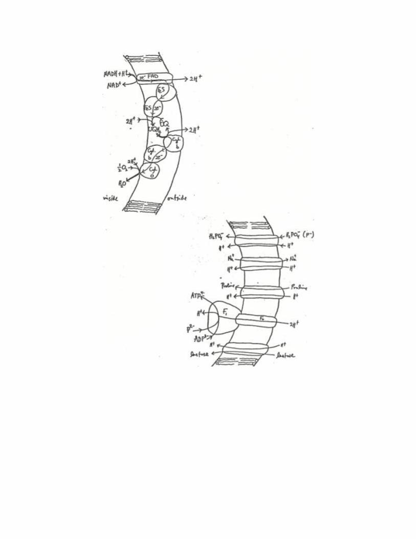
Electron transport in bacteria
The electron transport chain of bacteria is similar to that of mitochondria but simpler.
Thus, we will begin by considering electron transport in bacteria
In the figure the lipid bilayer is depicted by two curved arcs that make clear which side of the
bilayer is the inside of the bacterium and which is the outside. Embedded in the bacterial
membrane are many replicas of different types of protein complexes. One complex involves
proteins labeled with FAD (flavin adenine dinucleotide)
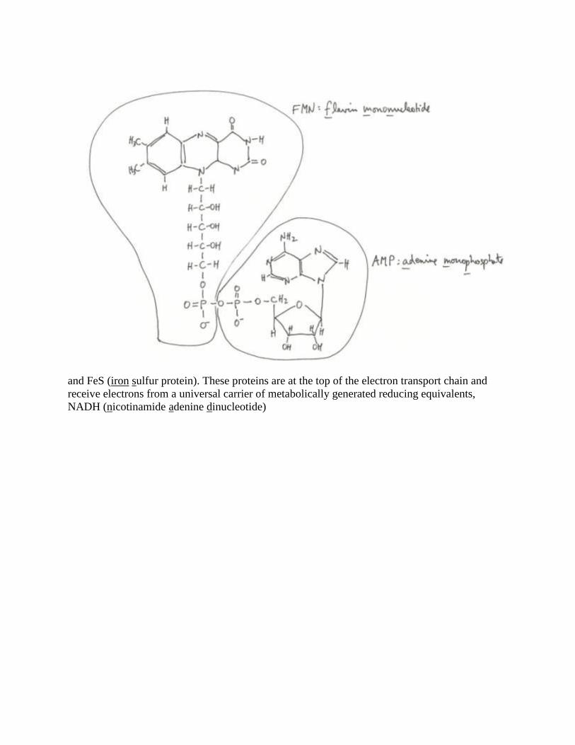
and FeS (iron sulfur protein). These proteins are at the top of the electron transport chain and
receive electrons from a universal carrier of metabolically generated reducing equivalents,
NADH (nicotinamide adenine dinucleotide)

This reduced molecule is generated by glycolysis and the citric acid cycle, energy metabolism
pathways discussed in [energy metabolism]. Two electrons are passed from NADH to the FAD
binding protein and then to the FeS proteins. These transfers are simple oxidation-reduction
reactions. However, some are pure electron transfers while others are transfers of both an
electron and a proton.
Indeed, three types of oxidation-reduction reactions occur in biology and all three are
exhibited in this intial stage of electron transport. FAD is reduced to FADH2 by a double
reduction that involves two electrons and two protons forming the two hydrogen atoms of
FADH2 as is depicted in the figure

NADH, on the other hand, gives up two electrons and only one proton when it is oxidized
by FAD to form NAD+ and FADH2. The extra proton needed by FADH2 for complete reduction
is taken up from a free proton on the inside of the bacterium (actually from a hydronium ion
H3O+).
223 FADHOHNADFADOHNADH
The FeS proteins, on the other hand, engage in pure electron exchanges, i.e. their oxidation-
reduction reactions are ferric-ferrous transitions involving the transfer of single electrons. Clearly
something must be done about the protons that appear in the first two steps. By positioning the
transfer of electrons from FADH2 to FeS adjacent to the exterior surface of the lipid bilayer, the
protons can be extruded to the exterior of the bacterium while the electrons are transferred to FeS
proteins. Thus, the arrangement of the proteins in the membrane results in a current-current
coupling of electron flow to proton flow in which protons are transported across the membrane.
This is called vectorial chemistry. Since the membrane is impermeable to protons (and to H3O+),
this creates a transmembrane electrochemical potential made up of a charge imbalance and a pH
imbalance. This is the essence of the chemiosmotic theory for the energized state of the intact
membrane. The two FeS proteins have different affinities for electrons. While their iron-sulfur
centers in isolation would have identical electron-transfer potentials, in the context of a
surrounding protein they have different potentials because the proteins are different. This permits
the sequential and preferential transport of electrons from one FeS protein to the next.
2121 2332 proteinSFeproteinSFeproteinSFeproteinSFe
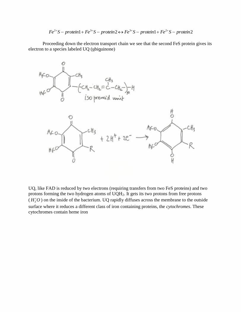
Proceeding down the electron transport chain we see that the second FeS protein gives its
electron to a species labeled UQ (ubiquinone)
UQ, like FAD is reduced by two electrons (requiring transfers from two FeS proteins) and two
protons forming the two hydrogen atoms of UQH2. It gets its two protons from free protons
( OH
3 ) on the inside of the bacterium. UQ rapidly diffuses across the membrane to the outside
surface where it reduces a different class of iron containing proteins, the cytochromes. These
cytochromes contain heme iron

and their elecron-transfer potentials are tuned by the surrounding protein, as in the case of the
different FeS proteins. These different cytochromes are labeled with subscripts accordingly. As
with the transfer of electrons from FADH2 to FeS proteins, the transfer of electrons from UQH2
to Cytb (cytochrome b) requires release of the protons to the exterior of the bacterium. Once
again we have vectorial chemistry and the current-current coupling of electron flow to proton
flow. Many replicas of the oriented cytochrome complex occurs in the bacterium’s plasma
membrane. In liver mitochondria there may be 5000 of these complexes and in heart
mitochondria the number is 20,000. They appear to be uniformily distributed so that there is one
every 400-500 nm2 of inner mitochondrial membrane area. The FeS complex and the cytochrome
complex are connected by the freely shuttling UQ. The final oxidation step is the oxidation of
cytochrome o by desolved molecular oxygen, O2. The product of this reduction is a molecule of
water. In mitochondria the process is somewhat more complicated and cytochrome c connects
the UQ-cytochrome c reductase complex to the cytochrome c oxidase complex by free diffusion
along the outside surface of the bilayer

Overall for bacteria, two electrons have traveled through the membrane from NADH to
H2O. They have remained on the inside of the bacterium while going downhill energetically
through a series of spontaneous oxidation-reduction reactions. At the same time one proton from
NADH and five protons from the interior of the bacterium have also been coupled to the electron
current. Two protons have ended up in H2O on the inside of the bacterium but the other four
have been transported vectorially across the membrane to create the energized membrane
potential. The energy inherent in the difference of electron-transfer potential between NADH and
O2 has been harvested as membrane energy captured in the disequilibrium of proton
concentrations on the inside and outside of the bacterium. This type of energy is readily available
for powering various transport processes and other membrane associated mechanisms but it is
not suitable for the emense demand for energy for synthetic purposes. This demand is met by the
universal energy currency for synthesis, ATP

ATP synthesis
ATP has been called the universal energy currency of cells. Indeed, it is the primary
source of energy for the synthesis of proteins, polynucleotides, polysaccharides and lipids. It is
also essential for a wide variety of allosteric control processes that are triggered by
phosphorylations, and dephosphorylations, of susceptible groups on proteins. In addition, ATP
plays a central role in the reaction cycles that result in the dynamics of actin and myosin
associations in muscle fibers and the dynamics of kinesin and dynein on microtubules. It is the
precursor to cyclic-AMP (adenosine monophosphate) that serves as the second messenger in
hormone actions. In other processes, GTP (guanodine triphosphate), CTP (cytodine triphosphate)
and UTP (uridine triphosphate) may be directly involved and may lose a phosphate. These
variants are recharged to triphosphates by ATP. Thus ATP does play many different essential
roles although the energized membrane does too. It is the energized membrane that is responsible
for the bulk of ATP synthesis (some ATP is generated by glycolysis).
The protein complex in the membrane that generates ATP is called an F-type ATPase. It
is a quite large protein complex with one portion, the Fo portion, embedded in the membrane and
spanning the entire width of the membrane, and with another portion, the F1 portion, attached to
the Fo portion on the inside of the membrane.
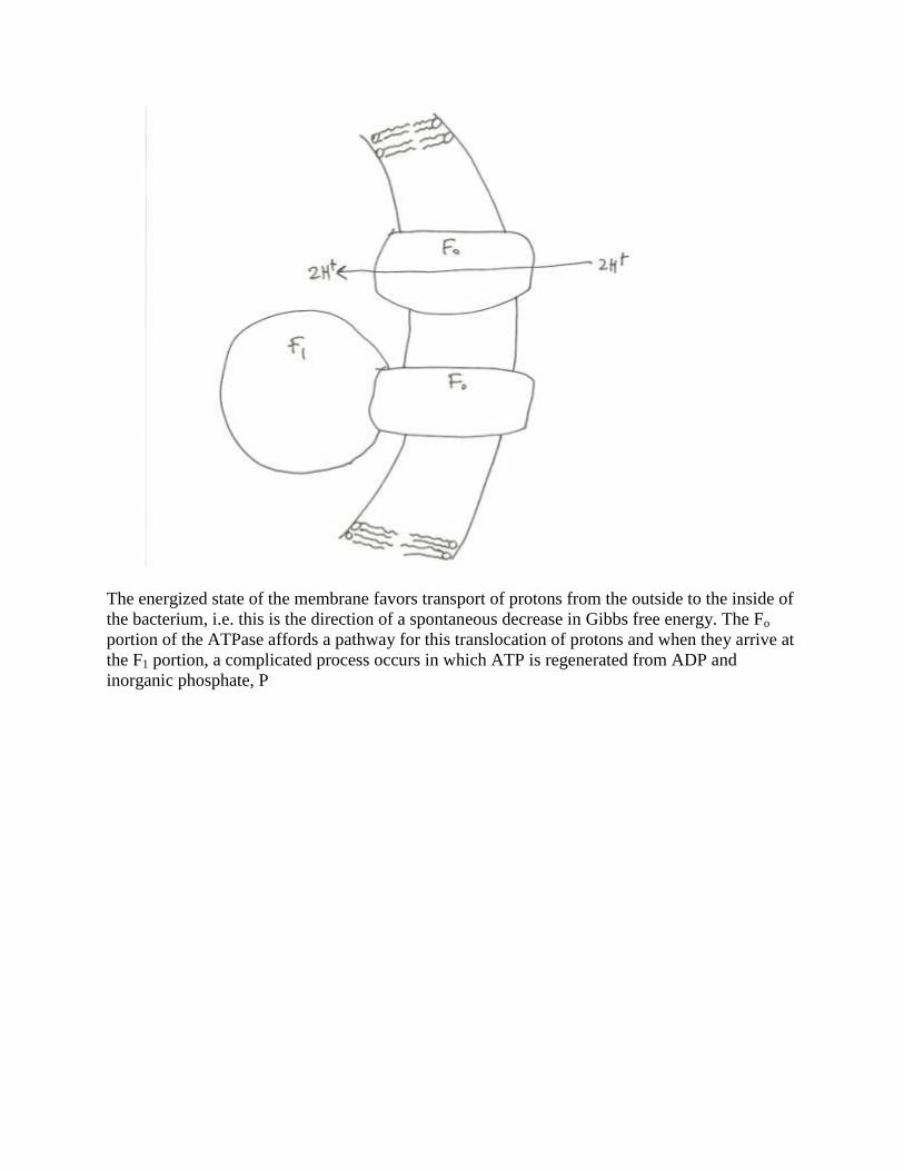
The energized state of the membrane favors transport of protons from the outside to the inside of
the bacterium, i.e. this is the direction of a spontaneous decrease in Gibbs free energy. The Fo
portion of the ATPase affords a pathway for this translocation of protons and when they arrive at
the F1 portion, a complicated process occurs in which ATP is regenerated from ADP and
inorganic phosphate, P

Just as there are many replicas of the FAD-FeS protein complexes and the cytochrome
complexes in the bacterial membrane, there are many replicas of the F-type ATPases. This
redundancy supports the huge demand for ATP turnover by the cell. Typically, a cell has enough
ATP for its energy and other needs for the order of a minute. Thus it must be regenerated from
metabolic energy sources. A 185 pound man at rest consumes and regenerates ATP at the rate of
~ 4 mol/hr (this is equivalent to 2 kg/hr) and at 10 times this rate during strenuous activity.
Biochemical thermodynamics
In order to make these energy transactions quantitatively precise, a review of biochemical
thermodynamics is needed. Oxidation-reduction (redox) processes always involve pairs of
molecules: one that is the oxidized form and one that is the reduced form of some underlying
molecular structure. For example, quinone (not to be confused with UQ which has an isoprenoid
tail) has the two forms

This redox pair is denoted by (Q/QH2), an example of the generic form (Aox/Ared) for the generic
molecular species A. Other examples of redox pairs include the ferri-ferro pair (Fe3+
/Fe2+
) and
the coenzyme NAD: (NAD+/NADH). The generic event is the transfer of electrons to the
oxidized form, thereby reducing it. For (Fe3+
/Fe2+
) that is all that happens, a pure electron
transfer. In (Q/QH2) a proton accompanies each of two electrons for a complete reduction. The
(NAD+/NADH) redox pair requires two electrons and one extra proton for reduction
NADHHeNAD 2

There is always an electron transfer, but there may or may not be a proton transfer too.
Typically, biology occurs at constant temperature, T, and pressure, P. Thus, the Gibbs
free energy, G, is the governing thermodynamic potential. The change in G, G, is what is
important. The second law of thermodynamics dictates that in a spontaneous process at constant
T and P G must be negative. Consider the cytochrome reaction that occurs in mitochondria
2332 FeCytFeCytFeCytFeCyt cbcb
in which an electron is transferred from Cytb to Cytc. The change in Gibbs free energy is written
]][[
]][[log3.2
32
23
10
0
FeCytFeCyt
FeCytFeCytRTGG
cb
cb
In this expression R is the gas constant given by the product of Avogadro’s number (6.0225 x
1023
molecules/mol) and Boltzmann’s constant kB (1.381 x 10⁻¹⁶
erg/K). This yields R = 8.317 x
107 ergs/K-mol = 1.987 cal/K-mol. The conversion identities 1 cal = 4.186 J (Joule) = 4.186 x
107 ergs have been used here. The 2.3 (actually 2.3025) comes from the conversion of Naperian
logarithms to base 10 logarithms. The expression [M] where M is a particular molecular species
denotes the concentration of M in moles per liter.
In standard state all reactants are at 1 molar by convention. Thus, at standard state

0GG
One mole of electrons has a charge of -1.0 F (Faradays) which is 9.648 x 10⁴ C (Coulombs).
Thus 1 F = 9.648 x 10⁴ J/V = 9.648 x 10
4 x 1/4.186 cal/V = 23.05 Kcal/V where V stands for
volts. Common practice is to express redox energetics in terms of electrical potential, i.e. volts
instead of in terms of Gibbs free energy. This is done by the identity
F
GE
Therefore the cytochrome redox reaction above is described by
]][[
]][[log3.2
32
23
10
0
FeCytFeCyt
FeCytFeCyt
F
RTEE
cb
cb
Since electrons are negatively charged, a spontaneous change is one in which E increases. The
quantity E0 is obtained from a table of redox potentials.

We find from the table that

E0 = 0.22 V for (CytcFe
3+/CytcFe
2+)
E0 = 0.12 V for (CytbFe
3+/CytbFe
2+)
Therefore, E0 = E
0final - E
0initial where final and initial are determined by the reduced member of
the pair. Thus, in this example, CytcFe2+
is the reduced product so that E0
final = 0.22 V. Therefore,
E0 = 0.22 V - 0.12 V = 0.10 V. This is a potential, not an energy. When multiplied by -F, an
energy is obtained: G0 = - 9648 J. This means that in standard state Cytb gives an electron to
Cytc.
A pH dependent redox reaction is the oxidation of ubiquinone by ferri-cyanide
OHcyanideFeUQcyanideFeUQHOH 3
23
22 2222
The redox potential change for this process is given by
23
2
22
10
0
2
2
23
2
2
3
22
10
0
]][[
]][[log
23.23.2
][]][[
][]][[log
23.2
cyanideFeUQH
cyanideFeUQ
F
RTpH
F
RTE
OHcyanideFeUQH
OHcyanideFeUQ
F
RTEE
Several points need to be emphasized about this expression. Firstly, the 2F in the first equality
reflects the fact that 2 electrons are transferred in this reaction. Secondly, since pH = -
log10[H3+O] and [H3
+O] is squared in the first line of the formula, the pH term in the second line
is multiplied by a factor of RT/F, not RT/2F. Thirdly, the [H2O] factors have been omitted in the
second line since the convention for the E0 values is that [H2O] is at standard state, i.e. has the
value 1, the true value of nearly 55.55 molar being built into the table entries automatically. At T
= 300K, a good approximation for 2.3 RT/F is 0.06 V or 60 mV (millivolt). Thus, each pH unit is
worth 60 mV. If protons are released during a reaction, then at pH 7, E is larger by 0.42 V per
mole of protons. If protons are consummed, then E is reduced.
G0 for H2 is zero by convention. In the redox pair (H
+/H2) H2 is on the product side and
H+ is on the reactant side. Therefore, at pH 7, E
0 for (H
+/H2) is - 0.42 V. Since pH 7 occurs
frequently, the convention is to write 0E = - 0.42 V. The prime on this redox potential signifies
that pH 7 applies. As an example consider the reduction of pyruvate to lactate by NADH
lactateOHNADpyruvateOHNADH
23
The redox potential change for this process is given by

]][[
]][[log
23.2
]][[
]][[log
23.2
23.2
]][][[
]][][[log
23.2
10
0
10
0
3
210
0
pyruvateNADH
lactateNAD
F
RTE
pyruvateNADH
lactateNAD
F
RTpH
F
RTE
pyruvateOHNADH
lactateOHNAD
F
RTEE
wherein 0E is determined from the table for pH 7, and the standard state for water is built into
these values 0E = - 0.19V - (- 0.32V) = 0.13V. Again the factors of 2 represent the
participation of 2 electrons in the reduction. Since only one external proton is required because
NADH provides two electrons and one proton, the pH term retains the 2 in the denominator.
Membrane potentials
Consider the schematic diagram representing the current-current coupling of electron
flow to proton translocation across the membrane
This figure captures the essence of what happens in bacteria and in mitochondria. The electrical
potential inside and outside the membrane compartment is denoted by in and out respectively.
Define by = out - in and pH by pH = pHout - pHin. Clearly, the result of active

electron transport is > 0 and pH < 0. The Gibbs free energy per particle is called the
chemical potential, . Therefore,
][
][ln||
in
outBinout
H
HTke
For a mole of protons, this expression needs to be multiplied by Avogadro’s number, NA, which
yields
pHRTFNA 3.2
The left hand side is defined to be Fp where p is called the protonmotive force. Clearly,
pHF
RTp 3.2
This terminology is misleading since p is not a force but an electrical potential. For the figure
above, p > 0. Since protons have positive charge, they will spontaneously move towards more
negative electrical potentials, the opposite of electrons. The tendency to do so is partly electrical,
, and partly osmotic or chemical, pHF
RT 3.2 . Peter Mitchell, the primary proponent of
this point of view early on, termed this chemiosmosis.
How big are p, , and pH for bacteria, mitochondria and chloroplasts?

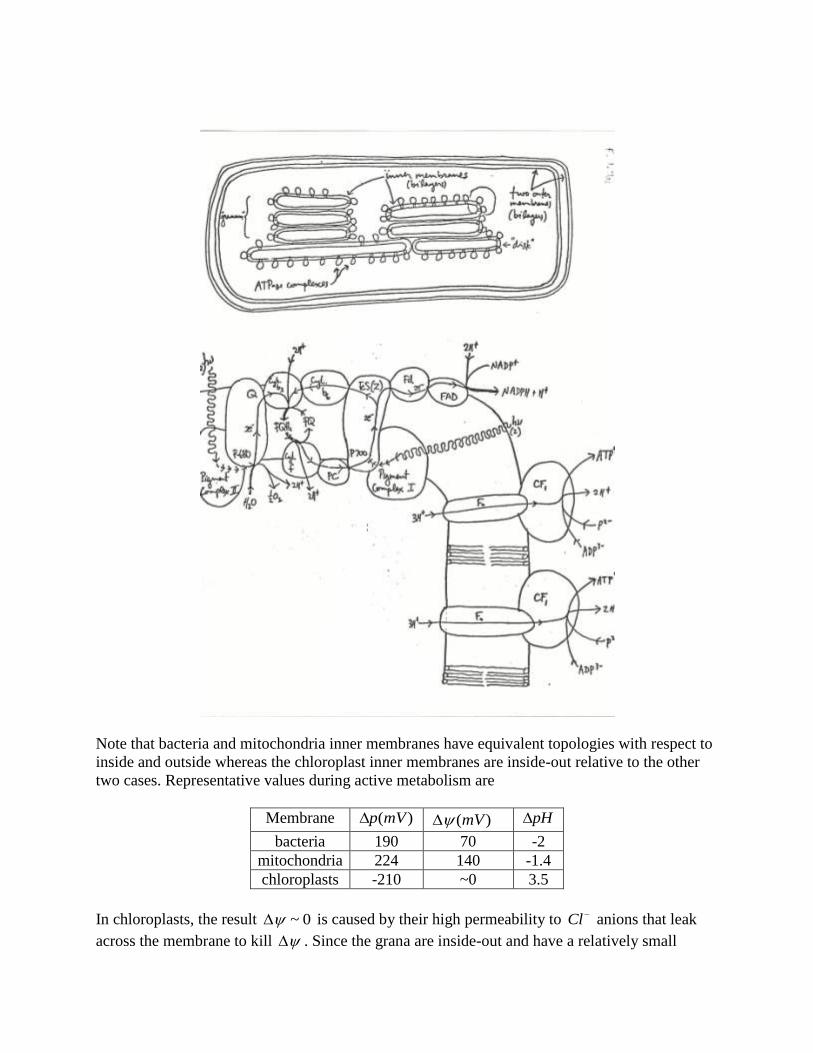
Note that bacteria and mitochondria inner membranes have equivalent topologies with respect to
inside and outside whereas the chloroplast inner membranes are inside-out relative to the other
two cases. Representative values during active metabolism are
Membrane )(mVp )(mV pH
bacteria 190 70 -2
mitochondria 224 140 -1.4
chloroplasts -210 ~0 3.5
In chloroplasts, the result 0~ is caused by their high permeability to Cl anions that leak
across the membrane to kill . Since the grana are inside-out and have a relatively small

interior volume, it is possible to create a larger pH than bacteria or mitochondria do that
compensates the 0~ .
These are steady state results, not equilibria. The energy required to move a proton
through an electrochemical potential p of approximately 200 mV is 1.6 x 10-19
x 0.2 V = 3.2 x
10-20
J = 3.2 x 10⁻¹³
ergs. This is about 8 kBT at T = 300 K. This energy must be supplied by the
electron transport chain energy.
The decomposition of p into and pHF
RT 3.2 permits experimental tests of
chemiosmosis that selectively check these two terms. Two distinct K+ transport systems are
ideally suited for this purpose, valinomycin and nigericin. Valinomycin, a dodecapeptide with
hydrophobic, lipophilic amino acid residues, binds K+ and can diffuse across the lipid membrane.
If a membrane is created by electron transport, then K+ will achieve an equilibrium
distribution across the mebrane with respect to this in which outKinK . Equilibrium
with respect to is given by
]ln[]ln[ ininoutout KRTFKRTF
This is equivalent to
RT
F
K
K
out
in exp
][
][
Since valinomycin transport has no H+ dependence, there is no dependence on the pH term in
p. The ionophore, nigericin, however, is a membrane associated polypeptide antiporter of K+
and H+.
inoutoutin HKHK
With steady state values of and pHF
RT 3.2 imposed by electron transport the overall
chemical potential change associated with this exchange can be written
0][
][ln||3.2||
in
outBB
K
KTkepHTke
in which the first two terms refer to protons going from outside to inside and the last two terms
refer to potassium ions going from inside to outside. As long as electron transport maintains pH
< 0 and nigericin is available for reverse flow of H+, K
+ ions will be transported outwardly until
an equilibrium for K+ ions is reached given by the expression above or its equivalent
pHK
K
in
out
3.2][
][ln

Note that in the case, 1][
][
out
in
K
K and in the pH case 1
][
][
in
out
K
K. With these two processes,
the two contributions to p can be separately measured.
Mitchell cycle for mitochondria
Peter Mitchell revived and championed the idea of chemiosmosis during the the 1960s
and 1970s. At first he had little support from others but eventually prevailed and was awarded
the Nobel prize in chemistry for his work in 1978. The problem he confronted was how electron
transport was coupled to ATP synthesis. This was a problem dating back to the 1940s. The
difficulty in finding a solution was that biochemists persisted in looking for chemical
intermediates such as are found in glycolysis. The techniques for looking for chemical
intermediates required breaking apart the bacterial or mitochondrial membranes. Since the
mechanism of coupling requires an intact membrane in order to maintain the chemiosmotic
electrochemical transmembrane potential, these methods failed to provide a solution. Mitchell’s
recognition of the importance of the coupling of electron current to proton current and
subsequent coupling of proton current to phosphorylation was the key to finding a solution.
In bacteria, the quinone (UQ) cycle translocates 2 H+’s for each pair of electrons
transported down the electron transport chain. In mitochondria, it appears that 4 H+’s are
translocated instead. Mitchell proposed a mechanism, called the Mitchell cycle, for this that is
now amply justified experimentally. In mitochondria, the quinone species is called CoQ
(coenzyme Q) and is virtually identical with the UQ of bacteria and the redox pair is
(CoQ/CoQH2). However, in the Mitchell cycle, a semiquinone plays a role. This species carries
an electron on one quinone oxygen atom and has a negative charge. It is denoted by CoQ . The
mitchell cycle operates in two stages called cycle 1 and cycle 2

In cycle 1, two FeS proteins of the upper portion of the electron transport chain together with 2
protons from the matrix side of the inner mitochondrial membrane reduce CoQ to CoQH2 on the
matrix side of the membrane. This CoQH2 freely diffuses across the membrane to the
intermembrane space side. It releases 2 protons to the outside and reduces a special FeS protein
with one electron leaving the semiquinone CoQ . The FeS protein reduces Cytc1. The
semiquinone transfers its one electron to CytbL and becomes the oxidized quinone CoQ. CytbL
reduces CytbH. The CoQ freely diffuses back across the membrane to the matrix side. The
arrangement of the cytochromes CytbL and CytbH in the membrane interior is such that when
CoQ gets back to the matrix side, it is partially reduced to the semiquinone CoQ by CytbH. Thus
the charged species CoQ does not freely diffuse across the membrane but is regenerated when
the neutral species CoQ has done so. This concludes cycle 1 with one CoQH2 having been
converted into CoQ , one Cytc1 having been reduced and two protons having been translocated
across the membrane. In cycle 2, another CoQH2 generated from two FeS proteins in the upper
portion of the electron transport chain along with two protons from the matrix diffuses across the
membrane to the intermembrane space side. Again, it releases two protons to the outside and
reduces one Cytc1 through the intermediation of a special FeS protein, creating a semiquinone.
This semiquinone in turn reduces CytbL which reduces CytbH and yields a fully oxidized CoQ
that diffuses freely across the mebrane back to the matrix side. The CoQ produced in cycle 1 is
now reduced by the reduced CytbH just created in cycle 2 and this requires two protons from the
matrix side. The result is regeneration of CoQH2. Thus, the overall net result is that one CoQH2
is converted into one CoQ, two oxidized Cytc1’s are reduced and two matrix protons are taken

up. The two protons from CoQH2 that are originally from the matrix and the two protons from
the matrix during cycle 2 are extruded to the outside for a total of four protons extruded for two
electrons transported to two Cytc1’s. The results in a more efficient harvesting of electron energy
as proton energy then occurs in the simple quinone cycle used by bacteria in which only two
protons are translocated for two electrons.
Transporters
The efflux of protons from the membrane compartment that results from current-current
coupling of electron flow and proton translocation builds up the chemiosmotic membrane proton
potential
pHF
RTp 3.2
This potential provides an impetus for protons to re-enter the compartment. To do so, however,
requires a functional pathway. Many such pathways exist and are created by specific proteins
embedded in the membrane. All sorts of current-current couplings of proton re-entry with other
solute species’ fluxes across the membrane occur. Each requires a specific protein transporter. In
the the following examples, the notation A = Aout - Ain will be used where A stands for any
molecule. It will be assumed that metabolism is at steady state and that the proton potential
across the membrane is maintained constant. The coupled solute will come to equilibrium with
respect to the nonequilibrium steady state value of p.
The figure below shows an example of obligate co-transport of a neutral solute species,
S0, into the membrane compartment
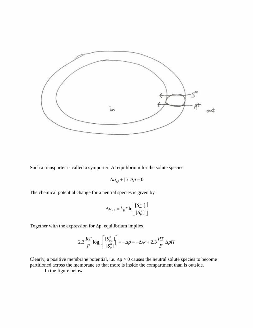
Such a transporter is called a symporter. At equilibrium for the solute species
0||0 peS
The chemical potential change for a neutral species is given by
][
][ln
0
0
0
in
outBS S
STk
Together with the expression for p, equilibrium implies
pHF
RTp
S
S
F
RT
in
out
3.2
][
][log3.2
0
0
10
Clearly, a positive membrane potential, i.e. p > 0 causes the neutral solute species to become
partitioned across the membrane so that more is inside the compartment than is outside.
In the figure below

a symporter that couples the re-entry of two protons to a neutral solute species is depicted. This
produces the equilibrium equation for the solute given by
pH
F
RTp
S
S
F
RT
in
out 3.222][
][log3.2
0
0
10
Clearly, a greater ratio of solute inside to outside is created when two protons are coupled to the
transport of one molecule of solute than is the case for one proton coupling. Notice that ratio of
solute concentrations outside to inside for a two proton symporter is the square of the ratio for a
one proton symporter.
There also exist transporters that are not coupled to obligate proton re-entry but that
couple directly to the electrical potential portion of p, . This requires that the transported
species be charged. For a cation, S+, transport to the inside takes place

In this case the equilibrium is given by
][
][log3.2 10
in
out
S
S
F
RT
because the chemical potential for the cation is given by
][
][ln||
in
outBS S
STke
and the electrical potential, , is maintained constant. Clearly, a positive membrane electrical
potential, i.e. > 0, will result in the influx of the cation.
Now consider a cationic symporter.

The equilibrium condition becomes
pHF
RT
S
S
F
RT
in
out 3.22][
][log3.2 10
Especially notice the factor of 2 in the electrical potential term caused by two positive charges
going inside.
Somewhat more subtle is the symport of an anion, S-.

The chemical potential for the anion is given by
][
][ln||
in
outBS S
STke
The condition for equilibrium for the anion is
0|| peS
which implies
pHF
RT
S
S
F
RT
in
out
3.2][
][log3.2 10
Since this symporter does not transport net charge the electrical potential portion of p does not
contribute and only the pH difference does.
An anion symporter requiring two protons

can be shown to lead to the equilibrium condition for the anion
pHF
RT
S
S
F
RT
in
out
3.22][
][log3.2 10
This there is a partial cancellation of the electrical potential terms and a doubling of the pH term.
There also exist protein transporters that move ions in the opposite direction to the proton
re-entry. These are called antiporters. Calcium is translocated by a calcium antiporter that is
coupled to two proton re-entry

The process may be represented by the vectorial reaction
inoutoutin HCaHCa 22 22
The equilibrium condition can be written as
ininoutoutB
outoutininB
epHeCaTk
epHeCaTk
||23.22||2]ln[
||23.22||2]ln[
2
2
This can be rewritten as
pHF
RT
Ca
Ca
F
RT
in
out
3.22][
][log3.2
2
2
10
Clearly, a lower pH on the outside compared to the inside will help drive calcium ions outside.
Similar cationic antiporters exist for potassium, K+, and for sodium, Na
+

Each of these antiporters couples just one proton to the cation flux. The equilibrium result for
sodium is
pHF
RT
Na
Na
F
RT
in
out
3.2][
][log3.2 10
Replacing Na with K gives the comparable result for potassium.
As an example, consider a cation symporter using one proton in a bacterium membrane
with = 70 mV and pH = -2. Exponentiation of the equilibrium result above for this case
yields
3108.514.5exp
3.22exp][
][
pH
F
RT
RT
F
S
S
in
out
Thus, the concentration of S+ on the inside is 171 times that on the outside.
Each of the examples described above occurs for some molecular species. A great many
transport systems exist in the membrane and they are responsible for the traffic of metabolites
into and out of the membrane compartment. The energy for these processes is supplied by the
chemiosmotic membrane potential.

Electron transport details
It was mentioned earlier that Cytc takes electrons from Cytc1 to the cytochrome oxidase
complex where the electrons ultimately reduce oxygen to water. Cytc does its job by freely
diffusing along the outer surface of the mitochondrial inner mebrane. Cytc is a highly conserved
protein that has a conformation that has remained constant for over a billion years. Cytc from any
eucaryotic species will react in vitro with cytochrome oxidase from any other species. The amino
acid sequences for over eighty Cytc species have been determined. 26 of the 104 amino acid
residues have been invariant for over 1.5 billion years. Cytochrome oxidase complex contains
copper atoms. These copper atoms are an integral part of the reaction center where oxygen is
reduced. The active site involves a complex of copper with sulfur and iron and the reaction takes
about one msec. The FeS proteins (also called ISPs for iron sulfur proteins) come in several
varieties that basically differ in the number of iron atoms involved in the iron sulfur complex. In
bacteria only, the simplest case occurs [Fe]
In [Fe] a single iron atom is coordinated by four sulfur atoms that are on cysteine residues of the
protein. In mitochondria, [2Fe-2S] and [4Fe-4S] complexes occur as well
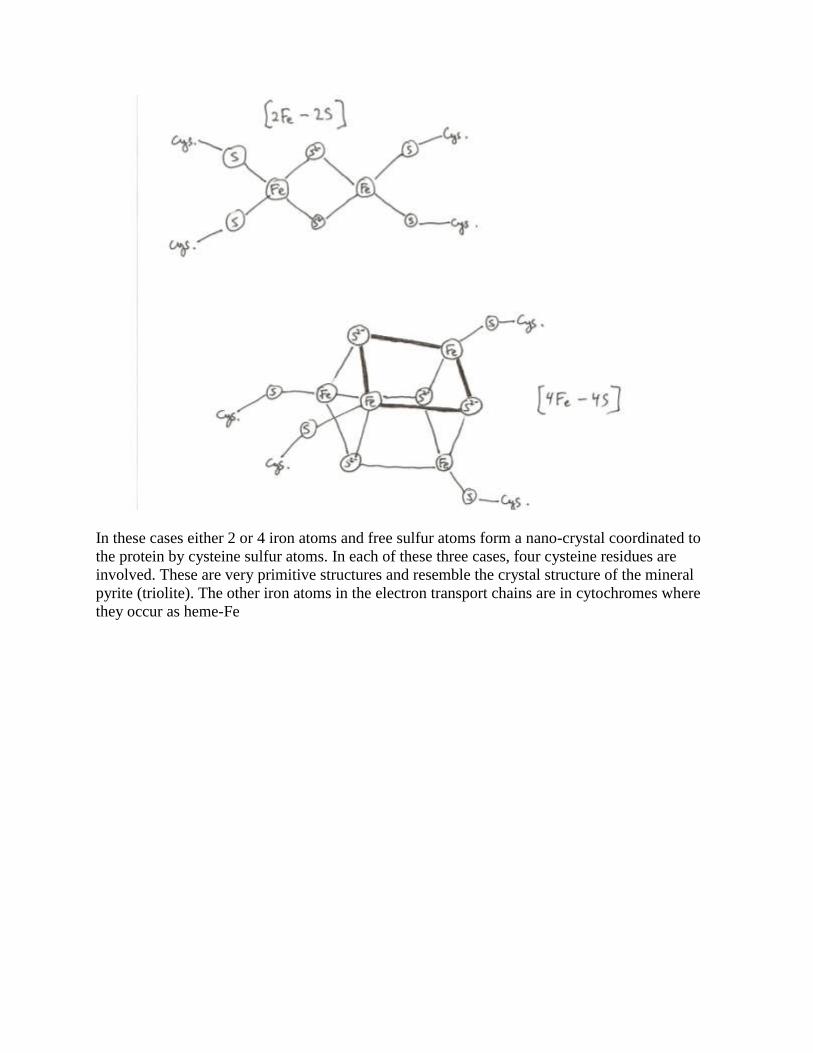
In these cases either 2 or 4 iron atoms and free sulfur atoms form a nano-crystal coordinated to
the protein by cysteine sulfur atoms. In each of these three cases, four cysteine residues are
involved. These are very primitive structures and resemble the crystal structure of the mineral
pyrite (triolite). The other iron atoms in the electron transport chains are in cytochromes where
they occur as heme-Fe

Heme-Fe not only occurs in cytochromes but also in the hemoglobins and myoglobins of blood.
In the latter cases, heme-Fe serves as a carrier of O2 molecules whereas in the cytochromes it is
the active site for redox exchanges of electrons.