Lipid patterns of edible fungi 18-suppl. 1978-08.pdf · identification of lipid classes. Materi als...
7
Xarstenia 18(supp!.) 1978 Lipid patterns of edible fungi D. TORLEY and E. V ADON-GYOREY TORLEY, D. A VADON-GY6REY, E. 1978; Lipid patterns of edible fungi.- t<arstenla 18 (suppl.). In the course of the investigation of the I ipids of fungi by column chromatography several neutra I and para I I i pi d c I asses have been separated which were ana 1 yzed by TLC. The distribution of the 1 ipid classes and the number of components in the fractions showed characteristic differences In the investigated mushroom species. Even members of the same fungus family differ from each other In the I ipid composition. Several Indicators were used to vi sua I i ze the spots: i ad i ne vapour 1 Dragendorf f reagent 1 L i eber mann-Burchard re- agent 1 n. i nhydr [ ne so lyt ion, Hanes reagent, iron-suI fosa I icy I i c acid reagent. UV rad i at; on. V. TiiiLtey t E. VepaMment o6 fl.Wchem.i.l..titq and Food Techno.l<Jgy, Technico.l UMv<M<-ty Budapu.t, Muegye.tem 3 H-1111 Budapu.t, HungaJty. Introducti on The literature dealing with fungal lipids is by means complete. At the beginning of the century Landsiedel & Bamberger (1), Rosenthal (2), Zellner and his co -workers (3, 4, 5) isolated sterols and cerebrosides from several fungus species. After a long'pause the research on the lipids of fungi has continued in the past two decades, so that in recent years considerable information on fungal lipids has accumulated. Brennan & co-workers (6) re v iewed this topic in 1975. Though with the introduction of the thin-layer chromatography and infrared and mass spectrometry extensive lipid analysis of fungi has started, the research is far from being finished. The published results show that in these investigations some lipid classes received more attention than others, and the information is not yet complete. In Hungary the lipids of edible fungi had not been investigated earlier, therefore we began only two years ago this research work on cultivated and wild mushrooms in We started with the search for suitable methods for the isolation and identification of lipid classes. Mater i al s and methods The cultivated mushrooms were provided by the trade and the wild growing species were collected in 1976 in the neighbourhood of Budapest. The mushrooms were homogenized with a double volume of acetone (w/2 v) and filtered with suction. After rehomogenizing with chloroform-methanol 2:1, the combined acetone and the combined chloroform -methanol filtrates were concentrated to dryness. The residue was shaken after the addition of chloroform -methanol-water 80:40:30 according to Felch et al. (7). Two phases were formed; the upper phase was dis- carded and the same amount of fresh upper phase was added. After miXing, the lower phase was concentrated to dryness and the residue ser ved as sample for the investigations. The isolation of the individual lipid clas s ec was performed by different chromatographic techniques. The identification was made partly with standards, partly on the basis of data in the literature. Analytical thin-layer chromatography (TLC) was performed on 20 x 20 em glass plates using Kieselgel G (according to Stahl). The best solvent systems were chosen by experiments. For the isolation of neutral lipids the n-hexane-diethylether-acetic acid 80:20:1 system was best suited. In the case of· polar lipids chloroform-methanol-water 65:25:4 and 65:25:2 systems were used after saturating the vessel at 25° C for 1 hour. The detection of the lipid classes wa s performed with iodine vapour (all lipids) and with the following spray reagents: modified Liebermann-Burchard reagent for sterols, ninhydrine solution for free amino groups, Dragendorff reagent for other nitrogen-containing compounds, reagent of Hanes for phosphate esters, orcinol reagent for carbohydrates, resorcinol reagent for carbohydrates . Res ults a nd discuss i on The separation of the polar and neutral lipids was performed by column chromatography. Kieselgur G column was prepared with chloroform and the residue of the mush- room dissolved in chloroform, was · applied to it. The column was eluted successively with chloro- form (neutral lipids) and methanol (polar lipids) . In the similar way a Kieselgur G column was prepared with
Transcript of Lipid patterns of edible fungi 18-suppl. 1978-08.pdf · identification of lipid classes. Materi als...

Xarstenia 18(supp!.) 1978
Lipid patterns of edible fungi
D. TORLEY and E. V ADON-GYOREY
TORLEY, D. A VADON-GY6REY, E. 1978; Lipid patterns of edible fungi.- t<arstenla 18 (suppl.).
In the course of the investigation of the I ipids of fungi by column chromatography several neutra I and para I I i pi d c I asses have been separated which were ana 1 yzed by TLC. The distribution of the 1 ipid classes and the number of components in the fractions showed characteristic differences In the investigated mushroom species. Even members of the same fungus family differ from each other In the I ipid composition. Several Indicators were used to vi sua I i ze the spots: i ad i ne vapour 1 Dragendorf f reagent 1 L i eber mann-Burchard reagent 1 n. i nhydr [ ne so lyt ion, Hanes reagent, iron-suI fosa I icy I i c acid reagent. UV rad i at; on.
V. TiiiLtey t E. Vadon-G~iiJtey, VepaMment o6 fl.Wchem.i.l..titq and Food Techno.l<Jgy, Technico.l UMv<M<-ty Budapu.t, Muegye.tem Jt~p 3 H-1111 Budapu.t, HungaJty.
Introducti on
The literature dealing with fungal lipids is by means
complete. At the beginning of the century Landsiedel
& Bamberger (1), Rosenthal (2), Zellner and his co
-workers (3, 4, 5) isolated sterols and cerebrosides
from several fungus species. After a long'pause the
research on the lipids of fungi has continued in the
past two decades, so that in recent years considerable
information on fungal lipids has accumulated. Brennan
& co-workers (6) re viewed this topic in 1975. Though with the introduction of the thin-layer chromatography
and infrared and mass spectrometry extensive lipid
analysis of fungi has started, the research is far
from being finished. The published results show that
in these investigations some lipid classes received
more attention than others, and the information is not
yet complete. In Hungary the lipids of edible fungi
had not been investigated earlier, therefore we began
only two years ago this research work on cultivated
and wild mushrooms in Hu~gary . We started with the
search for suitable methods for the isolation and
identification of lipid classes.
Mater i al s and methods
The cultivated mushrooms were provided by the trade
and the wild growing species were collected in 1976
in the neighbourhood of Budapest. The mushrooms were homogenized with a double
volume of acetone (w/2 v) and filtered with suction.
After rehomogenizing with chloroform-methanol 2:1,
the combined acetone and the combined chloroform
-methanol filtrates were concentrated to dryness. The
residue was shaken after the addition of chloroform
-methanol-water 80:40:30 according to Felch et al.
(7). Two phases were formed; the upper phase was dis
carded and the same amount of fresh upper phase was
added. After miXing, the lower phase was concentrated
to dryness and the residue served as sample for the investigations.
The isolation of the individual lipid clas s ec
was performed by different chromatographic techniques.
The identification was made partly with standards,
partly on the basis of data in the literature.
Analytical thin-layer chromatography (TLC) was
performed on 20 x 20 em glass plates using Kieselgel
G (according to Stahl). The best solvent systems were
chosen by experiments. For the isolation of neutral
lipids the n-hexane-diethylether-acetic acid 80:20:1
system was best suited. In the case of· polar lipids
chloroform-methanol-water 65:25:4 and 65:25:2 systems
were used after saturating the vessel at 25° C for 1
hour. The detection of the lipid classes was performed
with iodine vapour (all lipids) and with the following
spray reagents: modified Liebermann-Burchard reagent
for sterols, ninhydrine solution for free amino groups,
Dragendorff reagent for other nitrogen-containing
compounds, reagent of Hanes for phosphate esters,
orcinol reagent for carbohydrates, resorcinol reagent for carbohydrates .
Results a nd discuss i on
The separation of the polar and neutral lipids was
performed by column chromatography. Kieselgur G column was
prepared with chloroform and the residue of the mush
room extr~ction, dissolved in chloroform, was · applied
to it. The column was eluted successively with chloro
form (neutral lipids) and methanol (polar lipids) . In
the similar way a Kieselgur G column was prepared with

Karstenia l8(suppl . ) 1978 23
benzene and the elution was performed with benzene and
acetone. The eluates were · evaporated to dryness at room
temperature. The fractions obtained on the chloroform
and benzene columns were compared by TLC. No difference
was found in the number _and the Rf-values of.the
components. At the start line the spots appeared very
crowded; for better isolation the development was
repeated after drying at room temperature.
Better separation of the spots with one devel
opment on TLC plates was obtained by the use of sili
cic acid column (Silicic acid Koch & Light, 325 mesh) .
The chloroform solution of the residue of the extrac
tion of Ctitocybe nBbularis was applied to the column
prepared with chlorof~rm. The elution with chloroform
yielded two sharply separated neutral lipid fractions
(l a and l b). The polar lipids were eluted with
methanol (fraction 2).
Fig . 1 shows the thin-layer chromatography
plate (neutral lipids) treated with iodine vapour in
dicator. In Fig. 2 the same chromatogram is shown
after detection with the Liebermann-Burchard reagent,
which was slightly modified by us. The green spots
of the sterols appear instantly, in fraction 1 a there
may be seen a yellow fluorescent s pot, which appears
also i .n the control substance ergosterol.
Fig. 1. Isolation of neutral and polar lipids of
CZitocybe nebuZaPia on silicic acid column .
I. TLC of neutral lipids.
Indicator: iodine vapour.
Spotted from left to right: fraction l a,
ergosterol, cholesterol, stearic acid, fraction
1 b, extraction residue.
0
{J
0 c::a 0 0 0 •
0
• 0
•
D
•
IJ
0 0 0
0
0 0
•
Fig. 2. Isolation of neutral and polar lipids of
CZitocybe nebutaris on silicic acid column.
I. TLC of neutral lipids.
Indicator: Liebermann reagent.
Spotted from to right: fraction 1 a,ergosterol,
cholesterol, stearic acid, fraction 1 b, ex
traction residue.
0 0
-0 0 .:..-.
0 0 ::
0 0 § ~ 0 0 Q • Fig . 3 shows the chromatogram of the ext r ac
tion residue of C. nebuZaris, soya lecithin,fraction
2 (i.e. polar lipids) of the mushroom eluted from the
silicic acid column in 3 different concentrations,
and soya phosphatide standard after detection with
Fig. 3. Isolation of neutral and polar lipids of
CZitocybe nebutaPis on silicic acid column.
I. TLC of neutral lipids .
Indi cator : iodine vapour.
Spotted from left to right: extraction residue,
soya lecithin, fraction 2 (in three concentra
tions), soya phosphatide.
v 0 ..... 0 ()'" 0 (\
., Lj D 0 0 D
0 0 • 0 0 0 0
' '
D 0 0 0 \J 0 0 0 •

24 Karstenia l8(suppl.) 1978
iodine vapour. In Rig. 4 the same chromatogram is
shown after detection with orcinol reagent. The well
separated three spots are sterol glycosides and cer
ebrosides. In Fig. 5 the same c~ramatogram is shown
after detection with ammonium molybdate (Hanes rea
gent). (In a later experiment the spots of the mush
room extract were separated into several components
by column chromatography).
To compare more clearly the occurrence of the
free mine groups of ethanolamine and the other nitro
gen -containing compounds, the thin - layer chromatogram
was sprayed successively with two reagents: the left
part with ninhydrine, the right part with Dragendorff reagent (Fig. 6) . The chromatogram shows that c. nebu
laPis contains only very small amounts of phosph~idyl
ethanolamine .
Fig. 4. Isolation of neutral and polar lipids of
Clitooybe nebutaris on silicic acid column.
II. TLC of polar lipids .
Spotted from left to right: extraction residue,
soya lecithin, fraction 2(in three concentrations), soya phosphatide.
0
'
0 0
0
{)
0 0 • 0
s 0
-•
8 CJ
0
0
•
0
0
<g 0
To obtain clearer isolation of the lipid classes,
experiments were made with preparative thin-layer
chromatography. The extraction residue of the mus~
was applied to the plate in the form of a band on the
start line, and after developing the detection was
performed with iodine vapour. The isolated bands were
scraped off, and after the sublimation of the iodine
the lipid fractions were eluted from the silica gel
with chloroform-methanol-water (1:2 : 0.8). The evapo
rated material was examined by analytical TLC. By this
method the lipids of the cultivated mushroom Agaricus
bisporus could be separated in the first step into 8
fractions, from which a further 4-5 spots were iso
lated by analytical TLC.
Fig. 5. Isolation of neutral and polar lipids of Clitocybe nebularis on silicic acid column. II. TLC of polar lipids.
Spotted from left to right: extraction residue,
soya lecithin, fraction 2 (in three concentra-tions), soya phosphatide .
\J co 0 0 v 8 L1
0 0 0 () 0 0 0
0 0 0 g 0 0 0 0 0
I 0 0
0 0 0
0 0
B ~ 0 0 D 0 0 0
D 0 0 p Q Q 0
Fig. 6. Isolation of neutral and polar lipids of
Clitocybe nebularis on silicic acid column .
II. TLC of polar lipids.
0
0
Spotted from left to right: fraction 2, ex
traction residue, soya lecithin, soya phosphatide.
Indicator: ninhydrine .
Fraction 2, extraction residue, soya lecithin, soya phosphatide.
Indicator: Dragendorff reagent.
0
'
0
Q 0
0
0 0 6
0
0 0 0
(}
0 0
0 D
Karstenia 18(suppl .) 1978 25
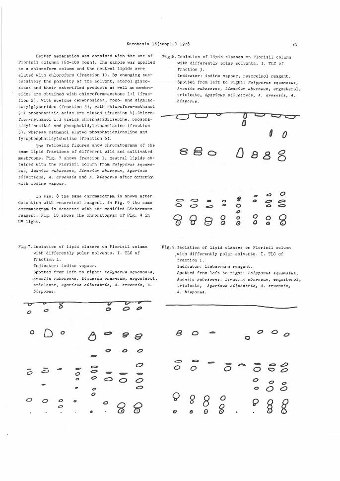
Better separation.was obtained with the use of
Florisil columns (60-100 mesh). The sample was applied
to a chloroform column and the neutral lipids were
eluted with chloroform (fraction 1). By changing suc
cessively the polarity of the solvent, sterol glyco
sides and their esterified products as well as cerebro
sides are obtained with chloroform-acetone 1:1 (frac
tion 2). With acetone cerebrosides, mono- and digalac
tosylglycerides (fraction 3), with chloroform-methanol
9:1 phosphatidic acids are eluted (fraction 4) . Chloro
form-methanol 1:1 yields phosphatidylserine, phospha
tidylinos itol and phosphatidylethanolamine (fraction
5), whereas methanol eluted phosphatidylcholine and
lysophosphatidylcholine (fraction 6).
The following figures show chromatograms of the
same lipid fractions of different wild and cultivated
mushrooms. Fig. 7 shows fraction 1, neutral lipids ob
tained with the Florisil column from Polyporus squama
sus, Amanita rubescens~ Cimacium eburneum, Agaricus
s i l. vatieus, A. arvens is and A. bisporus after detecti on
with iodine vapour.
In Fig. 8 the same chromatogram is shown after
detection with resorcinol reagent. In Fig . 9 the same
chromatogram is detected with the modified Liebermann
reagent. Fig. 10 shows the chromatogram of Fig. 9 in
uv light.
F~g.?.Isolation of lipid classes on Florisil column
with differently polar solvents. I. TLC of
fraction 1.
Indicator : iodine vapour.
Spotted from left to right: Potyporus squamo s us,
Amanita rubescens, Limacium eburneum, ergosterol,
trioleate, Agarieus si tvestris, A. arvensis, A.
bisporus .
0 0 0 v 0' • 0 0 0 p 0 0
0 D 0 6 c::;l 9 8
0 0 0 -~ 0 - - -0 c:::::>
0 ~ - - -0 0 c::::::> 0 0 0 0
0 0 0 0 0 0 g 0 ~ •
Fig.8. Isolation of lipid classes on Florisil column
with differently polar solvents. I. TLC of
fraction ,1.
Indicator: iodirie vapour, resorcinol reagent.
Spotted from left to right: Potyporus squamosus,
Amanita rubescens, Limacium eburneum, ergosterol,
trioleate, Agaricus sitvestris, A. arvensis, A.
bisporus.
CJ LJ C> 0 0
[J
c:;:, .:::> ... 0 f1 c <:::> ~ 0 0
~
CiJ 9 8 0 0 Q G
88
D 0
0 0 0
0 0 Q 0
0
0 0
0 p c:::>
g
Fig.9 . Isolation of lipid classes on Florisil column
with differently polar so l vents. I. TLC of
fraction 1 .
Indicator: Liebermann reagent.
Spotted from left to right: Potyporus squamosus,
Amanita rubescens, Limacium eburneum, ergosterol,
trioleate, Agaricus sitvestris, A. arvensis,
A. bisporus.
B 0 - 0 0 0 0
c::::> c:::. - ~ - - - ~ 0 0 0 0 0 0
0 0 0 0 0 0
Q 0 8 0 9 § g 0 0
Q e e GJ

26 Karstenia 18(suppl.) 1978
Fig . lO .Isolation of lipid classes on Florisil column
with differently polar solvents. II. TLC of
fraction 2.
Indicator: Liebermann reagent, i,lluminated with
uv light.
Spotted from left t.o right: Po ?..yporus squamosus,
Amanita rubescens, Limacium eburneum,ergosterol,
trioleate, Agaricus siZvestris, A. arvensis, A.
bisporus.
---<::)
-~ c::> -
--
-
0 0
Fig. 11 shows fraction 2 (polar lipids) of the
Florisil column detected by the modified Liebermann
reagent. The blue spots on the upper part of the
chromatogram cannot be ~etected in the original mush
room extracts; therefore they must be taken for arti
facts of the column chromatography.
Fig. 12 shows the carbohydrate-containing
components (fraction 3) of C. nebularis, Armillariella
mel~ea (grown on wood), A. mellea (grown on soil),
Clitocybe infundibuliformis~ Triohotoma _nudum and
Lepiota procera, detected with resorcinol reagent.
In Fig. 13 is shown the chromatogram of the
Florisil column fraction 3. The detection was made
first with iodine vapour, and after the sublimation
of the iodine with resorcinol reagent spray, heated
for some minutes to 100° C.
Fig. 14 shows the chromatogram of the phos
phatidic acid fraction (fraction 4). The detection
was made with ammonium molybdate solution (Hanes rea
gent). Fig. 15 shows fraction 5 containing phospha
tidylserine, phosphatidylinositol and phosphatidyl
ethanolamine, detected with iodine vapour, and Fig.
16 fraction 6 containing phosphatidylcholine and lyso
phosphatidylcholine, detected with ammonium molybdate.
The light blue spots in the lower part of the chromato
gram show the lysophosphatidylcholine content of the
fungi (Fig. 16) .
Fig.ll. Isolation of lipid classes on Florisil column
with differently polar solvents. II : TLC of
fraction 2.
0 0
Indicator: Liebermann reagent.
Spotted from left to right: Polyporua squamosus~
Amanita rubeacens~ Limacium eburneum,ergosterol,
trioleate, Agaricus ailveatria~ A. arvensis~
A. bisporua.
0 oo
- c:>
0
0 0 0 -
(!) 8 G> Q Q
Fig.l2Jsolation of lipid classes on Florisil column
with differently polar solvents. III. TLC of
fraction 3. Indicator : resorcinol reagent.
Spotted from left to right: Clitocybe nebularis,
Armittarietta mellea (grown on wood),A.mellea
(grown on soil), ergosterol, trioleate, Clito
cybe infundibiliformis~ Tricholoma nudum~Lepiota
procera.
v
0
-0
0
8 0
0 ao ~ ~
-o
- 0
0
0 0
0 0
0 0 &> !5 0 0
0
0 e 0 0
0
0 0
• 0

Karstenia l8(suppl.) 1978 27
Fig.l3~solation of lipid classes on Florisil column
with differently polar solvents. III. TLC of
fraction 3. Indicator: i od ine vapour, followed by resorcinol
reagent.
Spotted from left to right: Pol yporus squamosus~
Amanita rubescens, Limacium eburneum,ergosterol,
triolea te, Agaricus silvestris, A. arvensis,
A. bisporus.
0
0
0 0
Fig . l4 : Isolation of lipid classes on Florisil column with differently polar solvents. IV . TLC of
fraction 4. Indicator: Hanes reagent . Spotted from left to right: Polyporus squamosus
Amanita rubescens, Limaci.um eburneum, ergosterol
trioleate , Agaricus silvestris, A. arvensis,
A. bisporus.
v \7 VCJ (i <:::> C>' Z5 0 8 0
0 ~ 0
0 0
0 0 0 0 G 0
Fig.l5. Isolation of lipid classes on Florisil column
with differently polar solvents. V. TLC of
fraction 5. Indicator: iodine vapour.
Spotted from l eft to right; Polyporus aquamosus,
Amanita rubescena, Limacium eburneum,ergosterol,
trioleate, Agaricus sitveatria, A. arvenais,
A. biaporus.
u 0 6 u
~ 0 0 () -
6 [j () ' Cj
D • 0 0 0
0 0 0 0
0 D 0 0 0
0
0 10
0 0 Q • Fig.l6 . Isolation of lipid classes on Florisil column
with differently polar solvent•. VI . TLC of
fraction 6.
Indicator: Hanes reagent.
Spotted from left to right: Clitocybe nebularis,
Armillariella mellea (grown on wood), A . mellea
(grown on soil) , ergosterol, triolea te, Clito
cybe infundibilijormis, Tricholoma nudum,
Lepiota procera.
v II 0 'J -- mr CO>' -0 0 D 0 0 • 0 0 0
0 0 0 0
0 0 a 0
0 c
0
' • 0

28 Karstenia l8(suppl.) 1978
References
1. Landsiedel, A. & Bamberger, M. 1905: - Monatsh. Chern. 26: ll09.
2. Rosenthal, R. 1922: - Monatsh. Chern. 43: 237. 3- Zellner, J. 1911: - Monatsh. Chern. 32: 133, 1057.
4. Hartmann, E .
50: 193-5- Froeschl, N.
50: 201.
Zellner, J. 1928: - Monatsh. Chern.
Zellner, J. 1928: - Monatsh. Chern.
6. Brennan, P . J. et al. 1975: -Progress in the Chemistry of Fats and Other Lipids 14: 51 .
7- Felch, J., Lees, M. & Sloane Stanley, G. H. 1957: - J. Biol. Chern. 226: 497.


![KDIGO CLINICAL PRACTICE GUIDELINE FOR LIPID … · Suppl Table 1: Summary table of RCT examining the effect of exercise in CKD 5HD patients [continuous outcomes] Suppl Table 2: Summary](https://static.fdocuments.net/doc/165x107/5cb7fc8088c99348678c4e4e/kdigo-clinical-practice-guideline-for-lipid-suppl-table-1-summary-table-of.jpg)