Leukocyte Membrane-Coated Liquid Metal Nanoswimmers for ...
10
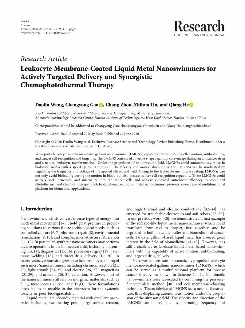
Research Article Leukocyte Membrane-Coated Liquid Metal Nanoswimmers for Actively Targeted Delivery and Synergistic Chemophotothermal Therapy Daolin Wang, Changyong Gao , Chang Zhou, Zhihua Lin, and Qiang He Key Laboratory of Microsystems and Microstructures Manufacturing, Ministry of Education, Micro/Nanotechnology Research Centre, Harbin Institute of Technology, 92 West Dazhi Street, Harbin 150080, China Correspondence should be addressed to Changyong Gao; [email protected] and Qiang He; [email protected] Received 1 April 2020; Accepted 27 May 2020; Published 24 June 2020 Copyright © 2020 Daolin Wang et al. Exclusive Licensee Science and Technology Review Publishing House. Distributed under a Creative Commons Attribution License (CC BY 4.0). We report a leukocyte membrane-coated gallium nanoswimmer (LMGNS) capable of ultrasound-propelled motion, antibiofouling, and cancer cell recognition and targeting. The LMGNS consists of a needle-shaped gallium core encapsulating an anticancer drug and a natural leukocyte membrane shell. Under the propulsion of an ultrasound field, LMGNSs could autonomously move in biological media with a speed up to 108.7 μms −1 . The velocity and motion direction of the LMGNSs can be modulated by regulating the frequency and voltage of the applied ultrasound field. Owing to the leukocyte membrane coating, LMGNSs can not only avoid biofouling during the motion in blood but also possess cancer cell recognition capability. These LMGNSs could actively seek, penetrate, and internalize into the cancer cells and achieve enhanced anticancer efficiency by combined photothermal and chemical therapy. Such biofunctionalized liquid metal nanoswimmer presents a new type of multifunctional platform for biomedical applications. 1. Introduction Nanoswimmers, which convert diverse types of energy into mechanical movement [1–5], hold great promise in provid- ing solutions to various future technological needs, such as controlled capture [6, 7], electronic repair [8], environmental remediation [9, 10], and complex microstructure fabrication [11, 12]. In particular, synthetic nanoswimmers may perform diverse operations in the biomedical field, including biosens- ing [13, 14], diagnostics [15, 16], precision surgery [17], laser tissue welding [18], and direct drug delivery [19, 20]. In recent years, various strategies have been employed to propel such micro/nanoswimmers, including chemical reaction [21, 22], light stimuli [23–25], and electric [26, 27], magnetism [28, 29], and acoustic [30, 31] actuation. However, most of the nanoswimmers still rely on inorganic materials, such as SiO 2 , mesoporous silicon, and Fe 3 O 4 ; these formulations often fail to be useable in the biosystem for the systemic toxicity or poor biodegradability. Liquid metal, a biofriendly material with excellent prop- erties including low melting point, large surface tension, and high thermal and electric conductivity [32–34], has emerged for stretchable electronics and soft robots [35–39]. In our previous work [40], we demonstrated a first example of the soft rod-like liquid metal nanoswimmers which could transform from rod to droplet, fuse together, and be degraded in both an acidic buffer and biomedium of cancer cells. To date, gallium-based liquid metal has aroused great interest in the field of biomedicine [41–43]. However, it is still a challenge to fabricate liquid metal-based nanoswim- mers with the capability of active motion, antibiofouling, and targeted drug delivery. Here, we demonstrate an acoustically propelled leukocyte membrane-coated gallium nanoswimmer (LMGNS), which can be served as a multifunctional platform for precise cancer therapy, as shown in Scheme 1. The biomimetic nanoswimmers were fabricated by combining the pressure- filter-template method [40] and cell membrane-cloaking technique. The as-fabricated LMGNS has a needle-like struc- ture, thus displaying autonomous motion under the propul- sion of the ultrasonic field. The velocity and direction of the LMGNSs can be regulated by alternating frequency and AAAS Research Volume 2020, Article ID 3676954, 10 pages https://doi.org/10.34133/2020/3676954
Transcript of Leukocyte Membrane-Coated Liquid Metal Nanoswimmers for ...
Research ArticleLeukocyte Membrane-Coated Liquid Metal Nanoswimmers forActively Targeted Delivery and SynergisticChemophotothermal Therapy
Daolin Wang, Changyong Gao , Chang Zhou, Zhihua Lin, and Qiang He
Key Laboratory of Microsystems and Microstructures Manufacturing, Ministry of Education,Micro/Nanotechnology Research Centre, Harbin Institute of Technology, 92 West Dazhi Street, Harbin 150080, China
Correspondence should be addressed to Changyong Gao; [email protected] and Qiang He; [email protected]
Received 1 April 2020; Accepted 27 May 2020; Published 24 June 2020
Copyright © 2020 Daolin Wang et al. Exclusive Licensee Science and Technology Review Publishing House. Distributed under aCreative Commons Attribution License (CC BY 4.0).
We report a leukocyte membrane-coated gallium nanoswimmer (LMGNS) capable of ultrasound-propelled motion, antibiofouling,and cancer cell recognition and targeting. The LMGNS consists of a needle-shaped gallium core encapsulating an anticancer drugand a natural leukocyte membrane shell. Under the propulsion of an ultrasound field, LMGNSs could autonomously move inbiological media with a speed up to 108.7μms−1. The velocity and motion direction of the LMGNSs can be modulated byregulating the frequency and voltage of the applied ultrasound field. Owing to the leukocyte membrane coating, LMGNSs cannot only avoid biofouling during the motion in blood but also possess cancer cell recognition capability. These LMGNSs couldactively seek, penetrate, and internalize into the cancer cells and achieve enhanced anticancer efficiency by combinedphotothermal and chemical therapy. Such biofunctionalized liquid metal nanoswimmer presents a new type of multifunctionalplatform for biomedical applications.
1. Introduction
Nanoswimmers, which convert diverse types of energy intomechanical movement [1–5], hold great promise in provid-ing solutions to various future technological needs, such ascontrolled capture [6, 7], electronic repair [8], environmentalremediation [9, 10], and complex microstructure fabrication[11, 12]. In particular, synthetic nanoswimmers may performdiverse operations in the biomedical field, including biosens-ing [13, 14], diagnostics [15, 16], precision surgery [17], lasertissue welding [18], and direct drug delivery [19, 20]. Inrecent years, various strategies have been employed to propelsuch micro/nanoswimmers, including chemical reaction [21,22], light stimuli [23–25], and electric [26, 27], magnetism[28, 29], and acoustic [30, 31] actuation. However, most ofthe nanoswimmers still rely on inorganic materials, such asSiO2, mesoporous silicon, and Fe3O4; these formulationsoften fail to be useable in the biosystem for the systemictoxicity or poor biodegradability.
Liquid metal, a biofriendly material with excellent prop-erties including low melting point, large surface tension,
and high thermal and electric conductivity [32–34], hasemerged for stretchable electronics and soft robots [35–39].In our previous work [40], we demonstrated a first exampleof the soft rod-like liquid metal nanoswimmers which couldtransform from rod to droplet, fuse together, and bedegraded in both an acidic buffer and biomedium of cancercells. To date, gallium-based liquid metal has aroused greatinterest in the field of biomedicine [41–43]. However, it isstill a challenge to fabricate liquid metal-based nanoswim-mers with the capability of active motion, antibiofouling,and targeted drug delivery.
Here, we demonstrate an acoustically propelled leukocytemembrane-coated gallium nanoswimmer (LMGNS), whichcan be served as a multifunctional platform for precisecancer therapy, as shown in Scheme 1. The biomimeticnanoswimmers were fabricated by combining the pressure-filter-template method [40] and cell membrane-cloakingtechnique. The as-fabricated LMGNS has a needle-like struc-ture, thus displaying autonomous motion under the propul-sion of the ultrasonic field. The velocity and direction of theLMGNSs can be regulated by alternating frequency and
AAASResearchVolume 2020, Article ID 3676954, 10 pageshttps://doi.org/10.34133/2020/3676954
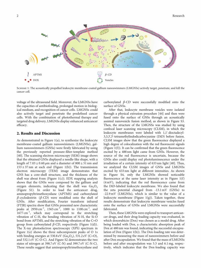
voltage of the ultrasound field. Moreover, the LMGNSs havethe capacities of antibiofouling, prolonged motion in biolog-ical medium, and recognition of cancer cells. LMGNSs couldalso actively target and penetrate the predefined cancercells. With the combination of photothermal therapy andtargeted drug delivery, LMGNSs display enhanced anticancerefficacy.
2. Results and Discussion
As demonstrated in Figure 1(a), to synthesize the leukocytemembrane-coated gallium nanoswimmers (LMGNSs), gal-lium nanoswimmers (GNSs) were firstly fabricated by usingthe previously reported pressure-filter-template method[40]. The scanning electron microscopy (SEM) image showsthat the obtained GNSs displayed a needle-like shape, with alength of 7:03 ± 0:60μm and a diameter of 800 ± 51 nm and153 ± 37 nm at each end (Figure 1(b)). The transmissionelectron microscopy (TEM) image demonstrates thatGNS has a core-shell structure, and the thickness of theshell was about 8 nm (Figure 1(c)). EDX mapping analysisshows that the GNSs were composed by the gallium andoxygen elements, indicating that the shell was Ga2O3(Figure S1). In order to load the anticancer drug,aminopropyltrimethoxysilane (APTMS) and carbonylatedβ-cyclodextrin (β-CD) were modified onto the shell ofGNSs. After modification, Fourier transform infrared(FTIR) spectra show that GNSs presented new characteristicpeaks at 2950 cm-1, 1565 cm-1, 1000 to 1100 cm-1, and1677 cm-1, which may correspond to the stretchingvibration of C-H, the bending vibration of N-H, the Si-Obond from APTMS, and the stretching vibration of the C=Ogroup from carbonylated β-CD, respectively (Figure 1(d)).The X-ray photoelectron spectroscopy (XPS) spectrum inFigure 1(e) shows the three subcomponent peaks of O 1swith binding energies at 530.8 eV (Ga2O3), 531.2 eV (C=O),and 532.1 eV (C-O-C). And Figure S2 demonstrates the twostates of nitrogen at 398.7 eV (C-N) and 399.7 eV (C-N-C).These results suggest that aminopropyltrimethoxysilane and
carbonylated β-CD were successfully modified onto thesurface of GNSs.
After that, leukocyte membrane vesicles were isolatedthrough a physical extrusion procedure [44] and then werefused onto the surface of GNSs through an acousticallyassisted nanovesicle fusion method, as shown in Figure S3.Then, the structure of the LMGNSs was studied by usingconfocal laser scanning microscopy (CLSM), in which theleukocyte membranes were labeled with 1,1'-dioctadecyl-3,3,3',3'-tetramethylindodicarbocyanine (DiD) before fusion.CLSM images show that the green fluorescence displayed ahigh degree of colocalization with the red fluorescent signals(Figure 1(f)). It can be confirmed that the green fluorescenceexcited by a 488nm light came from GNSs. However, thesource of the red fluorescence is uncertain, because theGNSs also could display red photoluminescence under theirradiation of a certain intensity of 633nm light [40]. Thus,we analyzed the CLSM images of GNSs and LMGNSsexcited by 633nm light at different intensities. As shownin Figure S4, only the LMGNSs showed noticeablefluorescence at the same laser intensity as in Figure 1(f)(4mV), indicating that the red fluorescence came fromthe DiD-labeled leukocyte membrane. We also found thatthe zeta potential changed from -13.1mV (GNSs) to-22.9mV (LMGNSs), which is similar to the value of aleukocyte membrane (Figure 1(g)). Taken together, theseresults demonstrate that leukocyte membrane vesicles fusedonto the surface of GNSs and LMGNSs were successfullyfabricated.
Then, these LMGNSs were explored to transport antican-cer drugs, and their drug-loading capacity was evaluated, inwhich doxorubicin (Dox) was chosen as a model drug. Afterbeing loaded with Dox, a characteristic absorption peak ofDox at 480nm was found, indicating the successful encapsu-lation of Dox (Figure 1(h)). The Dox-loading rate was deter-mined by measuring the mass of nanoswimmers before andafter Dox encapsulation. We found that the mass of LMGNSsbefore and after encapsulation was 5.3 and 6.1mg, respec-tively, which indicates that the Dox-loading capacity was
APTMS 𝛽–CD
DOX Vesicles
Scheme 1: The acoustically propelled leukocyte membrane-coated gallium nanoswimmers (LMGNSs) actively target, penetrate, and kill thecancer cell.
2 Research
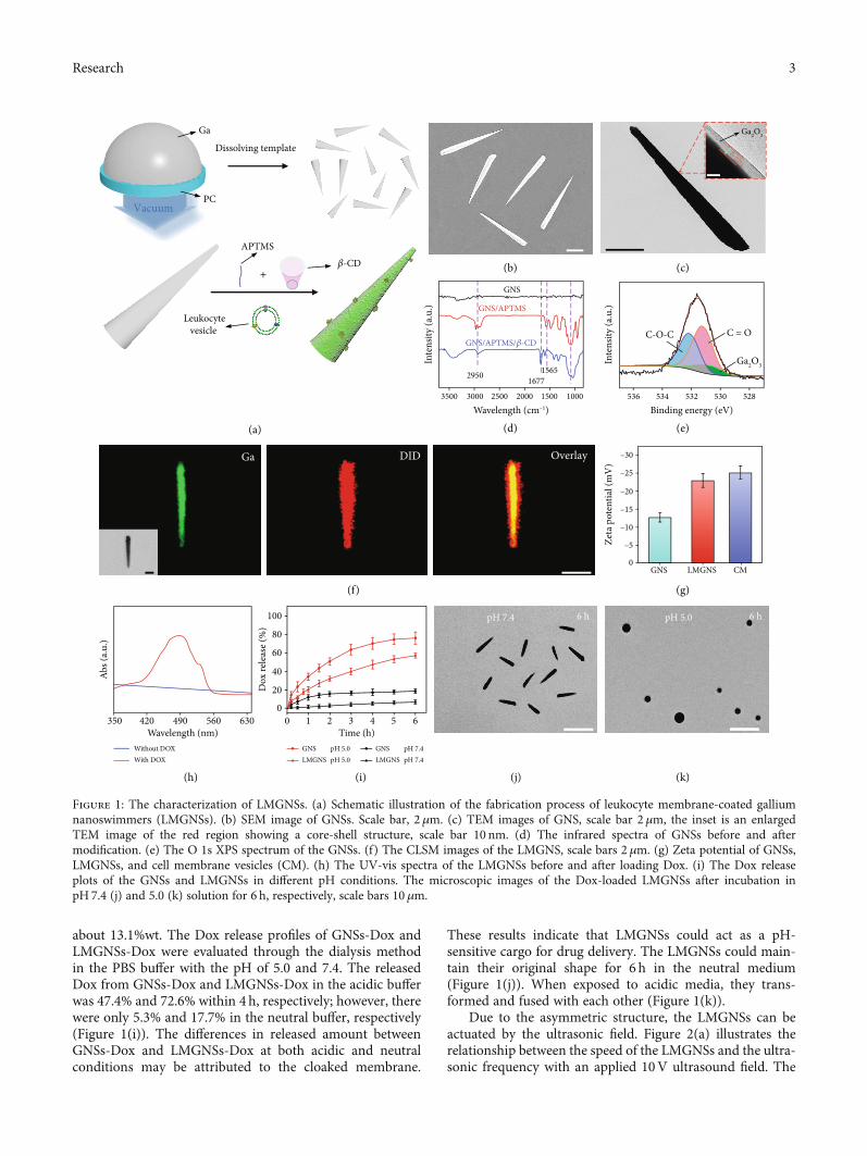
about 13.1%wt. The Dox release profiles of GNSs-Dox andLMGNSs-Dox were evaluated through the dialysis methodin the PBS buffer with the pH of 5.0 and 7.4. The releasedDox from GNSs-Dox and LMGNSs-Dox in the acidic bufferwas 47.4% and 72.6% within 4 h, respectively; however, therewere only 5.3% and 17.7% in the neutral buffer, respectively(Figure 1(i)). The differences in released amount betweenGNSs-Dox and LMGNSs-Dox at both acidic and neutralconditions may be attributed to the cloaked membrane.
These results indicate that LMGNSs could act as a pH-sensitive cargo for drug delivery. The LMGNSs could main-tain their original shape for 6 h in the neutral medium(Figure 1(j)). When exposed to acidic media, they trans-formed and fused with each other (Figure 1(k)).
Due to the asymmetric structure, the LMGNSs can beactuated by the ultrasonic field. Figure 2(a) illustrates therelationship between the speed of the LMGNSs and the ultra-sonic frequency with an applied 10V ultrasound field. The
Dissolving template
Vacuum
+
APTMS𝛽-CD
Leukocytevesicle
Ga
PC
Ga2O3
Ga2O3156516772950
GNS
GNS/APTMS
GNS/APTMS/𝛽-CD
3500 3000 2500
(a)
Wavelength (cm–1)In
tens
ity (a
.u.)
2000
(b)
(d) (e)
(f) (g)
(h) (i) (j) (k)
(c)
1500 1000
C-O-C C = O
536 534 532 530 528Binding energy (eV)
Inte
nsity
(a.u
.)
Ga DID Overlay
LMGNS GNS CM
–30
–25
–20
–15
–10
–5
0
Zeta
pot
entia
l (m
V)
Wavelength (nm)350 420 490 560
Without DOXWith DOX
630
Abs (
a.u.)
Time (h)6543210
GNS pH 5.0pH 5.0LMGNS
GNS pH 7.4pH 7.4LMGNS
0
20
40
60
80
100
Dox
rele
ase (
%)
pH 7.4 6 h pH 5.0 6 h
8 nm
Figure 1: The characterization of LMGNSs. (a) Schematic illustration of the fabrication process of leukocyte membrane-coated galliumnanoswimmers (LMGNSs). (b) SEM image of GNSs. Scale bar, 2μm. (c) TEM images of GNS, scale bar 2μm, the inset is an enlargedTEM image of the red region showing a core-shell structure, scale bar 10 nm. (d) The infrared spectra of GNSs before and aftermodification. (e) The O 1s XPS spectrum of the GNSs. (f) The CLSM images of the LMGNS, scale bars 2 μm. (g) Zeta potential of GNSs,LMGNSs, and cell membrane vesicles (CM). (h) The UV-vis spectra of the LMGNSs before and after loading Dox. (i) The Dox releaseplots of the GNSs and LMGNSs in different pH conditions. The microscopic images of the Dox-loaded LMGNSs after incubation inpH 7.4 (j) and 5.0 (k) solution for 6 h, respectively, scale bars 10 μm.
3Research
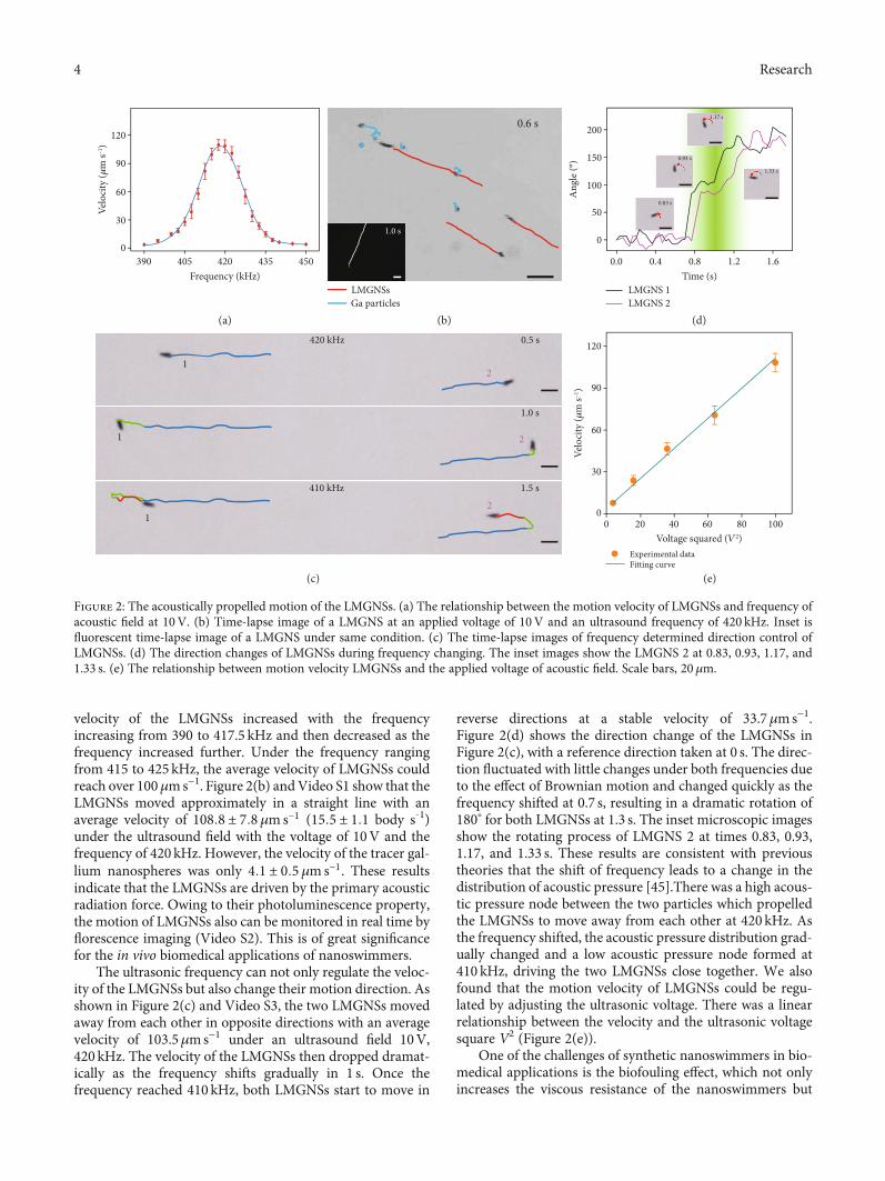
velocity of the LMGNSs increased with the frequencyincreasing from 390 to 417.5 kHz and then decreased as thefrequency increased further. Under the frequency rangingfrom 415 to 425 kHz, the average velocity of LMGNSs couldreach over 100μms−1. Figure 2(b) andVideo S1 show that theLMGNSs moved approximately in a straight line with anaverage velocity of 108:8 ± 7:8 μms−1 (15:5 ± 1:1 body s-1)under the ultrasound field with the voltage of 10V and thefrequency of 420 kHz. However, the velocity of the tracer gal-lium nanospheres was only 4:1 ± 0:5 μms−1. These resultsindicate that the LMGNSs are driven by the primary acousticradiation force. Owing to their photoluminescence property,the motion of LMGNSs also can be monitored in real time byflorescence imaging (Video S2). This is of great significancefor the in vivo biomedical applications of nanoswimmers.
The ultrasonic frequency can not only regulate the veloc-ity of the LMGNSs but also change their motion direction. Asshown in Figure 2(c) and Video S3, the two LMGNSs movedaway from each other in opposite directions with an averagevelocity of 103.5μms−1 under an ultrasound field 10V,420 kHz. The velocity of the LMGNSs then dropped dramat-ically as the frequency shifts gradually in 1 s. Once thefrequency reached 410 kHz, both LMGNSs start to move in
reverse directions at a stable velocity of 33.7μms−1.Figure 2(d) shows the direction change of the LMGNSs inFigure 2(c), with a reference direction taken at 0 s. The direc-tion fluctuated with little changes under both frequencies dueto the effect of Brownian motion and changed quickly as thefrequency shifted at 0.7 s, resulting in a dramatic rotation of180° for both LMGNSs at 1.3 s. The inset microscopic imagesshow the rotating process of LMGNS 2 at times 0.83, 0.93,1.17, and 1.33 s. These results are consistent with previoustheories that the shift of frequency leads to a change in thedistribution of acoustic pressure [45].There was a high acous-tic pressure node between the two particles which propelledthe LMGNSs to move away from each other at 420 kHz. Asthe frequency shifted, the acoustic pressure distribution grad-ually changed and a low acoustic pressure node formed at410 kHz, driving the two LMGNSs close together. We alsofound that the motion velocity of LMGNSs could be regu-lated by adjusting the ultrasonic voltage. There was a linearrelationship between the velocity and the ultrasonic voltagesquare V2 (Figure 2(e)).
One of the challenges of synthetic nanoswimmers in bio-medical applications is the biofouling effect, which not onlyincreases the viscous resistance of the nanoswimmers but
120
90
60
30
0
Velo
city
(𝜇m
s–1)
390 405 420Frequency (kHz)
(a)
(c) (e)
(b) (d)
435 450
Ga particles LMGNSs
0.6 s
1.0 s
1
1
12
1.0 s
2
2
1.5 s
0.5 s420 kHz
410 kHz
1.17 s
1.33 s
0.83 s
0.93 s
200
150
100
50
0
Ang
le (°
)
0.0 0.4 0.8Time (s)
1.2 1.6
LMGNS 2 LMGNS 1
120
90
Experimental dataFitting curve
60
30
00 20 40 60
Voltage squared (V2)
Velo
city
(𝜇m
s–1)
80 100
Figure 2: The acoustically propelled motion of the LMGNSs. (a) The relationship between the motion velocity of LMGNSs and frequency ofacoustic field at 10V. (b) Time-lapse image of a LMGNS at an applied voltage of 10V and an ultrasound frequency of 420 kHz. Inset isfluorescent time-lapse image of a LMGNS under same condition. (c) The time-lapse images of frequency determined direction control ofLMGNSs. (d) The direction changes of LMGNSs during frequency changing. The inset images show the LMGNS 2 at 0.83, 0.93, 1.17, and1.33 s. (e) The relationship between motion velocity LMGNSs and the applied voltage of acoustic field. Scale bars, 20μm.
4 Research
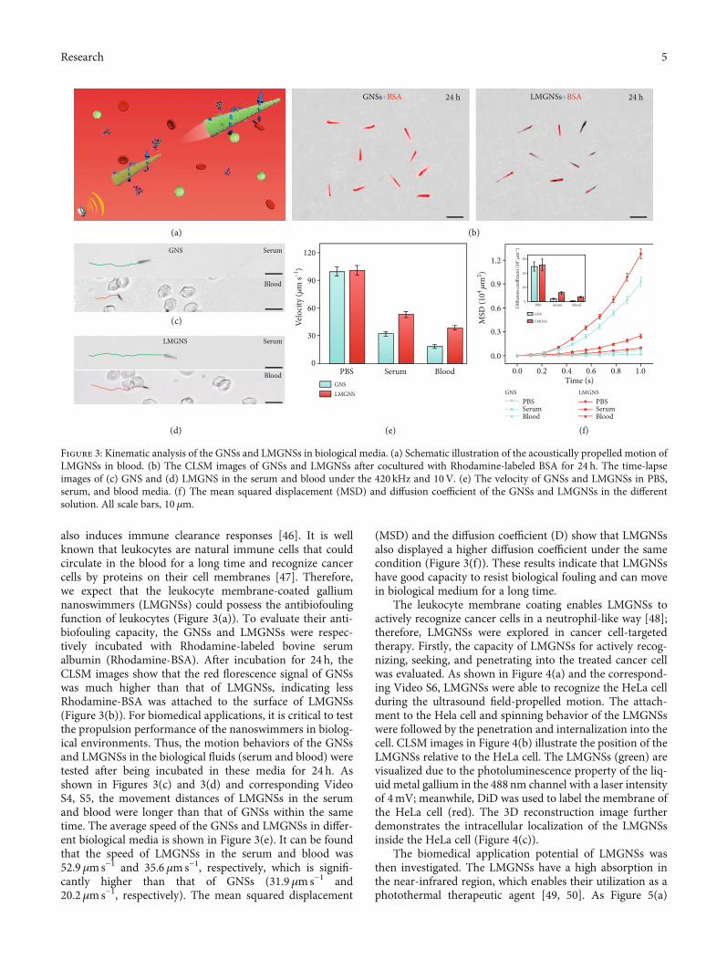
also induces immune clearance responses [46]. It is wellknown that leukocytes are natural immune cells that couldcirculate in the blood for a long time and recognize cancercells by proteins on their cell membranes [47]. Therefore,we expect that the leukocyte membrane-coated galliumnanoswimmers (LMGNSs) could possess the antibiofoulingfunction of leukocytes (Figure 3(a)). To evaluate their anti-biofouling capacity, the GNSs and LMGNSs were respec-tively incubated with Rhodamine-labeled bovine serumalbumin (Rhodamine-BSA). After incubation for 24 h, theCLSM images show that the red florescence signal of GNSswas much higher than that of LMGNSs, indicating lessRhodamine-BSA was attached to the surface of LMGNSs(Figure 3(b)). For biomedical applications, it is critical to testthe propulsion performance of the nanoswimmers in biolog-ical environments. Thus, the motion behaviors of the GNSsand LMGNSs in the biological fluids (serum and blood) weretested after being incubated in these media for 24h. Asshown in Figures 3(c) and 3(d) and corresponding VideoS4, S5, the movement distances of LMGNSs in the serumand blood were longer than that of GNSs within the sametime. The average speed of the GNSs and LMGNSs in differ-ent biological media is shown in Figure 3(e). It can be foundthat the speed of LMGNSs in the serum and blood was52.9μms−1 and 35.6μms−1, respectively, which is signifi-cantly higher than that of GNSs (31.9μms−1 and20.2μms−1, respectively). The mean squared displacement
(MSD) and the diffusion coefficient (D) show that LMGNSsalso displayed a higher diffusion coefficient under the samecondition (Figure 3(f)). These results indicate that LMGNSshave good capacity to resist biological fouling and can movein biological medium for a long time.
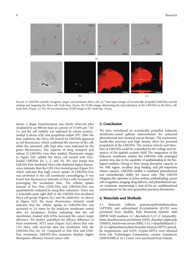
The leukocyte membrane coating enables LMGNSs toactively recognize cancer cells in a neutrophil-like way [48];therefore, LMGNSs were explored in cancer cell-targetedtherapy. Firstly, the capacity of LMGNSs for actively recog-nizing, seeking, and penetrating into the treated cancer cellwas evaluated. As shown in Figure 4(a) and the correspond-ing Video S6, LMGNSs were able to recognize the HeLa cellduring the ultrasound field-propelled motion. The attach-ment to the Hela cell and spinning behavior of the LMGNSswere followed by the penetration and internalization into thecell. CLSM images in Figure 4(b) illustrate the position of theLMGNSs relative to the HeLa cell. The LMGNSs (green) arevisualized due to the photoluminescence property of the liq-uid metal gallium in the 488nm channel with a laser intensityof 4mV; meanwhile, DiD was used to label the membrane ofthe HeLa cell (red). The 3D reconstruction image furtherdemonstrates the intracellular localization of the LMGNSsinside the HeLa cell (Figure 4(c)).
The biomedical application potential of LMGNSs wasthen investigated. The LMGNSs have a high absorption inthe near-infrared region, which enables their utilization as aphotothermal therapeutic agent [49, 50]. As Figure 5(a)
GNSs+BSA LMGNSs+BSA24 h 24 h
Blood
GNS
(a) (b)
(c)
(d) (e) (f)
Serum
Blood
SerumLMGNS
LMGNS GNS
BloodSerumPBS0
30
60
90
120
Velo
city
(𝜇m
s–1)
GNS LMGNS
CMGNS
GNS 0.6
0.3
0.0
0.0 0.2 0.4Time (s)
0.6 0.8 1.0
0.9
30
20
10
0PBS Serum Blood
1.2
PBSSerumBlood
PBSSerumBlood
MSD
(104 𝜇
m2 )
Diff
usio
n co
effici
ent (
103 𝜇
m–1
)
Figure 3: Kinematic analysis of the GNSs and LMGNSs in biological media. (a) Schematic illustration of the acoustically propelled motion ofLMGNSs in blood. (b) The CLSM images of GNSs and LMGNSs after cocultured with Rhodamine-labeled BSA for 24 h. The time-lapseimages of (c) GNS and (d) LMGNS in the serum and blood under the 420 kHz and 10V. (e) The velocity of GNSs and LMGNSs in PBS,serum, and blood media. (f) The mean squared displacement (MSD) and diffusion coefficient of the GNSs and LMGNSs in the differentsolution. All scale bars, 10 μm.
5Research
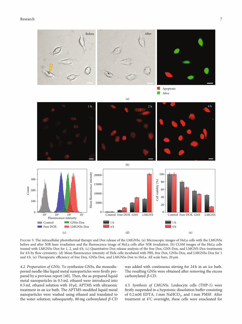
shows, a shape transformation was clearly observed afterirradiated by an 808nm laser at a power of 15mW·μm-2 for5 s, and the cell viability was indicated by calcein acetoxy-methyl (Calcein-AM) and propidium iodide (PI). After thelaser radiation, the HeLa cell treated by LMGNSs appearedas red fluorescence which confirmed the necrosis of the cell,while the untreated cells kept alive were indicated by thegreen fluorescence. The capacity of drug transport andrelease of LMGNSs were then studied. Fluorescent imagesin Figure 5(b) exhibit the HeLa cell treated with Dox-loaded LMGNSs for 1, 2, and 4h. We also found thatLMGNS-Dox-incubated HeLa cells displayed higher fluores-cence intensity than the GNS-Dox-treated group (Figure S5),which indicates that high cancer uptake of LMGNSs-Doxwas attributed to the cell membrane camouflaging. It wasfound that fluorescence intensity of HeLa cells increased byprolonging the incubation time. The cellular uptakeamount of free Dox, GNSs-Dox and LMGNSs-Dox wasquantitatively evaluated by using flow cytometry. There wasa noticeable peak right shift in the LMGNS-Dox-incubatedHeLa cell group (Figures 5(c) and S6, relatively). As shownin Figure 5(d), the mean fluorescence intensity resultindicates that the cellular uptake to LMGNSs-Dox wasincreased to 2.6 times by the leukocyte membrane coatedafter 4 h incubation, further indicating that the cellmembranes cloaked with GNSs increased the cancer targetefficiency. We further quantified the efficacy difference ofthese treatments. MTT assay (Figure 5(e)) shows that only15% HeLa cells survived after the incubation with theLMGNSs-Dox for 4 h. Compared to free Dox and GNS-Dox treatments, LMGSN-Dox treatment displays highertherapeutic efficiency toward cancer cells.
3. Conclusion
We have introduced an acoustically propelled leukocytemembrane-coated gallium nanoswimmer for enhancedphotothermal and chemical cancer therapy. The asymmetricneedle-like structure and high density allow for powerfulpropulsion of the LMGNSs. The motion velocity and direc-tion of LMGNSs could be controlled by the voltage and fre-quency of the applied acoustic field. The integration of theleukocyte membrane enables the LMGNSs with prolongedmotion time due to the capability of antibiofouling in the bio-logical medium. Owing to their strong absorptive capacity inthe NIR region, excellent drug loading, and pH-responsiverelease capacity, LMGNSs exhibit a combined photothermaland chemotherapy ability for cancer cells. This LMGNSintegrates the capacities of active motion, antibiofouling, cancercell recognition, imaging, drug delivery, and photothermal can-cer treatment, representing a state-of-the-art multifunctionalnanoswimmer for the next-generation precision theranostics.
4. Materials and Methods
4.1. Materials. Gallium, aminopropyltrimethoxysilane(APTMS), and carbonylated β-cyclodextrin (β-CD) werepurchased from Aladdin. Park Memorial Institute 1640(RPMI 1640) medium, 1,1′-dioctadecyl-3,3,3′,3′-tetramethy-lindo-dicarbocyanine perchlorate (DiD), dimethyl sulphoxide(DMSO), fetal bovine serum (FBS), 3-(4, 5-dimethylthiazol-2-yl)-2,5-diphenyltetrazolium bromide solution (MTT), penicil-lin streptomycin, and 0.25% Trypsin-EDTA were obtainedfrom Life Technologies Corporation; ceramic transducers(H4P101000, ϕ 10 ∗ 2mm) were purchased from Fukeda.
0 s
(a)
(b) (c)
0.5 s 1 s 2 s
Figure 4: LMGNSs actively recognize, target, and penetrate HeLa cell. (a) Time-lapse images of acoustically propelled LMGNSs activelyseeking and targeting the HeLa cell. Scale bars, 20μm. (b) CLSM images illustrating the internalization of the LMGNSs in the HeLa cell.Scale bars, 10 μm. (c) The 3D reconstruction CLSM image in (b). Scale bar, 10 μm.
6 Research
4.2. Preparation of GNSs. To synthesize GNSs, the monodis-persed needle-like liquid metal nanoparticles were firstly pre-pared by a previous report [40]. Then, the as-prepared liquidmetal nanoparticles in 0.5mL ethanol were introduced into0.5mL ethanol solution with 10μL APTMS with ultrasonictreatment in an ice bath. The APTMS-modified liquid metalnanoparticles were washed using ethanol and translated tothe water solution; subsequently, 80mg carbonylated β-CD
was added with continuous stirring for 24 h in an ice bath.The resulting GNSs were obtained after removing the excesscarbonylated β-CD.
4.3. Synthesis of LMGNSs. Leukocyte cells (THP-1) werefirstly suspended in a hypotonic dissolution buffer consistingof 0.2mM EDTA, 1mm NaHCO3, and 1mm PMSF. Aftertreatment at 4°C overnight, these cells were enucleated for
Before After
Alive Apoptosis
1 h 2 h 4 h
300
ControlFree DOX
GNSs-DoxLMGNSs-Dox
200
100
0102 103
Fluorescence intensity
Cou
nt
104
(c)
(b)
(a)
(d) (e)
105 LMGNS GNS Free DOX Control
20000
15000
1 h4 h
10000
5000
0
Mea
n flu
ores
cenc
e int
ensit
y
LMGNS GNS Free DOX Control
1 h4 h
0
20
40
60
80
100
Cel
l via
bilit
y (%
)
Figure 5: The intracellular photothermal therapy and Dox release of the LMGNSs. (a) Microscopic images of HeLa cells with the LMGNSsbefore and after NIR laser irradiation and the fluorescence image of HeLa cells after NIR irradiation. (b) CLSM images of the HeLa cellstreated with LMGNSs-Dox for 1, 2, and 4 h. (c) Quantitative Dox release analysis of the free Dox, GNS-Dox, and LMGNS-Dox treatmentsfor 4 h by flow cytometry. (d) Mean fluorescence intensity of Hela cells incubated with PBS, free Dox, GNSs-Dox, and LMGNSs-Dox for 1and 4 h. (e) Therapeutic efficiency of free Dox, GNSs-Dox, and LMGNSs-Dox to HeLa. All scale bars, 20 μm.
7Research
20 times by using a hand-held Dounce homogenizer. Afterthe above suspension was centrifuged at 3200 g at 4° C for5min, the leukocyte membrane precipitate was obtainedafter centrifugation at 150000 g at 4° C for 1 h. Subsequently,the isolated leukocyte membranes were physically extrudedthrough 1μm, 400 nm, 200nm, 100nm, and 50nm polycar-bonate membranes for 21 passes. The LMGNSs were thenfabricated by fusing the leukocyte membrane nanovesiclesonto the surface of GNSs following a published protocol[51]. Briefly, the GNSs and leukocyte nanovesicles weremixed and then sonicated by using an AutoscienceAS7240AT ultrasonic machine for 2 h in an ice bath. Afterremoving the excess leukocyte membrane nanovesicles,LMGNSs were obtained.
4.4. Acoustic Experiments. The acoustic-propelled motionof the LMGNSs was conducted by using an ultrasonicsetup, which is made of a function generator (TektronixAFG1062), signal amplifier (Toellner TOE 7607), andceramic transducer. And the sample cell is a cylinder with aheight of 1.5mm and a diameter of 5mm, and the ceramictransducer was pasted on the bottom of the sample cell.The LMGNSs were incubated in the biological media (water,PBS, serum, and blood) for 24h, and then a 15μL LMGNSsolution was dropped into the sample cell and covered by asquare coverslip. A frequency of 390-450 kHz and 0-10Vvoltage were applied to actuate the LMGNSs, which wasobserved by an Olympus optical microscope (OLYMPUSBX53).
4.5. Photothermal Treatment Experiments. The photothermaltherapy of LMGNSs was observed by the fluorescence micro-scope (Olympus IX 71). The HeLa cells with LMGNSs wereirradiated by an 808nm laser at the power of 15mW·μm-2
for 5 s. Then, the cell viability is determined by adding PIand Calcein-AM. Finally, the resulting fluorescence imagewas captured under a 488 nm light.
4.6. Flow Cytometry Analysis. The cellular uptake of the var-ious drug-loaded nanoswimmers was evaluated by using theflow cytometry analysis. GNSs-Dox, LMGNSs-Dox, and freeDox with a drug concentration of 10μgmL−1 was coculturedwith HeLa cells with for 1 h and 4h, respectively. After that,the free Dox and nanoswimmers were removed by washingwith PBS solution for 5 times. Then, HeLa cells were har-vested, and their fluorescence intensity was tested by flowcytometry (BD FACSAria).
4.7. MTT Assay. To evaluate their anticancer efficiencies,PBS, free Dox, GNSs-Dox and LMGNSs-Dox first coculturedwith HeLa cells for 1 h and 4h. Then, a drop of 20mL MTTsolution with the concentration of 5mgmL-1 was added intothe cell culture medium and incubated with HeLa cells for4 h. After removing the culture medium, 150mL DMSOwas dropped to HeLa cells, and the cell viability of HeLa cellswas measured at the absorbance wavelength of 570nm byusing a microplate reader.
Conflicts of Interest
The authors declare no competing financial interest.
Authors’ Contributions
Q. He and C. Y. Gao proposed and supervised the experi-ments; D. L. Wang conducted the most of the experimentalwork and wrote the original draft of the manuscript; Q. He,C. Y. Gao, D. L. Wang, C. Zhou, and Z. H. Lin discussedthe results.
Acknowledgments
This work was financially supported by the NationalNatural Science Foundation of China (No.: 21902037)and National Postdoctoral Program for Innovative Talents(BX201700065).
Supplementary Materials
Supplementary 1. Figure S1: SEM and EDX mapping imagesof the GNSs. Figure S2: N 1s XPS spectrum of the GNSs. Fig-ure S3: the schematic depicting the preparation process ofLMGNSs. Figure S4: CLSM images of GNS and LMGNSexcited by a 633 nm light with different intensity. Figure S5:CLSM images of the HeLa cells treated with Dox-loadedGNSs for 1, 2, and 4h. Figure S6: Quantitative Dox releaseanalysis after treatments for 1 h by flow cytometry.
Supplementary 2. Movie S1: the movement of LMGNSsunder 420 kHz, 10V ultrasound field.
Supplementary 3. Movie S2: the fluorescence movement ofLMGNS under 420 kHz, 10V ultrasound field.
Supplementary 4. Movie S3: the diverging motion ofLMGNSs by regulating the ultrasonic frequency.
Supplementary 5. Movie S4: the movement of GNS in theserum and blood solution.
Supplementary 6. Movie S5: the movement of LMGNS in theserum and blood solution.
Supplementary 7. Movie S6: the movement of LMGNSsactively target the HeLa cell.
References
[1] K. Ariga, J. Li, J. Fei, Q. Ji, and J. P. Hill, “Nanoarchitectonicsfor dynamic functional materials from atomic-/molecular-level manipulation to macroscopic action,” Advanced Mate-rials, vol. 28, no. 6, pp. 1251–1286, 2016.
[2] W. F. Paxton, K. C. Kistler, C. C. Olmeda et al., “Catalyticnanomotors: autonomous movement of striped nanorods,”Journal of the American Chemical Society, vol. 126, no. 41,pp. 13424–13431, 2004.
[3] U. Choudhury, D. P. Singh, T. Qiu, and P. Fischer, “Chemicalnanomotors at the gram scale form a dense active optorheolo-gical medium,” Advanced Materials, vol. 31, no. 12, article1807382, 2019.
[4] F. Peng, Y. Tu, J. C. M. van Hest, and D. A. Wilson, “Self-guided supramolecular cargo-loaded nanomotors with
8 Research
chemotactic behavior towards cells,” Angewandte ChemieInternational Edition, vol. 54, no. 40, pp. 11662–11665, 2015.
[5] H. Wang, J. G. S. Moo, and M. Pumera, “From nanomotors tomicromotors: the influence of the size of an autonomousbubble-propelled device upon its motion,” ACS Nano,vol. 10, no. 5, pp. 5041–5050, 2016.
[6] Y. Zhang, L. Zhang, L. Yang et al., “Real-time tracking offluorescent magnetic spore-based microrobots for remotedetection ofC. difftoxins,” Science Advances, vol. 5, no. 1,article eaau9650, 2019.
[7] T. Li, J. Li, K. I. Morozov et al., “Highly efficient freestylemagnetic nanoswimmer,” Nano Letters, vol. 17, no. 8,pp. 5092–5098, 2017.
[8] J. Li, O. E. Shklyaev, T. Li et al., “Self-propelled nanomotorsautonomously seek and repair cracks,” Nano Letters, vol. 15,no. 10, pp. 7077–7085, 2015.
[9] T. Xu, W. Gao, L.-P. Xu, X. Zhang, and S. Wang, “Fuel-freesynthetic micro-/nanomachines,” Advanced Materials,vol. 29, no. 9, article 1603250, 2017.
[10] J. Katuri, X. Ma, M. M. Stanton, and S. Sánchez, “Designingmicro- and nanoswimmers for specific applications,” Accountsof Chemical Research, vol. 50, no. 1, pp. 2–11, 2017.
[11] J. Palacci, S. Sacanna, A. P. Steinberg, D. J. Pine, and P. M.Chaikin, “Living crystals of light-activated colloidal surfers,”Science, vol. 339, no. 6122, pp. 936–940, 2013.
[12] H. Xie, M. Sun, X. Fan et al., “Reconfigurable magneticmicrorobot swarm: multimode transformation, locomotion,and manipulation,” Science Robotics, vol. 4, no. 28, articleeaav8006, 2019.
[13] Y. Su, Y. Ge, L. Liu et al., “Motion-based ph sensing based onthe cartridge-case-like micromotor,” ACS Applied Materials& Interfaces, vol. 8, no. 6, pp. 4250–4257, 2015.
[14] M. Zarei and M. Zarei, “Self-propelled micro/nanomotors forsensing and environmental remediation,” Small, vol. 14,no. 30, article 1800912, 2018.
[15] T. Li, X. Chang, Z. Wu et al., “Autonomous collision-free nav-igation of microvehicles in complex and dynamically changingenvironments,” ACS Nano, vol. 11, no. 9, pp. 9268–9275, 2017.
[16] A. Chałupniak, E. Morales-Narváez, and A. Merkoçi, “Microand nanomotors in diagnostics,” Advanced Drug DeliveryReviews, vol. 95, pp. 104–116, 2015.
[17] J. Shao, I. A. B. Pijpers, S. Cao et al., “Biomorphic engineeringof multifunctional polylactide stomatocytes toward therapeu-tic nano-red blood cells,” Advanced Science, vol. 6, no. 5, article1801678, 2019.
[18] W. He, J. Frueh, N. Hu, L. Liu, M. Gai, and Q. He, “Guidablethermophoretic janus micromotors containing gold nanoco-lorifiers for infrared laser assisted tissue welding,” AdvancedScience, vol. 3, no. 12, article 1600206, 2016.
[19] W. Gao, D. Kagan, O. S. Pak et al., “Cargo-towing fuel-freemagnetic nanoswimmers for targeted drug delivery,” Small,vol. 8, no. 3, pp. 460–467, 2012.
[20] J. Li, B. Esteban-Fernández de Ávila, W. Gao, L. Zhang, andJ. Wang, “Micro/nanorobots for biomedicine: delivery, sur-gery, sensing, and detoxification,” Science Robotics, vol. 2,no. 4, article eaam6431, 2017.
[21] F. Novotný, J. Plutnar, and M. Pumera, “Plasmonic self-propelled nanomotors for explosives detection via solution-based surface enhanced Raman scattering,” Advanced Func-tional Materials, vol. 29, no. 33, article 1903041, 2019.
[22] D. Xu, C. Zhou, C. Zhan et al., “Enzymatic micromotors as amobile photosensitizer platform for highly efficient on-chiptargeted antibacteria photodynamic therapy,” Advanced Func-tional Materials, vol. 29, no. 17, article 1807727, 2019.
[23] Y. Tu, F. Peng, J. M. Heuvelmans, S. Liu, R. J. M. Nolte, andD. A.Wilson, “Motion control of polymeric nanomotors basedon host-guest interactions,” Angewandte Chemie InternationalEdition, vol. 131, no. 26, pp. 8779–8783, 2019.
[24] R. Dong, Y. Cai, Y. Yang, W. Gao, and B. Ren, “Photocatalyticmicro/nanomotors: from construction to applications,”Accounts of Chemical Research, vol. 51, no. 9, pp. 1940–1947,2018.
[25] L. Xu, F. Mou, H. Gong, M. Luo, and J. Guan, “Light-drivenmicro/nanomotors: from fundamentals to applications,”Chemical Society Reviews, vol. 46, no. 22, pp. 6905–6926, 2017.
[26] G. Loget and A. Kuhn, “Electric field-induced chemical loco-motion of conducting objects,” Nature Communications,vol. 2, no. 1, 2011.
[27] J. Yan, M. Han, J. Zhang, C. Xu, E. Luijten, and S. Granick,“Reconfiguring active particles by electrostatic imbalance,”Nature Materials, vol. 15, no. 10, pp. 1095–1099, 2016.
[28] B. Jang, E. Gutman, N. Stucki et al., “Undulatory locomotionof magnetic multilink nanoswimmers,” Nano Letters, vol. 15,no. 7, pp. 4829–4833, 2015.
[29] X. Yan, Q. Zhou, M. Vincent et al., “Multifunctional biohybridmagnetite microrobots for imaging-guided therapy,” ScienceRobotics, vol. 2, no. 12, article eaaq1155, 2017.
[30] W. Wang, L. A. Castro, M. Hoyos, and T. E. Mallouk, “Auton-omousmotion of metallic microrods propelled by ultrasound,”ACS Nano, vol. 6, no. 7, pp. 6122–6132, 2012.
[31] B. E.-F. de Ávila, P. Angsantikul, J. Li et al., “Micromotor-enabled active drug delivery for in vivo treatment of stomachinfection,” Nature Communications, vol. 8, no. 1, p. 272, 2017.
[32] T. Daeneke, K. Khoshmanesh, N. Mahmood et al., “Liquidmetals: fundamentals and applications in chemistry,” Chemi-cal Society Reviews, vol. 47, no. 11, pp. 4073–4111, 2018.
[33] J. Zhang, Y. Yao, L. Sheng, and J. Liu, “Self-fueled biomimeticliquid metal mollusk,” Advanced Materials, vol. 27, no. 16,pp. 2648–2655, 2015.
[34] J. Yan, M. H. Malakooti, Z. Lu et al., “Solution processable liq-uid metal nanodroplets by surface-initiated atom transfer rad-ical polymerization,” Nature Nanotechnology, vol. 14, no. 7,pp. 684–690, 2019.
[35] M. D. Dickey, “Stretchable and soft electronics using liquidmetals,” Advanced Materials, vol. 29, no. 27, article 1606425,2017.
[36] Y. Chen, T. Zhou, Y. Li et al., “Robust fabrication of nonstick,noncorrosive, conductive graphene-coated liquid metal drop-lets for droplet-based, floating electrodes,” Advanced Func-tional Materials, vol. 28, no. 8, article 1706277, 2018.
[37] J. Wu, S.-Y. Tang, T. Fang, W. Li, X. Li, and S. Zhang, “Awheeled robot driven by a liquid-metal droplet,” AdvancedMaterials, vol. 30, no. 51, article 1805039, 2018.
[38] Y. Xin, H. Peng, J. Xu, and J. Zhang, “Ultrauniform embeddedliquid metal in sulfur polymers for recyclable, conductive, andself-healable materials,” Advanced Functional Materials,vol. 29, no. 17, article 1808989, 2019.
[39] Y. Wang, W. Duan, C. Zhou et al., “Phoretic liquid metalmicro/nanomotors as intelligent filler for targeted microweld-ing,” AdvancedMaterials, vol. 31, no. 51, article 1905067, 2019.
9Research
[40] D. Wang, C. Gao, W. Wang et al., “Shape-transformable,fusible rodlike swimming liquid metal nanomachine,” ACSNano, vol. 12, no. 10, pp. 10212–10220, 2018.
[41] Y. Lu, Q. Hu, Y. Lin et al., “Transformable liquid-metal nano-medicine,” Nature Communications, vol. 6, no. 1, 2015.
[42] Y. Lu, Y. Lin, Z. Chen et al., “Enhanced endosomal escape bylight-fueled liquid-metal transformer,” Nano Letters, vol. 17,no. 4, pp. 2138–2145, 2017.
[43] S. A. Chechetka, Y. Yu, X. Zhen, M. Pramanik, K. Pu, andE. Miyako, “Light-driven liquid metal nanotransformers forbiomedical theranostics,” Nature Communications, vol. 8,no. 1, 2017.
[44] C.-M. J. Hu, L. Zhang, S. Aryal, C. Cheung, R. H. Fang, andL. Zhang, “Erythrocyte membrane-camouflaged polymericnanoparticles as a biomimetic delivery platform,” Proceedingsof the National Academy of Sciences, vol. 108, no. 27,pp. 10980–10985, 2011.
[45] S. Ahmed, W. Wang, L. Bai, D. T. Gentekos, M. Hoyos, andT. E. Mallouk, “Density and shape effects in the acousticpropulsion of bimetallic nanorod motors,” ACS Nano,vol. 10, no. 4, pp. 4763–4769, 2016.
[46] C. Gao, Z. Lin, X. Lin, and Q. He, “Cell membrane-camouflaged colloid motors for biomedical applications,”Advanced Therapeutics, vol. 1, no. 5, article 1800056, 2018.
[47] A. Parodi, N. Quattrocchi, A. L. van de Ven et al., “Syntheticnanoparticles functionalized with biomimetic leukocyte mem-branes possess cell-like functions,” Nature Nanotechnology,vol. 8, no. 1, pp. 61–68, 2013.
[48] W. He, J. Frueh, Z. Wu, and Q. He, “How leucocyte cellmembrane modified janus microcapsules are phagocytosedby cancer cells,” ACS Applied Materials & Interfaces, vol. 8,no. 7, pp. 4407–4415, 2016.
[49] A. Kyrsting, P. M. Bendix, D. G. Stamou, and L. B.Oddershede, “Heat profiling of three-dimensionally opticallytrapped gold nanoparticles using vesicle cargo release,” NanoLetters, vol. 11, no. 2, pp. 888–892, 2011.
[50] W. He, J. Frueh, Z. Wu, and Q. He, “Leucocyte membrane-coated janus microcapsules for enhanced photothermal cancertreatment,” Langmuir, vol. 32, no. 15, pp. 3637–3644, 2016.
[51] Z. Wu, T. Li, W. Gao et al., “Cell-membrane-coated syntheticnanomotors for effective biodetoxification,” Advanced Func-tional Materials, vol. 25, no. 25, pp. 3881–3887, 2015.
10 Research